Embed Size (px)
Citation preview
Improving T cell therapy for relapsed EBV-negative Hodgkin’s Lymphoma by targeting upregulated
MAGE-A4
Shortened Running Title: MAGE-A4 T cell therapy for Hodgkin’s Lymphoma
Authors and Affiliations: Conrad R. Cruz1, Ulrike Gerdemann1, Ann M. Leen1, Jessica A. Shafer1,
Stephanie Ku1, Benjamin Tzou1, Terzah M. Horton2, Amanda Copeland3, Anas Younes3, Cliona M.
Rooney1, Helen E. Heslop1, Catherine M. Bollard1
1Center for Cell and Gene Therapy, Baylor College of Medicine, Texas Children’s Hospital, The Methodist
Hospital, Houston, Texas, 2Department of Pediatrics, Baylor College of Medicine, Houston, Texas, 3The
University of Texas MD Anderson Cancer Center, Houston, Texas
Corresponding Author Information:
Catherine M. Bollard, M.D.
Center for Cell and Gene Therapy
1102 Bates Street Suite 1750
Houston, Texas 77030 USA
Phone: 832-824-4781
Fax: 832-825-4732
Email: [email protected]
Author Financial Support: NIH SPORE Grant P50CA126752 AND Specialized Center of Research Award
from Leukemia & Lymphoma Society; CRYC - Bear Necessities Pediatric Cancer Foundation & Cancer
Prevention Research Institute of Texas; UG – Leukemia & Lymphoma Society; CMB – Leukemia &
Lymphoma Society, Gillson Longenbaugh Foundation, & Carl C. Anderson, Sr. and Marie Jo Anderson
Charitable Foundation
Keywords: T cell immunotherapy, relapsed Hodgkin’s Lymphoma, MAGE-A4, decitabine, epigenetic
modification of tumors, cancer testis antigens
Research. on December 31, 2019. © 2011 American Association for Cancerclincancerres.aacrjournals.org Downloaded from
Author manuscripts have been peer reviewed and accepted for publication but have not yet been edited. Author Manuscript Published OnlineFirst on September 9, 2011; DOI: 10.1158/1078-0432.CCR-11-1873
TRANSLATIONAL RELEVANCE
A subset of patients with Hodgkin’s lymphoma (HL) still relapses after conventional therapies and
presents with a poor prognosis. Several new treatments are being investigated for this group, and some
– like T cell immunotherapy – have shown great promise. Each therapy will, however, remain limited by
inherent difficulties: in the case of EBV-specific T cells, the absence of the relevant antigen in most
patients is a constraint on otherwise encouraging results. Choosing a different target antigen will
inevitably present similar obstacles; a synergistic second therapy is thus needed. Epigenetic-modifying
drugs have been shown to upregulate tumor antigen expression. Exploring combinations of new
therapies anticipated to have a better safety profile – while maintaining the multimodality approach
(the hallmark of successful cancer therapy), should provide adequate alternatives to current regimens.
This study validates such a therapy applicable to both autologous and allogeneic HSCT recipients.
Research. on December 31, 2019. © 2011 American Association for Cancerclincancerres.aacrjournals.org Downloaded from
Author manuscripts have been peer reviewed and accepted for publication but have not yet been edited. Author Manuscript Published OnlineFirst on September 9, 2011; DOI: 10.1158/1078-0432.CCR-11-1873
ABSTRACT
Rationale: Patients with Hodgkin’s Lymphoma (HL) relapsing after hematopoietic stem cell transplant
(HSCT) have limited options for long-term cure. We have shown that infused cytotoxic T cells (CTL)
targeting Epstein Barr virus (EBV)-derived proteins induced complete remissions in EBV+ HL patients. A
limitation of this approach is that up to 70% of relapsed HL tumors are EBV-negative. For these patients
an alternative is to target the cancer/testis antigen MAGE-A4 present in EBV antigen-negative HL
tumors. Furthermore, epigenetic modification by clinically available demethylating agents can enhance
MAGE-A4 expression in previously MAGE-negative tumors.
Experimental Design: We explored the feasibility of combining adoptive T cell therapy with epigenetic
modification of tumor antigen expression. We further characterized MAGE-A4-specific T cell phenotype
and function, and examined the effects of the epigenetic-modifying drug decitabine on these T cells.
Results: Cytotoxic T cells were generated specifically recognizing MAGE-A4 expressed by autologous HL
targets and tumor cell lines. Decitabine – previously shown to increase tumor antigen expression in
Hodgkin’s Lymphoma – did not compromise MAGE-A4 specific T cell phenotype and function. In patients
treated with decitabine, expanded MAGE-A4 specific T cells had a broader anti tumor T cell repertoire
consistent with increased antigen stimulation in vivo.
Conclusions: Adoptive transfer of MAGE-A4 specific T cells, combined with epigenetic-modifying drugs
to increase expression of the protein may improve treatment of relapsed Hodgkin’s Lymphoma.
Research. on December 31, 2019. © 2011 American Association for Cancerclincancerres.aacrjournals.org Downloaded from
Author manuscripts have been peer reviewed and accepted for publication but have not yet been edited. Author Manuscript Published OnlineFirst on September 9, 2011; DOI: 10.1158/1078-0432.CCR-11-1873
INTRODUCTION
While the majority of patients with Hodgkin’s Lymphoma (HL) respond favorably to conventional
chemotherapy and radiotherapy, an appreciable number relapse (1). For patients relapsing after
autologous and/or allogeneic hematopoietic stem cell transplantation (HSCT) (2), the prognosis is
especially poor. Moreover, the current success in HL treatment is balanced by the fact that many long-
term survivors develop life-threatening complications such as secondary malignancies and cardiac
toxicities (3, 4).
An attractive option to reduce the toxicity of standard therapy is to use tumor-specific T cells as
adoptive immunotherapy to augment the host response against the tumor cells. About 30% of HL
expresses EBV antigens that are suitable targets for adoptive T cell therapy. T cells specific for the EBV
associated proteins LMP1 and LMP2 expand in vivo following infusion, infiltrate tumor sites, decrease
viral load, and induce clinical remissions (5, 6). To develop adoptive immunotherapy for the majority of
HL patients with relapsed/refractory EBV-negative tumors, however, non-viral tumor associated
antigens must be targeted. Potential targets include cancer/testis antigens (CTA) which are particularly
attractive because of their selective expression on tumor cells. MAGE-A4, an HL-associated CTA, is
expressed only in malignant cells and in immune-privileged germ-line cells (7, 8). Like other members of
the MAGE-A family of proteins, it is putatively required by tumors to mediate anti-apoptotic functions
by interacting with p53 (9). CTA including MAGE-A4 are under epigenetic control and DNA
methyltransferase and histone deacetylase inhibitors, which restore expression of apoptosis and anti-
proliferation genes, have shown promise in several malignancies (10-16). We and others have shown
that a subset of EBV-negative HL express MAGE- A4, the only member of the MAGE family seen in this
malignancy (Supplementary Figure 1, 8), and that MAGE-A4 expression is enhanced by the
demethylating agent decitabine (10, 17). Furthermore epigenetic therapy mediates immune recognition
Research. on December 31, 2019. © 2011 American Association for Cancerclincancerres.aacrjournals.org Downloaded from
Author manuscripts have been peer reviewed and accepted for publication but have not yet been edited. Author Manuscript Published OnlineFirst on September 9, 2011; DOI: 10.1158/1078-0432.CCR-11-1873
of tumors by up regulating the expression of tumor-specific antigens like MAGE-A4 (10, 18, 19). Here
we validated a combined immune and epigenetic therapy approach to treat relapsed HL. We
hypothesized that epigenetic treatment would: (i) upregulate MAGE-A4 expression in malignant cells, (ii)
render tumor cells susceptible to the cytolytic effect of MAGE-A4-specific T cells without compromising
T cell function and (iii) broaden the tumor-specific immune response in vivo. We show that combining
decitabine with adoptive T cell therapy is a feasible and potentially effective immunotherapeutic
approach to treating relapsed HL.
Research. on December 31, 2019. © 2011 American Association for Cancerclincancerres.aacrjournals.org Downloaded from
Author manuscripts have been peer reviewed and accepted for publication but have not yet been edited. Author Manuscript Published OnlineFirst on September 9, 2011; DOI: 10.1158/1078-0432.CCR-11-1873

METHODS
Blood Donors and Tumor Cell Lines
Peripheral blood mononuclear cells (PBMCs) used to generate DCs, CTL lines and PHA blasts were
obtained from healthy volunteers and patients with HL after obtaining informed consent on Baylor
College of Medicine Institutional Review Board-approved protocols. Umbilical cord blood units were
obtained from the MD Anderson Cancer Cord Blood Bank. For in vivo immune monitoring studies, blood
was collected from patients enrolled under M. D. Anderson Cancer Center IRB approved protocols (#
2007-0536 and 2008-0769, NCT00543582 and NCT00866333, ClinicalTrials.Gov database). The EBV-
negative Hodgkin’s Lymphoma cell line L1236 was obtained from DSMZ (Braunschweig, Germany). Cells
were maintained in RPMI 1640 with 10% fetal bovine serum (FBS, Invitrogen, Carlsbad, CA) and 2 mM L-
glutamine (GlutaMAX, Gibco). The HLA type of L1236 was determined to authenticate this cell line, in
accordance with recommendations of the National Institutes of Health.
Immunohistochemistry
Patient samples were provided by the Texas Children’s Hospital Pathology Department. Cells suspended
in phosphate buffered saline (PBS) were placed on glass slides by cytospin centrifugation (Shandon
Cytospin Cytocentrifuge, Thermo Scientific, Waltham, MA) and immediately fixed by incubating for 15
minutes with 4% paraformaldehyde (BD Biosciences, Franklin Lakes, NJ). Antigen retrieval was achieved
by incubating slides in 0.3% Triton X-100 (GIBCO) for 5 minutes and then Digest ALL1 (Zymed, San
Francisco, CA) for 10 minutes at 37o C. Endogenous peroxidase activity is blocked in 3% hydrogen
peroxide. Immunohistochemistry was done using the Powervision+ kit (ImmunoVision Technologies,
Daly City, CA) according to the manufacturer’s instructions. For antigen detection, slides were first
incubated in preblock/diluent for 30 minutes as provided in the kit, and then incubated with anti-MAGE-
Research. on December 31, 2019. © 2011 American Association for Cancerclincancerres.aacrjournals.org Downloaded from
Author manuscripts have been peer reviewed and accepted for publication but have not yet been edited. Author Manuscript Published OnlineFirst on September 9, 2011; DOI: 10.1158/1078-0432.CCR-11-1873
A antibody – detecting MAGE A4 and other members of the MAGE family of proteins - (Abcam,
Cambridge, MA) diluted 1:50 in diluent for 1 hour at room temperature. The final step of detection was
done using an anti mouse/anti rabbit HRP polymer provided in the kit and detected using DBA.
Generation of MAGE-A4-specific cytotoxic T cells
Monocyte-derived dendritic cells presenting MAGE-A4 peptides were generated as previously described
(20), with some modifications. Briefly, PBMCs were obtained from Ficoll-gradient centrifugation of blood
from donors. The same protocol was used for all sources of T cells (healthy donors, Hodgkin’s
Lymphoma patients, and umbilical cord blood). CD14-positive cells were selected using magnetic cell
sorting as described by the manufacturer (Miltenyi, Bergisch Gladbach, Germany). Cells were then
cultured in DC media (CellGenix supplemented with 2 mM GlutaMAX TM-I) (CellGenix USA, Antioch, IL;
GlutaMAX, Invitrogen, Carlsbad, CA) with 800U/ml GM-CSF (Sargramostim Leukine, Immunex Corp.,
Seattle, WA) and 1,000U/ml IL-4 (R&D Systems, Minneapolis, MN) for 5 days. IL-4 and GM-CSF were
replenished on day 3. Dendritic cells were matured on day 5 using a maturation cocktail consisting of
800 U/mL GM-CSF, 1000 U/mL IL-4, 10 ng/mL IL-1β, 100 ng/mL IL-6, 10 ng/mL TNF-α (R&D Systems,
Minneapolis, MN), and 1 μg/mL PGE2 (Sigma, St. Louis, MO). On day 7, mature DCs were pulsed with an
overlapping peptide library spanning the MAGE-A4 protein (JPT Technologies, Berlin, Germany) for an
hour. After incubation, DCs were used to stimulate the CD14-negative fraction at a responder:
stimulator ratio of 10:1, along with the cytokines IL-7, IL-12 and IL-15 (at concentrations of 10 ng/mL
each) in CTL media (50% RPMI 1640, 50% Click’s media, 10% human AB serum, 2 mM GlutaMAX) .
Subsequent weekly stimulations were performed using MAGE-A4-pulsed DCs as antigen presenting cells,
again at responder: stimulator ratios of 10:1 with the addition of twice weekly feeds with the cytokines
IL-7 and IL-2 (50 U/mL) during the second stimulation, and IL-2 or IL-15 (5 ng/mL) in subsequent
stimulations. In healthy donors (n =12), we performed weekly stimulations for 4 weeks with MAGE-A4
Research. on December 31, 2019. © 2011 American Association for Cancerclincancerres.aacrjournals.org Downloaded from
Author manuscripts have been peer reviewed and accepted for publication but have not yet been edited. Author Manuscript Published OnlineFirst on September 9, 2011; DOI: 10.1158/1078-0432.CCR-11-1873
antigen-pulsed dendritic cells. After at least two stimulations, cells were harvested, counted and their
phenotype, specificity and function assessed.
Enzyme Linked Immunospot (ELISpot) Analysis
Interferon gamma enzyme-linked immunospot (ELISpot) analysis was used to evaluate the specificity of
expanded MAGE-A4 T cells in response to peptides (Jerini AG, Berlin, Germany) spanning the entire
MAGE-A4 protein. Briefly, plates were coated with anti-interferon capture antibodies, and 1x10e5 cells
were plated in each well. Peptide mixes or specific peptides were then added onto cells in duplicates or
triplicates. Irrelevant peptides (Aspf16) and staphylococcus enterotoxin B or phytohemaglutinnin (PHA)
were used as negative and positive controls, respectively. Their ability to elicit an interferon secretory
response was then independently assessed by Zellnet Consulting (New York, NY) and compared with
input cell numbers to obtain the frequency of tumor-specific T cells.
Flow Cytometry Phenotyping
Expanded MAGE-A4-specific T cells were also assessed for surface expression of CD3, CD4, CD8, CD25,
CD45RA, CD45RO, CCR7, CD27, CD28, CD69, and CD70 using fluorochrome-conjugated monoclonal
antibodies against these proteins (BD Biosciences, Franklin Lakes, New Jersey). T cells were harvested
and washed with cold phosphate-buffered saline (1X PBS, Sigma, St. Louis, MO) supplemented with 1%
FBS (FBS, Invitrogen, Carlsbad, CA). They were spun down and antibodies were added to the pellets and
allowed to bind at 4oC for 20 minutes in the dark. Cells were then washed twice with cold PBS with 1%
FBS and acquired on a FACSCalibur flow cytometer. Data was analyzed using Cell Quest software (Becton
Dickinson).
Research. on December 31, 2019. © 2011 American Association for Cancerclincancerres.aacrjournals.org Downloaded from
Author manuscripts have been peer reviewed and accepted for publication but have not yet been edited. Author Manuscript Published OnlineFirst on September 9, 2011; DOI: 10.1158/1078-0432.CCR-11-1873
Chromium Release Assay
MAGE-A4 T cells were tested for selective killing of MAGE-A4 targets using a chromium release
cytotoxicity assay. Briefly, 51Cr-labeled target cells (autologous PHA blasts or LCL lines pulsed with the
MAGE-A4 peptide mix) were co-cultured with effector cells that have been serially diluted to produce
the effector-to-target (E/T) ratios specified in the results. Target cells incubated in complete medium or
1% Triton X-100 (Sigma-Aldrich) were used to determine spontaneous and maximal 51Cr release,
respectively. After 4 to 6 hours, supernatants were harvested, and radioactivity was measured on a
gamma counter. Mean percentages of specific lysis of triplicate wells were calculated as 100 x
(experimental release – spontaneous release)/(maximal release – spontaneous release).
Incubation of T cells and tumor cells with Decitabine
In vitro experiments with the demethylating agent decitabine (5’ aza 2’ deoxycytidine) were performed
by adding 1 μM of the drug to cultured T cells. For experiments with MAGE-A4 specific T cells, generated
cells were cultured in the presence of the drug for 24 hours before assays (flow cytometry and IFNγ
ELISpot) were performed.
Evaluating the tumor specific immune response in HL patients receiving decitabine
Patients with relapsed and refractory HL enrolled on M. D. Anderson protocol # 2007-0536 were treated
with decitabine (75 mg/m2) daily x 5 days and oral MGCD-0103 therapy (85 mg fixed dose) for 3
consecutive weeks. Blood for tumor-specific immune analysis was collected pre decitabine, 2 days after
completion of decitabine, and following completion of a cycle. Patients enrolled on M. D. Anderson
protocol # 2008-0769 received entinostat starting at 10 mg and later increased to 15 mg orally self
administered every 2 weeks on 4 week cycles once weekly for three consecutive weeks. Blood for
immune analysis was collected pre therapy and 1 week following treatment.
Research. on December 31, 2019. © 2011 American Association for Cancerclincancerres.aacrjournals.org Downloaded from
Author manuscripts have been peer reviewed and accepted for publication but have not yet been edited. Author Manuscript Published OnlineFirst on September 9, 2011; DOI: 10.1158/1078-0432.CCR-11-1873

RESULTS
MAGE-A4 specific T cells can be expanded from healthy adult donors for use in adoptive
immunotherapy
To determine if MAGE-A4 specific T cells can be successfully expanded from different donor sources, we
generated allogeneic T cell lines from healthy adult donors and umbilical cord bloods and evaluated
their phenotype and function in vitro. Healthy donor T cells lines, predominantly CD8 cytotoxic T cells
(median 57%), and CD4 T helper cells (median 20%) expanded to sufficient numbers for clinical use
(Figure 1a,1b). A median of 69% of T cells had a memory (CD45RA-CD45RO+) phenotype while a median
of 7% had a naïve phenotype (CD45RA+CCR7+). Median expression of activation molecules was 24%
(CD27), 18% (CD69), and 14% (CD70) (Figure 1b). Specificity of the T cell lines was evaluated in IFNγ
ELISpot assays. A median response of 78 interferon gamma (IFNγ) spot forming cells (SFC)/1 x 105 cells
(mean 152; range 12-635) was observed in response to MAGE-A4 compared to a median of 4 IFNγ
SFC/1x105 cells (mean 4; range 0-30) in response to irrelevant peptides (Figure 1c). Both CD4+ and CD8+
T cells contributed to the interferon response (Supplementary Figure 2). Furthermore, generated
MAGE-A4 T cells killed MAGE-A4 pulsed autologous targets (PHA blasts), but not non-MAGE-A4
expressing autologous targets (Figure 1d, Table 1) or allogeneic PHA blasts (data not shown). MAGE-A4
T cells also killed HL cell lines expressing MAGE-A4 matched in at least one HLA allele (Figure 1e).
In contrast, it was only possible to expand 4/28 MAGE-A4 specific CTL lines from umbilical cord blood
(median 81 IFNγ SFC/1x105 cells; mean 74, range 20-115) for MAGE-A4 versus (median 6 IFNγ SFC; mean
12, range 0-37 for irrelevant peptides) (Supplementary Figure 3). Using overlapping peptide pools,
generated as previously described (21) we found that most MAGE-specific T cell lines mapped to 20mer
peptide epitopes near the C terminus of the protein. This region contains the majority of previously
Research. on December 31, 2019. © 2011 American Association for Cancerclincancerres.aacrjournals.org Downloaded from
Author manuscripts have been peer reviewed and accepted for publication but have not yet been edited. Author Manuscript Published OnlineFirst on September 9, 2011; DOI: 10.1158/1078-0432.CCR-11-1873

published MAGE-A4 T cell epitopes (22-29). Among the HLA A2 donors in our cohort, cytotoxic
responses were restricted to the HLA A2 allele (Supplementary Figure 4). We also consistently observed
a response against the 20-mer peptide – NPARYEFLWPRALAETSYV (Table 2). Further characterization
identified three novel HLA A2 restricted epitopes (YEFLWPRA, EFLWGPRAL, and RALAETSYV) within this
region (Figure 1f).
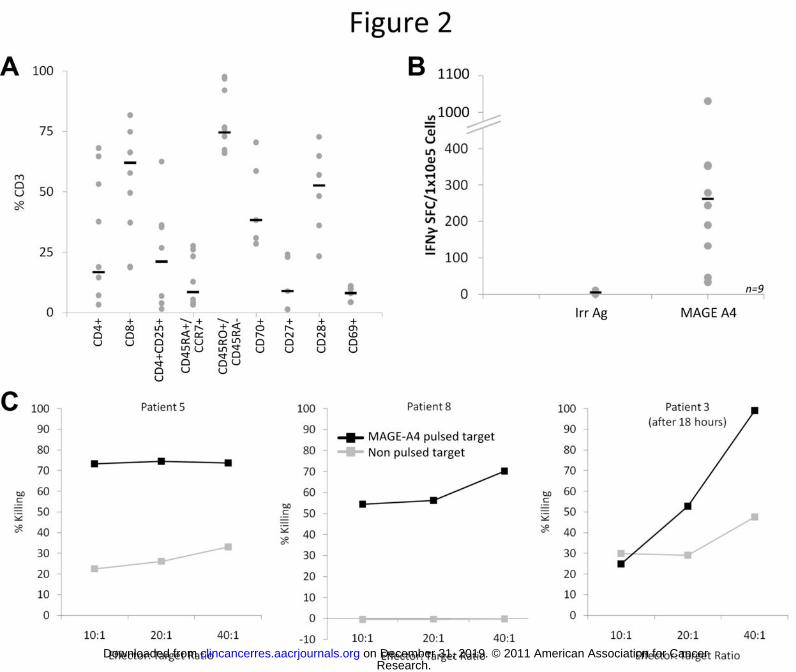
MAGE-A4 T cells can be generated from Hodgkin’s Lymphoma patients
Although relapsed HL patients typically have low blood counts, T cells specific for MAGE-A4 can be
expanded from HL patients. These cells phenotypically resembled T cell lines expanded from healthy
donors (Figure 2a). In T cells expanded from the peripheral blood of patients with HL (n=9) a median
244 SFC/1x105 cells (mean 296; range 32-1031) responded to MAGE-A4 peptides compared to a mean of
5 SFC/1x105 cells (mean 1.5; range 1-11) responding to irrelevant targets (Figure 2b). We also mapped
HL patient responses to identified 20mer epitopes and generated these epitope-specific T cells (Table 3).
Additionally, specific killing of MAGE-A4-pulsed autologous targets (but not non-pulsed autologous
targets) was seen in cytotoxicity assays (Figure 2c).
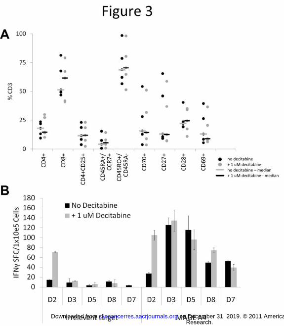
Decitabine has minimal effects on MAGE-A4 T cell phenotype and function
We studied the effects of decitabine in vitro on the MAGE-A4 specific T cells. Cell viability, measured by
trypan blue exclusion assays, showed a slight decrease in cell numbers (approximately 80%, data not
shown) following culture with decitabine. Importantly, however, treatment of the T cells with 1 μM
decitabine for 24 hours did not affect surface marker expression (Figure 3a) or IFNγ release by
expanded T cells (Figure 3b) in response to MAGE-A4 peptides.
Research. on December 31, 2019. © 2011 American Association for Cancerclincancerres.aacrjournals.org Downloaded from
Author manuscripts have been peer reviewed and accepted for publication but have not yet been edited. Author Manuscript Published OnlineFirst on September 9, 2011; DOI: 10.1158/1078-0432.CCR-11-1873

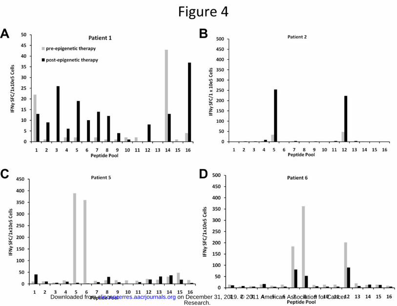
Patients with relapsed HL receiving epigenetic modifying agents have increased frequencies of MAGE-
A4 T cells
To evaluate the in vivo effects of decitabine on tumor antigen specific T cells, patient samples were
obtained before and after treatment with epigenetic-modifying drugs that included HDAC inhibitors
with or without decitabine. We hypothesized that the tumor-specific T cell immune response would
show a broader epitope specific T cell response or an increased cytokine response (signifying an increase
in the frequency of antigen-specific T cells). In patients receiving a decitabine containing regimen with a
clinical responses on PET scan, MAGE-specific T cells recognized a broader number of MAGE-A4 epitopes
(Figure 4a) and increased secretion of interferon gamma in response to MAGE-A4 antigens (Figure 4b)
after decitabine treatment. In patients not receiving a decitabine containing regimen, interferon gamma
response was decreased (Figure 4c and 4d). In other patients, no improvement in T cell reactivity to
MAGE-A4 antigen was observed (data not shown) following treatment with epigenetic modifying drugs,
suggesting that although decitabine may modulate immune function, the presence of other agents may
limit endogenous T cell responses to tumor antigens. These results support combining adoptive T cell
therapy with decitabine to upregulate CTA expression in HL relapsing after HSCT.
Research. on December 31, 2019. © 2011 American Association for Cancerclincancerres.aacrjournals.org Downloaded from
Author manuscripts have been peer reviewed and accepted for publication but have not yet been edited. Author Manuscript Published OnlineFirst on September 9, 2011; DOI: 10.1158/1078-0432.CCR-11-1873
DISCUSSION
Previous observations by our group showed that T cells targeting antigens expressed on malignant cells
in relapsed HL mediate clinical improvement (5) supporting the possibility that T cells recognizing CTAs
target a subset of cells that are more resistant to traditional cytotoxic therapies (10) . Epigenetic-
modifying drugs enhance tumor antigen expression and improve the cytotoxicity of antigen-specific T
cells (10). Based on these findings, we validate here an approach to improve the immunotherapeutic
potential of such adoptive T cell therapy by combining it with decitabine. We successfully expanded
MAGE-A4 specific CD8+ and CD4+ T cells from healthy donors and HL patients and showed that
decitabine did not restrict specificity or function of polyclonal T cell populations against MAGE-A4
targets in vitro.
In contrast to studies that have focused on single HLA -restricted epitopes to generate MAGE-A4 specific
T cells (21, 22), the peptide mixture we used elicits a broad T cell response. Using potent dendritic cells
as antigen presenting cells permitted the expansion of antigen-specific T cells from HL patients for
autologous use. We also evaluated the feasibility of combining decitabine with T cell immunotherapy for
relapsed HL and observed for the first time that tumor-specific T cells were largely unaffected by the
presence of drug.
MAGE-A4 is an attractive target antigen since it is expressed by HL Reed Sternberg cells, subject to
epigenetic regulation (7) (Supplementary Figure 1) and upregulated by DNA methyltransferase
inhibitors like decitabine (10, 23). Such MAGE-A4 upregulation by Reed Sternberg should enhance
tumor-specific T cell killing. Furthermore MAGE-A proteins may reduce malignant potential by inhibiting
the tumor suppressor p53 (9, 24). Finally, MAGE is expressed in a variety of malignancies (25-27),
Research. on December 31, 2019. © 2011 American Association for Cancerclincancerres.aacrjournals.org Downloaded from
Author manuscripts have been peer reviewed and accepted for publication but have not yet been edited. Author Manuscript Published OnlineFirst on September 9, 2011; DOI: 10.1158/1078-0432.CCR-11-1873
opening the possibility of extending this immunotherapeutic strategy to other hematological
malignancies and tumors.
The practical applicability of target antigens for immunotherapy depends on their immunogenicity and
the ability to expand cells from as many donor sources as possible. HSCT is a common setting for
adoptive transfer of either recipient or allogeneic donor T cells. Our MAGE-A4 specific T cell expansion
strategy is applicable for both donors and HL patients, even following immunosuppressive salvage high-
dose chemotherapy and autologous HSCT and despite the potential immunomodulatory effect of Reed-
Sternberg cells which produce cytokines that skew T helper cells towards a Th2 phenotype and can
mediate T cell apoptosis (1, 28, 29).
Umbilical cord blood is an emerging source of donor cells for HSCT, particularly useful for
underrepresented minorities and child recipients. Cord blood T cells have lower cytotoxic ability, as well
as greater rates of activation-induced cell death (30, 31), and the generation of MAGE-A4 T cells from
cord blood still presents a challenge for translation to the clinic. Although we successfully primed MAGE-
specific T cells from cord blood, the procedure was not robust with only 14% of the expanded CTL lines
showing specificity for MAGE-A4. This contrasts with our ability to reliably expanded virus-specific T cells
from umbilical cord blood (32, 33). The disparity may lie in the nature of the target antigen. Since
cancer/testis antigens like MAGE-A4 are expressed in the placenta (34), the constant interaction with
umbilical cord blood T cells in a highly tolerogenic environment may contribute to the lack of response
to CTA.
The percentage of tumor cells expressing the target antigen can limit the success of immunotherapy.
Since a single tumor target antigen is unlikely to be sufficient some investigators have targeting multiple
Research. on December 31, 2019. © 2011 American Association for Cancerclincancerres.aacrjournals.org Downloaded from
Author manuscripts have been peer reviewed and accepted for publication but have not yet been edited. Author Manuscript Published OnlineFirst on September 9, 2011; DOI: 10.1158/1078-0432.CCR-11-1873
tumor antigens (35). An alternative and complementary strategy is to increase expression of target
antigens in vivo using epigenetic-modifying drugs (36, 37) which upregulate CTA expression (38, 39).
Epigenetic-modifying drugs have synergistic effects on the immune system: enhanced T cell responses
occur in AML and myelodysplastic patients after treatment with decitabine (19), while a decitabine-
induced increase in Tregs may prevent GVHD without affecting the graft versus tumor effect (40).
Although the effects of demethylating agents on tumor cells have been extensively studied (11, 13),
effects of these drugs on the T cell effectors has not been explored. A murine transplant study suggests
that T cells are actively inhibited and inactivated by these agents (41) but these observations may not be
applicable to the adoptive transfer of ex vivo expanded antigen-specific T cells following epigenetic drug
treatment. Our results show that epigenetic drugs in concentrations exceeding expected treatment
levels does not adversely affect the phenotype and function of MAGE-A4 specific T cells in vitro, and
should not reduce efficacy of the adoptively transferred cells in vivo. Although we observed an inhibition
of proliferation and the abrogation of antigen specificity at continued exposures to higher >200ng/ml
(>1 μM) concentrations of decitabine, as well as cells exposed to longer term (>24 hour) culture with
decitabine, such a situation would not occur in vivo. Pharmacokinetic studies of AML and MDS patients
receiving decitabine describe maximum plasma concentrations of only 64-77 ng/mL (42). Moreover, we
plan to administer T cells after decitabine treatment, circumventing the effect of prolonged exposure to
the drug in the clinical setting.
We showed that patients with relapsed HL achieving clinical responses after decitabine also had
enhanced MAGE-specific T cell responses in vivo. However, although improved T cell responses were
seen following the administration of decitabine/HDACi regimens, this effect was not seen in patients
who received HDACi therapy alone. A variety of reasons can be postulated for this discrepancy, including
Research. on December 31, 2019. © 2011 American Association for Cancerclincancerres.aacrjournals.org Downloaded from
Author manuscripts have been peer reviewed and accepted for publication but have not yet been edited. Author Manuscript Published OnlineFirst on September 9, 2011; DOI: 10.1158/1078-0432.CCR-11-1873

the more pleiotropic effects of histone deacetylase inhibitors which affect chromatin structure and
interfere with the antigen presenting machinery (43). Further, the two classes of epigenetic modifying
drugs target different genes in T cells, with one class effectively activating T cells while another
suppresses them. The epigenetic modification of gene expression, while mediated by both DNA
methyltransferase inhibitors and histone deacetylase inhibitors, appears to have inherent gene
restrictions through mechanisms that remain incompletely understood. It is likely that targets for DNA
methyl transferase and HDAC inhibitors differ in T cells, although they mediate similar anti-tumor effects
in malignant cells. DNA methyl transferase inhibitors, for example, appear to mediate a pro-
inflammatory response by activating Th1 cells (44), while HDAC inhibitors result in opposite effects thus
limiting Th1 effector cell functions (45). Additionally, regulatory T cell (Treg) induced immune
suppression, is differentially affected by these classes of epigenetic-modifying drugs. Decitabine has
been reported to limit FOXP3 expression (46) while HDAC inhibitors enhance FOXP3+ Treg-mediated
suppressive activity (47).
One caveat to our findings is that epigenetic modification could have an impact on the autologous APC
used in their in vitro expansion. We did not address the effect of epigenetic modification on antigen
presenting cells and few studies have explored this possibility. One study (48) suggests that tumor-
infiltrating myeloid cells exposed to decitabine, differentiate into mature MHC class II-expressing
dendritic cells. Another study reports that antigen processing and presentation are unaffected by
treatment with histone deacetylase inhibitors (46). Nevertheless, to our knowledge, this is the first
demonstration of the validity of using decitabine to enhance the tumor-specific T cell immune response
in patients with relapsed HL. Results support the initiation of larger studies to correlate enhanced
tumor-specific immune responses with clinical response. Whether this effect is limited to MAGE A4 or is
also applicable to a host of other antigens (viral antigens and other tumor antigens) is still to be
Research. on December 31, 2019. © 2011 American Association for Cancerclincancerres.aacrjournals.org Downloaded from
Author manuscripts have been peer reviewed and accepted for publication but have not yet been edited. Author Manuscript Published OnlineFirst on September 9, 2011; DOI: 10.1158/1078-0432.CCR-11-1873
determined since the present study is limited by the small cell numbers we obtained from these heavily
pre treated patients.
The lack of endogenous T cells in Hodgkin’s Lymphoma patients following chemotherapy (49) highlights
the rationale for combining adoptive T cell therapy with epigenetic-modifying drugs. While we
successfully generated MAGE-A4 specific T cells from nine HL patients, no response occurred in other
patients tested (data not shown). Peripheral blood mononuclear cell and dendritic cell numbers were
reduced and T cells from these patients failed to expand after several stimulations, consistent with the
profound immune deficiency frequently seen in HL patients (49).
In conclusion our results highlight the important synergistic role that adoptively transferred tumor-
specific T cells could play with decitabine in the control of relapsed HL. Targeted immune based
treatment is a promising strategy for patients with HL to avoid the need for combination radiation and
chemotherapy which carry the risk of unacceptable long term effects (4).
Research. on December 31, 2019. © 2011 American Association for Cancerclincancerres.aacrjournals.org Downloaded from
Author manuscripts have been peer reviewed and accepted for publication but have not yet been edited. Author Manuscript Published OnlineFirst on September 9, 2011; DOI: 10.1158/1078-0432.CCR-11-1873
GRANT SUPPORT This work was supported in parts by NIH SPORE Grant P50CA126752 and a Specialized Center of
Research Award from the Leukemia Lymphoma Society. CRYC was also supported by the Bear
Necessities Pediatric Cancer Foundation and the Cancer Prevention Research Institute of Texas. UG was
supported by the Leukemia and Lymphoma Society. CMB was also supported by a career development
award from the Leukemia Lymphoma Society and awards from the Gillson Longenbaugh Foundation and
the Carl C. Anderson, Sr. and Marie Jo Anderson Charitable Foundation.
Research. on December 31, 2019. © 2011 American Association for Cancerclincancerres.aacrjournals.org Downloaded from
Author manuscripts have been peer reviewed and accepted for publication but have not yet been edited. Author Manuscript Published OnlineFirst on September 9, 2011; DOI: 10.1158/1078-0432.CCR-11-1873
REFERENCES CITED 1. Quddus F, Armitage JO. Salvage therapy for Hodgkin's lymphoma. Cancer J2009 Mar-Apr;15(2):161-3. 2. Sureda A. Autologous and allogeneic stem cell transplantation in Hodgkin's lymphoma. Hematol Oncol Clin North Am2007 Oct;21(5):943-60. 3. Castellino SM, Geiger AM, Mertens AC, Leisenring WM, Tooze JA, Goodman P, et al. Morbidity and mortality in long-term survivors of Hodgkin lymphoma: a report from the Childhood Cancer Survivor Study. Blood Feb 10;117(6):1806-16. 4. Dunleavy K, Bollard CM. Sobering realities of surviving Hodgkin lymphoma. Blood Feb 10;117(6):1772-3. 5. Bollard CM, Gottschalk S, Leen AM, Weiss H, Straathof KC, Carrum G, et al. Complete responses of relapsed lymphoma following genetic modification of tumor-antigen presenting cells and T-lymphocyte transfer. Blood2007 Oct 15;110(8):2838-45. 6. Bollard CM, Aguilar L, Straathof KC, Gahn B, Huls MH, Rousseau A, et al. Cytotoxic T lymphocyte therapy for Epstein-Barr virus+ Hodgkin's disease. J Exp Med2004 Dec 20;200(12):1623-33. 7. Simpson AJ, Caballero OL, Jungbluth A, Chen YT, Old LJ. Cancer/testis antigens, gametogenesis and cancer. Nat Rev Cancer2005 Aug;5(8):615-25. 8. Chambost H, Van Baren N, Brasseur F, Godelaine D, Xerri L, Landi SJ, et al. Expression of gene MAGE-A4 in Reed-Sternberg cells. Blood2000 Jun 1;95(11):3530-3. 9. Marcar L, Maclaine NJ, Hupp TR, Meek DW. Mage-A cancer/testis antigens inhibit p53 function by blocking its interaction with chromatin. Cancer Res Dec 15;70(24):10362-70. 10. Shafer JA, Cruz CR, Leen AM, Ku S, Lu A, Rousseau A, et al. Antigen-specific cytotoxic T lymphocytes can target chemoresistant side-population tumor cells in Hodgkin lymphoma. Leuk Lymphoma May;51(5):870-80. 11. Aribi A, Borthakur G, Ravandi F, Shan J, Davisson J, Cortes J, et al. Activity of decitabine, a hypomethylating agent, in chronic myelomonocytic leukemia. Cancer2007 Feb 15;109(4):713-7. 12. Fang F, Balch C, Schilder J, Breen T, Zhang S, Shen C, et al. A phase 1 and pharmacodynamic study of decitabine in combination with carboplatin in patients with recurrent, platinum-resistant, epithelial ovarian cancer. Cancer Sep 1;116(17):4043-53. 13. George RE, Lahti JM, Adamson PC, Zhu K, Finkelstein D, Ingle AM, et al. Phase I study of decitabine with doxorubicin and cyclophosphamide in children with neuroblastoma and other solid tumors: a Children's Oncology Group study. Pediatr Blood Cancer Oct;55(4):629-38. 14. Piekarz RL, Frye R, Turner M, Wright JJ, Allen SL, Kirschbaum MH, et al. Phase II multi-institutional trial of the histone deacetylase inhibitor romidepsin as monotherapy for patients with cutaneous T-cell lymphoma. J Clin Oncol2009 Nov 10;27(32):5410-7. 15. Mackay HJ, Hirte H, Colgan T, Covens A, MacAlpine K, Grenci P, et al. Phase II trial of the histone deacetylase inhibitor belinostat in women with platinum resistant epithelial ovarian cancer and micropapillary (LMP) ovarian tumours. Eur J Cancer Jun;46(9):1573-9. 16. Sigalotti L, Fratta E, Coral S, Cortini E, Covre A, Nicolay HJ, et al. Epigenetic drugs as pleiotropic agents in cancer treatment: biomolecular aspects and clinical applications. J Cell Physiol2007 Aug;212(2):330-44. 17. Maio M, Coral S, Fratta E, Altomonte M, Sigalotti L. Epigenetic targets for immune intervention in human malignancies. Oncogene2003 Sep 29;22(42):6484-8. 18. Moreno-Bost A, Szmania S, Stone K, Garg T, Hoerring A, Szymonifka J, et al. Epigenetic modulation of MAGE-A3 antigen expression in multiple myeloma following treatment with the demethylation agent 5-azacitidine and the histone deacetlyase inhibitor MGCD0103. Cytotherapy May;13(5):618-28.
Research. on December 31, 2019. © 2011 American Association for Cancerclincancerres.aacrjournals.org Downloaded from
Author manuscripts have been peer reviewed and accepted for publication but have not yet been edited. Author Manuscript Published OnlineFirst on September 9, 2011; DOI: 10.1158/1078-0432.CCR-11-1873
19. Goodyear O, Agathanggelou A, Novitzky-Basso I, Siddique S, McSkeane T, Ryan G, et al. Induction of a CD8+ T-cell response to the MAGE cancer testis antigen by combined treatment with azacitidine and sodium valproate in patients with acute myeloid leukemia and myelodysplasia. Blood Sep 16;116(11):1908-18. 20. Einsele H, Rauser G, Grigoleit U, Hebart H, Sinzger C, Riegler S, et al. Induction of CMV-specific T-cell lines using Ag-presenting cells pulsed with CMV protein or peptide. Cytotherapy2002;4(1):49-54. 21. Jia ZC, Ni B, Huang ZM, Tian Y, Tang J, Wang JX, et al. Identification of two novel HLA-A*0201-restricted CTL epitopes derived from MAGE-A4. Clin Dev Immunol;2010:567594. 22. Graff-Dubois S, Faure O, Gross DA, Alves P, Scardino A, Chouaib S, et al. Generation of CTL recognizing an HLA-A*0201-restricted epitope shared by MAGE-A1, -A2, -A3, -A4, -A6, -A10, and -A12 tumor antigens: implication in a broad-spectrum tumor immunotherapy. J Immunol2002 Jul 1;169(1):575-80. 23. Adair SJ, Hogan KT. Treatment of ovarian cancer cell lines with 5-aza-2'-deoxycytidine upregulates the expression of cancer-testis antigens and class I major histocompatibility complex-encoded molecules. Cancer Immunol Immunother2009 Apr;58(4):589-601. 24. Monte M, Simonatto M, Peche LY, Bublik DR, Gobessi S, Pierotti MA, et al. MAGE-A tumor antigens target p53 transactivation function through histone deacetylase recruitment and confer resistance to chemotherapeutic agents. Proc Natl Acad Sci U S A2006 Jul 25;103(30):11160-5. 25. Hussein YM, Gharib AF, Etewa RL, El-Shal AS, Abdel-Ghany ME, Elsawy WH. The melanoma-associated antigen-A3, -A4 genes: relation to the risk and clinicopathological parameters in breast cancer patients. Mol Cell Biochem May;351(1-2):261-8. 26. Cuffel C, Rivals JP, Zaugg Y, Salvi S, Seelentag W, Speiser DE, et al. Pattern and clinical significance of cancer-testis gene expression in head and neck squamous cell carcinoma. Int J Cancer Jun 1;128(11):2625-34. 27. Svobodova S, Browning J, MacGregor D, Pollara G, Scolyer RA, Murali R, et al. Cancer-testis antigen expression in primary cutaneous melanoma has independent prognostic value comparable to that of Breslow thickness, ulceration and mitotic rate. Eur J Cancer Feb;47(3):460-9. 28. Poppema S. Immunobiology and pathophysiology of Hodgkin lymphomas. Hematology Am Soc Hematol Educ Program2005:231-8. 29. Steingrimsdottir H, Gruber A, Bjorkholm M, Svensson A, Hansson M. Immune reconstitution after autologous hematopoietic stem cell transplantation in relation to underlying disease, type of high-dose therapy and infectious complications. Haematologica2000 Aug;85(8):832-8. 30. Canto E, Rodriguez-Sanchez JL, Vidal S. Distinctive response of naive lymphocytes from cord blood to primary activation via TCR. J Leukoc Biol2003 Dec;74(6):998-1007. 31. Chao NJ, Emerson SG, Weinberg KI. Stem cell transplantation (cord blood transplants). Hematology Am Soc Hematol Educ Program2004:354-71. 32. Hanley PJ, Cruz CR, Savoldo B, Leen AM, Stanojevic M, Khalil M, et al. Functionally active virus-specific T cells that target CMV, adenovirus, and EBV can be expanded from naive T-cell populations in cord blood and will target a range of viral epitopes. Blood2009 Aug 27;114(9):1958-67. 33. Micklethwaite KP, Savoldo B, Hanley PJ, Leen AM, Demmler-Harrison GJ, Cooper LJ, et al. Derivation of human T lymphocytes from cord blood and peripheral blood with antiviral and antileukemic specificity from a single culture as protection against infection and relapse after stem cell transplantation. Blood Apr 1;115(13):2695-703. 34. Fratta E, Coral S, Covre A, Parisi G, Colizzi F, Danielli R, et al. The biology of cancer testis antigens: putative function, regulation and therapeutic potential. Mol Oncol Apr;5(2):164-82. 35. Gerdemann U, Tripic T, Cruz CR, Vera J, Christin AS, Heslop HE, et al. Multi-tumor-antigen specific cytotoxic T lymphocytes for therapy of lymphoma. Accepted for publication at Molecular Therapy ed2011.
Research. on December 31, 2019. © 2011 American Association for Cancerclincancerres.aacrjournals.org Downloaded from
Author manuscripts have been peer reviewed and accepted for publication but have not yet been edited. Author Manuscript Published OnlineFirst on September 9, 2011; DOI: 10.1158/1078-0432.CCR-11-1873
36. Svane IM, Boesen M, Engel AM. The role of cytotoxic T-lymphocytes in the prevention and immune surveillance of tumors--lessons from normal and immunodeficient mice. Med Oncol1999 Dec;16(4):223-38. 37. Baron F, Storb R. The immune system as a foundation for immunologic therapy and hematologic malignancies: a historical perspective. Best Pract Res Clin Haematol2006;19(4):637-53. 38. Almstedt M, Blagitko-Dorfs N, Duque-Afonso J, Karbach J, Pfeifer D, Jager E, et al. The DNA demethylating agent 5-aza-2'-deoxycytidine induces expression of NY-ESO-1 and other cancer/testis antigens in myeloid leukemia cells. Leuk Res Jul;34(7):899-905. 39. Hambach L, Ling KW, Pool J, Aghai Z, Blokland E, Tanke HJ, et al. Hypomethylating drugs convert HA-1-negative solid tumors into targets for stem cell-based immunotherapy. Blood2009 Mar 19;113(12):2715-22. 40. Choi J, Ritchey J, Prior JL, Holt M, Shannon WD, Deych E, et al. In vivo administration of hypomethylating agents mitigate graft-versus-host disease without sacrificing graft-versus-leukemia. Blood Jul 8;116(1):129-39. 41. Sanchez-Abarca LI, Gutierrez-Cosio S, Santamaria C, Caballero-Velazquez T, Blanco B, Herrero-Sanchez C, et al. Immunomodulatory effect of 5-azacytidine (5-azaC): potential role in the transplantation setting. Blood Jan 7;115(1):107-21. 42. Cashen AF, Shah AK, Todt L, Fisher N, DiPersio J. Pharmacokinetics of decitabine administered as a 3-h infusion to patients with acute myeloid leukemia (AML) or myelodysplastic syndrome (MDS). Cancer Chemother Pharmacol2008 Apr;61(5):759-66. 43. Song W, Tai YT, Tian Z, Hideshima T, Chauhan D, Nanjappa P, et al. HDAC inhibition by LBH589 affects the phenotype and function of human myeloid dendritic cells. Leukemia Jan;25(1):161-8. 44. Quddus J, Johnson KJ, Gavalchin J, Amento EP, Chrisp CE, Yung RL, et al. Treating activated CD4+ T cells with either of two distinct DNA methyltransferase inhibitors, 5-azacytidine or procainamide, is sufficient to cause a lupus-like disease in syngeneic mice. J Clin Invest1993 Jul;92(1):38-53. 45. Brogdon JL, Xu Y, Szabo SJ, An S, Buxton F, Cohen D, et al. Histone deacetylase activities are required for innate immune cell control of Th1 but not Th2 effector cell function. Blood2007 Feb 1;109(3):1123-30. 46. Nagar M, Vernitsky H, Cohen Y, Dominissini D, Berkun Y, Rechavi G, et al. Epigenetic inheritance of DNA methylation limits activation-induced expression of FOXP3 in conventional human CD25-CD4+ T cells. Int Immunol2008 Aug;20(8):1041-55. 47. Tao R, de Zoeten EF, Ozkaynak E, Chen C, Wang L, Porrett PM, et al. Deacetylase inhibition promotes the generation and function of regulatory T cells. Nat Med2007 Nov;13(11):1299-307. 48. Daurkin I, Eruslanov E, Vieweg J, Kusmartsev S. Generation of antigen-presenting cells from tumor-infiltrated CD11b myeloid cells with DNA demethylating agent 5-aza-2'-deoxycytidine. Cancer Immunol Immunother May;59(5):697-706. 49. Kennedy-Nasser AA, Hanley P, Bollard CM. Hodgkin disease and the role of the immune system. Pediatr Hematol Oncol Apr;28(3):176-86.
Research. on December 31, 2019. © 2011 American Association for Cancerclincancerres.aacrjournals.org Downloaded from
Author manuscripts have been peer reviewed and accepted for publication but have not yet been edited. Author Manuscript Published OnlineFirst on September 9, 2011; DOI: 10.1158/1078-0432.CCR-11-1873
LIST OF TABLES AND FIGURES
TABLE 1. Cytolytic ability of 8 other donors is shown by their ability to lyse MAGE-A4-pulsed autologous
targets. Specific lysis was evaluated by standard Cr51 release assay, shown is the E:T ratio at 20:1.
TABLE 2. Responses of healthy donor-derived T cells to MAGE-A4 were mapped to 20mer regions of the
protein. Listed are the donors with their corresponding HLA types and 20mer regions recognized.
TABLE 3. Responses of Hodgkin’s lymphoma patient-derived T cells to MAGE-A4 were mapped to 20mer
regions of the protein.
FIGURE 1. Generation of MAGE-A4 specific T cells from healthy donors. A). Expansion of MAGE specific
T cells after 2 stimulations with MAGE pulsed antigen presenting cells. Fold change was calculated from
weekly cell counts using the trypan blue exclusion method. B). Phenotype of CD3+ cells after at least 2
stimulations was determined using flow cytometry of surface receptors listed on the x-axis. The median
is shown in black bars. C). Shows the specificity of MAGE-specific CTL lines generated from 15 donors as
measured by IFNγ release (IFNγELIspot) in response to stimulation with MAGE-A4 pepmix. Results are
expressed as SFC/1x105 input cells. Control was IFNγ release in response to stimulation with irrelevant
pepmix. The black bars show the median interferon gamma secretion. CTL lines were able to kill MAGE-
loaded autologous PHA blasts. D). and HLA A2 matched MAGE positive HL cell line L1236. E). Specific
lysis was evaluated by standard Cr51 release assay - E:T ratio of 40:1, 20:1, 10:1 and 5:1. F). Further
epitope mapping of the NPARYEFLWGPRALAETSYV 20mer commonly seen in A2+ donors show that 3
donors each recognize a unique and distinct 9mer epitopes as shown using IFNγ ELISPOT assay. Results
are expressed as SFC/1x105 input cells +/- SD.
Research. on December 31, 2019. © 2011 American Association for Cancerclincancerres.aacrjournals.org Downloaded from
Author manuscripts have been peer reviewed and accepted for publication but have not yet been edited. Author Manuscript Published OnlineFirst on September 9, 2011; DOI: 10.1158/1078-0432.CCR-11-1873
FIGURE 2: Generation of MAGE-A4 T cells from patients with Hodgkin’s lymphoma. A). Phenotype
analysis of cells obtained using flow cytometry are shown and are similar to the phenotype of CTL
derived from healthy donors. Black bars show the median percentage for each marker. B). Specificity of
the CTL was evaluated by IFNΓ ELIspot assay. CTL responses to MAGE-A4 pepmix versus irrelevant
pepmix are shown. Data from nine HL patients is shown. IFNγ positive Spot Forming Cells (SFC)/1x105
input cells are shown for each CTL line. The black bars show the median interferon gamma secretion for
each group. C). Cytolytic activity of evaluable T cell lines generated from Hodgkin’s lymphoma patients is
evaluated using autologous PHA blasts pulsed with the MAGE-A4 peptide mix as targets in a chromium
release assay.
FIGURE 3: MAGE specific T cell phenotype and function are unaffected by Decitabine. A). Phenotype of
the MAGE-specific T cells pre and post treatment with 5’aza-2’deoxycytidine is shown. B). T cell
secretion of interferon-gamma in response to MAGE-A4 peptides is shown pre and post decitabine
treatment. Black bars indicate the IFNγ response in CTL lines incubated with decitabine (gray bars)
versus without decitabine (black bars).
FIGURE 4: Increased frequency of MAGE-A4 specific T cells in vivo in patients receiving a regimen
containing decitabine. T cells obtained from the peripheral blood of HL patients taken before and after
treatment with an HDAC inhibitor with or without decitabine were expanded in vitro, and MAGE specific
responses were evaluated in an IFNγ ELISpot assay. Results are shown for patients who received
decitabine containing regimens (A and B) versus those who did not (C and D).
Research. on December 31, 2019. © 2011 American Association for Cancerclincancerres.aacrjournals.org Downloaded from
Author manuscripts have been peer reviewed and accepted for publication but have not yet been edited. Author Manuscript Published OnlineFirst on September 9, 2011; DOI: 10.1158/1078-0432.CCR-11-1873
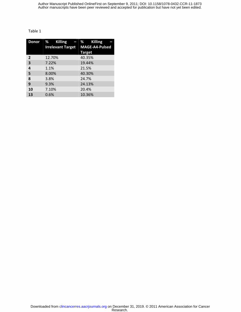
Table 1
Donor % Killing – Irrelevant Target
% Killing – MAGE-A4-Pulsed Target
2 12.70% 40.35% 3 7.22% 19.44% 4 1.1% 21.5% 5 8.00% 40.30% 8 3.8% 24.7% 9 9.3% 24.13% 10 7.10% 20.4% 13 0.6% 10.36%
Research. on December 31, 2019. © 2011 American Association for Cancerclincancerres.aacrjournals.org Downloaded from
Author manuscripts have been peer reviewed and accepted for publication but have not yet been edited. Author Manuscript Published OnlineFirst on September 9, 2011; DOI: 10.1158/1078-0432.CCR-11-1873

Table 2
Donor HLA Type Peptide Recognized
1 A2, A24, B8, B65 SASEEEIWEELGVMGVYDGR
2 A2, A26, B15, B44 NPARYEFLWGPRALAETSYV
3 A2, B7, B40 NPARYEFLWGPRALAETSYV
4 A3, A24, B40, B44 NPARYEFLWGPRALAETSYV
5 A1, A11, B8, B49 ETSYVKVLEHVVRVNARVRI
KVLEHVVRVNARVRIAYPSL
6 A2, B7, B40 NPARYEFLWGPRALAETSYV
7 A2, B60, B61 NPARYEFLWGPRALAETSYV
8 A3, A24, B8, B35 KVLEHVVRVNARVRIAYPSL
9 A1, A3, B8, B35 AYPSLAYPSLREAALLEEEE
11 --- IFPKTGLLIIVLGTIAMEGD
12 --- ELAHFLLRKYRAKELVTKAE
13 --- LEEVPAAESAGPPQSPQGAS
14 --- SNKVDELAHFLLRKYRAKEL
ELAHFLLRKYRAKELVTKAE
LLRKYRAKELVTKAEMLERV
RAKELVTKAEMLERVIKNYK
15 A3, A33, B14, B38 PRALAETSYVKVLEHVVRVN
Research. on December 31, 2019. © 2011 American Association for Cancerclincancerres.aacrjournals.org Downloaded from
Author manuscripts have been peer reviewed and accepted for publication but have not yet been edited. Author Manuscript Published OnlineFirst on September 9, 2011; DOI: 10.1158/1078-0432.CCR-11-1873
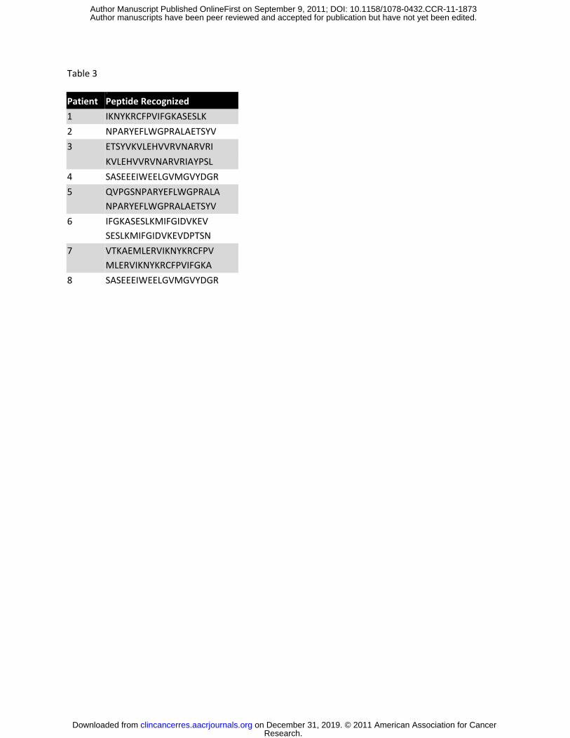
Table 3
Patient Peptide Recognized
1 IKNYKRCFPVIFGKASESLK
2 NPARYEFLWGPRALAETSYV
3 ETSYVKVLEHVVRVNARVRI
KVLEHVVRVNARVRIAYPSL
4 SASEEEIWEELGVMGVYDGR
5 QVPGSNPARYEFLWGPRALA NPARYEFLWGPRALAETSYV
6 IFGKASESLKMIFGIDVKEV SESLKMIFGIDVKEVDPTSN
7 VTKAEMLERVIKNYKRCFPV MLERVIKNYKRCFPVIFGKA
8 SASEEEIWEELGVMGVYDGR
Research. on December 31, 2019. © 2011 American Association for Cancerclincancerres.aacrjournals.org Downloaded from
Author manuscripts have been peer reviewed and accepted for publication but have not yet been edited. Author Manuscript Published OnlineFirst on September 9, 2011; DOI: 10.1158/1078-0432.CCR-11-1873

Published OnlineFirst September 9, 2011.Clin Cancer Res Conrad Russell Y Cruz, Ulrike Gerdemann, Ann M Leen, et al. Lymphoma by targeting upregulated MAGE-A4Improving T cell therapy for relapsed EBV negative Hodgkins
Updated version
10.1158/1078-0432.CCR-11-1873doi:
Access the most recent version of this article at:
Material
Supplementary
http://clincancerres.aacrjournals.org/content/suppl/2011/09/09/1078-0432.CCR-11-1873.DC1
Access the most recent supplemental material at:
Manuscript
Authoredited. Author manuscripts have been peer reviewed and accepted for publication but have not yet been
E-mail alerts related to this article or journal.Sign up to receive free email-alerts
Subscriptions
Reprints and
To order reprints of this article or to subscribe to the journal, contact the AACR Publications
Permissions
Rightslink site. Click on "Request Permissions" which will take you to the Copyright Clearance Center's (CCC)
.http://clincancerres.aacrjournals.org/content/early/2011/09/09/1078-0432.CCR-11-1873To request permission to re-use all or part of this article, use this link
Research. on December 31, 2019. © 2011 American Association for Cancerclincancerres.aacrjournals.org Downloaded from
Author manuscripts have been peer reviewed and accepted for publication but have not yet been edited. Author Manuscript Published OnlineFirst on September 9, 2011; DOI: 10.1158/1078-0432.CCR-11-1873