Embed Size (px)
Citation preview

EBV infection B cells and lymphomagenesis
Sridhar Chaganti

• How EBV infects B-cells
• How viral genes influence the infected B cell
• Differences and similarities between in vitro and in vivo infection
• How the virus exploits various stages of B-cell ontogeny to establish a persistent latent infection following primary infection
• How the virus may contribute to development of disease

• EBV: Human � herpes virus
• Humans are the only natural host for infection
• B-lymphotropic
• Establishes life long latency in B cells
• >90% of adults infected
• Most infected people remain healthy

EBV associated disease
• Non-malignant:– Infectious mononucleosis and its complications– Virus associated haemophagocytic syndrome– Immune haemolytic anaemias– Autoimmune phenomena
• Malignant:– Burkitt’s lymphoma– Hodgkin’s disease– LPD in the immunocompromised (eg PTLD, AIDS lymphomas)– Rare cases of T-cell and NK-cell lymphomas– Nasopharyngeal carcinoma– Certain cases of Gastric carcinoma

• In vitro infection:
– EBV causes activation and transformation of the infected B cellsinto lymphoblastoid cells
– Transformed cells proliferate indefinitely
– Establish permanently growing lymphoblastoid cell lines (LCLs); may be polyconal, oligoclonal or monoclonal
– Latent infection: EBV expressing all 9 latent genes (6 EBNAs and 3 LMPs): latency III or growth program
In vitro vs in vivo infection

Human B-cells: 7 days post infection with EBV in vitro
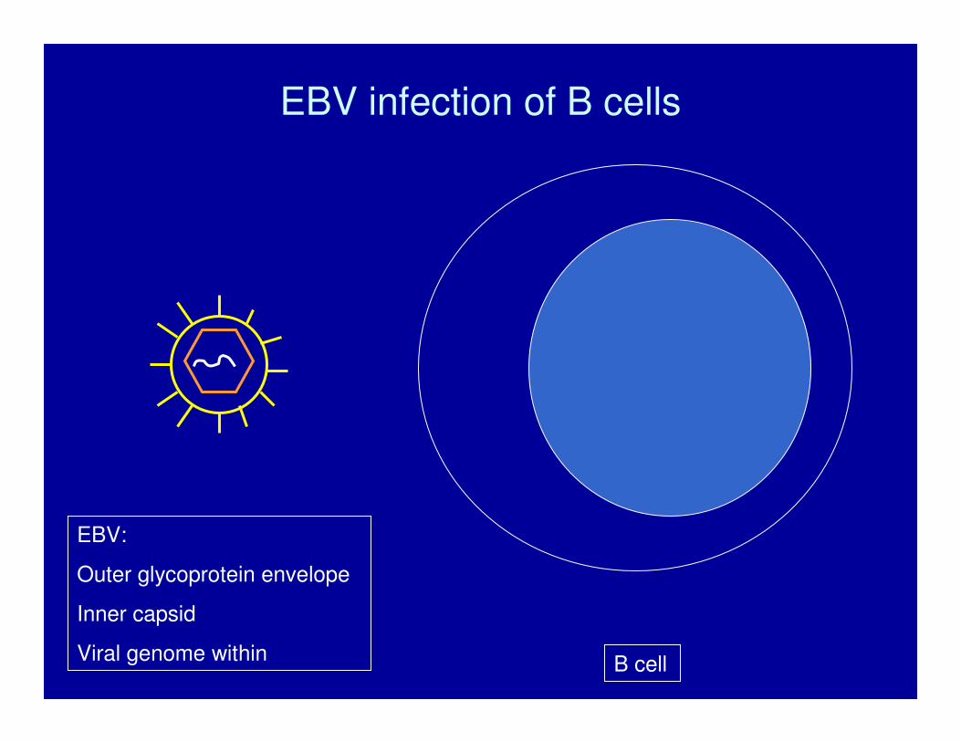
EBV infection of B cells
EBV:
Outer glycoprotein envelope
Inner capsid
Viral genome within B cell

EBV infection of B cells
EBV binds to B cells through CD21 (CR2 receptor) and HLA class-II molecule
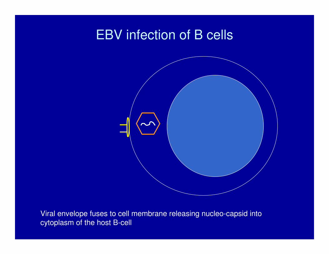
EBV infection of B cells
Viral envelope fuses to cell membrane releasing nucleo-capsid into cytoplasm of the host B-cell

EBV infection of B cells
Capsid fuses to nuclear membrane releasing viral genome into the nucleus of the host B-cell

EBV infection of B cells
Viral genome does not integrate into host B cell DNA but instead circularises to exist in an episomal form

EBV genome: representative diagram of the episomal form
EBV genome has several genes encoding the various viral proteins and antigens

EBV genome: linear form showing latent genes
Genes encoding nuclear antigens: EBNA1, 2, 3A, 3B, 3C, LP
Genes encoding membrane proteins: LMP1, LMP2A, LMP2B
Genes encoding EBERs
Latent genes
LP 2 3A 3B 3C 1 1 2A 2B

EBV genome: linear form showing latent genes and their promoters
Wp/Cp LMPp
Genes encoding nuclear antigens: EBNA1, 2, 3A, 3B, 3C, LP
Genes encoding membrane proteins: LMP1, LMP2A, LMP2B
Genes encoding EBERs
Latent genes
LP 2 3A 3B 3C 1 1 2A 2B

EBV genome: linear form showing latent genes and their promoters
Wp/Cp Qp LMPp
Genes encoding nuclear antigens: EBNA1, 2, 3A, 3B, 3C, LP
Genes encoding membrane proteins: LMP1, LMP2A, LMP2B
Genes encoding EBERs
Lytic genes; and their promoters are spread across the genome
Latent genes
LP 2 3A 3B 3C 1 1 2A 2B

EBV genome: linear form showing latent genes and their promoters
Wp/Cp Qp LMPp
B cell specific transcription factors bind to the promoter regions of the viral genome to initiate transcription of the down stream viral genes
LP 2 3A 3B 3C 1 1 2A 2B
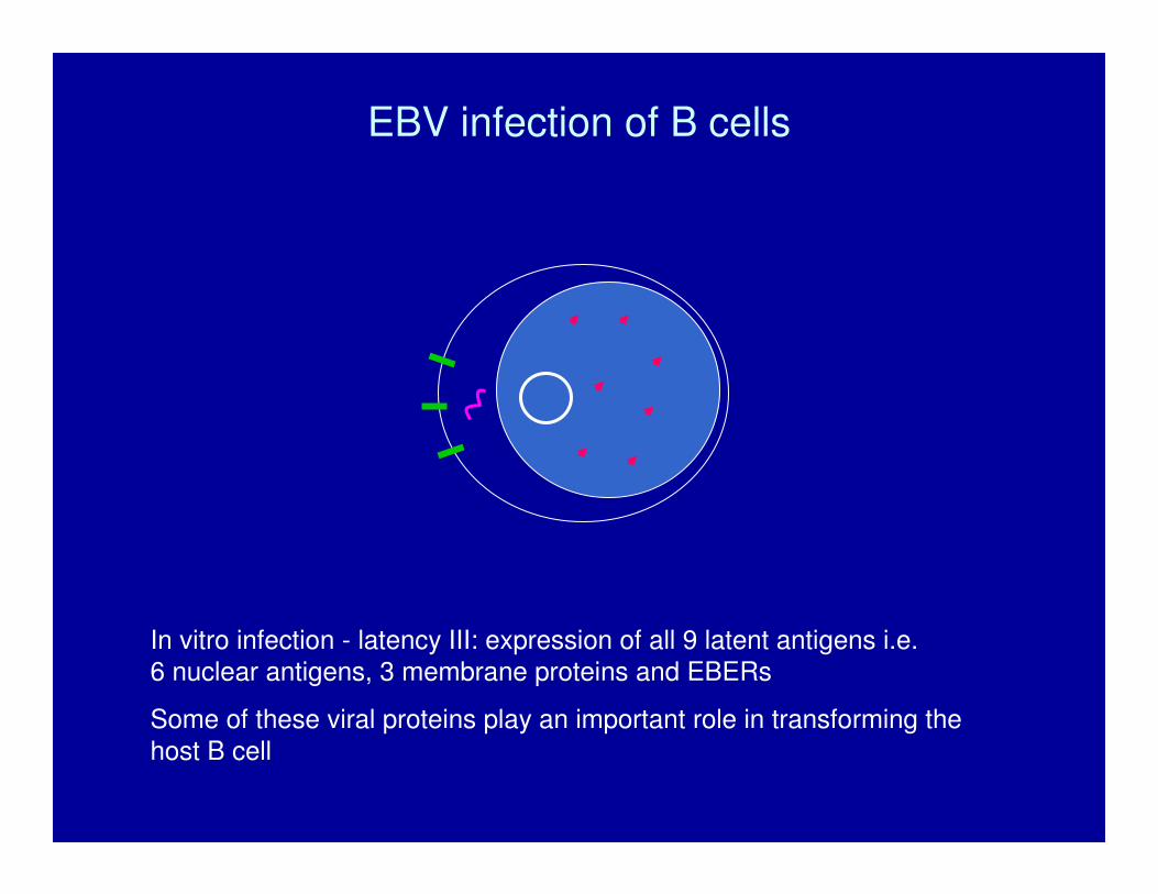
EBV infection of B cells
In vitro infection - latency III: expression of all 9 latent antigens i.e. 6 nuclear antigens, 3 membrane proteins and EBERs
Some of these viral proteins play an important role in transforming the host B cell

Functions of EBV latent antigens/proteins
• EBNA1: maintains viral genome when host cell divides
• EBNA2: – Important for transformation of B-cells– Initiates transcription of LMPs and increases transcription of
various cellular genes– Mimics the intracellular notch signalling pathway
• EBNA3 and LP: regulate EBNA2 functions

Functions of EBV latent antigens/proteins
• LMP1: – Main transforming protein of EBV– mimics constitutively activated CD40 receptor– Increases NF-kB activity
• LMP2A and 2B: – not essential for transformation– LMP2A mimics B-cell receptor and can rescue cells lacking a
functional BCR from apoptosis

EBV infection of B cells: latent infection
In latent infection, lytic genes are not expressed and there is no active viral replication
The virus divides with each cell division to maintain its copy number

EBV infection of B cells: lytic infection
During the phase of lytic infection:
EBV lytic genes are expressed: eg BZLF1, BHRF1, thymidine kinase and genes encoding capsid and envelop proteins
Leads to active genome replication
New virions are produced
Host cell lysis occurs

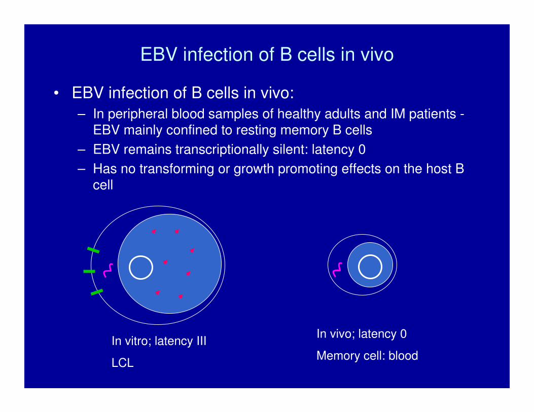
EBV infection of B cells in vivo
• EBV infection of B cells in peripheral blood:
– In peripheral blood samples of healthy adults and IM patients -EBV mainly confined to resting memory B cells
– EBV remains transcriptionally silent: latency 0
– Has no transforming or growth promoting effects on the host B cell
EBV infection of B cells in vivo
• EBV infection of B cells in vivo: – In peripheral blood samples of healthy adults and IM patients -
EBV mainly confined to resting memory B cells – EBV remains transcriptionally silent: latency 0– Has no transforming or growth promoting effects on the host B
cell
In vitro; latency III
LCL
In vivo; latency 0
Memory cell: blood
EBV infection of B cells
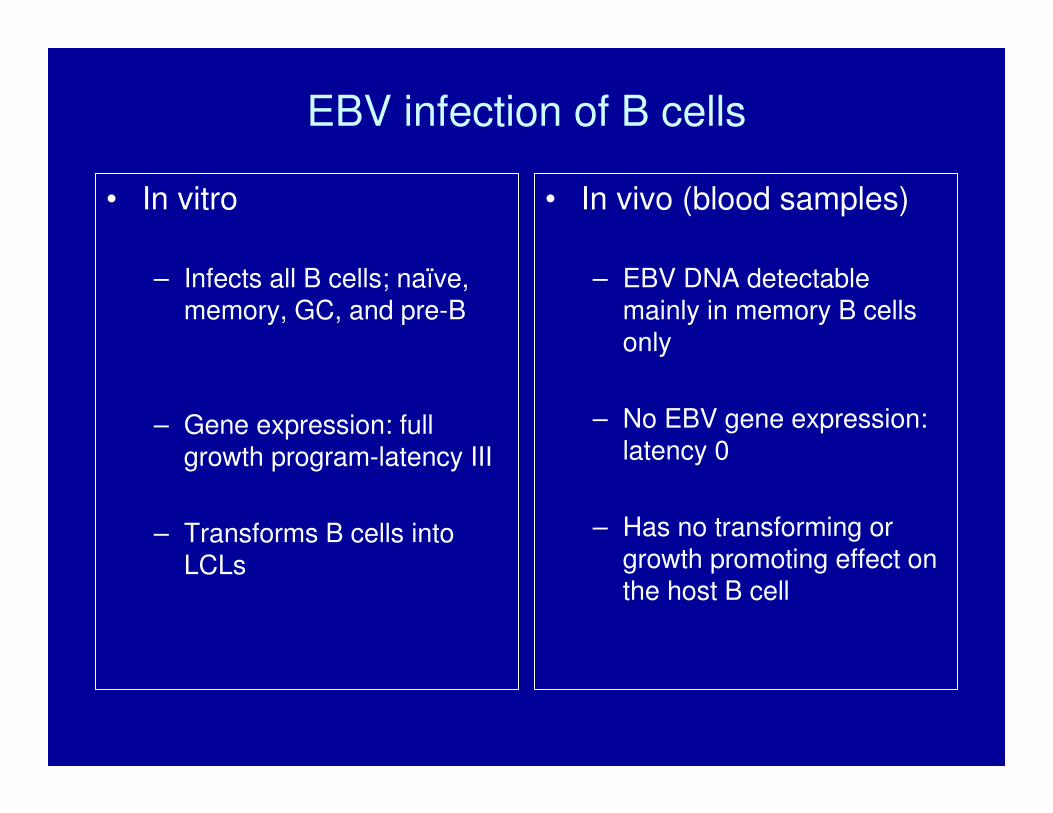
• In vitro
– Infects all B cells; naïve, memory, GC, and pre-B
– Gene expression: full growth program-latency III
– Transforms B cells into LCLs
• In vivo (blood samples)
– EBV DNA detectable mainly in memory B cells only
– No EBV gene expression: latency 0
– Has no transforming or growth promoting effect on the host B cell

EBV infection of B-cells in vivo
• However, EBV can infect other B cells in vivo, and express full growth program (latency III) or more restricted pattern of latent antigen expression (latency I or II)
– Evidence from study of EBV associated lymphomas and
– Limited evidence from studies on tonsils

EBV-associated tumours of B cell origin
Tumour Latency Cell of origin
Burkitt’s I: EBNA1, EBERs germinal centre B cellLymphoma
Hodgkin’s post-GC ‘crippled’ B cellDisease
PTLD III: EBNA1,2,3A,3B,3C,LP, LMP1, 2A, 2B
Naive, memory, or crippled ‘post-GC’ B cell
II: EBNA1, LMP1, LMP2A
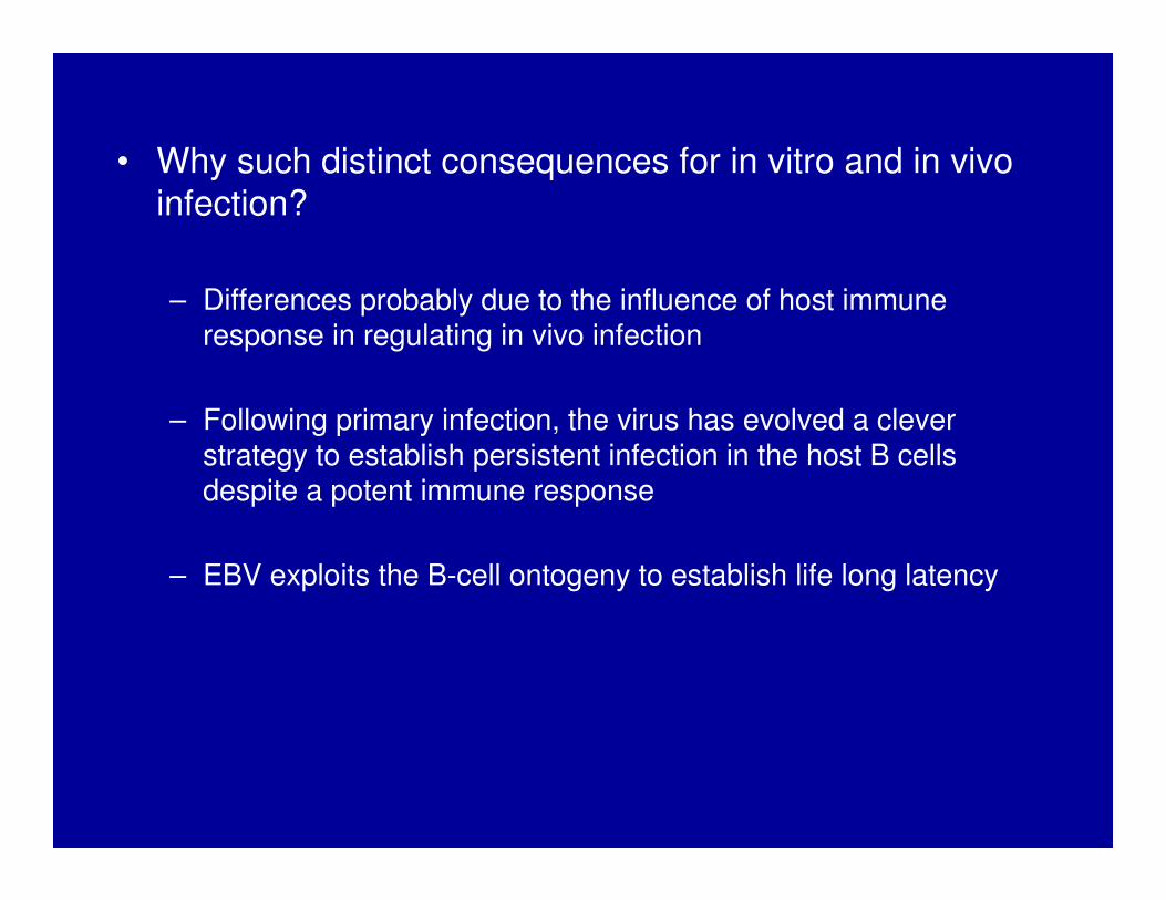
• Why such distinct consequences for in vitro and in vivo infection?
– Differences probably due to the influence of host immune response in regulating in vivo infection
– Following primary infection, the virus has evolved a clever strategy to establish persistent infection in the host B cells despite a potent immune response
– EBV exploits the B-cell ontogeny to establish life long latency

B cell differentiation in response to antigen
Source: R Kuppers; Nature reviews immunology; 2003
Naïve B cell sees antigen through it’s sIg (BCR) and proliferates to form GC
Antigen selected post-GC cells receive survival signals through CD40 ligand of T cells
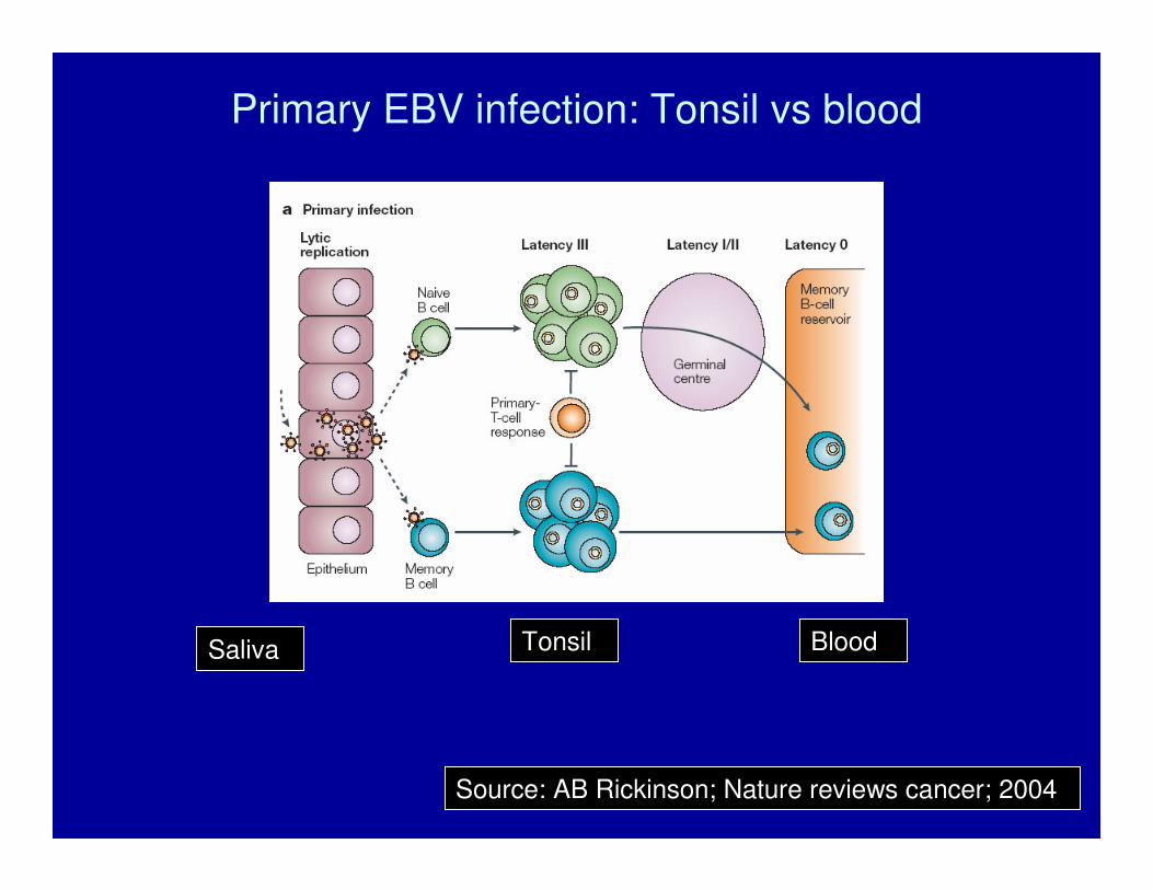
Source: AB Rickinson; Nature reviews cancer; 2004
Saliva Tonsil Blood
Primary EBV infection: Tonsil vs blood

Primary EBV infection: Tonsil vs blood
• Within tonsil, EBV probably can infect any B cell i.e. naïve, memory, or GC
• It probably expresses a full growth program
• Causes transformation and proliferation of infected cells
• Potent CD8 T-cell responses directed against EBNA3 antigens and lytic antigens: eliminates all proliferating lymphoblastoid cells
• Infected naïve cells may escape CD8 T-cell response by initiating a germinal centre ‘like’ reaction
• EBV down regulates it’s gene expression during this phase and assumes latency 0 when the cell emerges as a memory B cell

Source: DA Thorley-Lawson;NEJM;2004
EBV exploits B cell ontogeny to persist in memory B cells
EBNA2 activates the naïve EBV infected B cell and induces LMP2A and LMP1
LMP2A expression mimics constitutive signalling through the sIg (BCR)
EBV infected naïve B cell therefore undergoes a GC reaction
LMP1 expression mimics constitutively active CD40 receptor signalling

Source: DA Thorley-Lawson;NEJM;2004
Persistent EBV infection
Memory cells constantly re-circulate between blood and tonsils
Can proliferate and undergo terminal differentiation in tonsils
Terminal differentiation to plasma cells can induce lytic EBV replication

Clinical consequences of EBV infection
• Primary infection usually occurs in children or young adults
• May go undiagnosed or may present as infectious mononucleosis
• Self contained illness in the immunocompetent host leading to establishment of persistent B cell infection
• Complications are uncommon but may arise due to dysregulated immune response
• Persistent infection of no clinical consequence in most people
• EBV infection associated with malignancies in certain situations

EBV-associated tumours of B cell origin
Tumour Sub-type EBV assocn
Burkitt’s endemicEBNA1
Lymphoma sporadic Lat I
Hodgkin’s mixed cell/lymph.depl. EBNA1, LMP1, LMP2Disease
Lat II
Lymphomas of post-transplantimmunosuppressed
EBV Ag expression Latency
100%15-20%30-40%AIDS-assoc.
AIDS-assoc.>80%~70%
EBNAs 1, 2, 3A, 3B, 3C, LP Lat III
nodular sclerosinglymph. predominant
80%30%
nil
& LMP1, LMP2
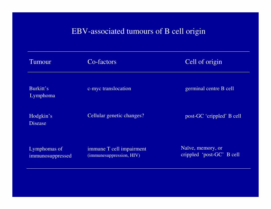
EBV-associated tumours of B cell origin
Tumour Co-factors Cell of origin
Burkitt’s c-myc translocation germinal centre B cellLymphoma
Hodgkin’s post-GC ‘crippled’ B cellDisease
Lymphomas of immune T cell impairment Naïve, memory, or crippled ‘post-GC’ B cellimmunosuppressed
Cellular genetic changes?
(immunesuppression, HIV)

Source: AB Rickinson; Nature reviews cancer; 2004
Saliva Tonsil Blood
Primary EBV infection: Tonsil vs blood
Uncontrolled proliferation of infected B cells in the immuno-compromised host can lead to LPD

B cell differentiation in response to antigen
Source: R Kuppers; Nature reviews immunology; 2003
EBV infection of the pre-apoptotic GC B cell may rescue it from apoptotic death by expression of LMP1 and LMP2

Role of EBV in B-cell lymphomas
• PTLD: – causative role – EBV usually expresses full growth program
• Burkitt and Hodgkin’s dis: – ? EBV innocent passenger in tumour cells– ? EBV plays a role in initial transformation– ? Tumour formation due to other cellular changes – ? On-going role or EBV in sustenance of the tumour

PTLD• Early onset tumours: <1 year post-transplant
– Polyclonal B cell proliferations or monoclonal tumours– May arise from naïve, memory or GC cells– Almost always EBV+, latency III
• Late onset tumours: >1 year post transplant– Mostly monoclonal– Most seem to arise from crippled GC cells– Only about 50% EBV+, usually latency III but variants seen
• Early tumours may respond to withdrawal of immune suppression
• Adoptive immunetherapy with EBV specific CTLs is possible in the setting of allogeneic SCT

Hodgkin’s disease
• About 40% EBV+, latency II (EBNA1, LMP1, LMP2A)
Wp/Cp Qp LMPp
LP 2 3A 3B 3C 1 1 2A 2B

HRS cell seems to arise from a crippled GC cell
EBV may have rescued this cell from apoptotic death
LMP2A mimics signalling through B-cell receptor: may be important in rescuing cell from apoptosis
LMP1 mimics CD40 signalling and causes strong activation of NF-kB: may be important in tumour sustenance
EBV- HRS cells also show a strong activation of NF-kB either by inactivation of IkB�, or amplification of REL gene
Hodgkin’s disease

Burkitt’s lymphoma
• Endemic in Africa (100% EBV associated)• sporadic in developed world (20% EBV associated)• Increased incidence in AIDS (40% EBV associated)
• Arises from GC cells
• c-myc proto-oncogene activation is the hallmark of all Burkitt’slymphoma; arises as an aberration of the SHM process
• Many tumours have mutations in tumour suppressor genes such as TP53 or retinoblastoma like gene

Burkitt’s lymphoma
• ?role of EBV
– Latency I (EBNA1 and EBERs only)
– EBNA1 has no growth promoting features
– EBV may have caused proliferation of B cells that are subsequently at an increased risk of acquiring c-myc translocation
Wp/Cp Qp LMPp
LP 2 3A 3B 3C 1 1 2A 2B

• Role of EBV in Burkitt’s:
– EBERs may inhibit pro-apoptotic effects of c-myc and selectively increase anti-apoptotic effects of c-myc
– EBV may upregulate TCL1 oncogene contributing to increased survival of tumour cells
• Role of Malaria and HIV in Burkitt’s lymphoma:
– Burkitt’s is endemic in malarious areas of Africa and has near 100% EBV association
– Burkitt’s occurs with increased frequency in AIDS and has 40% EBV association
– Both agents cause chronic B cell stimulation thereby increasing chances of a c-myc translocation
– They also increase EBV loads making it more likely that EBV infected B cells are recruited into the germinal centres

Can the presence of EBV in B cell lymphomas be exploited therapeutically?
• Induction of viral lytic cycle:– Can be done either pharmacologically or by inducing immediate early
lytic genes– Activates viral thymidine kinase which can phosphorylate Ganciclovir to
it’s cytotoxic form
• Inducing expression of immunodominant EBV genes:– DNA hypomethylating agents can de-repress lytic as well as
immunodominant latent EBV gene expression
• Targeting LMP1 and it’s downstream effects:– Antibodies or anti sense oligonucleotides to LMP1– Pharmocological blockade of NF-kB pathway

Summary
• EBV infects B cells in vitro and in vivo, expressing full growthprogram, causing transformation and proliferation of infected B cells
• Host CD8 T cell response eliminates B cells expressing lateny III antigens but proliferating naïve B cells can escape by initiating a GC reaction and emerging as memory B cells with down regulated EBV gene expression
• Dysregulations in the host T cell response or during GC transit of the EBV infected B cell, contribute to development of EBV associatedlymphomas.


![Nasopharynx€¦ · The Nasopharyngeal-Carcinoma (NPC) arises from the mucosal epithelium of the nasopharynx and is associated with an Epstein-Barr virus (EBV) infection [1]. EBV](https://img.pdfslide.us/doc/110x75/5f1d813d96302222034407ff/nasopharynx-the-nasopharyngeal-carcinoma-npc-arises-from-the-mucosal-epithelium.jpg)

![Smoking, Periodontitis and Vascular Disease -Collaboration ... · disease sufferers showed periodontal infection and 80% showed tonsil enlargement or pus attachment [9]. Allen believed](https://img.pdfslide.us/doc/110x75/5f37c4354da5c84b564be66e/smoking-periodontitis-and-vascular-disease-collaboration-disease-sufferers.jpg)