Embed Size (px)
Citation preview
JOURNAL OF VIROLOGY, Feb. 2008, p. 1748–1758 Vol. 82, No. 40022-538X/08/$08.00!0 doi:10.1128/JVI.02014-07Copyright © 2008, American Society for Microbiology. All Rights Reserved.
Hippocampal Poly(ADP-Ribose) Polymerase 1 and Caspase 3Activation in Neonatal Bornavirus Infection!
Brent L. Williams, Mady Hornig, Kavitha Yaddanapudi, and W. Ian Lipkin*Center for Infection and Immunity, Mailman School of Public Health of Columbia University, New York, New York
Received 12 September 2007/Accepted 19 November 2007
Infection of neonatal rats with Borna disease virus results in a characteristic behavioral syndrome andapoptosis of subsets of neurons in the hippocampus, cerebellum, and cortex (neonatal Borna disease [NBD]).In the NBD rat hippocampus, dentate gyrus granule cells progressively degenerate. Apoptotic loss of granulecells in NBD is associated with accumulation of zinc in degenerating neurons and reduced zinc in granule cellmossy fibers. Excess zinc can trigger poly(ADP-ribose) polymerase 1 (PARP-1) activation, and PARP-1activation can mediate neuronal death. Here, we evaluate hippocampal PARP-1 mRNA and protein expressionlevels, activation, and cleavage, as well as apoptosis-inducing factor (AIF) nuclear translocation and execu-tioner caspase 3 activation, in NBD rats. PARP-1 mRNA and protein levels were increased in NBD hippocampi.PARP-1 expression and activity were increased in granule cell neurons and glia with enhanced ribosylation ofproteins, including PARP-1 itself. In contrast, levels of poly(ADP-ribose) glycohydrolase mRNA were decreasedin NBD hippocampi. PARP-1 cleavage and AIF expression were also increased in astrocytes in NBD hip-pocampi. Levels of activated caspase 3 protein were increased in NBD hippocampi and localized to nuclei,mossy fibers, and dendrites of granule cell neurons. These results implicate aberrant zinc homeostasis,PARP-1, and caspase 3 activation as contributing factors in hippocampal neurodegeneration in NBD.
Borna disease virus (BDV) is a nonsegmented, negative-sense, single-stranded RNA virus that persistently infects thecentral nervous systems (CNS) of and causes behavioral dis-turbances in a wide range of mammalian and avian species (18,25). Experimental infection of adult immunocompetent Lewisrats causes a severe meningoencephalitis and a progressivemovement disorder that may be associated with detected al-terations of the dopamine system and immune-mediated dam-age (29, 52). In contrast, newborn rats infected with BDV(neonatal borna disease [NBD]) do not mount an overt cellularimmune response yet have prominent neuronal loss; pro-nounced astrogliosis and microgliosis; altered cytokine, neuro-trophic factor, and neurotrophic factor receptor gene expres-sion; abnormal development of brain monoaminergic systems;neuronal and astrocytic endoplasmic reticulum (ER) stress;and disturbances of learning, mood, and behavior (11, 31, 38,45, 62, 67). Although BDV is noncytolytic, NBD is attended byapoptotic degeneration of neurons that undergo substantialpostnatal maturation, especially in the hippocampus (HC), cer-ebellum (CBLM), and cortex (31, 60). Neuronal loss in theCBLM is associated with the induction of ER stress in Purkinjecells, expression of the proapototic molecule C/EBP homolo-gous protein (CHOP), and deficient expression of ER qualitycontrol molecules. However, apoptosis of HC dentate gyrusgranule cell neurons (DGNs) is not associated with the clearsigns of ER disturbances found in other brain regions (62).Thus, the molecular mechanisms contributing to HC neurode-generation in NBD remain unclear and may be distinct fromthose in the CBLM.
BDV preferentially infects the limbic system, including theHC, where the highest viral load is consistently reported inNBD rats (10, 25). DGNs in the HC are extensively affected,with continuing apoptotic loss and eventual dissolution of thegranule cell layer by postnatal day 45 (PND45) to PND55 (10,31, 67). In NBD, zinc accumulates in the somata of degener-ating DGNs in conjunction with zinc depletion in granule cellmossy fibers, decreased levels of mossy fiber zinc transporter 3expression, astrocytic induction of metallothioneins, subcellu-lar redistribution of metallothionein III, and sprouting ofmossy fibers into the inner molecular layer of the dentate gyrus(61). Neuronal zinc translocation plays a causal role in hip-pocampal neurodegeneration in seizure, ischemia, braintrauma, and hypoglycemia models (20, 36, 53, 54, 55). How-ever, the mechanism by which excess zinc mediates neuronaldeath has not been clearly defined. Excess zinc can inhibit keyglycolytic enzymes, induce p75NTR and the p75NTR-associateddeath executor, and induce oxidative stress and DNA damage,leading to activation of poly(ADP-ribose) polymerase 1(PARP-1) (35, 43, 49, 50). Zinc deficiency also induces apop-tosis, a process that is at least partially dependent on caspase 3activation (57). Findings that both excess and deficient zincculminate in cell death highlight the importance of cellular zinchomeostasis in maintaining cell viability.
Zinc and PARP-1 activation are linked by studies demon-strating PARP-1 activation and cell death following in vitroneuronal exposure to zinc and abrogation of zinc-induced celldeath by PARP-1 inhibitors (35, 50, 51, 58). PARP-1 partici-pates in diverse physiological reactions, such as DNA damagerepair, transcription, cell death, recombination, regulation ofchromosome structure, cell differentiation and proliferation,and microglial activation (33, 48). When activated by DNAdamage, PARP-1 consumes NAD! to synthesize polymers ofADP-ribose (PAR) onto acceptor proteins, including PARP-1
* Corresponding author. Mailing address: Center for Infection andImmunity, Mailman School of Public Health, Columbia University, 722West 168th Street, Room 1801, New York, NY 10032. Phone: (212)342-9033. Fax: (212) 342-9044. E-mail: [email protected].
! Published ahead of print on 5 December 2007.
1748
at COLUM
BIA UNIVERSITY on June 4, 2008 jvi.asm
.orgDownloaded from
itself, histones, p53, and DNA topoisomerases (16). WhilePAR catabolism is an extensive posttranslational modification,it is transient due to the unique PAR-degrading activity ofpoly(ADP-ribose) glycohydrolase (PARG). Thus, the con-certed action of PARP-1 and PARG is critical in maintainingthe levels of PAR required for diverse cellular processes (7).
Despite its function in DNA repair, overactivation ofPARP-1 may lead to cellular NAD! depletion, energy failure,mitochondrial-to-nuclear translocation of apoptosis-inducingfactor (AIF), and cell death (2, 13, 65). PARP-1 can alsoinfluence neuronal injury by regulating the brain inflammatoryresponse. Microglia are the resident immune cells of the CNSthat migrate to the site of neuronal damage, where they secretecytokines and free radicals that may contribute to CNS injury.Microglial activation and proliferation are dependent onPARP-1 and its interactions with the transcription factor nu-clear factor "B (NF-"B) (14, 33).
While a role for PARP-1 activation in neurodegenerationhas been described in animal models of excitotoxicity, isch-emia, traumatic brain injury, and CNS inflammation (59, 64),few studies have examined the contribution of PARP-1 to viralpathogenesis. PARP-1 ADP-ribosylates simian virus 40 (SV40)large T antigen and is activated by the SV40 minor structuralprotein VP3, facilitating SV40 release from infected cells; fa-cilitates retroviral integration into the host genome; modifiescore proteins of adenovirus; is activated during Sindbis virusinfection, leading to cell death; and is involved in polyomavirusVP1 removal from viral DNA, promoting early viral gene ex-pression (4, 12, 19, 21, 24, 27, 39).
In this study, we examine the expression, distribution, acti-vation, and cleavage of PARP-1, as well as AIF expression andcaspase 3 activation, in NBD rat brains. Our results implicateenhanced PARP-1 expression and PARP-1 and caspase 3 ac-tivation as contributing factors in HC neurodegeneration andthe glial response to persistent BDV infection of the CNS.
MATERIALS AND METHODS
Animals and virus inoculation. Lewis rat dams were obtained from CharlesRiver Laboratories (Wilmington, MA). Within 12 h of birth, Lewis rat pups (n #30) were inoculated in the right cerebral hemisphere with 50 $l of 5 % 103 tissueculture infectious units of BDV strain He/80-1 (NBD) or phosphate-bufferedsaline (control: sham inoculated). The rats were sacrificed at PND28 for nucleicacid, protein, and anatomic analyses.
RNA extraction. At PND28 postinoculation, NBD (n # 7) and control (n # 5)rats were terminally anesthetized with CO2. The HC were immediately dissected,snap frozen in TRIzol (Invitrogen, Carlsbad, CA), and stored at &80°C. Follow-ing extraction using standard protocols, RNA was quantitated by UV spectro-photometry.
Quantitative Sybr green real-time PCR. Intron/exon-spanning PCR primersspecific for rat PARP, PARG, and porphobilinogen deaminase (PBGD) as ahousekeeping gene control were designed for real-time PCR using Primer Ex-press 1.0 software (Applied Biosystems, Foster City, CA) (Table 1). PCR stan-dards for determining copy numbers of target transcripts were cloned into thevector pGEM-T easy (Promega Corporation, Madison, WI). Linearized plasmidswere quantitated by UV spectroscopy, and 10-fold serial dilutions were createdin water containing Saccharomyces cerevisiae tRNA (1 ng/$l). RNA from the HCof individual animals was used for real-time PCR assays. cDNA was synthesizedusing TaqMan reverse transcription reagents (Applied Biosystems) from 2 $gRNA per 100-$l reaction mixture from the HC of each of seven NBD rats andfive control rats; each sample was assayed in triplicate. Each 25-$l amplificationreaction mixture contained 10 $l template cDNA, 12.5 $l Sybr green master mix(Applied Biosystems), and gene-specific primers at the concentrations indicatedin Table 1. The thermal-cycling profile using a Model 7700 sequence detectorsystem (Applied Biosystems) consisted of stage 1, one cycle at 50°C for 2 min;
stage 2, 1 cycle at 95°C for 10 min; and stage 3, 45 cycles at 95°C for 15 secondsand 60°C for 1 min. A PBGD fragment was amplified in triplicate reactions byreal-time PCR on the same plate as the gene of interest. The mean concentrationof PBGD in each sample was used to control for the integrity of input RNA andto normalize the values of target gene expression to those of the housekeepinggene expression. The final results were expressed as the mean number of copiesper 200 ng total RNA for PARP and PARG relative to values obtained forPBGD RNA.
Western blot analysis. Individual HC were dissected from PND28 NBD (n #4) and control (n # 4) rats and homogenized in ice-cold hypotonic cell lysis buffer(20 mM HEPES, pH 7.9, 400 mM NaCl, 1 mM EDTA, 1 mM EGTA, and 1 mMdithiothreitol) containing protease inhibitors (Complete Mini EDTA-free tab-lets; Roche Molecular Biochemicals, Indianapolis, IN) and incubated on ice for30 min. The homogenates were centrifuged at 15,000 % g for 20 min at 4°C. Thesupernatants containing proteins were collected, and the protein concentrationswere estimated by Bradford assay (Bio-Rad, Hercules, CA). Protein lysates (30$g) in sample buffer (10 mM Tris-HCl, pH 7.5, 10 mM EDTA, 20% [vol/vol]glycerol, 1% [wt/vol] sodium dodecyl sulfate, 0.005% [wt/vol] bromophenol blue,100 mM dithiothreitol, 1% [vol/vol] beta-mercaptoethanol) were boiled for 5 minand size fractionated by 10% sodium dodecyl sulfate-polyacrylamide gel electro-phoresis. The proteins were transferred to nitrocellulose membranes using asemidry blotting apparatus (Owl Separation Systems, Portsmouth, NH). Themembranes were blocked in 2% nonfat milk powder in 20 mM Tris-HCl, pH 7.6,137 mM NaCl, 0.1% Tween 20 (TTBS) overnight at room temperature andincubated with rabbit anti-PARP antibody (Ab) (1:200; H-250; Santa Cruz Bio-technology), mouse anti-PAR monoclonal Ab (MAb) (1:400; 10H; Alexis Bio-chemicals, San Diego, CA), or rabbit anti-cleaved caspase 3 Ab (1:500; Asp175;Cell Signaling Technology, Danvers, MA), in TTBS with 1% nonfat milk for 2 hat room temperature. The membranes were washed three times for 10 min eachtime with TTBS prior to incubation with peroxidase-conjugated goat anti-mouseimmunoglobulin G (IgG) (1:2,000; Bio-Rad) or goat anti-rabbit IgG (1:2,000;Bio-Rad) in TTBS with 1% nonfat dry milk for 1 h at room temperature. Themembranes were developed using the ECL Western blot detection system (Am-ersham Biosciences, Arlington Heights, IL) and scanned for chemiluminescenceusing a Storm 840 imager (Molecular Dynamics, Sunnyvale, CA). The blots werestripped and reprobed with mouse anti-GAPDH (glyceraldehyde-3-phosphatedehydrogenase) MAb (Ambion, Austin, TX), as a housekeeping gene and forloading control and normalization. Protein bands were quantitated using ImageQuant software (v.1.0; Molecular Dynamics).
TUNEL. Cellular-DNA fragmentation was labeled in brain sections by termi-nal deoxynucleotidyltransferase (TdT)-mediated dUTP-biotin nick end labeling(TUNEL), using diaminobenzidine as a chromogen (22). Cryostat sections werefixed in 4% paraformaldehyde and treated with 1 $g/ml proteinase K (Roche,Indianapolis, IN) for 5 min at 37°C. The sections were washed with phosphate-buffered saline (PBS), fixed in paraformaldehyde, treated with 0.3% hydrogenperoxide in 0.1 M phosphate buffer for 20 min, and dehydrated through gradedethanol solutions. The sections were covered with a mixture of 1 mM biotinylated16-dUTP (Roche), 10 mM dATP (Roche), TdT enzyme (Promega, Madison,WI), 5% TdT buffer (Promega), and distilled H2O and incubated at 37°C for 1 h.After the sections were washed in PBS, visualization of the reaction was carriedout using a Vectastain Elite ABC kit (Vector Laboratories, Burlingame, CA) and
TABLE 1. Primers used in this work
Gene (accession no.) Primer pairs (5'–3')[reaction concn]
Ampliconsize (bp)
PARP-1 (NM_013063) For: CGCTCAAGGCTCAGAACGAG [100 nM]
130
Rev: CAGGATTGCGGACTCTCCA [100 nM]
PARG (AB019366) For: GTTCCAAAACCGTTTCCAACA [300 nM]
115
Rev: GCAGTTCGCTCACCATTCTCA [300 nM]
PBGD (X06827) For: ATTCGGGGAAACCTCAACACC [300 nM]
157
Rev: CTGACCCACAGCATACATGCAT [300 nM]
VOL. 82, 2008 PARP-1 AND CASPASE 3 IN NBD 1749
at COLUM
BIA UNIVERSITY on June 4, 2008 jvi.asm
.orgDownloaded from
diaminobenzidine as a substrate (Vector Laboratories) according to the manu-facturer’s instructions. The slides were counterstained with hematoxylin,mounted, and visualized with a light microscope at %100 magnification. Forcounting of TUNEL-positive neurons, approximately 700 granule cells per den-tate gyrus from the dorsal and ventral blades, as well as the dorsal-ventraljunction, were analyzed for each PND28 control (n # 4) and NBD (n # 4) ratat %100 magnification. Cells with intense brown nuclear staining or brownstaining of fragmented DNA within the cell were scored as TUNEL positive.
Histological and IF analyses. Under CO2 anesthesia, PND28 NBD (n # 4)and control (n # 4) rats were perfused via left ventricular puncture with PBS (1ml/g body weight), followed by buffered 4% paraformaldehyde (1 ml/g bodyweight). Their brains were postfixed in 4% paraformaldehyde overnight at 4°Cand cryoprotected with graded sucrose solutions. Cryostat sections (14 $m) werecollected onto glass slides (Super Frost Plus; Fisher Scientific, Pittsburgh, PA).Timm’s staining and immunofluorescence (IF) microscopy were carried out aspreviously described (61). The following primary Abs were used for IF: rabbitanti-BDV N protein (1:1,500) (62), rabbit anti-PARP Ab (1:50; H-250; SantaCruz Biotechnology), mouse anti-PAR MAb (1:50; 10H; Alexis Biochemicals),rabbit anti-PARP p85 fragment Ab (1:50; Promega Corporation, Madison,WI), rabbit anti-cleaved caspase 3 Ab (1:50; Asp175; Cell Signaling Technology),rabbit anti-glial fibrillary acidic protein (GFAP) Ab (1:200; Dako Cytomation,Carpinteria, CA), mouse anti-GFAP MAb cocktail (1:30; BD Pharmingen, San
Diego, CA), mouse anti-neuronal nuclei (NeuN) MAb (1:100; Chemicon Inter-national, Temecula, CA), mouse OX-42 MAb (1:100; Chemicon), rabbit anti-Iba1 Ab (2 $g/ml; Wako Pure Chemicals Industries, Ltd., Richmond, VA), rabbitanti-AIF (1:50; Chemicon), and goat anti-AIF (1:30; D-20; Santa Cruz Biotech-nology). Secondary Abs were Cy3-conjugated anti-mouse, anti-rabbit, or anti-goat IgG (1:200; Jackson Immunoresearch Laboratories, Inc., West Grove, PA)and/or Cy2-conjugated anti-mouse or anti-rabbit IgG (1:200; Jackson Immunore-search).
Statistical analysis. The significance of observed differences between NBDand control groups was assessed by analysis of variance (ANOVA) for TUNEL-positive cell counts, real-time PCR, and Western immunoblot analysis. Analysiswas carried out using StatView software (v.5.0.1; SAS Institute Inc., Cary, NC).Values were considered to be significant when P was (0.05.
RESULTS
Dentate gyrus granule cell neurodegeneration: zinc and apop-tosis. Progressive apoptotic loss of DGNs is a hallmark ofNBD. Consistent with our previous report, Timm’s staining forzinc (a dark-brown reaction product) was normal in control
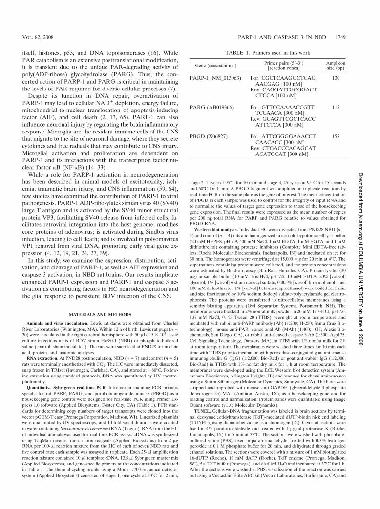
FIG. 1. Granule cell degeneration, zinc distribution, and apoptosis in the NBD HC. (A) Timm’s staining (dark-brown stain) in PND28 controlrat dentate gyrus. gcl, granule cell layer; iml, inner molecular layer; mf, mossy fibers. (B) Timm’s staining in PND28 NBD rat dentate gyrus; notethe accumulation of zinc in DGNs (arrows), decreased intensity of zinc stain in mossy fibers, and increased intensity of zinc stain in the iml.(C) Representative TUNEL of PND28 control rat DGNs. (D) Representative TUNEL of PND28 NBD rat DGNs; note the numerous TUNEL-positive neurons (brown stain). (E) TUNEL-positive DGNs with pyknotic nuclei (arrow) and signs of nuclear fragmentation (arrowhead).(F) Quantitation of TUNEL-positive DGNs in control (n # 4; 0.75 ) 0.479 cells) and PND28 NBD (n # 4; 80 ) 17.213 cells) rats (ANOVA, P #0.0037). The data are presented as the mean number of DGNs per dentate gyrus ) standard error of the mean. The asterisk indicates a P valueof (0.05. (G and H) IF staining for BDV nucleoprotein in control (G) and NBD (H) rat DGNs. (I and J) IF staining for BDV nucleoprotein incontrol (I) and NBD (J) rat hippocampal CA1 region pyramidal neurons. Bars # 100 $m.
1750 WILLIAMS ET AL. J. VIROL.
at COLUM
BIA UNIVERSITY on June 4, 2008 jvi.asm
.orgDownloaded from
rats (Fig. 1A; note the prominent Timm’s stain in the mossyfibers). In PND28 NBD rats, zinc accumulated in the somata ofDGNs (Fig. 1B) and was associated with increased Timm’sstaining in the inner molecular layer and decreased Timm’sstaining in the mossy fibers (Fig. 1B) (61). TUNEL in HC ofindividual control rats revealed few, if any, TUNEL-positiveDGNs (Fig. 1C). In contrast, numerous TUNEL-positiveDGNs were present in PND28 NBD rats (Fig. 1D). TUNEL-labeled DGNs in NBD rats had apoptotic morphology consis-tent with the morphology of degenerating DGNs harboringzinc in their somata (61), including nuclear pyknosis (Fig. 1E)and nuclear/chromatin fragmentation (Fig. 1E). Quantitationof TUNEL-positive DGNs in control and NBD rat HC re-vealed rare TUNEL-positive DGNs in control rats (0.75 )0.479 cells/dentate gyrus), while approximately 10% of allDGNs were TUNEL positive in NBD rats (80 ) 17.213cells/dentate gyrus) (Fig. 1F). Whereas immunostaining re-
vealed only scattered infected DGNs (Fig. 1G and H),nearly all pyramidal neurons of CA1 harbored BDV antigen(Fig. 1I and J).
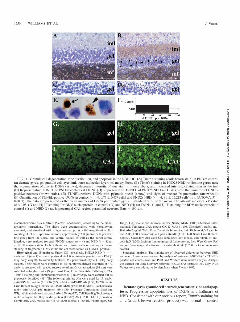
PARP-1 mRNA and protein expression in PND28 NBD rats.Although PARP-1 protein has been shown to colocalize withzinc-laden DGNs in NBD rats (61), PARP-1 expression hasnot been quantitated. Sybr green real-time PCR demon-strated increases in PARP-1 mRNA in PND28 NBD HCrelative to control rat HC (Fig. 2A) (3.18-fold; ANOVA, P #0.044). The increase in PARP-1 mRNA was correlated withincreased PARP-1 protein levels. Protein extracts from con-trol and NBD HC were analyzed by Western blotting.PARP-specific Abs detected a single band of 116 kDa cor-responding to the expected size of PARP-1 in both controland NBD HC extracts (Fig. 2B). Quantitation of PARP-1band densities normalized to GAPDH (housekeeping con-trol) revealed increased PARP-1 protein levels in NBD HC
FIG. 2. PARP-1 mRNA and protein expression in NBD and control rat brains. (A) Real-time PCR analysis of PARP-1 mRNA in HC of PND28control (n # 5) and NBD (n # 7) rats. PARP-1 mRNA in HC was significantly increased for NBD relative to control rats (3.18-fold increase inNBD; ANOVA, P # 0.044). The asterisk indicates a P value of (0.05. The error bars indicate standard errors of the means (SEM). (B) Rep-resentative Western immunoblots for PARP-1 protein (top) in extracts from control or NBD rat HC. Corresponding immunoblot signals forGAPDH are shown (bottom). The bar graph shows the determination of the PARP-1 protein band density relative to the GAPDH signal in extractsfrom control (n # 4) or NBD (n # 4) rat HC. HC PARP-1 protein levels were significantly increased in extracts from NBD rats relative to controls(1.68-fold increase in NBD; ANOVA, P # 0.012). PARP-1 IF in control (C, E, and G) and NBD (D, F, and H) rat HC. Note the faint stainingof DGNs in control rats (C and E) and increased staining of DGNs in NBD rats (D and F). Few PARP-1-positive cells were apparent in themolecular layers of control rats (C and G) compared to numerous PARP-1-positive cells scattered throughout the molecular layers of NBD rats(D and H). gcl, granule cell layer; ml, molecular layer; Hi, hilus. Bars # 100 $m.
VOL. 82, 2008 PARP-1 AND CASPASE 3 IN NBD 1751
at COLUM
BIA UNIVERSITY on June 4, 2008 jvi.asm
.orgDownloaded from
relative to control rats (1.68-fold; ANOVA, P # 0.012)(Fig. 2B).
HC expression of PARP-1 was assessed in brain sectionsof PND28 control and NBD rats by IF microscopy. In con-trol rats, the PARP-1 signal was weak in cells of the HC(Fig. 2C). In NBD rats, the PARP-1 signal was increased inscattered cells throughout the HC (Fig. 2D). DGNs stainedfaintly for PARP-1 in control rats (Fig. 2E). In NBD, nu-merous DGNs were strongly immunoreactive for PARP-1(Fig. 2F). Few glial cells in the molecular layer of the den-tate gyrus of control rats were immunoreactive for PARP-1(Fig. 2G). In NBD rats, the density of PARP-1 immunore-active glial cells in the molecular layer was increased relativeto control rats (Fig. 2H).
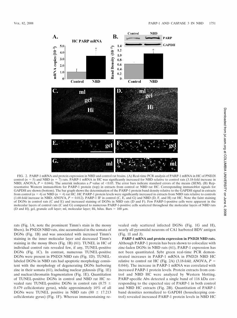
PARP-1 activation in HC. PARP-1 catalyzes the additionof long chains of PAR onto target proteins, including itself,using NAD! as the substrate. In order to assess the ribosy-lation status of specific proteins in the HC of NBD rats, weanalyzed HC protein lysates from control and NBD rats byWestern immunoblotting using Abs to PAR and GAPDH(loading control) (Fig. 3A). In both control and NBD rats,the major band that was recognized by the PAR antibody
was 116 kDa, consistent with the mass of PARP-1. Addi-tional 50-kDa and 25-kDa bands were present in NBD ratlysates. Individual band densities were quantitated and nor-malized to GAPDH. This analysis revealed significant in-creases in the 116-kDa (representing PARP-1) (1.74-fold;ANOVA, P # 0.05), the 50-kDa (8.25-fold; ANOVA, P #0.02), and the 25-kDa (9.15-fold; ANOVA, P # 0.03) spe-cies in NBD relative to control rats (Fig. 3B).
Homeostatic control of poly(ADP-ribosyl)ation is regulatedby the enzyme PARG, which rapidly hydrolyzes PAR units intofree ADP-ribose. To assess whether altered PARG expressioncould contribute to increased PAR levels, we evaluated PARGmRNA levels in the HC of PND28 control and NBD rats. Sybrgreen real-time PCR revealed a significant decrease in PARGmRNA in NBD rats relative to controls (1.4-fold; ANOVA,P # 0.041) (Fig. 3C).
IF analysis revealed moderate PAR staining in control ratHC, especially in DGNs (Fig. 3D). The intensity of the PARsignal was increased in numerous DGNs of PND28 NBD rats(Fig. 3E). The density of PAR-stained glial cells in the molec-ular layer of the dentate gyrus of control rats (Fig. 3F) waslower than in NBD rats (Fig. 3G).
FIG. 3. Enhanced poly(ADP-ribosyl)ation of proteins in NBD rats. (A) Protein lysates from HC of PND28 control (n # 4) and NBD (n # 4)rats were evaluated by Western analysis using Abs specific for PAR. (B) Determination of protein band densities relative to that for GAPDHrevealed increased ribosylation of PARP-1 (116 kDa; 1.74-fold increase; ANOVA, P # 0.05), an unknown 50-kDa protein (8.25-fold increase;ANOVA, P # 0.02), and an unknown 25-kDa protein (9.15-fold increase; ANOVA, P # 0.03). The asterisks indicate P values of (0.05. The errorbars indicate SEM. (C) Real-time PCR analysis of PARG mRNA in HC of PND28 control (n # 5) or NBD (n # 7) rats. PARG mRNA wassignificantly decreased for NBD rats relative to the results for control rats (1.4-fold decrease; ANOVA, P # 0.041). (D to G) IF analysis for PARin DGNs (D and E) and the dentate molecular layer (F and G) of PND28 control (D and F) and NBD (E and G) rats. Note the enhanced IF inDGNs and cells in the molecular layer of NBD relative to control rats. gcl, granule cell layer; ml, molecular layer. Bars # 100 $m.
1752 WILLIAMS ET AL. J. VIROL.
at COLUM
BIA UNIVERSITY on June 4, 2008 jvi.asm
.orgDownloaded from
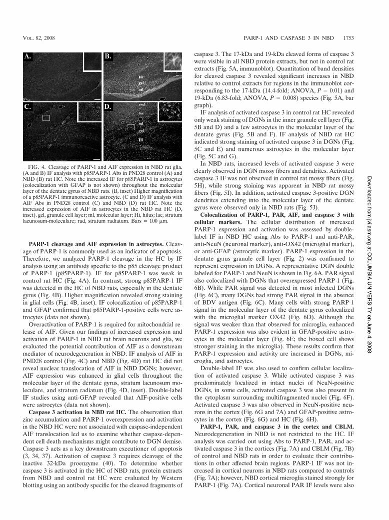
PARP-1 cleavage and AIF expression in astrocytes. Cleav-age of PARP-1 is commonly used as an indicator of apoptosis.Therefore, we analyzed PARP-1 cleavage in the HC by IFanalysis using an antibody specific to the p85 cleavage productof PARP-1 (p85PARP-1). IF for p85PARP-1 was weak incontrol rat HC (Fig. 4A). In contrast, strong p85PARP-1 IFwas detected in the HC of NBD rats, especially in the dentategyrus (Fig. 4B). Higher magnification revealed strong stainingin glial cells (Fig. 4B, inset). IF colocalization of p85PARP-1and GFAP confirmed that p85PARP-1-positive cells were as-trocytes (data not shown).
Overactivation of PARP-1 is required for mitochondrial re-lease of AIF. Given our findings of increased expression andactivation of PARP-1 in NBD rat brain neurons and glia, weevaluated the potential contribution of AIF as a downstreammediator of neurodegeneration in NBD. IF analysis of AIF inPND28 control (Fig. 4C) and NBD (Fig. 4D) rat HC did notreveal nuclear translocation of AIF in NBD DGNs; however,AIF expression was enhanced in glial cells throughout themolecular layer of the dentate gyrus, stratum lacunosum mo-leculare, and stratum radiatum (Fig. 4D, inset). Double-labelIF studies using anti-GFAP revealed that AIF-positive cellswere astrocytes (data not shown).
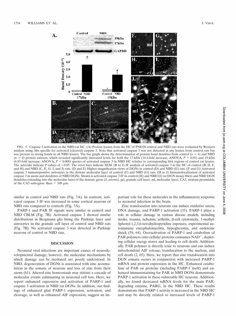
Caspase 3 activation in NBD rat HC. The observation thatzinc accumulation and PARP-1 overexpression and activationin the NBD HC were not associated with caspase-independentAIF translocation led us to examine whether caspase-depen-dent cell death mechanisms might contribute to DGN demise.Caspase 3 acts as a key downstream executioner of apoptosis(3, 34, 37). Activation of caspase 3 requires cleavage of theinactive 32-kDa proenzyme (40). To determine whethercaspase 3 is activated in the HC of NBD rats, protein extractsfrom NBD and control rat HC were evaluated by Westernblotting using an antibody specific for the cleaved fragments of
caspase 3. The 17-kDa and 19-kDa cleaved forms of caspase 3were visible in all NBD protein extracts, but not in control ratextracts (Fig. 5A, immunoblot). Quantitation of band densitiesfor cleaved caspase 3 revealed significant increases in NBDrelative to control extracts for regions in the immunoblot cor-responding to the 17-kDa (14.4-fold; ANOVA, P # 0.01) and19-kDa (6.83-fold; ANOVA, P # 0.008) species (Fig. 5A, bargraph).
IF analysis of activated caspase 3 in control rat HC revealedonly weak staining of DGNs in the inner granule cell layer (Fig.5B and D) and a few astrocytes in the molecular layer of thedentate gyrus (Fig. 5B and F). IF analysis of NBD rat HCindicated strong staining of activated caspase 3 in DGNs (Fig.5C and E) and numerous astrocytes in the molecular layer(Fig. 5C and G).
In NBD rats, increased levels of activated caspase 3 wereclearly observed in DGN mossy fibers and dendrites. Activatedcaspase 3 IF was not observed in control rat mossy fibers (Fig.5H), while strong staining was apparent in NBD rat mossyfibers (Fig. 5I). In addition, activated caspase 3-positive DGNdendrites extending into the molecular layer of the dentategyrus were observed only in NBD rats (Fig. 5J).
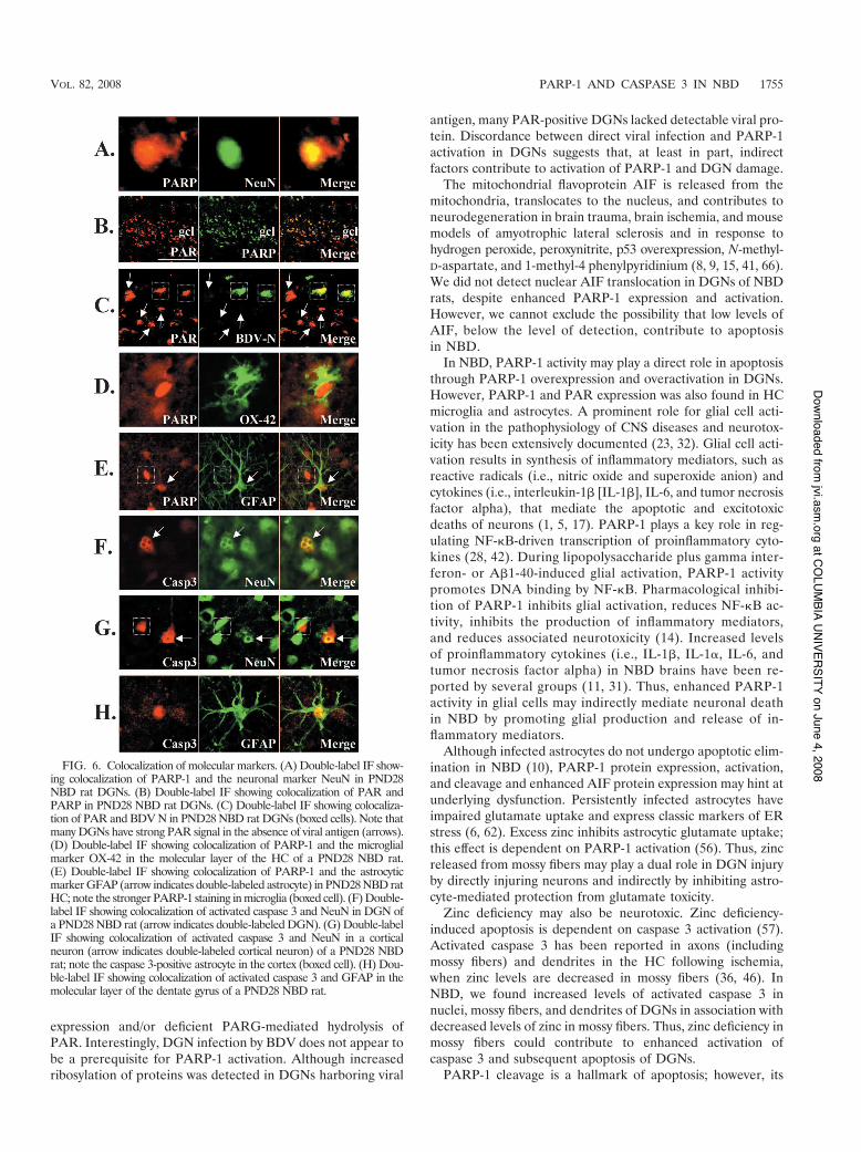
Colocalization of PARP-1, PAR, AIF, and caspase 3 withcellular markers. The cellular distribution of increasedPARP-1 expression and activation was assessed by double-label IF in NBD HC using Abs to PARP-1 and anti-PAR,anti-NeuN (neuronal marker), anti-OX42 (microglial marker),or anti-GFAP (astrocytic marker). PARP-1 expression in thedentate gyrus granule cell layer (Fig. 2) was confirmed torepresent expression in DGNs. A representative DGN doublelabeled for PARP-1 and NeuN is shown in Fig. 6A. PAR signalalso colocalized with DGNs that overexpressed PARP-1 (Fig.6B). While PAR signal was detected in most infected DGNs(Fig. 6C), many DGNs had strong PAR signal in the absenceof BDV antigen (Fig. 6C). Many cells with strong PARP-1signal in the molecular layer of the dentate gyrus colocalizedwith the microglial marker OX42 (Fig. 6D). Although thesignal was weaker than that observed for microglia, enhancedPARP-1 expression was also evident in GFAP-positive astro-cytes in the molecular layer (Fig. 6E; the boxed cell showsstronger staining in the microglia). These results confirm thatPARP-1 expression and activity are increased in DGNs, mi-croglia, and astrocytes.
Double-label IF was also used to confirm cellular localiza-tion of activated caspase 3. While activated caspase 3 waspredominately localized in intact nuclei of NeuN-positiveDGNs, in some cells, activated caspase 3 was also present inthe cytoplasm surrounding multifragmented nuclei (Fig. 6F).Activated caspase 3 was also observed in NeuN-positive neu-rons in the cortex (Fig. 6G and 7A) and GFAP-positive astro-cytes in the cortex (Fig. 6G) and HC (Fig. 6H).
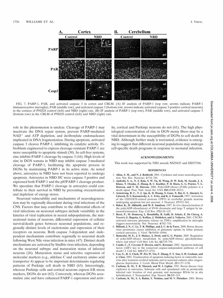
PARP-1, PAR, and caspase 3 in the cortex and CBLM.Neurodegeneration in NBD is not restricted to the HC. IFanalysis was carried out using Abs to PARP-1, PAR, and ac-tivated caspase 3 in the cortices (Fig. 7A) and CBLM (Fig. 7B)of control and NBD rats in order to evaluate their contribu-tions in other affected brain regions. PARP-1 IF was not in-creased in cortical neurons in NBD rats compared to controls(Fig. 7A); however, NBD cortical microglia stained strongly forPARP-1 (Fig. 7A). Cortical neuronal PAR IF levels were also
FIG. 4. Cleavage of PARP-1 and AIF expression in NBD rat glia.(A and B) IF analysis with p85PARP-1 Abs in PND28 control (A) andNBD (B) rat HC. Note the increased IF for p85PARP-1 in astrocytes(colocalization with GFAP is not shown) throughout the molecularlayer of the dentate gyrus of NBD rats. (B, inset) Higher magnificationof a p85PARP-1 immunoreactive astrocyte. (C and D) IF analysis withAIF Abs in PND28 control (C) and NBD (D) rat HC. Note theincreased expression of AIF in astrocytes in the NBD rat HC (D,inset). gcl, granule cell layer; ml, molecular layer; Hi, hilus; lac, stratumlacunosum-moleculare; rad, stratum radiatum. Bars # 100 $m.
VOL. 82, 2008 PARP-1 AND CASPASE 3 IN NBD 1753
at COLUM
BIA UNIVERSITY on June 4, 2008 jvi.asm
.orgDownloaded from
similar in control and NBD rats (Fig. 7A). In contrast, acti-vated caspase 3 IF was increased in some cortical neurons ofNBD rats compared to controls (Fig. 7A).
PARP-1 and PAR IF signals were similar in control andNBD CBLM (Fig. 7B). Activated caspase 3 showed similardistributions in Bergmann glia lining the Purkinje layer andastrocytes in the granule cell layer of control and NBD rats(Fig. 7B). No activated caspase 3 was detected in Purkinjeneurons of control or NBD rats.
DISCUSSION
Neonatal viral infections are important causes of neurode-velopmental damage; however, the molecular mechanisms bywhich damage can be mediated are poorly understood. InNBD, degeneration of DGNs is associated with zinc accumu-lation in the somata of neurons and loss of zinc from theiraxons (61). Altered zinc homeostasis may initiate a cascade ofmolecular events culminating in neuronal cell loss. Here, wereport enhanced expression and activation of PARP-1 andcaspase 3 activation in NBD rat DGNs. In addition, our find-ings of enhanced glial PARP-1 expression, activation, andcleavage, as well as enhanced AIF expression, suggest an im-
portant role for these molecules in the inflammatory responseto neonatal infection in the brain.
Zinc translocation into neurons can induce oxidative stress,DNA damage, and PARP-1 activation (35). PARP-1 plays arole in cellular damage in various disease models, includingstroke, trauma, ischemia, arthritis, *-cell cytotoxicity, 1-methyl-4phenyl-1,2,3,6-tetrahydropyridine exposure, experimental au-toimmune encephalomyelitis, hypoglycemia, and endotoxicshock (59, 64). Overactivation of PARP-1 and catabolism ofPAR polymers onto cellular proteins consumes NAD!, deplet-ing cellular energy stores and leading to cell death. Addition-ally, PAR polymer is directly toxic to neurons and can inducemitochondrial AIF release, translocation to the nucleus, andcell death (2, 65). Here, we report that zinc translocation intoDGN somata occurs in conjunction with increased PARP-1mRNA and protein expression in the HC. Enhanced catabo-lism of PAR on proteins (including PARP-1 itself) and en-hanced immunostaining for PAR in NBD DGNs demonstratePARP-1 activation in these vulnerable HC neurons. Addition-ally, we found decreased mRNA levels for the main PAR-degrading enzyme, PARG, in the NBD HC. These resultsdemonstrate that PARP-1 activity is increased in the NBD HCand may be directly related to increased levels of PARP-1
FIG. 5. Caspase 3 activation in the NBD rat HC. (A) Protein lysates from the HC of PND28 control and NBD rats were evaluated by Westernanalysis using Abs specific for activated (cleaved) caspase 3. Note that activated caspase 3 was not detected in any lysates from control rats butwas present as strong bands in all NBD lysates. The bar graph shows the determination of protein band densities from control (n # 4) and NBD(n # 4) protein extracts, which revealed significantly increased levels for both the 17-kDa (14.4-fold increase; ANOVA, P # 0.01) and 19-kDa(6.83-fold increase; ANOVA, P # 0.008) species of activated caspase 3 in NBD HC relative to corresponding blot regions of control rat lysates.The asterisks indicate P values of (0.05. The error bars indicate SEM. (B to J) IF analysis of activated caspase 3 in the HC of control (B, D, F,and H) and NBD (C, E, G, I, and J) rats. (D and E) Higher-magnification views of DGNs in control (D) and NBD (E) rats. (F and G) Activatedcaspase 3 immunopositive astrocytes in the dentate molecular layer of control (F) and NBD (G) rats. (H to J) Immunolocalization of activatedcaspase 3 in axons and dendrites of NBD DGNs. Shown is activated caspase 3 IF in control (H) and NBD (I) rat DGN mossy fibers and NBD DGNdendrites extending into the molecular layer of the dentate gyrus (J, arrows). gcl, granule cell layer; ml, molecular layer; CA3, stratum pyramidaleof the CA3 subregion. Bars # 100 $m.
1754 WILLIAMS ET AL. J. VIROL.
at COLUM
BIA UNIVERSITY on June 4, 2008 jvi.asm
.orgDownloaded from
expression and/or deficient PARG-mediated hydrolysis ofPAR. Interestingly, DGN infection by BDV does not appear tobe a prerequisite for PARP-1 activation. Although increasedribosylation of proteins was detected in DGNs harboring viral
antigen, many PAR-positive DGNs lacked detectable viral pro-tein. Discordance between direct viral infection and PARP-1activation in DGNs suggests that, at least in part, indirectfactors contribute to activation of PARP-1 and DGN damage.
The mitochondrial flavoprotein AIF is released from themitochondria, translocates to the nucleus, and contributes toneurodegeneration in brain trauma, brain ischemia, and mousemodels of amyotrophic lateral sclerosis and in response tohydrogen peroxide, peroxynitrite, p53 overexpression, N-methyl-D-aspartate, and 1-methyl-4 phenylpyridinium (8, 9, 15, 41, 66).We did not detect nuclear AIF translocation in DGNs of NBDrats, despite enhanced PARP-1 expression and activation.However, we cannot exclude the possibility that low levels ofAIF, below the level of detection, contribute to apoptosisin NBD.
In NBD, PARP-1 activity may play a direct role in apoptosisthrough PARP-1 overexpression and overactivation in DGNs.However, PARP-1 and PAR expression was also found in HCmicroglia and astrocytes. A prominent role for glial cell acti-vation in the pathophysiology of CNS diseases and neurotox-icity has been extensively documented (23, 32). Glial cell acti-vation results in synthesis of inflammatory mediators, such asreactive radicals (i.e., nitric oxide and superoxide anion) andcytokines (i.e., interleukin-1* [IL-1*], IL-6, and tumor necrosisfactor alpha), that mediate the apoptotic and excitotoxicdeaths of neurons (1, 5, 17). PARP-1 plays a key role in reg-ulating NF-"B-driven transcription of proinflammatory cyto-kines (28, 42). During lipopolysaccharide plus gamma inter-feron- or A*1-40-induced glial activation, PARP-1 activitypromotes DNA binding by NF-"B. Pharmacological inhibi-tion of PARP-1 inhibits glial activation, reduces NF-"B ac-tivity, inhibits the production of inflammatory mediators,and reduces associated neurotoxicity (14). Increased levelsof proinflammatory cytokines (i.e., IL-1*, IL-1+, IL-6, andtumor necrosis factor alpha) in NBD brains have been re-ported by several groups (11, 31). Thus, enhanced PARP-1activity in glial cells may indirectly mediate neuronal deathin NBD by promoting glial production and release of in-flammatory mediators.
Although infected astrocytes do not undergo apoptotic elim-ination in NBD (10), PARP-1 protein expression, activation,and cleavage and enhanced AIF protein expression may hint atunderlying dysfunction. Persistently infected astrocytes haveimpaired glutamate uptake and express classic markers of ERstress (6, 62). Excess zinc inhibits astrocytic glutamate uptake;this effect is dependent on PARP-1 activation (56). Thus, zincreleased from mossy fibers may play a dual role in DGN injuryby directly injuring neurons and indirectly by inhibiting astro-cyte-mediated protection from glutamate toxicity.
Zinc deficiency may also be neurotoxic. Zinc deficiency-induced apoptosis is dependent on caspase 3 activation (57).Activated caspase 3 has been reported in axons (includingmossy fibers) and dendrites in the HC following ischemia,when zinc levels are decreased in mossy fibers (36, 46). InNBD, we found increased levels of activated caspase 3 innuclei, mossy fibers, and dendrites of DGNs in association withdecreased levels of zinc in mossy fibers. Thus, zinc deficiency inmossy fibers could contribute to enhanced activation ofcaspase 3 and subsequent apoptosis of DGNs.
PARP-1 cleavage is a hallmark of apoptosis; however, its
FIG. 6. Colocalization of molecular markers. (A) Double-label IF show-ing colocalization of PARP-1 and the neuronal marker NeuN in PND28NBD rat DGNs. (B) Double-label IF showing colocalization of PAR andPARP in PND28 NBD rat DGNs. (C) Double-label IF showing colocaliza-tion of PAR and BDV N in PND28 NBD rat DGNs (boxed cells). Note thatmany DGNs have strong PAR signal in the absence of viral antigen (arrows).(D) Double-label IF showing colocalization of PARP-1 and the microglialmarker OX-42 in the molecular layer of the HC of a PND28 NBD rat.(E) Double-label IF showing colocalization of PARP-1 and the astrocyticmarker GFAP (arrow indicates double-labeled astrocyte) in PND28 NBD ratHC; note the stronger PARP-1 staining in microglia (boxed cell). (F) Double-label IF showing colocalization of activated caspase 3 and NeuN in DGN ofa PND28 NBD rat (arrow indicates double-labeled DGN). (G) Double-labelIF showing colocalization of activated caspase 3 and NeuN in a corticalneuron (arrow indicates double-labeled cortical neuron) of a PND28 NBDrat; note the caspase 3-positive astrocyte in the cortex (boxed cell). (H) Dou-ble-label IF showing colocalization of activated caspase 3 and GFAP in themolecular layer of the dentate gyrus of a PND28 NBD rat.
VOL. 82, 2008 PARP-1 AND CASPASE 3 IN NBD 1755
at COLUM
BIA UNIVERSITY on June 4, 2008 jvi.asm
.orgDownloaded from
role in the phenomenon is unclear. Cleavage of PARP-1 mayinactivate the DNA repair system, prevent PARP-mediatedNAD! and ATP depletion, and deribosylate endonucleasesimplicated in DNA fragmentation. During apoptosis, activatedcaspase 3 cleaves PARP-1, inhibiting its catalytic activity. Fi-broblasts engineered to express cleavage-resistant PARP-1 aremore susceptible to apoptotic stimuli (30). In cell-free systems,zinc inhibits PARP-1 cleavage by caspase 3 (44). High levels ofzinc in DGN somata in NBD may inhibit caspase 3-mediatedcleavage of PARP-1, facilitating the apoptotic process inDGNs by maintaining PARP-1 in its active state. As notedabove, astrocytes in NBD have not been reported to undergoapoptosis. Astrocytes in NBD HC were caspase 3 positive andexpressed both PARP-1 and the p85PARP-1 cleavage product.We speculate that PARP-1 cleavage in astrocytes could con-tribute to their survival in NBD by preventing overactivationand depletion of energy stores.
Neuronal vulnerability and mechanisms of neurodegenera-tion may be regionally discordant during viral infections of theCNS. Factors that may contribute to the differential effects ofviral infections on neuronal subtypes include variability in thekinetics of viral replication in neural subpopulations, the mat-urational status of neurons, differential expression of cellularsurvival/death genes between neuronal populations, and re-gionally distinct levels of excitotoxins and expression of theirreceptors on neurons. Both caspase 3-dependent and -inde-pendent mechanisms contribute regionally to neuronal deathfollowing West Nile virus infection in mice (47). Distinct deathmechanisms are activated by Sindbis virus infection, dependingon the neuronal subtype and maturation status of infectedneurons (26). Maturation status and selective expression ofmolecular markers (e.g., aldolase C and excitatory amino acidtransporter 4) appear to be important determinants regulatingpatterns of Purkinje cell death in NBD (63). Interestingly,whereas Purkinje cells and cortical neurons express ER stressmarkers, DGNs do not (62). Conversely, whereas DGNs accu-mulate zinc and have enhanced PARP-1 expression and activ-
ity, cortical and Purkinje neurons do not (61). The high phys-iological concentration of zinc in DGN mossy fibers may be avital determinant in the susceptibility of DGNs to cell death inNBD. Although further study is warranted, evidence is emerg-ing to suggest that different neuronal populations may undergocell-specific death programs in response to neonatal infection.
ACKNOWLEDGMENTS
This work was supported by NIH awards NS29425 and HD37546.
REFERENCES
1. Allan, S. M., and N. J. Rothwell. 2001. Cytokines and acute neurodegenera-tion. Nat. Rev. Neurosci. 2:734–744.
2. Andrabi, S. A., N. S. Kim, S. W. Yu, H. Wang, D. W. Koh, M. Sasaki, J. A.Klaus, T. Otsuka, Z. Zhang, R. C. Koehler, P. D. Hurn, G. G. Poirier, V. L.Dawson, and T. M. Dawson. 2006. Poly(ADP-ribose) (PAR) polymer is adeath signal. Proc. Natl. Acad. Sci. USA 103:18308–18313.
3. Armstrong, R. C., T. J. Aja, K. D. Hoang, S. Gaur, X. Bai, E. S. Alnemri, G.Litwack, D. S. Karanewsky, L. C. Fritz, and K. J. Tomaselli. 1997. Activationof the CED3/ICE-related protease CPP32 in cerebellar granule neuronsundergoing apoptosis but not necrosis. J. Neurosci. 17:553–562.
4. Baksi, K., H. Alkhatib, and M. E. Smulson. 1987. In vivo characterization ofthe poly(ADP-ribosylation) of SV40 chromatin and large T antigen by im-munofractionation. Exp. Cell Res. 172:110–123.
5. Bezzi, P., M. Domercq, L. Brambilla, R. Galli, D. Schols, E. De Clercq, A.Vescovi, G. Bagetta, G. Kollias, J. Meldolesi, and A. Volterra. 2001. CXCR4-activated astrocyte glutamate release via TNF+: amplification by microgliatriggers neurotoxicity. Nat. Neurosci. 4:702–710.
6. Billaud, J. N., C. Ly, T. R. Phillips, and J. C. de la Torre. 2000. Borna diseasevirus persistence causes inhibition of glutamate uptake by feline primarycortical astrocytes. J. Virol. 74:10438–10446.
7. Bonicalzi, M. E., J. F. Haince, A. Droit, and G. G. Poirier. 2005. Regulationof poly(ADP-ribose) metabolism by poly(ADP-ribose) glycohydrolase:where and when? Cell Mol. Life Sci. 62:739–750.
8. Cande, C., F. Cecconi, P. Dessen, and G. Kroemer. 2002. Apoptosis-inducingfactor (AIF): key to the conserved caspase-independent pathways of celldeath? J. Cell Sci. 115:4727–4734.
9. Cao, G., R. S. Clark, W. Pei, W. Yin, F. Zhang, F. Y. Sun, S. H. Graham, andJ. Chen. 2003. Translocation of apoptosis-inducing factor in vulnerable neu-rons after transient cerebral ischemia and in neuronal cultures after oxygen-glucose deprivation. J. Cereb. Blood Flow Metab. 23:1137–1150.
10. Carbone, K. M., T. R. Moench, and W. I. Lipkin. 1991. Borna disease virusreplicates in astrocytes, Schwann cells and ependymal cells in persistentlyinfected rats: location of viral genomic and messenger RNAs by in situhybridization. J. Neuropathol. Exp. Neurol. 50:205–214.
11. Carbone, K. M., S. A. Rubin, Y. Nishino, and M. V. Pletnikov. 2001. Borna
FIG. 7. PARP-1, PAR, and activated caspase 3 in cortex and CBLM. (A) IF analysis of PARP-1 (top row; arrows indicate PARP-1immunoreactive microglia), PAR (middle row), and activated caspase 3 (bottom row; arrows indicate activated caspase 3-positive cortical neurons)in the cortices of PND28 control (left) and NBD (right) rats. (B) IF analysis of PARP-1 (top row), PAR (middle row), and activated caspase 3(bottom row) in the CBLM of PND28 control (left) and NBD (right) rats.
1756 WILLIAMS ET AL. J. VIROL.
at COLUM
BIA UNIVERSITY on June 4, 2008 jvi.asm
.orgDownloaded from

disease: virus-induced neurobehavioral disease pathogenesis. Curr. Opin.Microbiol. 4:467–475.
12. Carbone, M., A. Reale, A. Di Sauro, O. Sthandier, M. I. Garcia, R. Maione,P. Caiafa, and P. Amati. 2006. PARP-1 interaction with VP1 capsid proteinregulates polyomavirus early gene expression. J. Mol. Biol. 363:773–785.
13. Chiarugi, A. 2002. Poly(ADP-ribose) polymerase: killer or conspirator? The‘suicide hypothesis’ revisited. Trends Pharmacol. Sci. 23:122–129.
14. Chiarugi, A., and M. A. Moskowitz. 2003. Poly(ADP-ribose) polymerase-1activity promotes NF-"B-driven transcription and microglial activation: im-plication for neurodegenerative disorders. J. Neurochem. 85:306–317.
15. Chu, C. T., J. H. Zhu, G. Cao, A. Signore, S. Wang, and J. Chen. 2005.Apoptosis inducing factor mediates caspase-independent 1-methyl-4-phe-nylpyridinium toxicity in dopaminergic cells. J. Neurochem. 94:1685–1695.
16. D’Amours, D., S. Desnoyers, I. D’Silva, and G. G. Poirier. 1999. Poly(ADP-ribosyl)ation reactions in the regulation of nuclear functions. Biochem. J.342:249–268.
17. Dawson, V. L., and T. M. Dawson. 1998. Nitric oxide in neurodegeneration.Prog. Brain Res. 118:215–229.
18. de la Torre, J. C. 1994. Molecular biology of Borna disease virus: prototypeof a new group of animal viruses. J. Virol. 68:7669–7675.
19. Dery, C. V., G. de Murcia, D. Lamarre, N. Morin, G. G. Poirier, and J.Weber. 1986. Possible role of ADP-ribosylation of adenovirus core proteinsin virus infection. Virus Res. 4:313–329.
20. Frederickson, C. J., M. D. Hernandez, S. A. Goik, J. D. Morton, and J. F.McGinty. 1988. Loss of zinc staining from hippocampal mossy fibers duringkainic acid induced seizures: a histofluorescence study. Brain Res. 446:383–386.
21. Gaken, J. A., M. Tavassoli, S. U. Gan, S. Vallian, I. Giddings, D. C. Darling,J. Galea-Lauri, M. G. Thomas, H. Abedi, V. Schreiber, J. Menissier-deMurcia, M. K. Collins, S. Shall, and F. Farzaneh. 1996. Efficient retroviralinfection of mammalian cells is blocked by inhibition of poly(ADP-ribose)polymerase activity. J. Virol. 70:3992–4000.
22. Gavrieli, Y., Y. Sherman, and S. A. Ben-Sasson. 1992. Identification ofprogrammed cell death in situ via specific labeling of nuclear DNA fragmen-tation. J. Cell Biol. 119:493–501.
23. Gonzalez-Scarano, F., and G. Baltuch. 1999. Microglia as mediators ofinflammatory and degenerative diseases. Annu. Rev. Neurosci. 22:219–240.
24. Gordon-Shaag, A., Y. Yosef, M. Abd El-Latif, and A. Oppenheim. 2003.The abundant nuclear enzyme PARP participates in the life cycle ofsimian virus 40 and is stimulated by minor capsid protein VP3. J. Virol.77:4273–4282.
25. Gosztonyi, G., and H. Ludwig. 1995. Borna disease—neuropathology andpathogenesis. Curr. Top. Microbiol. Immunol. 190:39–73.
26. Griffin, D. E. 2005. Neuronal cell death in alphavirus encephalomyelitis.Curr. Top. Microbiol. Immunol. 289:57–77.
27. Ha, H. C., K. Juluri, Y. Zhou, S. Leung, M. Hermankova, and S. H. Snyder.2001. Poly(ADP-ribose) polymerase-1 is required for efficient HIV-1 inte-gration. Proc. Natl. Acad. Sci. USA 98:3364–3368.
28. Ha, H. C., L. D. Hester, and S. H. Snyder. 2002. Poly(ADP-ribose) polymer-ase-1 dependence of stress-induced transcription factors and associated geneexpression in glia. Proc. Natl. Acad. Sci. USA 99:3270–3275.
29. Hatalski, C. G., W. F. Hickey, and W. I. Lipkin. 1998. Evolution of theimmune response in the central nervous system following infection withBorna disease virus. J. Neuroimmunol. 90:137–142.
30. Herceg, Z., and Z. Q. Wang. 1999. Failure of poly(ADP-ribose) polymerasecleavage by caspases leads to induction of necrosis and enhanced apoptosis.Mol. Cell. Biol. 19:5124–5133.
31. Hornig, M., H. Weissenbock, N. Horscroft, and W. I. Lipkin. 1999. Aninfection-based model of neurodevelopmental damage. Proc. Natl. Acad.Sci. USA 96:12102–12107.
32. Iadecola, C., and M. Alexander. 2001. Cerebral ischemia and inflammation.Curr. Opin. Neurol. 14:89–94.
33. Kauppinen, T. M., and R. A. Swanson. 2005. Poly(ADP-ribose) polymer-ase-1 promotes microglial activation, proliferation, and matrix metallopro-teinase-9-mediated neuron death. J. Immunol. 174:2288–2296.
34. Keane, R. W., A. Srinivasan, L. M. Foster, M. P. Testa, T. Ord, D. Nonner,H. G. Wang, J. C. Reed, D. E. Bredesen, and C. Kayalar. 1997. Activation ofCPP32 during apoptosis of neurons and astrocytes. J. Neurosci. Res. 48:168–180.
35. Kim, Y. H., and J. Y. Koh. 2002. The role of NADPH oxidase and neuronalnitric oxide synthase in zinc-induced poly(ADP-ribose) polymerase activa-tion and cell death in cortical culture. Exp. Neurol. 177:407–418.
36. Koh, J. Y., S. W. Suh, B. J. Gwag, Y. Y. He, C. Y. Hsu, and D. W. Choi. 1996.The role of zinc in selective neuronal death after transient global cerebralischemia. Science 272:1013–1016.
37. Kuida, K., T. S. Zheng, S. Na, C. Kuan, D. Yang, H. Karasuyama, P. Rakic,and R. A. Flavell. 1996. Decreased apoptosis in the brain and prematurelethality in CPP32-deficient mice. Nature 384:368–372.
38. Narayan, O., S. Herzog, K. Frese, H. Scheefers, and R. Rott. 1983. Behav-
ioral disease in rats caused by immunopathological responses to persistentBorna virus in the brain. Science 220:1401–1403.
39. Nargi-Aizenman, J. L., C. M. Simbulan-Rosenthal, T. A. Kelly, M. E. Smul-son, and D. E. Griffin. 2002. Rapid activation of poly(ADP-ribose) polymer-ase contributes to Sindbis virus and staurosporine-induced apoptotic celldeath. Virology 293:164–171.
40. Nicholson, D. W., A. Ali, N. A. Thornberry, J. P. Vaillancourt, C. K. Ding, M.Gallant, Y. Gareau, P. R. Griffin, M. Labelle, Y. A. Lazebnik, N. A. Munday,S. M. Raju, M. E. Smulson, T. Yamin, V. L. Yu, and D. K. Miller. 1995.Identification and inhibition of the ICE/CED-3 protease necessary for mam-malian apoptosis. Nature 376:17–18.
41. Oh, Y. K., K. S. Shin, and S. J. Kang. 2006. AIF translocates to the nucleusin the spinal motor neurons in a mouse model of ALS. Neurosci. Lett.406:205–210.
42. Oliver, F. J., J. Menissier-de Murcia, C. Nacci, P. Decker, R. Andriantsito-haina, S. Muller, G. de la Rubia, J. C. Stoclet, and G. de Murcia. 1999.Resistance to endotoxic shock as a consequence of defective NF-"B activa-tion in poly(ADP-ribose) polymerase-1 deficient mice. EMBO J. 18:4446–4454.
43. Park, J. A., J. Y. Lee, T. A. Sato, and J. Y. Koh. 2000. Co-induction ofp75NTR and p75NTR-associated death executor in neurons after zinc ex-posure in cortical culture or transient ischemia in the rat. J. Neurosci.20:9096–9103.
44. Perry, D. K., M. J. Smyth, H. R. Stennicke, G. S. Salvesen, P. Duriez, G. G.Poirier, and Y. A. Hannun. 1997. Zinc is a potent inhibitor of the apoptoticprotease, caspase-3. A novel target for zinc in the inhibition of apoptosis.J. Biol. Chem. 272:18530–18533.
45. Pletnikov, M. V., S. A. Rubin, G. J. Schwartz, K. M. Carbone, and T. H.Moran. 2000. Effects of neonatal rat Borna disease virus (BDV) infection onthe postnatal development of the brain monoaminergic systems. Brain Res.Dev. Brain Res. 119:179–185.
46. Rami, A., S. Jansen, I. Giesser, and J. Winckler. 2003. Post-ischemic acti-vation of caspase-3 in the rat hippocampus: evidence of an axonal anddendritic localisation. Neurochem. Int. 43:211–223.
47. Samuel, M. A., J. D. Morrey, and M. S. Diamond. 2007. Caspase 3-depen-dent cell death of neurons contributes to the pathogenesis of West Nile virusencephalitis. J. Virol. 81:2614–2623.
48. Schreiber, V., F. Dantzer, J. C. Ame, and G. de Murcia. 2006. Poly(ADP-ribose): novel functions for an old molecule. Nat. Rev. Mol. Cell Biol.7:517–528.
49. Sensi, S. L., H. Z. Yin, S. G. Carriedo, S. S. Rao, and J. H. Weiss. 1999.Preferential Zn2! influx through Ca2!-permeable AMPA/kainate channelstriggers prolonged mitochondrial superoxide production. Proc. Natl. Acad.Sci. USA 96:2414–2419.
50. Sheline, C. T., M. M. Behrens, and D. W. Choi. 2000. Zinc-induced corticalneuronal death: contribution of energy failure attributable to loss of NAD!
and inhibition of glycolysis. J. Neurosci. 20:3139–3146.51. Sheline, C. T., H. Wang, A. L. Cai, V. L. Dawson, and D. W. Choi. 2003.
Involvement of poly ADP ribosyl polymerase-1 in acute but not chronic zinctoxicity. Eur. J. Neurosci. 18:1402–1409.
52. Solbrig, M. V., G. F. Koob, J. N. Joyce, and W. I. Lipkin. 1996. A neuralsubstrate of hyperactivity in Borna disease: changes in brain dopamine re-ceptors. Virology 222:332–338.
53. Suh, S. W., J. W. Chen, M. Motamedi, B. Bell, K. Listiak, N. F. Pons, G.Danscher, and C. J. Frederickson. 2000. Evidence that synaptically-releasedzinc contributes to neuronal injury after traumatic brain injury. Brain Res.852:268–273.
54. Suh, S. W., R. B. Thompson, and C. J. Frederickson. 2001. Loss of vesicularzinc and appearance of perikaryal zinc after seizures induced by pilocarpine.Neuroreport 12:1523–1525.
55. Suh, S. W., P. Garnier, K. Aoyama, Y. Chen, and R. A. Swanson. 2004. Zincrelease contributes to hypoglycemia-induced neuronal death. Neurobiol.Dis. 16:538–545.
56. Suh, S. W., K. Aoyama, C. C. Alano, C. M. Anderson, A. M. Hamby, andR. A. Swanson. 2007. Zinc inhibits astrocyte glutamate uptake by activationof poly(ADP-ribose) polymerase-1. Mol. Med. 13:344–349.
57. Truong-Tran, A. Q., J. Carter, R. E. Ruffin, and P. D. Zalewski. 2001. Therole of zinc in caspase activation and apoptotic cell death. Biometals 14:315–330.
58. Virag, L., and C. Szabo. 1999. Inhibition of poly(ADP-ribose) synthetase(PARS) and protection against peroxynitrite-induced cytotoxicity by zincchelation. Br. J. Pharmacol. 126:769–777.
59. Virag, L., and C. Szabo. 2002. The therapeutic potential of poly(ADP-ribose) polymerase inhibitors. Pharmacol. Rev. 54:375–429.
60. Weissenbock, H., M. Hornig, W. F. Hickey, and W. I. Lipkin. 2000. Micro-glial activation and neuronal apoptosis in Bornavirus infected neonatalLewis rats. Brain Pathol. 10:260–272.
61. Williams, B. L., K. Yaddanapudi, C. M. Kirk, A. Soman, M. Hornig, andW. I. Lipkin. 2006. Metallothioneins and zinc dysregulation contribute toneurodevelopmental damage in a model of perinatal viral infection. BrainPathol. 16:1–14.
62. Williams, B. L., and W. I. Lipkin. 2006. Endoplasmic reticulum stress and
VOL. 82, 2008 PARP-1 AND CASPASE 3 IN NBD 1757
at COLUM
BIA UNIVERSITY on June 4, 2008 jvi.asm
.orgDownloaded from
neurodegeneration in rats neonatally infected with Borna disease virus. J. Vi-rol. 80:8613–8626.
63. Williams, B. L., K. Yaddanapudi, M. Hornig, and W. I. Lipkin. 2007. Spatio-temporal analysis of Purkinje cell degeneration relative to parasagittalexpression domains in a model of neonatal viral infection. J. Virol. 81:2675–2687.
64. Yu, S. W., H. Wang, T. M. Dawson, and V. L. Dawson. 2003. Poly(ADP-ribose) polymerase-1 and apoptosis inducing factor in neurotoxicity. Neuro-biol. Dis. 14:303–317.
65. Yu, S. W., S. A. Andrabi, H. Wang, N. S. Kim, G. G. Poirier, T. M. Dawson,and V. L. Dawson. 2006. Apoptosis-inducing factor mediates poly(ADP-
ribose) (PAR) polymer-induced cell death. Proc. Natl. Acad. Sci. USA 103:18314–18319.
66. Zhang, X., J. Chen, S. H. Graham, L. Du, P. M. Kochanek, R. Draviam, F.Guo, P. D. Nathaniel, C. Szabo, S. C. Watkins, and R. S. Clark. 2002.Intranuclear localization of apoptosis-inducing factor (AIF) and large scaleDNA fragmentation after traumatic brain injury in rats and in neuronalcultures exposed to peroxynitrite. J. Neurochem. 82:181–191.
67. Zocher, M., S. Czub, J. Schulte-Monting, J. C. de La Torre, and C. Sauder.2000. Alterations in neurotrophin and neurotrophin receptor gene expres-sion patterns in the rat CNS following perinatal Borna disease virus infec-tion. J. Neurovirol. 6:462–477.
1758 WILLIAMS ET AL. J. VIROL.
at COLUM
BIA UNIVERSITY on June 4, 2008 jvi.asm
.orgDownloaded from