Embed Size (px)
Citation preview
Lack of Poly(ADP-Ribose) Polymerase-1 Gene Product
Enhances Cellular Sensitivity to Arsenite
Anuradha Poonepalli,1,2Lakshmidevi Balakrishnan,
1Aik Kia Khaw,
1Grace Kah Mun Low,
1
Manikandan Jayapal,3Rabindra N. Bhattacharjee,
4,5Shizuo Akira,
4,5
Adayabalam S. Balajee,6and M. Prakash Hande
1,2
1Genome Stability Laboratory, Department of Physiology; 2Oncology Research Institute; 3Molecular and Cellular ImmunologyLaboratory, Department of Physiology, Yong Loo Lin School of Medicine, National University of Singapore, Singapore;4Department of Host Defense, Research Institute for Microbial Diseases, Osaka University; 5Exploratory Research forAdvanced Technology, Japan Science and Technology Corp., Osaka, Japan; and 6Center for Radiological Research,Columbia University, New York, New York
Abstract
Arsenite (As3+) has long been known to induce cancer andother degenerative diseases. Arsenite exerts its toxicity in partby generating reactive oxygen species. Identification of geneticfactors that contribute to arsenic mutagenicity and carcino-genicity is critical for the treatment and prevention of arsenicexposure in human population. As poly(ADP-ribose) polymer-ase (PARP) is critical for genomic DNA stability, role of PARP-1was evaluated in arsenic-induced cytotoxic and genotoxiceffects. Our study revealed that telomere attrition, probablyowing to arsenite-induced oxidative stress, was much morepronounced in PARP-1�/� mouse embryonic fibroblasts (MEF;40%) compared with PARP-1+/+ MEFs (10-20%). Correlationobserved between telomere reduction and apoptotic death inPARP-1 null cells strongly indicates that the telomere attritionmight be a trigger for enhanced apoptotic death after arsenitetreatment. Elevated DNA damage detected by alkaline cometassay points to an impaired repair ability of arsenite-inducedDNA lesions in PARP-1�/� MEFs. Consistent with elevatedDNA damage, increased micronuclei induction reflecting grossgenomic instability was also observed in arsenite-treatedPARP-1�/� MEFs. Microarray analysis has revealed thatarsenite treatment altered the expression of about 311 genesmajority of which have known functions in cellular responsesto stress/external stimulus and cell growth and/or mainte-nance. Our results suggest an important role for PARP-1 geneproduct in the maintenance of chromosome-genome stabilityin response to arsenite-induced DNA damage. (Cancer Res 2005;65(23): 10977-83)
Introduction
Poly(ADP-ribose) polymerase-1 (PARP-1), the best-characterizedmember of the PARP family is an abundant nuclear zinc fingerprotein found in most eukaryotes. PARP-1 primarily functions as aDNA damage sensor (1, 2) by recognizing and binding with highaffinity to both ssDNA and dsDNA breaks that arise directly orindirectly as byproducts of ongoing DNA repair process (1, 2).Further PARP-1 also facilitates the access of other DNA repair factors
to the sites of DNA damage (3, 4). PARP-1 is also known for its abilityto modulate the cellular responses either to survive or to undergoapoptotic death, depending on the extent of DNA damage (5).Arsenite is a significant environmental concern worldwide
especially in some parts of the United States as well as inArgentina, Canada, India, Japan, Thailand, Taiwan, and Bangladesh.Chronic exposure to inorganic arsenite is associated with hepaticinjury, peripheral neuropathy, and a wide variety of cancers (6).Many different modes of arsenite-induced genotoxicity have beenidentified, including oxidative stress, altered DNA repair andmethylation mechanisms, altered cell proliferation, and abnormalgene amplification (6). Recently, very low concentrations ofarsenite have been shown to inhibit poly(ADP) ribosylation ofproteins in mammalian cells (7).Our previous study showed that the PARP-1-deficient mice had
drastically shortened telomeres with high chromosomal instability(8). In addition, PARP-1 deficiency also induced telomeredysfunction and tumor development in mice with a p53 mutantbackground (9). PARP-1 thus seems to function in regulatingtelomere length as well as telomeric end capping. In view of itsimportance in both DNA repair and chromosome stability, thepresent study was undertaken to determine whether PARP-1 is animportant genetic factor responsible for arsenic-induced cytotox-icity in mammalian cells. Our study indicates that arsenite-inducedcell death, telomere attrition, and genomic instability are greatlyenhanced in PARP-1 null cells, and that PARP-1 is required forcellular resistance to arsenite exposure.
Materials and Methods
Cell culture and sodium arsenite treatment. PARP-1+/+ and PARP-1�/�
mouse embryonic fibroblasts (MEFs; kindly provided by Dr. Zhao-Qi Wang)
were cultured following the procedure described earlier (10). Cells in
exponential growth phase (at about 70% confluence) were exposed to dosesof sodium arsenite [As3+; Sigma, St. Louis, MO; 1.5 Ag/mL (11.5 Amol/L) or
3.0 Ag/mL (23 Amol/L)], and the cells were treated for 24 or 48 hours. These
same doses were used for all the experiments described below. Several
earlier studies have shown that doses in the range of 5 to 20 Amol/L ofsodium arsenite were found to show moderate effect level for the induction
of sister chromatid exchanges and micronuclei in mammalian systems (11).
Doses in the range of 1.5 and 10 Ag/mL were required to induce
chromosome aberrations in mouse lymphoma cells (12). Additionally, adose of 1.5 Ag/mL is considered pertinent to our study, as the arsenic level
is quite high in some Asian countries. A higher dose of 3.0 Ag/mL was used
to induce sufficient oxidative damage (13, 14) for enabling us to show theeffect in a DNA repair–deficient system. Two independent sets of MEFs for
each genotype were used in the experiments. The data were pooled and
presented.
Note: A. Poonepalli and L. Balakrishnan contributed equally to this work.Requests for reprints: M. Prakash Hande, Genome Stability Laboratory,
Department of Physiology, Faculty of Medicine, National University of Singapore,Block MD9, 2 Medical Drive, Singapore 117597, Singapore. Phone: 65-6874-3664; Fax:65-6778-8161; E-mail: [email protected].
I2005 American Association for Cancer Research.doi:10.1158/0008-5472.CAN-05-2336
www.aacrjournals.org 10977 Cancer Res 2005; 65: (23). December 1, 2005
Research Article
Research. on June 18, 2018. © 2005 American Association for Cancercancerres.aacrjournals.org Downloaded from
Flow fluorescence in situ hybridization for telomere measurement.After a continuous treatment with As3+ for 24 or 48 hours, cells were
trypsinized and washed with 1� PBS/0.1% bovine serum albumin (BSA).Cells were then probed with a telomere sequence–specific peptide nucleic
acid (PNA) by fluorescence in situ hybridization (FISH). Telomere length
was measured by flow cytometry as explained elsewhere (15).Alkaline single-cell gel electrophoresis (Comet) assay. Cells were
treated with As3+ for 30 minutes and 24 hours with the doses mentioned
earlier. The treated cells were harvested by trypsinization, washed in ice-
cold 1� PBS, and resuspended in HBSS with 10% DMSO with EDTA. Thecells were then suspended in (0.75%) molten low melting point agarose
(at 37jC) and immediately pipetted onto the comet slides (Trevigen,
Gaithersburg, MD). Electrophoresis was done as per vendor’s suggestions.
After electrophoresis, slides were briefly rinsed in neutralization buffer(500 mmol/L Tris-HCl, pH 7.5), air-dried, and stained with SYBR green dye.
The tail moment of the comets was generated using the Metasystems
(Altlussheim, Germany) analysis software ‘‘Comet imager version 1.2.’’ Fiftyrandomly chosen comets were analyzed per sample. The extent of DNA
damage observed was expressed as tail moment, which corresponded to the
fraction of the DNA in the tail of the comet.
Fluorescence in situ hybridization analysis of chromosomes. AfterAs3+ treatment for the specified time intervals, cells were released from
the treatment and allowed to grow for 24 hours in the absence of arsenite.
Cells were arrested at mitosis by treatment with colcemid (0.1 Ag/mL).
The cells were then incubated with a hypotonic solution of sodium citrateat 37jC for 20 minutes followed by fixation in Carnoy’s fixative. FISH was
done using telomere-specific PNA probe labeled with Cy3, and the cells
were counterstained with 4V,6-diamidino-2-phenylindole (Vectashield;
refs. 8, 15). Fifty metaphases were captured using the Zeiss Axioplan 2imaging fluorescence microscope and analyzed using the in situ imaging
software (Metasystems).
Cytokinesis-blocked micronucleus assay. Cells, after treatment for 24and 48 hours with As3+, were incubated with cytochalasin B (Sigma, 5 Ag/mL) for an additional 22 hours. The cells were then trypsinized and
subsequently fixed using a combination of both Carnoy’s fixative (acetic
acid/methanol, 1:3) and three to four drops of formaldehyde (to fix the
cytoplasm). Fixed cells were dropped onto clean slides and stained with3 Ag/mL of acridine orange, which differentially stains cytoplasm and
nucleus (16, 17). One thousand binucleated cells were scored for each
sample.
Cell cycle analysis. Control and As3+-treated cells were washed with 1�PBS/0.1% BSA and fixed in 70% ethanol. The fixed cells were later stained
with propidium iodide/RNase A. Samples were then analyzed by flow
cytometry at 488-nm excitation E and 610-nm emission E. Approximately
10,000 events per sample were collected, and the data was analyzed usingWINMDI software.
Microarray gene chip analysis. PARP-1+/+ and PARP-1�/� cells were
treated with 1.5 Ag/mL of As3+ for 24 hours. Total RNA was extracted
(RNeasy kit, Qiagen, Hilden, Germany), and double-stranded cDNA wassynthesized from 5 Ag of total RNA using Superscript system (Invitrogen,
Carlsbad, CA) primed with T7-(dT)-24 primer. For biotin-labeled cRNA
synthesis, in vitro transcription reaction was done in the presence of T7RNA polymerase and biotinylated ribonucleotides (Enzo Diagnostics,
Farmingdale, NY). The cRNA product was purified (RNeasy kit, Qiagen),
fragmented, and hybridized to Affymetrix GeneChip Mouse Genome 430
2.0 in a Gene chip hybridization oven 640 (Affymetrix, Inc., Santa Clara,CA) as per the Gene Chip Expression Analysis manual (Affymetrix). After
16 hours of hybridization, the gene chips were washed and stained using
the Affymetrix Fluidic station and scanned by Gene Array Scanner
(Affymetrix). Image data were normalized and statistically analyzedusing Gene Spring 7.2 (Silicon Genetics, Redwood City, CA). There were
311 differentially (P < 0.05, one-way ANOVA) expressed genes, and they
were annotated according to Gene Ontology: Biological Process.Subsequent data analysis involved agglomerative average-linkage hier-
archical clustering for finding different patterns and levels of gene
expression.
Statistical analysis. Statistical comparisons between and among thegroups were made using two-way ANOVA, Student’s t test, and contingency
tables analysis (m2 test and Fisher’s exact test) using Microsoft Excel 2003
(Microsoft Corp., Redmond, WA). The difference was considered to be
statistically significant when P < 0.05.
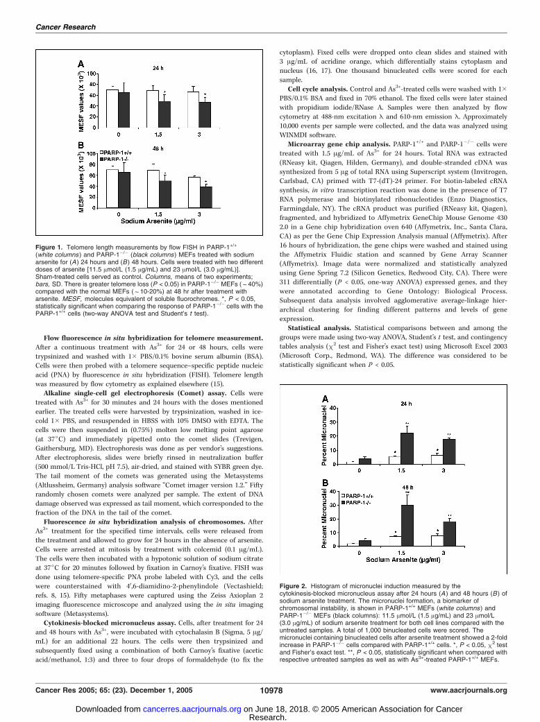
Figure 1. Telomere length measurements by flow FISH in PARP-1+/+
(white columns ) and PARP-1�/� (black columns) MEFs treated with sodiumarsenite for (A) 24 hours and (B ) 48 hours. Cells were treated with two differentdoses of arsenite [11.5 Amol/L (1.5 Ag/mL) and 23 Amol/L (3.0 Ag/mL)].Sham-treated cells served as control. Columns, means of two experiments;bars, SD. There is greater telomere loss (P < 0.05) in PARP-1�/� MEFs (f40%)compared with the normal MEFs (f10-20%) at 48 hr after treatment witharsenite. MESF, molecules equivalent of soluble fluorochromes. *, P < 0.05,statistically significant when comparing the response of PARP-1�/� cells with thePARP-1+/+ cells (two-way ANOVA test and Student’s t test).
Figure 2. Histogram of micronuclei induction measured by thecytokinesis-blocked micronucleus assay after 24 hours (A ) and 48 hours (B ) ofsodium arsenite treatment. The micronuclei formation, a biomarker ofchromosomal instability, is shown in PARP-1+/+ MEFs (white columns) andPARP-1�/� MEFs (black columns): 11.5 Amol/L (1.5 Ag/mL) and 23 Amol/L(3.0 Ag/mL) of sodium arsenite treatment for both cell lines compared with theuntreated samples. A total of 1,000 binucleated cells were scored. Themicronuclei containing binucleated cells after arsenite treatment showed a 2-foldincrease in PARP-1�/� cells compared with PARP-1+/+ cells. *, P < 0.05, m2 testand Fisher’s exact test. **, P < 0.05, statistically significant when compared withrespective untreated samples as well as with As3+-treated PARP-1+/+ MEFs.
Cancer Research
Cancer Res 2005; 65: (23). December 1, 2005 10978 www.aacrjournals.org
Research. on June 18, 2018. © 2005 American Association for Cancercancerres.aacrjournals.org Downloaded from
Results
Arsenite-induced telomere attrition was greater inPARP-1�/� mouse embryonic fibroblasts. Arsenite has beensuggested to be a potent inducer of oxidative stress and DNAdamage (14), and telomere shortening has been attributed tooxidative stress (18, 19). Our earlier studies have implicatedPARP-1 in telomere maintenance (8, 9). We therefore investigatedthe effect of arsenite treatment on telomere length in the absenceof the PARP-1 gene product. There was no significant differencein telomere length in both the cell types studied (Fig. 1A and B)at 24 hours after treatment, but the extent of telomere attritionwas significantly (P < 0.05, two-way ANOVA) greater in PARP-1�/�
cells (30-40% loss) compared with the PARP-1+/+ cells (10-20%loss; Fig. 1A and B) at 48 hours after treatment. This interestingobservation led us further to examine whether the telomere losstriggered by As3+ treatment enhances the chromosome instability.
To test this possibility, micronuclei analysis was done because itis a reliable indicator of chromosomal damage and genomicinstability (20, 21). Micronuclei formation is due to the exclusionof chromosomes or chromosomal fragments from the daughternuclei. Consistent with telomere loss, PARP-1�/� cells exhibited a2- to 3-fold increase (P < 0.05, m2/Fisher’s exact test) in thenumber of micronuclei containing binucleated cells at 24 and 48hours after sodium arsenite treatment compared with PARP-1+/+
cells (Fig. 2A and B). FISH using telomeric PNA probe wasemployed to analyze the chromosomal aberrations, particularlychromosome end-to-end fusions. Chromosomes with criticallyshort telomeres are unstable and often result in end-to-end
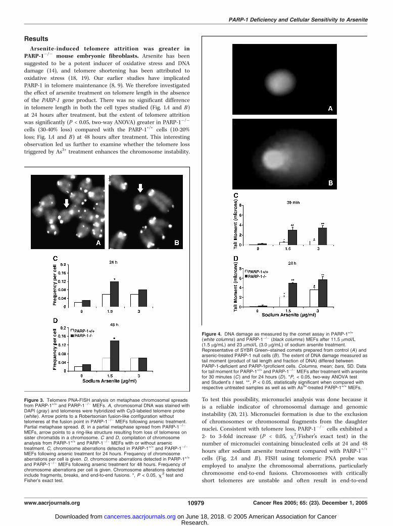
Figure 3. Telomere PNA-FISH analysis on metaphase chromosomal spreadsfrom PARP-1+/+ and PARP-1�/� MEFs. A, chromosomal DNA was stained withDAPI (gray ) and telomeres were hybridized with Cy3-labeled telomere probe(white ). Arrow points to a Robertsonian fusion-like configuration withouttelomeres at the fusion point in PARP-1�/� MEFs following arsenic treatment.Partial metaphase spread. B, in a partial metaphase spread from PARP-1�/�
MEFs, arrow points to a ring-like structure resulting from loss of telomeres onsister chromatids in a chromosome. C and D, compilation of chromosomeanalysis from PARP-1+/+ and PARP-1�/� MEFs with or without arsenictreatment. C, chromosome aberrations detected in PARP-1+/+ and PARP-1�/�
MEFs following arsenic treatment for 24 hours. Frequency of chromosomeaberrations per cell is given. D, chromosome aberrations detected in PARP-1+/+
and PARP-1�/� MEFs following arsenic treatment for 48 hours. Frequency ofchromosome aberrations per cell is given. Chromosome alterations detectedinclude fragments, breaks, and end-to-end fusions. *, P < 0.05, C 2 test andFisher’s exact test.
Figure 4. DNA damage as measured by the comet assay in PARP-1+/+
(white columns ) and PARP-1�/� (black columns ) MEFs after 11.5 Amol/L(1.5 Ag/mL) and 23 Amol/L (3.0 Ag/mL) of sodium arsenite treatment.Representative of SYBR Green–stained comets prepared from control (A ) andarsenic-treated PARP-1 null cells (B). The extent of DNA damage measured astail moment (product of tail length and fraction of DNA) differed betweenPARP-1-deficient and PARP-1proficient cells. Columns, mean; bars, SD. Datafor tail moment for PARP-1+/+ and PARP-1�/� MEFs after treatment with arsenitefor 30 minutes (C ) and for 24 hours (D ). *P, < 0.05, two-way ANOVA testand Student’s t test. **, P < 0.05, statistically significant when compared withrespective untreated samples as well as with As3+-treated PARP-1+/+ MEFs.
PARP-1 Deficiency and Cellular Sensitivity to Arsenite
www.aacrjournals.org 10979 Cancer Res 2005; 65: (23). December 1, 2005
Research. on June 18, 2018. © 2005 American Association for Cancercancerres.aacrjournals.org Downloaded from
fusions and chromosomal aberrations. Higher numbers of chromo-some aberrations were detected in PARP-1�/� MEFs at 1.5 Ag/mLdose at both the time points. Typical fusions, such as Robertsonianfusion-like structures and ring like structures (Fig. 3A and B), weredetected in the As3+ -treated PARP-1�/� MEFs, which are bestindicators of telomere loss and dysfunction. However, arsenitetreatment did not increase the frequency of chromosome end-to-end fusions in PARP-1 null cells as expected, but the frequency ofgross chromosome aberrations (which includes end-to-end fusions,chromosome breaks, and fragments) observed was higher inPARP-1�/� cells than PARP-1+/+ cells (Fig. 3C and D). Statisticallysignificant increase (P < 0.05, m2 test) in the total number ofchromosome aberrations was observed at a dose of 1.5 Ag/mLin PARP-1�/� MEFs at both 24 and 48 hours following treatment.Cells lacking PARP-1 displayed elevated DNA damage. The
extent of arsenic induced DNA damage and repair was estimatedby alkaline single-cell gel electrophoresis popularly known ascomet assay (Fig. 4A and B). The comet assay was done underalkaline condition (pH >13), to estimate all types of DNA damage,including double-strand breaks, single-strand breaks, and alkalilabile sites. PARP-1�/� cells reflected enhanced DNA damageinduction compared with PARP-1+/+ cells after arsenite treatment(Fig. 4C and D). DNA damage induced by 30 minutes of treatmentof As3+ was 5- to 6-fold (P < 0.001, two-way ANOVA test andStudent’s t test) more in PARP-1�/�cells than PARP-1+/+ cells.Similarly, arsenic exposure for 24 hours resulted in an increased tail
moment, indicating the impaired repair efficiency of arsenite-induced DNA damage in PARP-1 null cells (Fig. 4D).PARP-1�/� mouse embryonic fibroblasts are more sensitive
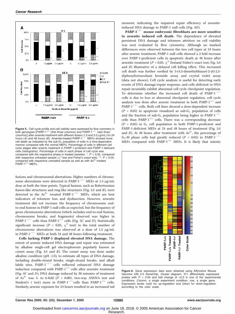
to arsenite induced cell death. The dependence of elevatedpersistent DNA damage and telomere attrition on cell viabilitywas next evaluated by flow cytometry. Although no markeddifferences were observed between the two cell types at 24 hoursafter arsenic treatment, PARP-1 null cells showed a 2-fold increaseover PARP-1-proficient cells in apoptotic death at 48 hours afterarsenite treatment (P < 0.05, m2 Testand Fisher’s exact test; Fig. 5Aand B) illustrative of a delayed cell killing effect. This increasedcell death was further verified by 3-(4,5-dimethylthiazol-2-yl)-2,5-diphenyltetrazolium bromide assay and crystal violet assay(data not shown). Cell cycle analysis is useful for detecting earlyevents of DNA damage/repair response, and cells deficient in DNArepair invariably exhibit abnormal cell cycle checkpoint regulation.To determine whether the increased cell death of PARP-1�/�
cells is due to loss or abnormal checkpoint regulation, cell cycleanalysis was done after arsenic treatment in both PARP-1+/+ andPARP-1�/� cells. Both cell lines showed a dose-dependent increase(P < 0.05) in apoptosis visualized as sub-G1 population of cellsand the fraction of sub-G1 population being higher in PARP-1�/�
cells than PARP-1+/+ cells. There was a corresponding decrease(P < 0.05) in G1 cell population in both PARP-1-proficient andPARP-1-deficient MEFs at 24 and 48 hours of treatment (Fig. 5Aand B). At 48 hours after treatment with As3+, the percentage ofG2-M phase cells had greatly reduced (P < 0.05) in PARP-1�/�
MEFs compared with PARP-1+/+ MEFs. It is likely that mitotic
Figure 5. Cell cycle profile and cell viability were assessed by flow cytometry inboth genotypes [PARP-1+/+ (first three columns ) and PARP-1�/� (last threecolumns )] after arsenite treatment with different doses (1.5 and 3.0 Ag/mL) for 24hours (A ) and 48 hours (B). Arsenite-treated PARP-1�/� MEFs showed morecell death as indicated by the sub-G1 population of cells in a time-dependentmanner compared with the normal MEFs. Percentage of cells in different cellcycle stages after arsenic treatment in PARP-1-proficient and PARP-1-deficientcells (histograms ). Percentage of cells in each phase of cell cycle wascompared with the respective phase in treated samples. *, P < 0.05, comparedwith respective untreated sample (m2 test and Fisher’s exact test). **, P < 0.05,compared with respective untreated sample as well as with As3+-treatedPARP-1+/+ MEFs.
Figure 6. Gene expression data were obtained using Affymetrix MouseGenome 430 2.0 GeneChip. Cluster diagram, 311 differentially expressedprobes with P < 0.05 and fold change of >2.0 in one of the experimentalconditions. Column, a single experiment condition; row, a single gene.Expression levels (red) for up-regulation and (blue ) for down-regulationaccording to the color scale.
Cancer Research
Cancer Res 2005; 65: (23). December 1, 2005 10980 www.aacrjournals.org
Research. on June 18, 2018. © 2005 American Association for Cancercancerres.aacrjournals.org Downloaded from
catastrophe occurs increasingly in PARP-1�/� cells, leading to increasedapoptotic death detected by an increase in sub-G1 cell population.Differential gene expression patterns in PARP-1+/+ and
PARP-1�/� cells after arsenite treatment. To study thedifferential gene expression patterns in PARP-1+/+ and PARP-1�/�
MEFs with or without arsenite treatment, microarray technologywas employed. Using this technology, the expression pattern ofover 34,000 genes was analyzed and compared with untreatedcontrols. PARP-1+/+ and PARP-1�/� MEFs were treated with1.5 Ag/mL of As+3 for 24 hours and then subjected to gene
expression studies. Microarray analysis was focused on genes thatwere altered with a >2-fold change with P < 0.05. A one-wayANOVA test was done to find distinct groups of genes that weresignificantly changed. Analysis has indicated that there are about311 genes, which are differentially expressed among the differentgroups (control versus treated, PARP-1�/� versus PARP-1+/+). Thesegenes belong to different biological processes, such as apoptosis/cell death, physiologic processes, and response to stress/externalagents. The data are presented as a hierarchical clustering inFig. 6 for the genes with differential expression in arsenic-treated
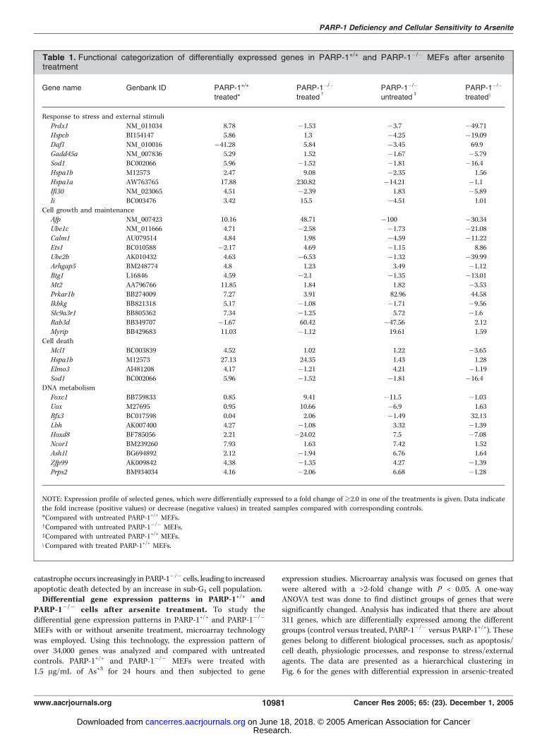
Table 1. Functional categorization of differentially expressed genes in PARP-1+/+ and PARP-1�/� MEFs after arsenitetreatment
Gene name Genbank ID PARP-1+/+
treated*PARP-1�/�
treatedc
PARP-1�/�
untreatedb
PARP-1�/�
treatedx
Response to stress and external stimuliPrdx1 NM_011034 8.78 �1.53 �3.7 �49.71
Hspcb BI154147 5.86 1.3 �4.25 �19.09
Daf1 NM_010016 �41.28 5.84 �3.45 69.9Gadd45a NM_007836 5.29 1.52 �1.67 �5.79
Sod1 BC002066 5.96 �1.52 �1.81 �16.4
Hspa1b M12573 2.47 9.08 �2.35 1.56
Hspa1a AW763765 17.88 230.82 �14.21 �1.1Ifi30 NM_023065 4.51 �2.39 1.83 �5.89
Ii BC003476 3.42 15.5 �4.51 1.01
Cell growth and maintenance
Afp NM_007423 10.16 48.71 �100 �30.34Ube1c NM_011666 4.71 �2.58 �1.73 �21.08
Calm1 AU079514 4.84 1.98 �4.59 �11.22
Ets1 BC010588 �2.17 4.69 �1.15 8.86
Ube2b AK010432 4.63 �6.53 �1.32 �39.99Arhgap5 BM248774 4.8 1.23 3.49 �1.12
Btg1 L16846 4.59 �2.1 �1.35 �13.01
Mt2 AA796766 11.85 1.84 1.82 �3.53Prkar1b BB274009 7.27 3.91 82.96 44.58
Ikbkg BB821318 5.17 �1.08 �1.71 �9.56
Slc9a3r1 BB805362 7.34 �1.25 5.72 �1.6
Rab3d BB349707 �1.67 60.42 �47.56 2.12Myrip BB429683 11.03 �1.12 19.61 1.59
Cell death
Mcl1 BC003839 4.52 1.02 1.22 �3.65
Hspa1b M12573 27.13 24.35 1.43 1.28Elmo3 AI481208 4.17 �1.21 4.21 �1.19
Sod1 BC002066 5.96 �1.52 �1.81 �16.4
DNA metabolismFoxc1 BB759833 0.85 9.41 �11.5 �1.03
Uox M27695 0.95 10.66 �6.9 1.63
Rfx3 BC017598 0.04 2.06 �1.49 32.13
Lbh AK007400 4.27 �1.08 3.32 �1.39Hoxd8 BF785056 2.21 �24.02 7.5 �7.08
Ncor1 BM239260 7.93 1.63 7.42 1.52
Ash1l BG694892 2.12 �1.94 6.76 1.64
Zfp99 AK009842 4.38 �1.35 4.27 �1.39Prps2 BM934034 4.16 �2.06 6.68 �1.28
NOTE: Expression profile of selected genes, which were differentially expressed to a fold change of z2.0 in one of the treatments is given. Data indicate
the fold increase (positive values) or decrease (negative values) in treated samples compared with corresponding controls.
*Compared with untreated PARP-1+/+ MEFs.cCompared with untreated PARP-1�/� MEFs.bCompared with untreated PARP-1+/+ MEFs.xCompared with treated PARP-1+/+ MEFs.
PARP-1 Deficiency and Cellular Sensitivity to Arsenite
www.aacrjournals.org 10981 Cancer Res 2005; 65: (23). December 1, 2005
Research. on June 18, 2018. © 2005 American Association for Cancercancerres.aacrjournals.org Downloaded from
samples. It is clear from the data that there are several genes,which are up-regulated in both PARP-1+/+ and PARP-1�/�
following arsenic treatment. Differentially expressed genes wereclassified according to Gene Ontology: Biological Process.Expression profile of selected genes, which were differentiallyexpressed to a fold change of z2.0 in one of the treatments, isgiven in Table 1. We have also identified candidate genes, whichare in apoptosis and cell cycle pathways following treatmentwith arsenic. Differences observed in the expression patterns ofthese genes (data not shown) between the PARP-1-proficient andPARP-1-deficient cells warrant further investigation.
Discussion
In this study, PARP-1-deficient cells showed a greater telomereattrition than in PARP-1-proficient cell lines at doses of 11.5 and23 Amol/L. Use of low concentrations (<1.0 Amol/L) of arsenitehave been shown to promote telomerase activity with or withoutan increment in telomere length (22). High concentrations ofarsenite (>1.0 Amol/L) used in this study have shown a rapid anddramatic loss of telomeric DNA, leading to apoptotic cell death.Although inhibition of telomerase alone by arsenite may causetelomere shortening over numerous population doublings, rapidtelomere loss was observed in PARP-1�/� cells after only 24 to 48hours of sodium arsenite treatment. There are several possibleexplanations for this observation. Rapid telomere loss may be dueto the susceptibility of the hexametric repeat structure oftelomeric DNA (triple-G-containing structures) to oxidativedamage (23, 24), as available evidences indicate that oxidativestress has the potency to break polyguanosine sequences intelomeric repeats, leading to telomere loss (19, 25). Arsenite, beingone of the efficient inducers of oxidative stress (13, 14, 26), canalso break polyguanosine sequences in telomeric repeats, result-ing in rapid telomere loss (27). Additionally, DNA repair in thetelomeric DNA was shown to be less efficient than the rest ofthe genome (25), further exacerbating the telomere attrition inthe absence of the PARP-1 gene product. In view of theinteractions of PARP-1 with the participants of the base excisionrepair pathway (BER; refs. 28, 29), it is likely that PARP-1deficiency may be a contributing factor to the rapid telomereshortening due to perturbations in the cellular processes of DNAdamage recognition and repair. Consistent with this, deficienciesin both shortlong- and long-patch BER pathways have beenreported in PARP-1-deficient cells (28). This study (28) shows thatboth uracil and 8-oxoguanine are poorly repaired in PARP-1-deficient cells compared with wild-type cells. Arsenite has beenshown to induce a wide variety of oxidized base lesions, including8-hydroxyguanine (30). Furthermore, acute arsenic treatmentinhibits the activity of human 8-hydroxyguanine glycosylase(OGG1) enzyme (30). Hence, a fundamental BER deficiency inthe absence of PARP-1 is presumably responsible for theenhanced sensitivity of PARP-1 null cells to arsenite treatment.Recently, arsenic mutagenicity has been shown to involve mito-chondrial DNA damage (31), and it is presently unclear whetherPARP-1 is also involved in BER process in mitochondrial DNA.Earlier studies have implicated telomere shortening as an
important checkpoint to limit the potential of human cells toproliferate (32). Shortening of telomeres triggers an apoptoticresponse in a p53-dependent manner (33). Telomere dysfunctioncoupled with chromosome instability due to inefficient DNA repairmight be responsible for increased cell death in PARP-1-deficient
cells. The cellular hypersensitivity of PARP-1 null cells thereforeunequivocally projects the role of PARP-1 as a survival factoragainst arsenic exposure. One of the earliest responses to ionizingradiation– and alkylating agent–induced DNA damage is thepoly(ADP) ribosylation of many nuclear proteins, which ismediated chiefly by PARP-1 and to a lesser extent by the PARPfamily of proteins (2, 34). Arsenite was shown to inhibit poly(ADP)ribosylation of proteins (7) in mammalian cells, and it is presentlyunclear whether the lack of poly(ADP) ribosylation of proteinsaffects the DNA repair process. As PARP-1 is implicated in diverserepair pathways, particularly BER, it is reasonable to assume thatthe increased cell death, telomere attrition, and genomic instabilityobserved in PARP-1 null cells is due to inefficient repair of oxidizedbase lesions.Telomere length maintenance has also been implicated in cancer
development due to reactivation of telomerase in tumor cells.Telomerase inhibition is therefore emerging as a promisingapproach in cancer chemotherapy (35, 36). Treatment of cancercell lines with telomerase inhibitors induces telomere shorteningand halts cellular proliferation (35). The importance of PARP-1 intelomere integrity suggests that PARP-1 inhibitors may present amyriad of potential therapeutic applications, especially in cancertreatment (37, 38). Seimiya et al. (37) showed that the combinationof inhibitors to the PARP family protein found at the telomeres,tankyrase, might serve as an effective anticancer therapy approach(36, 37). Because PARP-1 deficiency increases the cytotoxicity ofarsenite treatment, PARP-1 inhibitors could also be used incombination with other DNA-damaging agents to increase thecytotoxicity of cancer cells (39).Gene expression studies have revealed differential expression of
>300 genes following arsenic treatment in PARP-1+/+ and PARP-1�/�
MEFs. Genes that are up-regulated or down-regulated followingtreatment with arsenic in both cell types include genes thatparticipate in diverse biological processes, such as cell death, signaltransduction, response to stress/external stimulus, and cell growthand/or maintenance. As many as 36 genes involved in response toexternal stimulus were differentially expressed in the treatedsamples. More importantly, about 39 genes, which are importantfor cell growth and/or maintenance, were altered in theirexpression profiles. This clearly indicates that PARP-1 has a rolein the above cellular processes, which warrants further validationand investigation. Differential expression in these genes mayprovide biomarkers for gaining insights into mode of arsenictoxicity. Efforts are under way to identify and characterize specificDNA repair and cell cycle pathway(s) in the pathobiology of arsenicexposure. Taken together, our study identifies PARP-1 as one ofthe important genetic factors responsible for mediating the toxiceffects of arsenite.
Acknowledgments
Received 7/5/2005; revised 8/22/2005; accepted 9/22/2005.Grant support: Academic Research Fund; National University of Singapore; Office
of Life Sciences; National University Medical Institutes, Singapore; and NationalMedical Research Council, Ministry of Health, Singapore.
The costs of publication of this article were defrayed in part by the payment of pagecharges. This article must therefore be hereby marked advertisement in accordancewith 18 U.S.C. Section 1734 solely to indicate this fact.
We thank Drs. Zhao-Qi Wang and Wei-Min Tong (IARC, Lyon, France) for thePARP-1-proficient and PARP-1-deficient MEFs, Prof. Tom K. Hei (ColumbiaUniversity, New York, NY) for his advise on the use of sodium arsenite for studyingthe oxidative damage, and Adam Ng Tsan Sheng for his assistance with statisticalanalysis.
Cancer Research
Cancer Res 2005; 65: (23). December 1, 2005 10982 www.aacrjournals.org
Research. on June 18, 2018. © 2005 American Association for Cancercancerres.aacrjournals.org Downloaded from
References1. Jeggo PA. DNA repair: PARP—another guardian angel?Curr Biol 1998;8:R49–51.
2. Shall S, de Murcia G. Poly(ADP-ribose) polymerase-1:what have we learned from the deficient mouse model?Mutat Res 2000;460:1–15.
3. Yung TM, Sato S, Satoh MS. Poly(ADP-ribosyl)ation asa DNA damage-induced post-translational modificationregulating poly(ADP-ribose) polymerase-1-topoisomeraseI interaction. J Biol Chem 2004;279:39686–96.
4. Vispe S, Yung TM, Ritchot J, Serizawa H, Satoh MS. Acellular defense pathway regulating transcriptionthrough poly(ADP-ribosyl)ation in response to DNAdamage. Proc Natl Acad Sci U S A 2000;97:9886–91.
5. Huber A, Bai P, de Murcia JM, de MG. PARP-1, PARP-2and ATM in the DNA damage response: functionalsynergy in mouse development. DNA Repair (Amst) 2004;3:1103–8.
6. Kitchin KT, Ahmad S. Oxidative stress as a possiblemode of action for arsenic carcinogenesis. Toxicol Lett2003;137:3–13.
7. Hartwig A, Pelzer A, Asmuss M, Burkle A. Very lowconcentrations of arsenite suppress poly(ADP-ribosyl)a-tion in mammalian cells. Int J Cancer 2003;104:1–6.
8. d’ adda di Fagagna F, Hande MP, Tong WM, LansdorpPM, Wang ZQ, Jackson SP. Functions of poly(ADP-ribose) polymerase in controlling telomere length andchromosomal stability. Nat Genet 1999;23:76–80.
9. Tong WM, Hande MP, Lansdorp PM, Wang ZQ. DNAstrand break-sensing molecule poly(ADP-Ribose) poly-merase cooperates with p53 in telomere function,chromosome stability, and tumor suppression. Mol CellBiol 2001;21:4046–54.
10. Wang ZQ, Auer B, Sting L, et al. Mice lacking ADPRTand poly(ADP-ribosyl)ation develop normally but aresusceptible to skin disease. Genes Dev 1995;9:509–20.
11. Fan SR, Ho ICH, Yeoh LFY, Lin CJ. Lee TC. Squaleneinhibits sodium arsenite-induced sister chromatidexchanges and micronuclei in Chinese hamster ovary-K1 cells. Mutat Res 1996;368:165–9.
12. Moore MM, Harrington-Brock K, Doerr CL. Relativegenotoxic potency of arsenic and its methylatedmetabolites. Mutat Res 1997;386:279–90.
13. Hei TK, Liu SX, Waldren C. Mutagenicity of arsenic in
mammalian cells: role of reactive oxygen species. ProcNatl Acad Sci U S A 1998;95:8103–7.
14. Kessel M, Liu SX, Xu A, Santella R, Hei TK. Arsenicinduces oxidative DNA damage in mammalian cells. MolCell Biochem 2002;234–5:301–8.
15. Hande MP, Balajee AS, Tchirkov A, Wynshaw-Boris A,Lansdorp PM. Extra-chromosomal telomeric DNA incells from Atm(�/�) mice and patients with ataxia-telangiectasia. Hum Mol Genet 2001;10:519–28.
16. Hande MP, Boei JJ, Natarajan AT. Induction andpersistence of cytogenetic damage in mouse splenocytesfollowing whole-body X-irradiation analysed by fluores-cence in situ hybridization. II. Micronuclei. Int J RadiatBiol 1996;70:375–83.
17. Hande MP, Boei JJ, Natarajan AT. Induction andpersistence of cytogenetic damage in mouse splenocytesfollowing whole-body X-irradiation analysed by fluores-cence in situ hybridization. III. Chromosome malsegre-gation/aneuploidy. Mutagenesis 1997;12:125–31.
18. Saretzki G, Von Zglinicki T. Replicative aging,telomeres, and oxidative stress. Ann N Y Acad Sci 2002;959:24–9.
19. Von Zglinicki T. Oxidative stress shortens telomeres.Trends Biochem Sci 2002;27:339–44.
20. Fenech M. Chromosomal biomarkers of genomicinstability relevant to cancer. Drug Discov Today 2002;7:1128–37.
21. Fenech M. Biomarkers of genetic damage for cancerepidemiology. Toxicology 2002;181–2:411–6.
22. Zhang TC, Schmitt MT, Mumford JL. Effects ofarsenic on telomerase and telomeres in relation to cellproliferation and apoptosis in human keratinocytes andleukemia cells in vitro . Carcinogenesis 2003;24:1811–7.
23. Le PF, Schreiber V, Dherin C, de MG, Boiteux S.Poly(ADP-ribose) polymerase-1 (PARP-1) is required inmurine cell lines for base excision repair of oxidativeDNA damage in the absence of DNA polymerase h. J BiolChem 2003;278:18471–7.
24. Petersen S, Saretzki G, Von Zglinicki T. Preferentialaccumulation of single-stranded regions in telomeres ofhuman fibroblasts. Exp Cell Res 1998;239:152–60.
25. Evans MD, Cooke MS. Factors contributing to theoutcome of oxidative damage to nucleic acids. Bio-Essays 2004;26:533–42.
26. Liu SX, Athar M, Lippai I, Waldren C, Hei TK.
Induction of oxyradicals by arsenic: implication formechanism of genotoxicity. Proc Natl Acad Sci U S A2001;98:1643–8.
27. Liu L, Trimarchi JR, Navarro P, Blasco MA, Keefe DL.Oxidative stress contributes to arsenic-induced telo-mere attrition, chromosome instability, and apoptosis.J Biol Chem 2003;278:31998–2003.
28. Dantzer F, de La RG, Menissier-de MJ, Hostomsky Z,de MG, Schreiber V. Base excision repair is impaired inmammalian cells lacking poly(ADP-ribose) polymerase-1.Biochemistry 2000;39:7559–69.
29. Sanderson RJ, Lindahl T. Down-regulation of DNArepair synthesis at DNA single-strand interruptions inpoly(ADP-ribose) polymerase-1 deficient murine cellextracts. DNA Repair (Amst) 2002;1:547–58.
30. Mei N, Kunugita N, Hirano T, Kasai H. Acute arsenite-induced 8-hydroxyguanine is associated with inhibitionof repair activity in cultured human cells. BiochemBiophys Res Commun 2002;297:924–30.
31. Liu SX, Davidson MM, Tang X, et al. Mitochondrialdamage mediates genotoxicity of arsenic in mammaliancells. Cancer Res 2005;65:3236–42.
32. Maser RS, DePinho RA. Connecting chromosomes,crisis, and cancer. Science 2002;297:565–9.
33. Saretzki G, Sitte N, Merkel U, Wurm RE, Von ZT.Telomere shortening triggers a p53-dependent cell cyclearrest via accumulation of G-rich single stranded DNAfragments. Oncogene 1999;18:5148–58.
34. Burkle A. Poly(APD-ribosyl)ation, a DNA damage-driven protein modification and regulator of genomicinstability. Cancer Lett 2001;163:1–5.
35. Shay JW, Wright WE. Telomerase: a target for cancertherapeutics. Cancer Cell 2002;2:257–65.
36. Shay JW, Wright WE. Mechanism-based combinationtelomerase inhibition therapy. Cancer Cell 2005;7:1–2.
37. Seimiya H, Muramatsu Y, Ohishi T, Tsuruo T.Tankyrase 1 as a target for telomere-directed molecularcancer therapeutics. Cancer Cell 2005;7:25–37.
38. Tentori L, Portarena I, Graziani G. Potential clinicalapplications of poly(ADP-ribose) polymerase (PARP)inhibitors. Pharmacol Res 2002;45:73–85.
39. Calabrese CR, Almassy R, Barton S, et al. Anticancerchemosensitization and radiosensitization by the novelpoly(ADP-ribose) polymerase-1 inhibitor AG14361.J Natl Cancer Inst 2004;96:56–67.
PARP-1 Deficiency and Cellular Sensitivity to Arsenite
www.aacrjournals.org 10983 Cancer Res 2005; 65: (23). December 1, 2005
Research. on June 18, 2018. © 2005 American Association for Cancercancerres.aacrjournals.org Downloaded from
2005;65:10977-10983. Cancer Res Anuradha Poonepalli, Lakshmidevi Balakrishnan, Aik Kia Khaw, et al. Enhances Cellular Sensitivity to ArseniteLack of Poly(ADP-Ribose) Polymerase-1 Gene Product
Updated version
http://cancerres.aacrjournals.org/content/65/23/10977
Access the most recent version of this article at:
Cited articles
http://cancerres.aacrjournals.org/content/65/23/10977.full#ref-list-1
This article cites 37 articles, 10 of which you can access for free at:
Citing articles
http://cancerres.aacrjournals.org/content/65/23/10977.full#related-urls
This article has been cited by 4 HighWire-hosted articles. Access the articles at:
E-mail alerts related to this article or journal.Sign up to receive free email-alerts
Subscriptions
Reprints and
To order reprints of this article or to subscribe to the journal, contact the AACR Publications
Permissions
Rightslink site. (CCC)Click on "Request Permissions" which will take you to the Copyright Clearance Center's
.http://cancerres.aacrjournals.org/content/65/23/10977To request permission to re-use all or part of this article, use this link
Research. on June 18, 2018. © 2005 American Association for Cancercancerres.aacrjournals.org Downloaded from