Embed Size (px)
Citation preview
1
A role for Poly (ADP-ribose) polymerase (PARP) in the
transcriptional regulation of the melanoma growth
stimulatory activity (CXCL1) gene expression.
Chaitanya Nirodi2, Subir NagDas2, Steven P. Gygi4, Gary Olson2, Ruedi
Aebersold3 and Ann Richmond1,2
Department of Veterans Affairs1 and Vanderbilt University School of Medicine,
Department of Cell Biology 2, Nashville, TN 37232. Institute of Systems Biology, Seattle,
WA. Department of Cell Biology, Harvard Medical School, Boston, MA 02115.
Corresponding Author:Ann Richmond, Ph.D.Department of Cancer BiologyVanderbilt University School of MedicineMCN T-2212Nashville, TN 37232
Running Title : Role of PARP in the transcriptional activation of CXCL1.
JBC Papers in Press. Published on December 8, 2000 as Manuscript M009897200 by guest on M
ay 3, 2018http://w
ww
.jbc.org/D
ownloaded from
2
Abstract
The melanoma growth stimulatory activity/growth regulated protein, CXCL1, is
constitutively expressed at high levels during inflammation and progression of
melanocytes into malignant melanoma. It has been previously shown that CXCL1 over-
expression in melanoma cells is due to increased transcription as well as stability of the
CXCL1 message. The transcription of CXCL1 is regulated through several cis acting
elements including Sp1, NF-κB , HMGI(Y) and the IUR element (–94 to –78 nt) which
lies immediately upstream to the NF-κB element. Earlier it has been shown that the IUR
is necessary for basal and cytokine-induced transcription of the CXCL1 gene. UV-
crosslinking and South-western blot analyses indicate that the IUR oligonucleotide probe
selectively binds a 115 kDa protein. In this study, the IUR element has been further
characterized. We show here that proximity of the IUR element to the adjacent NF-κB
element is critical to its function as a positive regulatory element. Using binding site
oligonucleotide affinity chromatography, we have selectively purified the 115 kDa IUR-
F. MS/MS/MALDI spectroscopy and amino acid analysis as well as microcapillary
reverse phase chromatography electrospray ionization tandem mass spectrometry
identified this protein as the 114 kDa, Poly (ADP-ribose) polymerase (PARP 1).
Furthermore, 3−aminobenzamide, an inhibitor of PARP-specific ADP-ribosylation,
inhibits CXCL1 promoter activity and reduces levels of CXCL1 mRNA. The data point
to the possibility that PARP may be a co-activator of CXCL1 transcription.
by guest on May 3, 2018
http://ww
w.jbc.org/
Dow
nloaded from
3
The abbreviations used are
CXCL1. Melanoma growth stimulatory activity/growth related protein; IUR, immediate
upstream region; IL-1, interleukin-1; TNF, tumor necrosis factor, LPS, lipopolysaccharide; RPE,
retinal pigment epithelial; NF-κB, nuclear factor κB; I- κB, inhibitor of NF- κB ; IKK, I- κB
kinase; PARP, poly ADP-ribose polymerase; 3-AB, 3-amino benzamide; EMSA, electrophoretic
mobility shift assay.
by guest on May 3, 2018
http://ww
w.jbc.org/
Dow
nloaded from
4
Introduction
The melanoma growth stimulatory activity/growth regulated protein (CXCL1)
chemokine plays an important role in wound healing, inflammation and tumorigenesis
(1). The CXCL1 gene is not constitutively expressed in normal retinal pigment epithelial
cells (RPE) but can be induced by cytokines such as interleukin-1 (IL-1) and tumor
necrosis factor α (TNF α) and bacterial products such as lipopolysaccharide (LPS). In
contrast, Hs294T malignant melanoma cells exhibit high, constitutive levels of CXCL1
mRNA. IL-1 treatment of Hs294T cells does not significantly increase the transcription
of the gene, although it does appear to stabilize CXCL1 mRNA (2).
Transcription of the CXCL1 gene is regulated through a 306 bp minimal promoter
comprising four cis-acting elements which include the TATA box (-25 to –30 nt), a NF-
κB binding site (-67 to – 77 nt), an AT-rich HMGI(Y) binding element nested within the
NF-κB site, an immediate upstream region, IUR, (-78 to – -93 nt) and a GC-rich SP1
binding site (-117 to –128 nt) (3). In RPE cells, IL-1 increases nuclear levels of NF-κB
p65 (Rel A) and NF-κB p50 subunits (4). This is due to the IL-1 induced activation of
the I-κB kinases (IKK1/IKK2) and subsequent phosphorylation, ubiquitination and
degradation of the IKK substrate, I-κB. In Hs294T cells, constitutively high nuclear
levels of the p50 and p65/RelA proteins can be correlated with constitutive activity of
IKK1/ IKK2 and enhanced degradation of the I-κB protein. (5,6). The NF-κB element
therefore represents a crucial, inducible component of the putative CXCL1
enhanceosome.
The IUR is an approximately 20 bp sequence which is located immediately
upstream of the NF-κB site in the CXCL1 promoter. Previously we demonstrated that
by guest on May 3, 2018
http://ww
w.jbc.org/
Dow
nloaded from
5
this element is essential for basal as well as cytokine expression of the CXCL1 gene. In
particular, point mutations within a putative TCGAT motif abolished basal and IL-1
induced transcription in reporter gene assays with RPE and Hs294T cells. Furthermore, in
electrophoretic mobility shift assays (EMSA), these mutations blocked the ability of this
element to compete with a constitutive, IUR-specific complex in RPE and Hs294T
nuclear extracts (7). UV-crosslinking and Southwestern blot analyses revealed that at
least one protein having a relative molecular size of 115 kDa bound the IUR element in a
sequence-specific (8). In this study the 115 kDa IUR-specific protein has been purified
by binding-site oligonucleotide affinity chromatography. Peptide sequence analysis
identifies this protein as Poly (ADP-ribose) polymerase (PARP).
PARP is a 114-115 kDa nuclear DNA binding protein, which catalyses the
transfer of long, branched ADP-ribose chains to either itself or different classes of target
proteins involved in chromatin decondensation, DNA replication, DNA repair and gene
expression (9,10) ADP-ribosylation by PARP affects such cellular processes as
apoptosis, necrosis, cellular differentiation and malignant transformation (11). PARP
appears to have dual functions in the regulation of transcription. PARP-mediated ADP-
ribosylation of the transcription factors TATA-binding protein (TBP), Yin-Yang (YY1),
Sp1 (12), NF-κB (13), and p53 (14) and alters the sequence-specific DNA binding of
these protein and is thought to cause a reversible silencing of transcription. On the other
hand, PARP has been shown to enhance activator-dependent transcription in vitro (15).
More recently, PARP has been shown to activate muscle-specific gene expression by
interacting with sequences in the MCAT element of the cardiac troponin T promoter and
by ADP-ribosylating the MCAT-specific Transcription Enhancer Factor 1 (TEF-1) (16).
by guest on May 3, 2018
http://ww
w.jbc.org/
Dow
nloaded from
6
In addition, PARP increases the on-rate binding of nuclear factors to the PAX-6 gene
enhancer (17). Furthermore, PARP has been shown to activate transcription of genes
involved in cell proliferation in cooperation with the transcription factor, B-MYB (18)
and potentiates the transcriptional activation by the human T-cell Leukemia virus type 1
Tax protein (19).
In this study, the IUR element of the CXCL1 promoter has been further
characterized. Evidence presented here indicates that the IUR is a positive cis-acting
element and its activity is dependent on its contiguity with the adjacent NF-κB element A
115 kDa IUR-specific activity has been purified by binding site oligonucleotide affinity
chromatography and identified as PARP. A specific PARP inhibitor, 3-aminobenzamide,
inhibits CXCL1 promoter activity and decreases the endogenous levels of CXCL1
mRNA in a dose-dependent manner. We propose that PARP is a potential co-activator of
CXCL1 gene transcription. by guest on May 3, 2018
http://ww
w.jbc.org/
Dow
nloaded from
7
Materials and Methods
Cell Lines and treatments
Hs294T melanoma cells are a continuous cell line established from a human melanoma
metastatic to the lymph node. These cells were obtained from American Type Culture
Collection (Rockville, MD). RPE cells are normal retinal pigment epithelial cells that
were cultured by Dr. Glenn Jaffe from the North Carolina Organ Donor and Eye Bank
within 24 h of death. RPE and Hs294T cells were cultured as previously described (7). In
one series of experiments, Hs294T cells were incubated at 37 °C in the absence of serum
for 48 h, during which period, cells received the indicated doses of 3-aminobenzamide
(Sigma-Aldrich, St. Louis, MO) at 24 h intervals.
Sequence of oligonucleotides
The wildtype IUR probe, 2xIUR, used in electrophoretic mobility shift assays and
Southwestern blot analyses had the upper strand sequence
5’ccatcgatctggaactccggttcgatctggaactccggatgc 3’ and contained two copies of the IUR
sequence which are underscored. Sequences between the two IUR repeats and those
flanking the probe were included to optimize binding. A mutant IUR oligonucleotide,
2xmIUR, which contained mutations in the TCGAT motif of the IUR element had the
upper strand sequence, 5’ccaAGTaCctggaactccggtAGTaCctggaactccggatgc 3’. Upper
case characters indicate nucleotide replacements in the TCGAT motif while the
underscored sequences define the two copies of IUR element.
The oligonucleotide used to multimerize the IUR sequence for DNA affinity
chromatography had the upper strand sequence 5’-
by guest on May 3, 2018
http://ww
w.jbc.org/
Dow
nloaded from
8
gggatcgatctggaactccgggatcgatctggaactcc – 3’ and the lower strand sequence 5’-
cccggagttccagatcgatcccggagttccagatcgat -3’. The underscored characters represent the
two copies of the IUR sequence. The two strands when annealed form cohesive ends and
were ligated with T4 Polynucleotide Kinase (Promega, Madison, WI) to generate the
multimerized IUR DNA comprising up to 24 tandem repeats of the 2x IUR sequence. A
similar strategy was employed to generate the mulitmerized mutant IUR (mIUR) DNA
using the upper strand sequence 5’- gggaAGTaCctggaactccgggaAGTaCctggaactcc – 3’.
The upper case characters represent nucleotide replacements in the TCGAT motif, while
the underscored characters outline the IUR element repeats.
Reporter and Expression vectors
The CXCL1 minimal promoter region (-306 to +45 nt) was inserted in the pGL2 Basic
vector (Promega) either in the correct orientation (pWT.Luc) or in the opposite
orientation (pREV.Luc) relative to the transcription start site. To separate the IUR
element from the NF-κB site, pWT.Luc was modified to include an Nco I site between
the two elements using the GeneEditor in vitro site-directed mutagenesis sytem
(Promega, Madison, WI). The mutagenic oligonucleotide employed for this purpose had
the sequence: 5’ tcgggatcgatctggaactccATgggaatttccctggcc 3’. (The characters in upper
case represent the two nucleotide insertion that was necessary to create the Nco I site
which is underscored). The modified construct was termed pIN:2 to represent a two
nucleotide insertion between the the IUR and NF-κB sites. Similarly, mutant promoters
containing a 6 bp (pIN:6) and 12 bp insertion (pIN:12) were generated using the
mutagenic oligonucleotides 5’ gggatcgatctggaactccGGATCCgggaatttccctggcc 3’ and 5’
by guest on May 3, 2018
http://ww
w.jbc.org/
Dow
nloaded from
9
tcgggatcgatctggaactccGGATCCTCTAGAgggaatttccctggcc 3’. (Characters in upper case
represent the BamHI site and the BamHI/XbaI sites introduced to create the 6 and 12 bp
separations). To generate the promoter with a 25 bp insertion (pIN: 25), a 25 bp
oligonucleotide having the sequence 5’ catggcagtgagcgcaacgcaattac 3’ and flanked by
Nco I sites, was inserted in the Nco I site of pIN:2. A clone pIN:50, in which the 25 bp
insert had dimerized, was also selected. To generate the pGL2.mIUR construct, the
TCGAT motif in the wild type promoter was mutated to AGTAC by using a mutagenic
oligonucleotide 5’ ttccttccggactcgggaAGTaCctggaactccgggaatt 3’. All constructs were
confirmed by restriction analysis and automated plasmid sequencing.
Reagents :
Anti-PARP antibodies were from Santacruz Biotechnology, Santacruz, CA. The PARP
enzyme was purchased from R & D Systems Minneapolis, MN.
Electrophoretic Mobility Shift Assays.
Nuclear extracts were prepared as described previously (7). All extracts contained a 1 x
concentration of Complete protease inhibitor cocktail (Roche Diagnostics, Indianapolis,
IN). The 2xIUR or the 2xmIUR probes were radio-labeled by extension of an annealed
primer, 5’ gcatccggagttcca 3 with the Klenow fragment of E. coli DNA polymerase,
dNTPs and α32P dCTP.A typical binding reaction involved a 15 min pre-incubation with
10 µg of nuclear extract, 2 µg of non-specific competitor poly dIdC, 200 ng single-
stranded oligonucleotide, 20 mM Hepes-NaOH (pH 7.6), 100 mM NaCl, 1 mM DTT, 2%
glycerol, followed by a 20 min incubation with 50,000 cpm (40 femtomoles) of radio-
by guest on May 3, 2018
http://ww
w.jbc.org/
Dow
nloaded from
10
labeled probe. In oligonucleotide competitions, 1000- fold molar excess of cold, double-
stranded oligonucleotide was added to the pre-incubation mix. Complexes were resolved
by electrophoresis for 2 h at 170 volts on a 6% native, polyacrylamide gel, which was
later dried and processed for autoradiography. EMSA with affinity-purified fractions
contained 50 ng of poly dIdC and 100 ng of cold, double-stranded 2mR oligonucleotide
to ensure specificity of binding in the fractions. In cases where purified PARP was used,
the reaction mixture contained 50 ng of poly dIdC and about 5-10 femtomoles of pure
enzyme (cat # 4667-50-01) obtained from R&D Systems, Minneapolis, MN.
Southwestern and Western Blot Analysis
Nuclear extracts (25 µg) were heated at 90 °C for 3 minutes in 50 mM Tris.Cl (pH:6.8),
100 mM DTT, 2% SDS and 10% glycerol, resolved on 4% stacking/8% resolving SDS-
polyacrylamide gel and electrophoretically transferred to nitrocellulose membranes
(Biorad Laboratories). The membranes were rocked for 30 minutes in PBS, blocked for 4
h at RT with buffer A (20 mM Hepes-NaOH, 50 mM NaCl, 12.5 mg/ml skim milk
powder, 2.5 mg/ml bovine serum albumin, 100 µg/ml native salmon sperm DNA) and
incubated overnight at RT in up to 2 ml of Buffer A + 107 cpm (8-10 pmoles) of radio-
labeled 2xIUR or 2xmIUR probes. Membranes were subjected to three washes of 15
minutes each at RT in a buffer containing 20 mM Hepes-NaOH, 50 mM NaCl, 1g/L skim
milk powder 0.025% Nonidet P40, prior to drying and autoradiography.
For western analysis, membranes were probed with 800 µg/10 ml of anti-PARP
rabbit polyclonal antibody (cat # : sc-7150) from SantaCruz Antibodies, Santa Cruz, CA
using procedures recommended by the antibody manufacturer and the signal was
by guest on May 3, 2018
http://ww
w.jbc.org/
Dow
nloaded from
11
visualized by enhanced chemiluminescence (ECL) assay (Amersham Pharmacia Biotech)
according to the manufacturer’s recommendations.
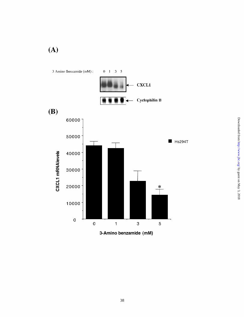
Northern Analysis :
Total RNA from Hs294T cells treated over a 48 h period with various doses of 3-
aminobenzamide was isolated using the Ultraspec RNA isolation system (Biotecx
Laboratories, Houston, TX) according to the manufacturer’s protocol. 20 µg/lane of
purified RNA was used in northern analysis which was performed exactly as described
previously (2). After probing for levels of CXCL1 mRNA, blots were stripped by boiling
for 10 minutes in water and re-probed with radiolabeled, cyclophilin cDNA. Bands
corresponding to the CXCL1 and cyclophilin were analysed by phosphorimager using
ImageQuant software (Molecular Dynamics). Values for CXCL1 mRNA were
normalized against cyclophilin mRNA. The experiment was repeated three times in
triplicate. Mean normalized values obtained from samples receiving the highest dose of
3-AB were compared with values from samples that received DMSO alone and were
found to be statistically different as determined by Student’s T-test (paired) .
Transient Transfection and Reporter Activity assay :
Hs294T were plated in 60-mm dishes at a density of 2 X 10⁵. The following day, cells at
nearly 70% confluency were transfected using the Lipofectamine/Plus method (Life
Technologies, Rockville, MD) with 1 µg of the appropriate reporter construct containing
either the wild type CXCL1 promoter (WT) or promoters containing insertions between
the IUR and NF-κB elements or nucleotide replacements within the TCGAT motif of
by guest on May 3, 2018
http://ww
w.jbc.org/
Dow
nloaded from
12
the IUR element (mIUR.Luc). In addition, all samples received 1µg of pRSV-β-gal.
Cells were harvested 48 h after transfection and luciferase activity was measured using
the luciferase assay system (Promega) and Monolight 2010 Luminometer (Analytical
Luminiscence Laboratory, San Diego, CA
In transfection experiments where 3-Aminobenzamide was used, cells were first
transfected with 1 µg of pRSV-β-gal reporter as well as reporter constructs driven by
either the wild type (CXCL1.Luc) or the mutant CXCL1 (mIUR.Luc) promoters. Six
hours after transfection, the medium was replaced by medium containing either DMSO or
3 -Aminobenzamide within a dose range of 1-5 M. A second change of DMSO or 3-AB
containing medium was given at 24 h after transfection. Cells were harvested 48 h after
transfection and processed as described above.
All values were normalized to β-galactosidase expression to correct for
transfection efficiency. Each experiment was repeated three separate times in triplicate.
In promoter mutagenesis experiments, mean normalized values from samples showing
the highest inhibition of promoter activity were compared to those obtained from samples
transfected with the wild type promoter and were found to be significantly different
according to the Student’s paired T-test. Similarly, mean normalized values of the
samples treated with the highest dose of 3-AB were compared with values obtained of the
sample containing the wild type promoter treated with DMSO alone and were found to be
significantly different according to Student’s paired T-test (p < 0.01). Statistical
measurements were performed using Microsoft Excel software.
by guest on May 3, 2018
http://ww
w.jbc.org/
Dow
nloaded from
13
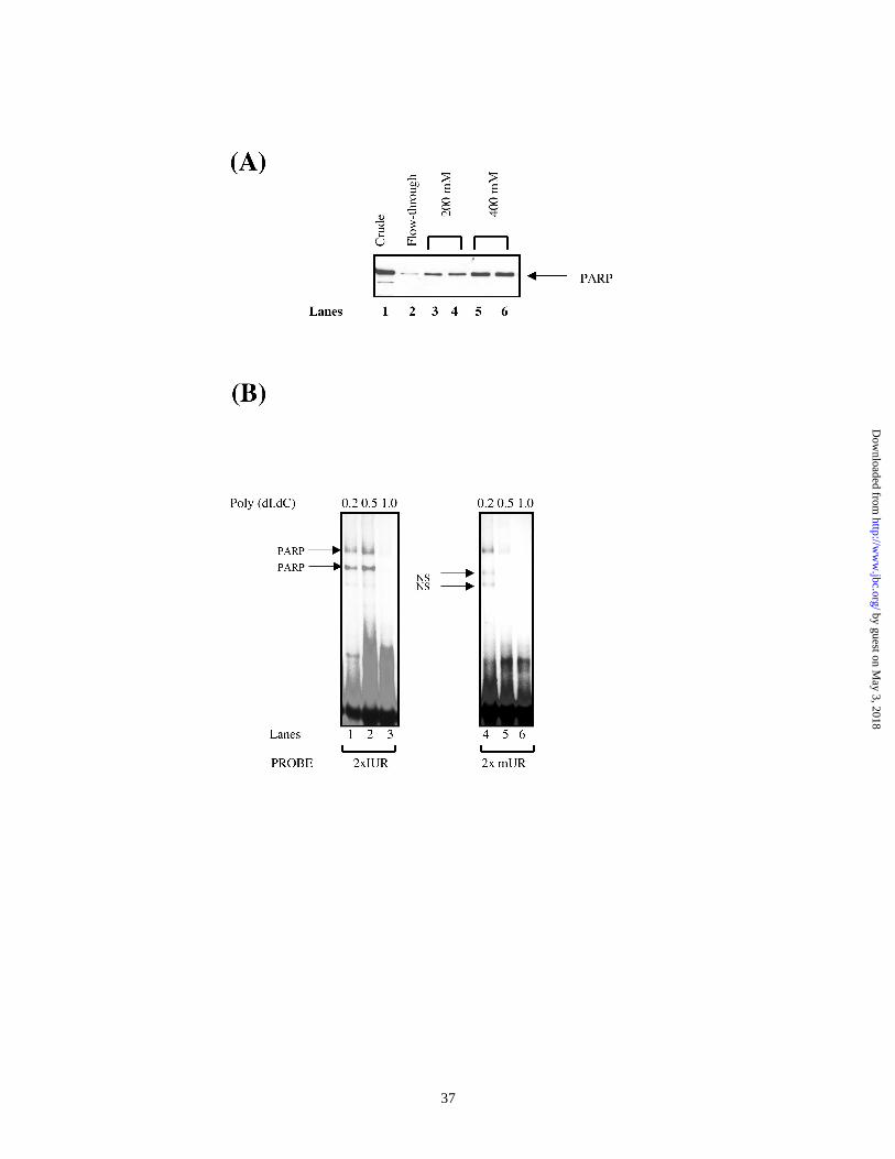
Purification of the 115 kDa IUR-specific protein :
Binding site oligonucleotide affinity chromatography was performed using a modified
protocol of Kadonaga et al.(20). Briefly, multimers of the IUR were cross-linked to
cyanogen bromide-activated agarose to generate the IUR-agarose column. Similarly, a
control column containing mIUR multimers (mIUR–agarose) was made. Cells equivalent
to 60 L HeLa suspension culture was obtained from National Cell Culture Center,
Minneapolis, MN. Approximately 2.5 g of nuclear extract was prepared from these cells.
First, non-specific DNA binding proteins were competed with poly dI:dC at a protein :
DNA ratio of 30 : 1, incubated at room temperature for 6 h and eliminated by high speed
centrifugation. Unbound proteins in the supernatant were subjected to binding site
oligonucleotide chromatography which involved two passages through a 3ml settled bed-
volume of the mutant IUR-agarose column, an overnight incubation with a 3 ml bed-
volume of IUR-agarose resin, followed by two passes through the same column. After 5
washes of 3ml each in a 50 mM NaCl containing buffer, fractions were eluted from the
IUR-agarose column by a step-gradient of buffers containing 0.2 to 1.0 M NaCl. In
EMSA and Southwestern assays, the 400 mM NaCl fraction consistently tested positive
for an activity with a relative molecular size of 115 kDa that specifically bound the
2xIUR probe. SDS-PAGE and silver staining revealed selective enrichment of the 115
kDa protein.
In order to further purify the 115 kDa protein, the 400 mM NaCl fraction was
pooled, dialyzed against water, lyophilized and reconstituted in the 1x SDS-PAGE
loading dye. The sample was then subjected to continuous-elution SDS-PAGE on a 7%
polyacrylamide gel using a Model 491 Prep Cell (Bio-Rad Laboratories, Hercules, CA).
by guest on May 3, 2018
http://ww
w.jbc.org/
Dow
nloaded from
14
Electro-elution was performed at a flow-rate of 0.1ml/min. Aliquots of electro-eluted
fractions were screened by electrophoresis on 4% stacking/10% resolving SDS-PAGE
gels and polypeptides were detected by silver-staining. Fractions that contained the 115
kDa protein were pooled and lyophilized. Reconstituted sample was re-electrophoresed
through a 4% stacking/8% resolving SDS-PAGE gel and the band containing the 115
kDa protein was excised and sent for sequencing. Two different, nearly homogenous,
preparations of the 115 kDa protein were sequenced separately in two different
laboratories; by microcapillary reverse phase chromatography electrospray ionization
tandem mass spectrometry using an ion trap mass spectrometer (LCQ, Finnigan MAT,
San Jose, CA) in the laboratory of Dr. R. Aebersold at the University of Washington,
Seattle, WA and by MS/MS MALDI at the sequencing facility at the W.M. Keck
Foundation, Yale University, New Haven, CT.
by guest on May 3, 2018
http://ww
w.jbc.org/
Dow
nloaded from
15
Results
The activity of the CXCL1 promoter depends on the position and orientation of the
IUR element:
The IUR element in the CXCL1 promoter plays an important role in the basal and
cytokine induced expression of the CXCL1 gene. The IUR contains a TCGAT motif
designated here as T1C2G3A4T5. Previously, we showed that a T5-C5 point mutation
inhibits basal and IL-1-induced CXCL1 promoter activity. In order to further characterize
the IUR element the effect of multiple replacements in the TCGAT motif on CXCL1
promoter activity was examined. In addition we investigated whether dislocating the IUR
relative to the NF-κB site has any effect on transcriptional activity of the CXCL1
promoter. We created reporter gene constructs which either retained the 306 bp CXCL1
promoter in the wild type configuration (WT) or had a 2, 6, 12, or 25 bp insertion
between the IUR and NF-κB elements (IN:2, IN:6, IN:12 and IN:25, respectively). Two
additional promoter mutants were also constructed, one which reversed the entire 350 bp
CXCL1 promoter relative to the luciferase reporter (Rev) and a second, which had a
T1C2G3A4T5 to A1G2T3A4C5 conversion in the IUR element (mIUR). These constructs
were then transfected into Hs294T malignant melanoma cells and luciferase activity
measured 48 hours later. Results in Figure 1A indicate that transcription from the CXCL1
promoter is inhibited by nearly 90% when the entire promoter is oriented in a direction
opposite to the transcription start site (Rev). Furthermore, a 75% inhibition in promoter
activity is observed when the T1C2G3A4T5 motif in the IUR element is replaced by
A1G2T3A4C5 (mIUR) . A similar inhibition of promoter activity is seen when the IUR
and NF-κB elements are separated by insertions (Figure 1B). Although promoter activity
by guest on May 3, 2018
http://ww
w.jbc.org/
Dow
nloaded from
16
appears to be unaffected when the IUR and NF-κB elements are separated by a 2 bp
insert. However, inserts that dislocate the IUR element from the NF-κB site, by 6, 12,
and 25 bp strongly inhibit promoter activity in a distance dependent manner. This
inhibition, however, is independent of helical phase of the two elements, because
introduction of a half (IN:6) or two and a half helical turn (IN:25) has the same effect as a
full helical turn (IN:12), ruling out the possibility that the insert-mediated inhibition of
CXCL1 promoter activity is due to a change in the helical phase between the two
elements. Together, the data suggest that the IUR is a positive cis acting element and that
the transcriptional activity of the CXCL1 promoter is strictly dependent on the distance
of the IUR element relative to the NF-κB site.
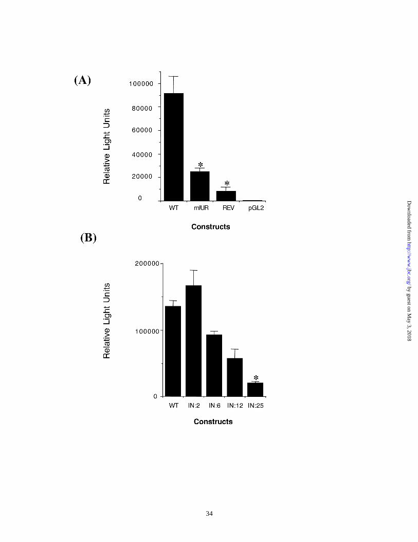
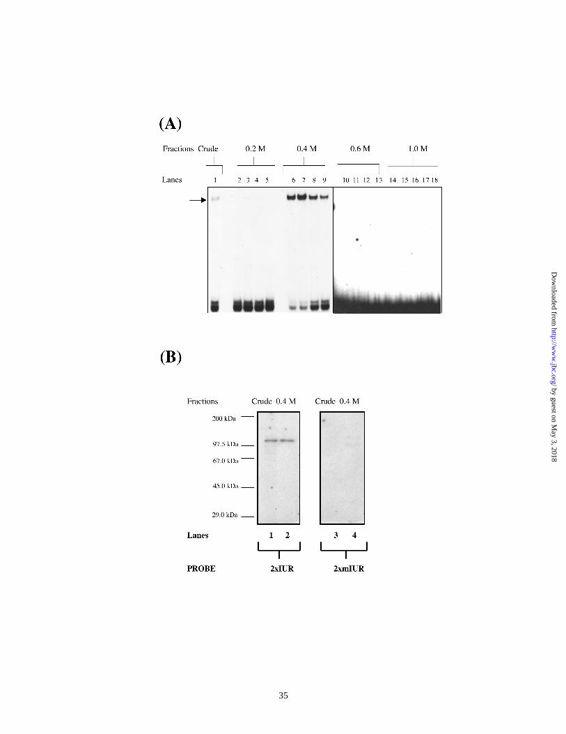
Purification and identification of 115 kDa IUR binding protein :
We have previously demonstrated that the IUR element binds a 115 kDa protein
in Southwestern blot assays (8). In order to identify the 115 kDa protein, which binds the
IUR, we purified the IUR specific factor by binding site oligonucleotide affinity
chromatography. HeLa nuclear extract proved to be a convenient and abundant source for
both proteins. We obtained frozen pellets of HeLa cells from 63 L suspension cultures
from the National Tissue Culture Center. The yield of nuclear extract from a 63 L
suspension culture was about 1.5 – 2.0 g. The IUR-specific activities were reproducibly
purified to near homogeneity in three separate experiments. Making use of the following
protocol, nuclear extract from HeLa cells was first challenged with non-specific
competitor DNA and chromatographed through the mutant-IUR-agarose column which
contained multimers of the double-stranded mIUR oligonucleotide. The unbound fraction
by guest on May 3, 2018
http://ww
w.jbc.org/
Dow
nloaded from
17
was then chromatographed through an IUR-agarose column, which contained multimers
of double-stranded IUR oligonucleotide. Bound fractions from both columns were
washed and eluted by a step gradient of NaCl ranging from 50 mM to 1000 mM. Proteins
specific to the IUR element emerged in the unbound (flow-through) fraction of the
mutant-IUR column and eluates from the bound fraction of this column consistently
tested negative for IUR-specific activity in EMSA (data not shown). Eluates from the
IUR-agarose column were assayed for IUR-specific activity by EMSA and Southwestern
blot analysis. Figure 2 is a representative of three separate experiments. The results from
an EMSA (Figure 2A) indicate that an IUR-specific activity reproducibly eluted in the
400 mM fraction of the IUR-agarose column. This correlated well with results from
Southwestern blot analysis (Figure 2B) where a 115 kDa protein in the 400 mM fraction
bound the 2xIUR element in a sequence specific manner. Proteins from bound and
unbound fractions from the IUR-agarose column were resolved by SDS-PAGE (Figure
2C) and detected by silver-staining. SDS-PAGE analysis consistently showed that the
400 mM fraction of the IUR-agarose column essentially contains 5 bands with relative
molecular sizes of 180 kDa, 115 kDa, 85 kDa, 42 and 29 kDa. Of these, the 115 kDa
band was considerably enriched in this fraction.
In order to further purify the 115 kDa protein, the 400 mM IUR-agarose fractions
were pooled, dialyzed and lyophilized to dryness. The reconstituted 400 mM fraction
was then electrophoresed through a preparative SDS-PAGE miniprep cell and 265
fractions of 1.0 ml each were collected. Electro-eluted fractions were analyzed by SDS-
PAGE/silver-staining. Figure 2D shows results of such an experiment. The 115 kDa
protein electro-elutes reproducibly in fractions 135-160 of the minprep cell.
by guest on May 3, 2018
http://ww
w.jbc.org/
Dow
nloaded from
18
Two separate preparations of the 115 kDa protein, purified to near homogeneity, were
independently sequenced at two different institutions : by MS/MS/MALDI mass
spectroscopy at W.M. Keck Foundation, Yale University, New Haven, CT and by
microcapillary reverse phase chromatography electrospray ionization tandem mass
spectrometry using an ion trap mass spectrometer (LCQ, Finnigan MAT, San Jose, CA)at
University of Washington, Seattle, WA by S. Gygi and R. Aebersold). Each preparation
had about 10 - 20 picomoles of the 115 kDa protein. Both sources identified the 115 kDa
as the human poly ADP ribose polymerase (PARP) (EC Number: 2.4.2.30).
The DNA-affinity purified 115 kDa protein is PARP :
We tested the 400 mM fraction from the IUR agarose column by Western blot
analysis (Figure 3A) to determine whether or not the 115 kDa is the poly ADP ribose
polymerase (PARP). Anti-PARP antibodies successfully detected a 115 kDa protein in
the fraction thus confirming that the affinity purified 115 kDa protein is PARP.
Recombinant PARP binds the CXCL1 IUR element in a sequence-specific manner.
Results in Figure 2 demonstrate that the 115 kDa protein (PARP) emerged in the
unbound fraction of the mutant-IUR agarose column and eluted in the 400 mM fraction
bound to IUR agarose column. This strongly suggested that the PARP protein could
discriminate between the mutant and wild-type IUR sequences. To verify this, we used
purified, recombinant PARP in EMSA and compared PARP binding to 2xIUR and
2xmIUR probes (Figure 3B). The use of poly dIdC in the reaction mixture was found to
be necessary since the recombinant protein was not a homogenous preparation.
by guest on May 3, 2018
http://ww
w.jbc.org/
Dow
nloaded from
19
Specificity to IUR was observed at relatively high concentrations of the non-specific
competitor DNA. Three shifted complexes could be detected with the 2xIUR probe. Two
complexes, which were specific to 2xIUR, did not bind the 2xmIUR probe. This result
indicates that PARP can form a sequence-specific complex with the IUR element of
CXCL1. We would like to point out that the complex generated by the purified
recombinant protein appears to differ in mobility and intensity to the one obtained from
crude nuclear extracts. Moreover, there are two IUR specific complexes generated with
the recombinant protein as opposed to a single complex observed with crude nuclear
extracts. The reasons for these discrepancies are unclear at this time. However, it is likely
that the complex observed in crude nuclear extract could be a composite of more than one
protein in addition to PARP.
3-aminobenzamide (3-AB) decreases levels of CXCL1 mRNA in Hs294T cells.
PARP catalyses the transfer of multiple ADP-ribose units to target nuclear
proteins. This ADP-ribose transferase activity is believed to inhibit DNA binding as well
as trans-activation functions of the target proteins. 3-Aminobenzamide (3AB), a specific
PARP inhibitor, blocks ADP-ribose transferase functions of PARP. We asked the
question whether inhibition of PARP catalytic activity by 3AB might have any effect on
CXCL1 expression in Hs294T cells.
Hs294T cells were either left untreated or treated with 3AB in a dose range of 1.0
mM to 5.0 mM. Forty-eight hours later, cells were harvested and RNA from these
samples was isolated. CXCL1 mRNA levels were measured by Northern analysis (Figure
4A). Values, normalized against levels of an internal control cyclophilin B, are
graphically represented in Figure 4B. Results indicate that 3-AB treatment inhibited
by guest on May 3, 2018
http://ww
w.jbc.org/
Dow
nloaded from
20
CXCL1 mRNA levels by nearly 60 % over untreated or DMSO-treated controls. The data
suggest that PARP-mediated ADP-ribosylation is essential for CXCL1 gene expression.
The ADP-ribose transferase activity of PARP, has been shown to be involved in
negatively regulating gene expression by abrogating the DNA binding of such factors as
NF-κB, YY1, C/EBPβ and Sp1. In addition ADP-ribosylation of these factors does not
appear to require DNA binding of PARP. It is therefore possible that the effect of 3-AB
on CXCL1 mRNA levels may be indirect and could involve other trans activators of the
CXCL1 promoter such as NF-κB and Sp1.
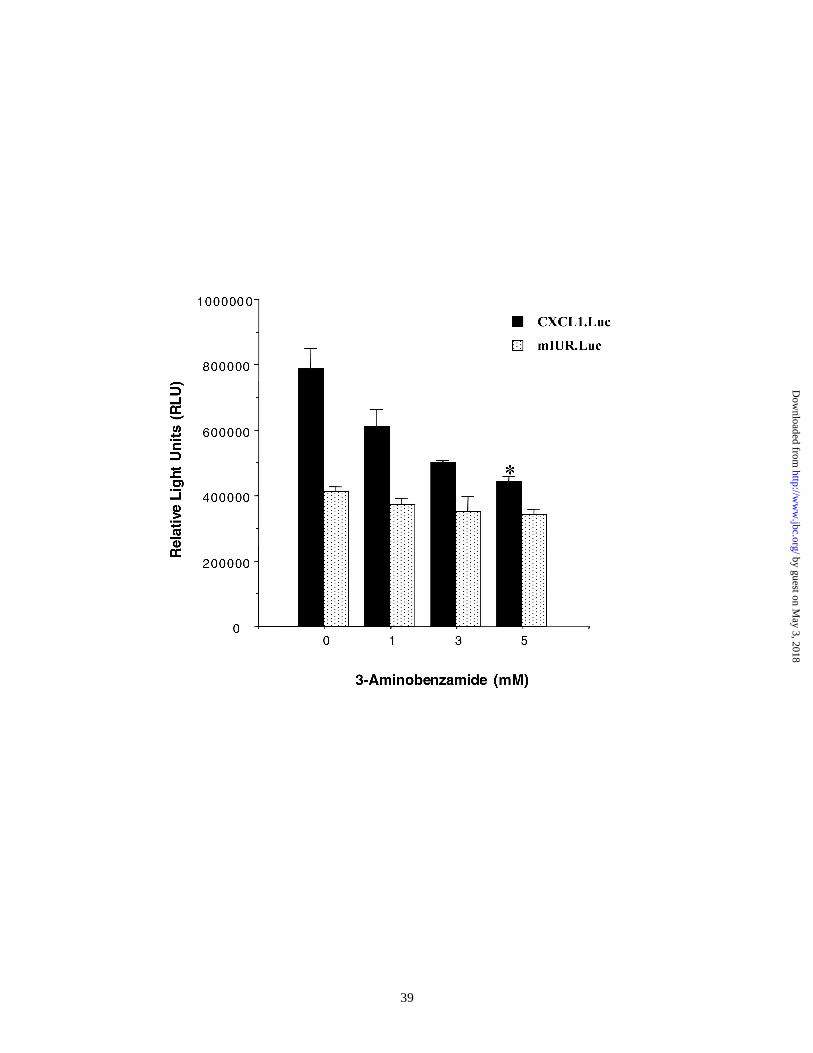
To test the possibility that the effect of 3-AB on CXCL1 mRNA levels is at the
level of the CXCL1 promoter and involved the IUR element, luciferase reporter
constructs driven by either the wild type CXCL1 promoter or a promoter containing
replacements in the IUR element of this promoter were transiently transfected in Hs294T
cells and cells were treated with 3-AB in a dose range of 1-5 mM. Cells were harvested
48 h after transfection and luciferase activity was measured. Figure 5 shows a
representative of four independent experiments performed in duplicate. The data indicate
that 3-AB inhibited CXCL1 promoter activity in a dose dependent manner. Maximum
inhibition (50%) was observed at 5 mM 3-AB. There was no effect on the mutant IUR
promoter (mIUR.Luc) although the mIUR promoter contained intact binding sites for NF-
κB, HMGI(Y)and Sp1. The results in Figure 2 show that the 115 kDa protein identified
to be PARP bound the wild type IUR but not the mutant mIUR sequence and that
recombinant PARP specifically bound the IUR but not the mIUR sequence (Figure 4).
Results shown in Figure 5 further demonstrate that the PARP specific inhibitor, 3-AB
specifically inhibits the wild type CXCL1, but not the mIUR promoter, indicating that
by guest on May 3, 2018
http://ww
w.jbc.org/
Dow
nloaded from
21
PARP mediated ADP ribosylation is involved in the trans-activation of the CXCL1
promoter. This rules out the possibility that 3-AB mediated inhibition of CXCL1 mRNA
levels is due to abrogation of other trans-acting factors of the CXCL1 promoter.
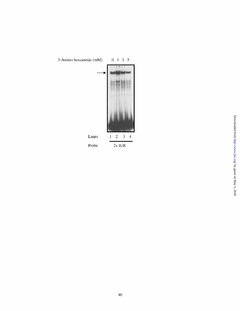
Binding of PARP to the IUR element does not require ADP-ribosylation:
Since 3-AB treatment had a dramatic effect on endogenous CXCL1 mRNA levels as well
as on the CXCL1 promoter activity in reporter assays, we considered the possibility that
binding of PARP to the IUR element might involve the ADP-ribosylation activity of the
factor. Hs294T cells were treated with 3-AB within a dose range of 1-5 mM. Cells were
harvested after 48 h of 3-AB treatment and nuclear extracts were prepared. Figure 6
shows a representative EMSA experiment in which nuclear extracts from DMSO treated
or 3-AB treated cells were probed with radiolabeled 2xIUR oligonucleotide. The results
indicate that there was no difference in the binding of the IUR-specific protein, PARP,
with 3-AB treatment. The data suggest that ADP-ribosylation may not be involved in
binding of PARP to the IUR element.
The data presented in this study provide evidence that the IUR element is a positive cis-
acting element in the CXCL1 promoter whose function depends on its contiguity with the
adjacent NF-kB element. The IUR binds a 115 kDa factor which has been purified and
identified as PARP. Although binding of PARP does not require the ADP ribose
transferase function of PARP, this activity appears to be important for its ability to
transactivate the CXCL1 promoter and induce CXCL1 mRNA levels. A model for the
regulation of CXCL1 transcription is proposed .
by guest on May 3, 2018
http://ww
w.jbc.org/
Dow
nloaded from
22
Discussion
The CXCL1 gene is expressed at a high constitutive level in several disorders
including acute and chronic inflammation and malignant melanoma. Our earlier findings
indicate that the CXCL1 gene is under the control of a 306 bp minimal promoter
containing four cis acting elements, including a 20 bp region called the IUR, located
immediately upstream to the NF-κB binding sequence (7). In an effort to understand the
mechanism by which the CXCL1 gene is regulated, we have further characterized the
IUR element in the CXCL1 promoter and examined its role in CXCL1 gene regulation.
To examine whether the IUR has a positive or negative role in CXCL1 regulation,
we first made mutations in the CXCL1 promoter which either altered the TCGAT motif
or separated it from the adjacent NF-κB element. When tested in luciferase reporter
assays, these mutations effected an almost 50% inhibition in CXCL1 promoter activity.
The data indicate that the IUR is a positive regulatory sequence in the CXCL1 promoter
and that the contiguity of the IUR and NF-κB element is critical for optimal promoter
activity.
In electrophoretic mobility shift assays, we previously identified constitutive
binding to the IUR element and detected a 115 kDa protein which bound to the IUR
element in southwestern blot analyses. Mutations which altered the TCGAT motif
eliminated binding in both assays suggesting that these interactions were specific to the
TCGAT motif of the IUR element (8). Taken together with our current observations from
reporter assays, the evidence points to the possibility that p115 is a positive
transcriptional regulator of the CXCL1 gene.
by guest on May 3, 2018
http://ww
w.jbc.org/
Dow
nloaded from
23
In this study we have purified the 115 kDa protein binding to the IUR element
using a two-step procedure involving binding site oligonucleotide affinity
chromatography and electrophoretic separation through a Biorad mini-prep cell. Pre-
incubation of the nuclear extract through a mutant-IUR-sepharose matrix eliminated non-
specific proteins and ensured that bound fractions eluting from the wild-type IUR-
sepharose matrix represented proteins that selectively bound the wild-type but not the
mutant IUR oligonucleotide.
Peptide sequence analysis has established the identity of the 115 kDa IUR-
specific protein as the Poly (ADP-ribose) polymerase (PARP). We show here by western
blot analysis that the 115 kDa PARP is enriched in the IUR affinity purified fraction, that
peptide sequence of the purified 115 kDa matches that of PARP, that a commercial
preparation of the PARP enzyme can discriminate between the wild-type and mutant IUR
sequences and treatment of cells with 3-Amino benzamide (3-AB), a specific PARP
inhibitor, significantly reduces CXCL1 mRNA levels in Hs294T cells and inhibits
CXCL1 promoter activity in transient transfection assays. These data indicate that PARP
transactivates the CXCL1 promoter and its ADP ribose transferase activity is essential for
this process.
The ability of PARP to activate or suppress specific gene expression appears to be
dependent on at least two factors: (1) the ADP-ribose transferase activity of PARP (12).
(2) DNA binding of PARP (13). Reversible repression of gene expression by PARP can
be correlated with its catalytic activity and may not involve DNA binding of the enzyme.
PARP-catalyzed ADP-ribosylation of the TATA binding protein (TBP), Yin-Yang-1
(YY1), p53, NF-kB, Sp1, CREB (12), Oct-1 (21)and retinoid X receptors (22) has been
by guest on May 3, 2018
http://ww
w.jbc.org/
Dow
nloaded from
24
shown to occur before DNA binding and prevented the formation of transcriptionally
active complexes. In contrast, DNA-bound PARP appears to be associated with trans-
activation. PARP has been identified as the active component of the transcription factor
TFIIC (19) In addition, PARP has been demonstrated to enhance activator-dependent
transcription during which, DNA binding, but not the ADP-ribose transferase activity, of
the unmodified PARP seemed to be necessary for transcriptional activation (15). More
recently, PARP has been shown to trans-activate the cardiac Troponin T promoter by
binding to a 5’ TGTTG 3’ sequence in the MCAT cis element and cooperating with the
muscle-specific TEF-1 transcription factor (16). In the latter case, both DNA binding and
ADP-ribose transferase functions appear to be essential for coactivation of the troponin
gene promoter.
We examined the possibility that PARP-mediated ADP-ribosylation plays a role
in the regulation of CXCL1 gene expression. We addressed this question by examining
the effects of 3-Aminobenzamide, a specific inhibitor of PARP-mediated ADP-
ribosylation, on (1) CXCL1 mRNA levels, (2) CXCL1 promoter activity and (3) specific
binding to the IUR oligonucleotide probe. We show here that inhibition by 3AB reduces
CXCL1 mRNA levels and blocks CXCL1 promoter activity up to 50 % over untreated
controls in a dose dependent manner. The level of inhibition seen with 3AB appears to be
similar to that observed when the TCGAT motif in the CXCL1 promoter is altered. In
addition, the activity of the mutant-IUR promoter appears to be insensitive to 3-AB
treatment indicating that 3AB effects target events associated with the TCGAT motif.
Inhibition of PARP-mediated ADP-ribosylation, however had no effect on the complex
associated with the IUR probe in EMSA indicating that although ADP-ribosylation is
by guest on May 3, 2018
http://ww
w.jbc.org/
Dow
nloaded from
25
required for trans-activation of the CXCL1 promoter, it may not be necessary for binding
to the IUR element in the promoter.
Our data indicate the following : (1) contiguity with NF-κB element appears to be
necessary for trans-activation through the IUR element; (2) the 115 kDa PARP binds to
the IUR element in the CXCL1 promoter; (3) PARP is associated with trans-activation of
the CXCL-1 promoter; (4) the ADP ribose transferase activity is necessary for trans
activation; (5) this activity is not necessary for binding of PARP to the IUR element.
Based on these findings we have proposed a model for the regulation of the CXCL1
promoter
We have uncovered a novel role for the PARP in the regulation of the CXCL1
chemokine gene. The CXCL1 minimal promoter, under the control of five cis-acting
elements including the Sp1, IUR, NF-κB, HMGI(Y) and TATA box binding sites, is a
template for a multi-protein complex, the CXCL1 enhanceosome. The TCGAT motif of
the IUR element and the 115 kDa PARP, which specifically binds the IUR, appear to
possess distinct positive roles in CXCL1 regulation. PARP appears to be associated with
the trans-activation of the gene. One mechanism of PARP-mediated trans-activation of
the CXCL1 promoter may involve the NF-κB site because dislocation of the IUR element
from this site inhibits trans-activation of the promoter, possibly by disrupting interactions
between PARP and Rel factors. We have no evidence of protein-protein interactions
between PARP and the NF-κB factors at this time. However, a recent report has
demonstrated that NF-κB dependent transcription activation is severely compromised in
mice that are deficient in PARP (13). PARP deficient mice appear to have extensive skin
disorder and extreme sensitivity to X-ray irradiation, a phenotype closely resembling the
by guest on May 3, 2018
http://ww
w.jbc.org/
Dow
nloaded from
26
one observed in mice deficient for IKK2, the kinase involved in the NF-κB activation
pathway. Based on the findings by Hassa et al., PARP deficiency may contribute to a
failure in NF-κB signaling (13).
In a separate study we have identified a second protein binding to the IUR
element which is also specific to the TCGAT motif. Preliminary evidence indicates that
the 170 kDa IUR-specific factor could be a repressor of CXCL1 transcription. A second
mechanism of PARP mediated trans-activation could involve displacement or
inactivation by ADP-ribosylation of the putative 170 kDa trans-repressor of the CXCL1
promoter binding to a site at or near the IUR element
Tumor models involving PARP are yet to be developed. In this context, the role
of PARP as an anti-apoptotic factor may be important. During the onset of apoptosis,
PARP is cleaved into two fragments, 85 and 29 kDa, by caspase-3, a crucial component
of the apoptotic cascade and this is thought to contribute to cell death (24). However,
direct evidence for PARP as an agent of cell proliferation is forthcoming. In this regard,
the DNA binding and transcriptional regulatory properties of PARP may be more
revealing. PARP has been previously shown to modulate DNA binding and specificity of
the anti-tumor factor, p53 (14). More of such evidence will be crucial towards our
understanding of its role in cell proliferation and tumorigenesis. Our findings that PARP
binds the CXCL1 promoter and is likely involved in its trans-activation, therefore,
present an important step in that direction.
Future investigations in this area could examine interactions between PARP and
other constituents of the CXCL1 enhanceosome, compare levels or post-translational
modification status of these factors in normal and melanoma cells and trace the signaling
by guest on May 3, 2018
http://ww
w.jbc.org/
Dow
nloaded from
27
pathways that modulate their activity. These investigations could elucidate the role for
PARP in disorders such as chronic inflammation and melanoma.
Acknowledgements:
This work was supported by the Department of Veterans Affairs and grants from the NCI
(CA56704) to Ann Richmond. We acknowledge the expert technical contribution of
Steve Gygi and Ruedi Aebersold at the University of Washington, Seattle, WA for the
MALDI Mass Spectroscopy sequence analysis of the 115 kDa protein. Technical support
from the Vanderbilt Ingram Cancer Center grant (CA68485) and the Skin Diseases
Research Center grant (5P30AR4194) is acknowledged. We are indebted to Roland Stein
and Steve Hann for their critical reading of the manuscript and helpful suggestions.
Technical assistance from Amy Pruitt, Ben Johnston, and Neepa Ray were enormously
helpful.
by guest on May 3, 2018
http://ww
w.jbc.org/
Dow
nloaded from
28
List of References
1. Richmond, A. and Shattuck, R.L. (1996) in Chemoattractant Ligands and their
Receptors (Horuk, R., ed) pp. 87-124, CRC Press, Boca Raton.
2. Shattuck, R.L., Wood, L.D., Jaffe, G.J., and Richmond, A. (1994) Mol.Cell.Biol. 14,
791-802.
3. Wood, L.D., Farmer, A.A., and Richmond, A. (1995) Nucleic Acids Research 23,
4210-4219.
4. Wood, L.D., Shattuck, R.L., and Richmond, A. (1992) J.Biol.Chem. 270, 30619-
30626.
5. Shattuck-Brandt, R.L. and Richmond, A. (1997) Cancer Res. 57, 3032-3039.
6. Devalaraja, M.N., Wang, D.Z., Ballard, D.W., and Richmond, A. (1999) Cancer Res.
59, 1372-1377.
7. Wood, L.D. and Richmond, A. (1995) J.Biol.Chem. 270, 30619-30626.
8. Luan, J., Shattuck-Brandt, R., Haghnegahdar, H., Owen, J.D., Strieter, R., Burdick,
M., Nirodi, C., Beauchamp, D., Johnson, K.N., and Richmond, A. (1997)
J.Leuko.Biol. 62, 588-597.
9. Lindahl, T. (1995) J.Cell Sci.Suppl. 19:73-7, 73-77.
10. Berger, N.A. (1985) Radiat.Res. 101, 4-15.
11. Lindahl, T., Satoh, M.S., Poirier, G.G., and Klungland, A. (1995)
Trends.Biochem.Sci. 20 , 405-411.
by guest on May 3, 2018
http://ww
w.jbc.org/
Dow
nloaded from
29
12. Oei, S.L., Griesenbeck, J., Ziegler, M., and Schweiger, M. (1998) Biochemistry 37,
1465-1469.
13. Hassa, P.O. and Hottiger, M.O. (1999) Biol.Chem. 380, 953-959.
14. Wesierska-Gadek, J., Schmid, G., and Cerni, C. (1996) Biochem. Biophys. Res.
Commun. 224 , 96-102.
15. Meisterernst, M., Stelzer, G., and Roeder, R.G. (1997) Proc.Natl.Acad.Sci.U.S.A. 94,
2261-2265.
16. Butler, A.J. and Ordahl, C.P. (1999) Mol.Cell Biol. 19, 296-306.
17. Plaza,S.; Aumercier,M.; Bailly,M.; Dozier,C.; Saule,S. (1999) Oncogene, 18, 1041-
1052.
18. Cervellera,M.N.; Sala,A. (2000) J. Biol.Chem. 275, 10692-10696.
19. Anderson, M.G., Scoggin, K.E., Simbulan-Rosenthal, C.M., and Steadman, J.A.
(2000) J.Virol. 74, 2169-2177.
20. Kadonaga, J.T. and Tjian, R. (1986) Proc.Natl.Acad.Sci.USA 83, 5889-5893.
21. Nie, J., Sakamoto, S., Song, D., Qu, Z., Ota, K., and Taniguchi, T. (1998) FEBS Lett.
424, 27-32.
22. Miyamoto, T., Kakizawa, T., and Hashizume, K. (1999) Mol.Cell Biol. 19, 2644-
2649.
23. Tewari, M., Quan, L.T., O'Rourke, K., Desnoyers, S., Zeng, Z., Beidler, D.R., Poirier,
G.G., Salvesen, G.S., and Dixit, V.M. (1995) Cell 81, 801-809.
by guest on May 3, 2018
http://ww
w.jbc.org/
Dow
nloaded from
30
Figure Legends.
Figure 1. The IUR element is a positive cis element which requires the TCGAT motif as
well as contiguity with the adjacent NF-κB element :(A) 5 x 10⁵ Hs294T cells were
transfected with RSV-β-Gal vector and one of the following luciferase reporter
constructs (1) a reporter construct lacking the CXCL1 promoter (pGL2); (2) a
reporter construct driven by the wild-type CXCL1 promoter in the correct orientation
relative to the transcription start site (WT); (3) a reporter construct with the CXCL1
promoter in the opposite direction (REV); (4) a mutant CXCL1 promoter in which the
TCGAT motif was altered to AGTAC (mIUR). (B) 5 x 10⁵ Hs294T cells were
transfected with RSV-β-Gal vector as well as luciferase reporter constructs driven by
the wild type CXCL1 promoter or mutant promoter which had an insertion of 2 bp
(IN:2), 6 bp (IN: 6), 12 bp (IN:12) or 25 bp (IN:25) between the IUR and the NF-κb
elements. 48 h after transfection, cells were harvested and luciferase activity was
measured and normalized to β-galactosidase activity. Shown here is a representative
of three independent experiments performed each time in triplicate. Mean normalized
values obtained from the sample showing the highest inhibition in promoter activity
were compared to those from samples transfected with the wild type promoter and
were found to be significantly different according to Student’s T-test (paired). The
asterisk (*) indicates p< 0.01.
Figure 2. Binding site oligonucleotide affinity purification of the 115 kDa IUR-specific
protein. In Electrophoretic mobility shift assays (A), crude HeLa nuclear extract (2
µg) (lane 1) or 200 ng bound fractions eluting at 0.2 M NaCl (lanes 2-5), 0.4 M NaCl
by guest on May 3, 2018
http://ww
w.jbc.org/
Dow
nloaded from
31
(lanes 7-10), 0.6 M NaCl (lanes 11-14) or 1 M NaCl (lanes 16-18) from the IUR-
agarose column, were tested for binding to the 2R probe in EMSA. All samples
contained a 250-fold molar excess of cold oligonucleotide corresponding to the 2mR
probe. (B) Southwestern blot analysis : Polypeptides in crude HeLa nuclear extract
(25 µg) (lanes 1 and 3) or the 0.4 M NaCl fraction (100 ng) eluted from the IUR-
agarose column were separated on 8% SDS-PAGE gels, trans-blotted on
nitrocellulose membranes and probed with radio-labeled oligonucleotides
corresponding to either the 2R (lanes 1 and 2) or the 2mR (lanes 3 and 4) probes. The
relative molecular size is indicated on the left. (C) Silver-stained SDS-PAGE profile
of the 0.4 M NaCl fraction. analysis : Polypeptides in crude HeLa nuclear extract
(500 ng) or the 0.4 M NaCl fraction (100 ng) eluted from the IUR-agarose column
was separated on 8% SDS-PAGE gels and the protein was visualized by silver-
staining. (D) SDS-PAGE electro-elution : The 0.4 M IUR-agarose eluate was then
fractionated by electro-elution through a 7% SDS-PAGE using the Model 491 Prep
Cell (Biorad). Fractions were collected and every 8th fraction was electrophoresed on
an 8% SDS-PAGE gel and proteins were stained by silver-staining.
Figure 3. PARP can bind the IUR element : (A) Western Blot analysis : crude HeLa
nuclear extract (25 µg), unbound/flow-through fraction from the IUR-agarose column
(25 µg) and 100 ng of 0.2 M (lanes 3 and 4) or 0.4 M (lanes 5 and 6) IUR-agarose
fractions were separated on 8% SDS-PAGE gels and probed with anti-PARP
antibody. Arrow (→) indicates the relative mobility of the PARP protein. (B) EMSA :
Partially purified, commercially available PARP was tested for binding to the 2xIUR
(lanes 1-3) or the 2xmIUR (lanes 4-6) probes. Non-specific complexes were
by guest on May 3, 2018
http://ww
w.jbc.org/
Dow
nloaded from
32
competed with poly dI:dC in the range of 0.2- 1.0 µg. The arrow (→) indicates the
specific complex formed by PARP with the 2xIUR probe, while NS represents non-
specific complexes.
Figure 4. 3AB inhibits MGSA/GRO expression (A) Northern analysis : Hs294T cells
were treated for 48 h with indicated concentrations of 3-aminobenzamide (3-AB).
RNA from samples was resolved on a 1.4% formaldehyde-agarose gel, trans-blotted
to nitrocellulose membrane and probed with an MGSA/GRO cDNA probe (A, top
panel). Blots were stripped and re-probed with a cDNA probe for cyclophilin B
transcripts (A, bottom panel). Blots were densitometrically scanned using a
phosphorimager (Molecular Dynamics). Values for MGSA/GRO mRNA were
normalized against those for cyclophilin mRNA. (B) A graphical representative of 6
independent experiments, each performed in triplicate is shown. The results in all 6
experiments was qualitatively identical. Error bars represent SD values. The mean
normalized value obtained from samples receiving the highest dose of 3AB was
compared to those from samples receiving DMSO and were found to be significantly
different according to Student’s paired T-test. The asterisk indicates p<0.01.
Figure 5. 3-AB inhibits CXCL1 promoter activity : 5 x 10⁵ cells were first
transfected with 1 µg of pRSV-β-gal reporter as well as reporter constructs driven by
either the wild type (MGSA.Luc) or the mutant CXCL1 promoter (mIUR.Luc). Six hours
after transfection, the medium was replaced by medium containing either DMSO or 3 -
AB within a concentration range of 1-5 mM. Cells were harvested 48 h after transfection
and luciferase activity was measured. Values obtained were normalized to β-
by guest on May 3, 2018
http://ww
w.jbc.org/
Dow
nloaded from
33
galactosidase activity. The experiment was performed three times in triplicate. Error bars
represent standard deviations. The mean normalized values obtained from samples
receiving the highest dose of 3AB as compared to those from samples receiving DMSO
were significantly different according to Student’s paired T-test The asterisk indicates
p<0.01.
Figure 6. 3-AB has no effect on binding to the IUR element. Hs 294T cells were treated
with either DMSO alone or with 1-5 mM 3-AB for 48 h. Cells were harvested and
nuclear extracts were made. 10 µg of nuclear extract was incubated with 32P labeled
2xIUR oligonucleotide probe in the presence of 1 – 2 µg of poly dI.dC. The reaction
mixtures electrophoresed on 6% native polyacrylamide gels which were then dried and
processed for autoradiography. The arrow indicates the specific complex associated with
the IUR probe. Shown here is a representative of three independent experiments. Results
were qualitatively similar in all three experiments.
by guest on May 3, 2018
http://ww
w.jbc.org/
Dow
nloaded from
RichmondChaitanya S Nirodi, Subir NagDas, Steven P Gygi, Gary Olson, Ruedi Aebersold and Ann
melanoma growth stimulatory activity (CXCL1) gene expressionA role for poly ADP-ribose polymerase (PARP) in the transcriptional regulation of
published online December 8, 2000J. Biol. Chem.
10.1074/jbc.M009897200Access the most updated version of this article at doi:
Alerts:
When a correction for this article is posted•
When this article is cited•
to choose from all of JBC's e-mail alertsClick here
by guest on May 3, 2018
http://ww
w.jbc.org/
Dow
nloaded from


![Untersuchungen zum Wirkmechanismus von 6-Amino-11,12 ... · PARP Poly [ADP-ribose] polymerase PBGD Porphobilinogen deaminase PBS Phosphate buffered saline PCR Polymerase chain reaction](https://img.pdfslide.us/doc/110x75/5d5cbcc088c9939b368b7c27/untersuchungen-zum-wirkmechanismus-von-6-amino-1112-parp-poly-adp-ribose.jpg)

![Novel therapies are changing treatment paradigms in ... · Polyadenosine diphosphate [ADP]-ribose polymerase (PARP) is a nuclear enzyme that aids the repair of single-strand DNA breaks](https://img.pdfslide.us/doc/110x75/60f47096160be920b7480ca6/novel-therapies-are-changing-treatment-paradigms-in-polyadenosine-diphosphate.jpg)


![Flyer PARP FAmily NP V01 - biolinks k.k.1].pdf · Tomorrow’s Reagents Manufactured Today® International Edition The PARP Family T highlight PRODUCT FLYER The PARP Family](https://img.pdfslide.us/doc/110x75/5cb9946788c993f37c8c0cfc/flyer-parp-family-np-v01-biolinks-kk-1pdf-tomorrows-reagents-manufactured.jpg)