Embed Size (px)
Citation preview
1
Supplementary information
Structure of mammalian poly (ADP-ribose) glycohydrolase reveals a flexible tyrosine
clasp as a unique substrate-binding element In-Kwon Kim, James R. Kiefer, Chris M. Ho, Roderick A. Stegeman, Scott Classen, John A. Tainer, and Tom
Ellenberger
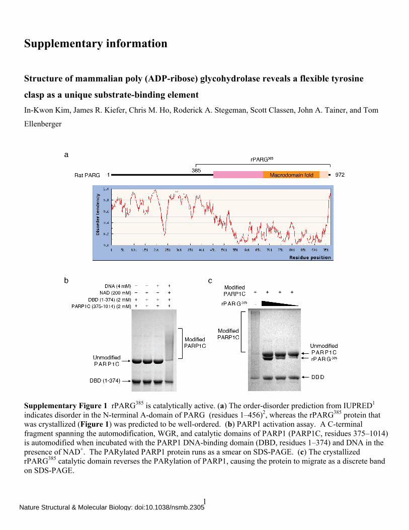
Supplementary Figure 1 rPARG385 is catalytically active. (a) The order-disorder prediction from IUPRED1 indicates disorder in the N-terminal A-domain of PARG (residues 1–456)2, whereas the rPARG385 protein that was crystallized (Figure 1) was predicted to be well-ordered. (b) PARP1 activation assay. A C-terminal fragment spanning the automodification, WGR, and catalytic domains of PARP1 (PARP1C, residues 375–1014) is automodified when incubated with the PARP1 DNA-binding domain (DBD, residues 1–374) and DNA in the presence of NAD+. The PARylated PARP1 protein runs as a smear on SDS-PAGE. (c) The crystallized rPARG385 catalytic domain reverses the PARylation of PARP1, causing the protein to migrate as a discrete band on SDS-PAGE.
Nature Structural & Molecular Biology: doi:10.1038/nsmb.2305
2
Nature Structural & Molecular Biology: doi:10.1038/nsmb.2305
3
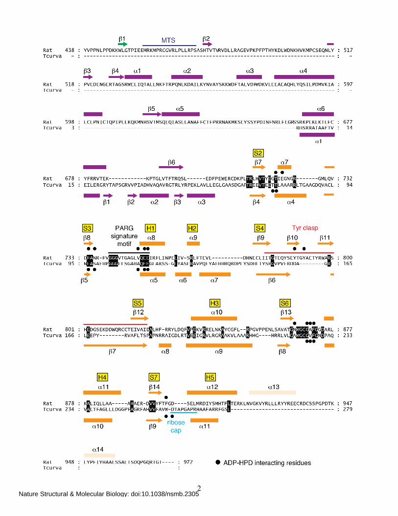
Supplementary Figure 2 Structure-based alignment of rat PARG and T. curvata glycohydrolase. The conserved strands (S2, S3, etc.) and helices (H1, H2, etc.) of a canonical macro domain fold are shown with the sequences of rat PARG and T. curvata glycohydrolase aligned by superposition of the crystal structures. Residues in contact with the ADP-HPD ligand (•) are primarily located within the connecting segments joining strands S2, S3, S6, and S7 of the macro domain fold. A polypeptide segment in the T. curvata glycohydrolase, which we term the ribose cap (cyan), completely blocks the 2′-OH of the adenosine ribose and restricts its activity to exo-glycosidic cleavage of PAR polymers3.
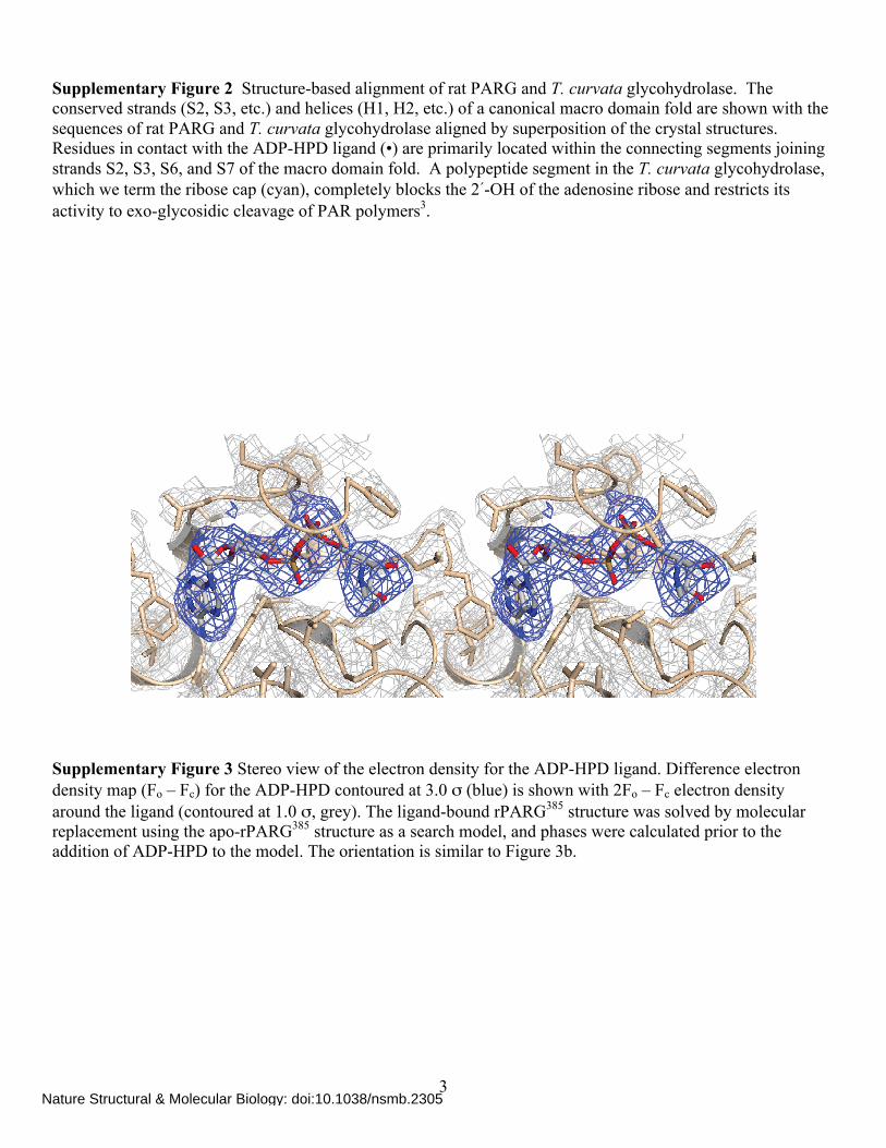
Supplementary Figure 3 Stereo view of the electron density for the ADP-HPD ligand. Difference electron density map (Fo – Fc) for the ADP-HPD contoured at 3.0 σ (blue) is shown with 2Fo – Fc electron density around the ligand (contoured at 1.0 σ, grey). The ligand-bound rPARG385 structure was solved by molecular replacement using the apo-rPARG385 structure as a search model, and phases were calculated prior to the addition of ADP-HPD to the model. The orientation is similar to Figure 3b.
Nature Structural & Molecular Biology: doi:10.1038/nsmb.2305
4
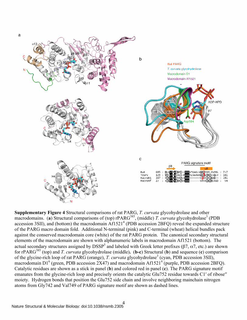
Supplementary Figure 4 Structural comparisons of rat PARG, T. curvata glycohydrolase and other macrodomains. (a) Structural comparisons of (top) rPARG385, (middle) T. curvata glycohydrolase3 (PDB accession 3SII), and (bottom) the macrodomain Af15214 (PDB accession 2BFQ) reveal the expanded structure of the PARG macro domain fold. Additional N-terminal (pink) and C-terminal (wheat) helical bundles pack against the conserved macrodomain core (white) of the rat PARG protein. The canonical secondary structural elements of the macrodomain are shown with alphanumeric labels in macrodomain Af1521 (bottom). The actual secondary structures assigned by DSSP5 and labeled with Greek letter prefixes (β7, α7, etc.) are shown for rPARG385 (top) and T. curvata glycohydrolase (middle). (b-c) Structural (b) and sequence (c) comparison of the glycine-rich loop of rat PARG (orange), T. curvata glycohydrolase3 (cyan, PDB accession 3SII), macrodomain D16 (green, PDB accession 2X47) and macrodomain Af15214 (purple, PDB accession 2BFQ). Catalytic residues are shown as a stick in panel (b) and colored red in panel (c). The PARG signature motif emanates from the glycine-rich loop and precisely orients the catalytic Glu752 residue towards C1′ of ribose′′ moiety. Hydrogen bonds that position the Glu752 side chain and involve neighboring mainchain nitrogen atoms from Gly742 and Val749 of PARG signature motif are shown as dashed lines.
Nature Structural & Molecular Biology: doi:10.1038/nsmb.2305
5
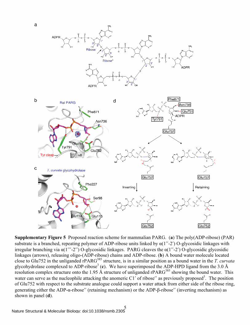
Supplementary Figure 5 Proposed reaction scheme for mammalian PARG. (a) The poly(ADP-ribose) (PAR) substrate is a branched, repeating polymer of ADP-ribose units linked by α(1ʹ′ʹ′-2ʹ′) O-glycosidic linkages with irregular branching via α(1ʹ′ʹ′ʹ′-2ʹ′ʹ′) O-glycosidic linkages. PARG cleaves the α(1ʹ′ʹ′-2ʹ′) O-glycosidic glycosidic linkages (arrows), releasing oligo-(ADP-ribose) chains and ADP-ribose. (b) A bound water molecule located close to Glu752 in the unliganded rPARG385 structure, is in a similar position as a bound water in the T. curvata glycohydrolase complexed to ADP-ribose3 (c). We have superimposed the ADP-HPD ligand from the 3.0 Å resolution complex structure onto the 1.95 Å structure of unliganded rPARG385 showing the bound water. This water can serve as the nucleophile attacking the anomeric C1ʹ′ of riboseʹ′ʹ′ as previously proposed3. The position of Glu752 with respect to the substrate analogue could support a water attack from either side of the ribose ring, generating either the ADP-α-riboseʹ′ʹ′ (retaining mechanism) or the ADP-β-riboseʹ′ʹ′ (inverting mechanism) as shown in panel (d).
Nature Structural & Molecular Biology: doi:10.1038/nsmb.2305
6
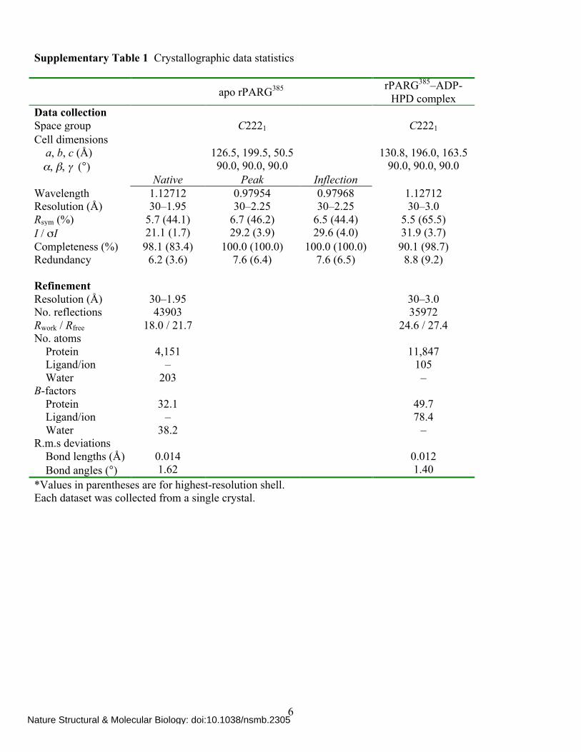
Supplementary Table 1 Crystallographic data statistics apo rPARG385 rPARG385–ADP-
HPD complex Data collection Space group C2221 C2221 Cell dimensions a, b, c (Å) 126.5, 199.5, 50.5 130.8, 196.0, 163.5 α, β, γ (°) 90.0, 90.0, 90.0 90.0, 90.0, 90.0 Native Peak Inflection Wavelength 1.12712 0.97954 0.97968 1.12712 Resolution (Å) 30–1.95 30–2.25 30–2.25 30–3.0 Rsym (%) 5.7 (44.1) 6.7 (46.2) 6.5 (44.4) 5.5 (65.5) I / σI 21.1 (1.7) 29.2 (3.9) 29.6 (4.0) 31.9 (3.7) Completeness (%) 98.1 (83.4) 100.0 (100.0) 100.0 (100.0) 90.1 (98.7) Redundancy 6.2 (3.6) 7.6 (6.4) 7.6 (6.5) 8.8 (9.2) Refinement Resolution (Å) 30–1.95 30–3.0 No. reflections 43903 35972 Rwork / Rfree 18.0 / 21.7 24.6 / 27.4 No. atoms Protein 4,151 11,847 Ligand/ion – 105 Water 203 – B-factors Protein 32.1 49.7 Ligand/ion – 78.4 Water 38.2 – R.m.s deviations Bond lengths (Å) 0.014 0.012 Bond angles (°) 1.62 1.40 *Values in parentheses are for highest-resolution shell. Each dataset was collected from a single crystal.
Nature Structural & Molecular Biology: doi:10.1038/nsmb.2305
7
SUPPLEMENTARY REFERENCES
1. Dosztanyi, Z., Csizmok, V., Tompa, P. & Simon, I. IUPred: web server for the prediction of intrinsically unstructured regions of proteins based on estimated energy content. Bioinformatics 21, 3433-4 (2005).
2. Patel, C.N., Koh, D.W., Jacobson, M.K. & Oliveira, M.A. Identification of three critical acidic residues of poly(ADP-ribose) glycohydrolase involved in catalysis: determining the PARG catalytic domain. Biochem J 388, 493-500 (2005).
3. Slade, D. et al. The structure and catalytic mechanism of a poly(ADP-ribose) glycohydrolase. Nature 477, 616-20 (2011).
4. Karras, G.I. et al. The macro domain is an ADP-ribose binding module. EMBO J 24, 1911-20 (2005). 5. Kabsch, W. & Sander, C. Dictionary of protein secondary structure: pattern recognition of hydrogen-
bonded and geometrical features. Biopolymers 22, 2577-637 (1983). 6. Chen, D. et al. Identification of macrodomain proteins as novel O-acetyl-ADP-ribose deacetylases. J
Biol Chem 286, 13261-71 (2011).
Nature Structural & Molecular Biology: doi:10.1038/nsmb.2305