Embed Size (px)
Citation preview
Guideline
Management of epithelial precancerous conditions and lesions inthe stomach (MAPS II): European Society of GastrointestinalEndoscopy (ESGE), European Helicobacter and Microbiota StudyGroup (EHMSG), European Society of Pathology (ESP), andSociedade Portuguesa de Endoscopia Digestiva (SPED) guidelineupdate 2019
Authors
Pedro Pimentel-Nunes1,2, 3, Diogo Libânio1,2, Ricardo Marcos-Pinto2,4, Miguel Areia2, 5, Marcis Leja6,
Gianluca Esposito7, Monica Garrido4, Ilze Kikuste6, Francis Megraud8, Tamara Matysiak-Budnik9, Bruno Annibale7,
Jean-Marc Dumonceau10, Rita Barros11, 12, Jean-François Fléjou13, Fátima Carneiro11, 12, 14, Jeanin E. van Hooft15,
Ernst J. Kuipers16, Mario Dinis-Ribeiro1,2
Institutions
1 Gastroenterology Department, Portuguese Oncology
Institute of Porto, Portugal
2 Center for Research in Health Technologies and
Information Systems (CINTESIS), Faculty of Medicine,
Porto, Portugal
3 Surgery and Physiology Department, Faculty of
Medicine of the University of Porto, Porto, Portugal,
4 Department of Gastroenterology, Porto University
Hospital Centre, Institute of Biomedical Sciences,
University of Porto (ICBAS/UP), Portugal
5 Gastroenterology Department, Portuguese Oncology
Institute of Coimbra, Portugal
6 Institute of Clinical and Preventive Medicine, University
of Latvia, Digestive Diseases Center, GASTRO, Riga,
Latvia
7 Department of Medicine, Surgery and Translational
Medicine University Hospital Sant’Andrea, University
Sapienza Roma, Rome, Italy
8 INSERM U1053, Université de Bordeaux and CHU
Pellegrin, Laboratoire de Bacteriologie, Bordeaux,
France
9 IMAD, Hepato-Gastroenterology and Digestive
Oncology, CHU de Nantes, University of Nantes, France
10 Gedyt Endoscopy Center, Buenos Aires, Argentina
11 Institute of Molecular Pathology and Immunology at
the University of Porto (Ipatimup), Porto, Portugal
12 Instituto de Investigação e Inovação em Saúde (i3S),
University of Porto, Porto, Portugal
13 Service d’Anatomie Pathologique, Hôpital Saint-
Antoine, AP-HP, Faculté de Médecine Sorbonne
Université, Paris, France
14 Pathology Department, Centro Hospitalar de São João
and Faculty of Medicine, Porto, Portugal
15 Department of Gastroenterology and Hepatology,
Amsterdam UMC, University of Amsterdam, The
Netherlands
16 Department of Gastroenterology and Hepatology,
Erasmus MC University Medical Center, Rotterdam,
The Netherlands
Bibliography
DOI https://doi.org/10.1055/a-0859-1883
Published online: 0.0.2019 | Endoscopy 2019; 51: 365–388
© Georg Thieme Verlag KG Stuttgart · New York
ISSN 0013-726X
Corresponding author
Pedro Pimentel-Nunes, MD PhD, Gastroenterology
Department, Portuguese Oncology Institute of Porto, Rua
Dr. Bernardino de Almeida, 4200-072 Porto, Portugal
Fax: +351-22-5513646
Supplementary material
Online content viewable at:
https://doi.org/10.1055/a-0859-1883
Pimentel-Nunes Pedro et al. MAPS II … Endoscopy 2019; 51
1 IntroductionGastric cancer is still a major world problem, ranking fifth forincidence and third for cancer-related mortality worldwide inthe latest published global cancer statistics [1]. Even thoughearly recognition and treatment is possible, most cases are di-agnosed at a late stage and thus most patients with a diagnosisof gastric cancer die of the disease [1]. Screening and surveil-lance of people at risk may decrease gastric cancer mortalityby allowing early detection and treatment, often by endoscopyinstead of more invasive surgery, and have therefore been re-commended [2, 3].
In 2012, the European Society of Gastrointestinal Endoscopy(ESGE), the Sociedade Portuguesa de Endoscopia Digestiva(SPED), the European Helicobacter and Microbiota Study Group(EHMSG), and the European Society of Pathology (ESP) pro-duced the first international guideline on the management ofprecancerous conditions and lesions in the stomach (MAPS) [4,5]. Its recommendations were then presented in various coun-tries, and were adapted and translated in some. Moreover, theMAPS Guideline was incorporated into ESGE guidelines on qual-ity parameters for upper gastrointestinal (GI) endoscopy [6].
This document aims to update the first MAPS guideline (re-ferred to here as MAPS I) and to summarize current evidence onthe management of patients with precancerous conditions andlesions, focusing on the evidence published after 2010.
MAIN RECOMMENDATIONS
Patients with chronic atrophic gastritis or intestinal meta-
plasia (IM) are at risk for gastric adenocarcinoma. This
underscores the importance of diagnosis and risk stratifica-
tion for these patients. High definition endoscopy with
chromoendoscopy (CE) is better than high definition
white-light endoscopy alone for this purpose. Virtual CE
can guide biopsies for staging atrophic and metaplastic
changes and can target neoplastic lesions. Biopsies should
be taken from at least two topographic sites (antrum and
corpus) and labelled in two separate vials. For patients
with mild to moderate atrophy restricted to the antrum
there is no evidence to recommend surveillance. In patients
with IM at a single location but with a family history of gas-
tric cancer, incomplete IM, or persistent Helicobacter pylori
gastritis, endoscopic surveillance with CE and guided biop-
sies may be considered in 3 years. Patients with advanced
stages of atrophic gastritis should be followed up with a
high quality endoscopy every 3 years. In patients with dys-
plasia, in the absence of an endoscopically defined lesion,
immediate high quality endoscopic reassessment with CE
is recommended. Patients with an endoscopically visible le-
sion harboring low or high grade dysplasia or carcinoma
should undergo staging and treatment. H. pylori eradication
heals nonatrophic chronic gastritis, may lead to regression
of atrophic gastritis, and reduces the risk of gastric cancer
in patients with these conditions, and it is recommended.
H. pylori eradication is also recommended for patients with
neoplasia after endoscopic therapy. In intermediate to high
risk regions, identification and surveillance of patients with
precancerous gastric conditions is cost-effective.
SOURCE AND SCOPE
This Guideline is an official statement of the EuropeanSociety of Gastrointestinal Endoscopy (ESGE), the Euro-pean Helicobacter and Microbiota Study Group (EHMSG),the European Society of Pathology (ESP), and the Socie-dade Portuguesa de Endoscopia Digestiva (SPED). Basedon new evidence, it makes recommendations on the diag-nostic assessment and management of individuals withatrophic gastritis, intestinal metaplasia and dysplasia ofthe stomach, updating the 2012 MAPS guideline.
ABBREVIATIONS
AGREE Appraisal of Guidelines for Research andEvaluation
AUC area under the curveCE chromoendoscopyCI confidence intervalCOX cyclo-oxygenaseEGC early gastric cancerEHMSG European Helicobacter and Microbiota Study
GroupESD endoscopic submucosal dissectionESGE European Society of Gastrointestinal
EndoscopyESP European Society of PathologyGI gastrointestinalGRADE Grading of Recommendations Assessment,
Development, and EvaluationHD-WLE high definition white-light endoscopyHGD high grade dysplasiaHR hazard ratioIM intestinal metaplasiaLGD low grade dysplasiaMAPS Management of precancerous conditions and
lesions in stomachNBI narrow-band imagingNSAID nonsteroidal anti-inflammatory drugOLGA Operative Link on Gastritis AssessmentOLGIM Operative Link on Gastritis Assessment based
on Intestinal MetaplasiaOR odds ratioRCT randomized controlled trialRR relative riskSIR standardized incidence ratioSPED Sociedade Portuguesa de Endoscopia
Digestiva
Guideline
Pimentel-Nunes Pedro et al. MAPS II … Endoscopy 2019; 51
Scope
Management (diagnostic assessment, treatment, and surveil-lance) of individuals with atrophic gastritis, intestinal metapla-sia, and dysplasia of the stomach.
2 MethodsThese recommendations were developed according to the Ap-praisal of Guidelines for Research and Evaluation (AGREE) pro-cess for the development of clinical practice guidelines [7]. InOctober 2016, on behalf of ESGE, EHMSG, ESP, and SPED, thecoordinators of the previous 2012 Guideline (MAPS I) assem-bled a panel of European gastroenterologists and pathologistsin order to produce an updated guideline, MAPS II.
Working groups were set up to cover the following topics:(1) Definitions and prevalence; (2) Endoscopic diagnosis;(3) Biopsies and histology; (4) Noninvasive assessment; (5) Fol-low-up; (6) Helicobacter pylori treatment; (7) Other therapies;(8) Management; and (9) Cost-effectiveness. (See online-onlySupplementary material.)
The evidence-based Delphi process was applied to developconsensus statements. First, key questions were agreed andstatements were proposed by the MAPS II coordinators (P.P.N.and M.D.R.), considering previous MAPS I statements and po-tential changes to previous recommendations. Each workinggroup considered their statements, and changed these accord-ing to evidence if necessary. A literature search was done usingPubMed (until March 2018) with a focus on articles publishedafter the MAPS I literature search (November 2010). Each work-ing group rated the quality level of the available evidence andthe strength of recommendations using the Grading of Recom-mendations Assessment, Development, and Evaluation(GRADE) process [8, 9]. The MAPS II coordinators evaluatedand grouped each statement and evidence in a single docu-ment with all the necessary bibliography. This document wasthen sent to every participant and statements were votedupon online. At this stage, changes were made if necessary andstatements with less than 75% agreement were excluded. Afinal version with the consensus recommendations (▶Table1)was sent to and approved by every author. Finally, the manu-script was reviewed by two members of the ESGE GoverningBoard and sent for further comments to the National Societiesand Individual Members. Suggestions were considered, andafter agreement on a final version the manuscript was submit-ted for publication.
3 Definitions and prevention aims3.1 Gastric carcinogenesis
Intestinal-type gastric adenocarcinoma represents the finaloutcome of the inflammation–atrophy–metaplasia–dysplasia–carcinoma sequence, known as the Correa cascade [10–14].
Chronic atrophic gastritis and intestinal metaplasia (IM) areconsidered to be precancerous conditions because they inde-pendently confer a risk for development of gastric cancer andconstitute the background in which dysplasia and adenocarci-noma may occur [11, 15–17]. Diverse efforts have been madeto stage or classify individuals according to the severity and/orextent of these changes. Advanced stages of atrophic gastritisshould be defined as significant (moderate to marked) atrophyor as IM (as the best and more reliable marker of atrophy) af-fecting both antral and corpus mucosa.In MAPS I, the Operative Link on Gastritis Assessment (OLGA),and Operative Link on Gastritis Assessment based on IntestinalMetaplasia (OLGIM) systems were proposed for staging of atro-phy and IM, respectively. A large body of evidence, consolidatedin a recent meta-analysis, is now available ascertaining OLGA/OLGIM reliability, with minor differences between the two sys-tems regarding predictive value for gastric cancer risk [18]. Arecent study pointed out that the likelihood for progression togastric cancer of high versus low OLGIM stages is two times thatof high versus low OLGA stages [19]. As the diagnosis of atrophicgastritis needs grading of the severity of gland loss and thisshows poor inter- and intraobserver agreement, we recom-mend that OLGIM should be preferred whenever the aim isstaging of mucosal changes [19–24]. OLGIM can be widelyapplied with higher accuracy and cost-effectiveness, and alsohas lower technical requirements regarding orientation of
STATEMENT
1 Patients with chronic atrophic gastritis or intestinalmetaplasia are at risk for gastric adenocarcinoma.High quality evidence (100% agree [94% strongly ormoderately agree]).
STATEMENT
2 Histologically confirmed intestinal metaplasia is themost reliable marker of atrophy in gastric mucosa.High quality evidence (100% agree [100% strongly ormoderately agree]).
RECOMMENDATION
3 Patients with advanced stages of gastritis, that is,atrophy and/or intestinal metaplasia affecting both antraland corpus mucosa, should be identified as they are con-sidered to be at higher risk for gastric adenocarcinoma.Moderate quality evidence, strong recommendation(94% agree [94% strongly or moderately agree]).
RECOMMENDATION
4 High grade dysplasia and invasive carcinoma should beregarded as the outcomes to be prevented when patientswith chronic atrophic gastritis or intestinal metaplasia aremanaged.Moderate quality evidence, strong recommendation(100% agree [100% strongly or moderately agree]).
Pimentel-Nunes Pedro et al. MAPS II … Endoscopy 2019; 51
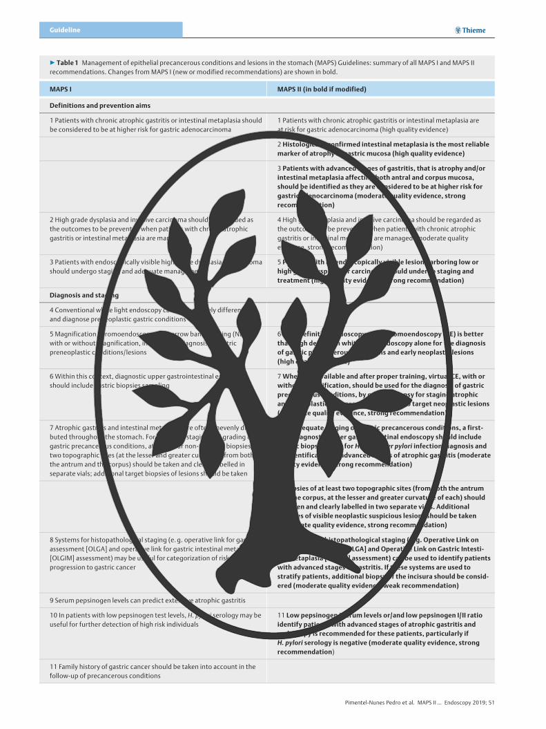
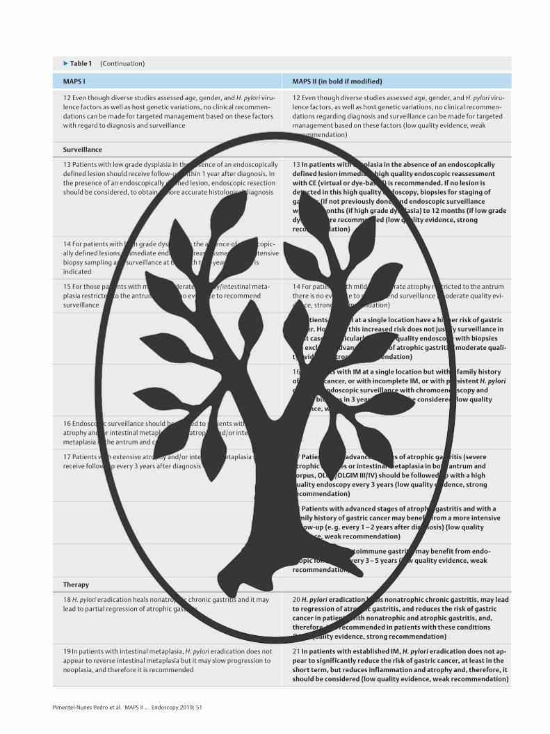
▶ Table 1 Management of epithelial precancerous conditions and lesions in the stomach (MAPS) Guidelines: summary of all MAPS I and MAPS IIrecommendations. Changes from MAPS I (new or modified recommendations) are shown in bold.
MAPS I MAPS II (in bold if modified)
Definitions and prevention aims
1 Patients with chronic atrophic gastritis or intestinal metaplasia shouldbe considered to be at higher risk for gastric adenocarcinoma
1 Patients with chronic atrophic gastritis or intestinal metaplasia areat risk for gastric adenocarcinoma (high quality evidence)
2 Histologically confirmed intestinal metaplasia is the most reliablemarker of atrophy in gastric mucosa (high quality evidence)
3 Patients with advanced stages of gastritis, that is atrophy and/orintestinal metaplasia affecting both antral and corpus mucosa,should be identified as they are considered to be at higher risk forgastric adenocarcinoma (moderate quality evidence, strongrecommendation)
2 High grade dysplasia and invasive carcinoma should be regarded asthe outcomes to be prevented when patients with chronic atrophicgastritis or intestinal metaplasia are managed
4 High grade dysplasia and invasive carcinoma should be regarded asthe outcomes to be prevented when patients with chronic atrophicgastritis or intestinal metaplasia are managed (moderate qualityevidence, strong recommendation)
3 Patients with endoscopically visible high grade dysplasia or carcinomashould undergo staging and adequate management
5 Patients with an endoscopically visible lesion harboring low orhigh grade dysplasia or carcinoma should undergo staging andtreatment (high quality evidence, strong recommendation)
Diagnosis and staging
4 Conventional white light endoscopy cannot accurately differentiateand diagnose preneoplastic gastric conditions
5 Magnification chromoendoscopy and narrow band imaging (NBI),with or without magnification, improve the diagnosis of gastricpreneoplastic conditions/lesions
6 High definition endoscopy with chromoendoscopy (CE) is betterthan high definition white-light endoscopy alone for the diagnosisof gastric precancerous conditions and early neoplastic lesions(high quality evidence)
6 Within this context, diagnostic upper gastrointestinal endoscopyshould include gastric biopsies sampling
7Whenever available and after proper training, virtual CE, with orwithout magnification, should be used for the diagnosis of gastricprecancerous conditions, by guiding biopsy for staging atrophicand metaplastic changes and by helping to target neoplastic lesions(moderate quality evidence, strong recommendation)
7 Atrophic gastritis and intestinal metaplasia are often unevenly distri-buted throughout the stomach. For adequate staging and grading ofgastric precancerous conditions, at least four non-targeted biopsies oftwo topographic sites (at the lesser and greater curvature, from boththe antrum and the corpus) should be taken and clearly labelled inseparate vials; additional target biopsies of lesions should be taken
8 For adequate staging of gastric precancerous conditions, a first-time diagnostic upper gastrointestinal endoscopy should includegastric biopsies both for Helicobacter pylori infection diagnosis andfor identification of advanced stages of atrophic gastritis (moderatequality evidence, strong recommendation)
9 Biopsies of at least two topographic sites (from both the antrumand the corpus, at the lesser and greater curvature of each) shouldbe taken and clearly labelled in two separate vials. Additionalbiopsies of visible neoplastic suspicious lesions should be taken(moderate quality evidence, strong recommendation)
8 Systems for histopathological staging (e. g. operative link for gastritisassessment [OLGA] and operative link for gastric intestinal metaplasia[OLGIM] assessment) may be useful for categorization of risk ofprogression to gastric cancer
10 Systems for histopathological staging (e. g. Operative Link onGastritis Assessment [OLGA] and Operative Link on Gastric Intesti-nal Metaplasia [OLGIM] assessment) can be used to identify patientswith advanced stages of gastritis. If these systems are used tostratify patients, additional biopsy of the incisura should be consid-ered (moderate quality evidence, weak recommendation)
9 Serum pepsinogen levels can predict extensive atrophic gastritis
10 In patients with low pepsinogen test levels, H. pylori serology may beuseful for further detection of high risk individuals
11 Low pepsinogen I serum levels or/and low pepsinogen I/II ratioidentify patients with advanced stages of atrophic gastritis andendoscopy is recommended for these patients, particularly ifH. pylori serology is negative (moderate quality evidence, strongrecommendation)
11 Family history of gastric cancer should be taken into account in thefollow-up of precancerous conditions
Pimentel-Nunes Pedro et al. MAPS II … Endoscopy 2019; 51
Guideline
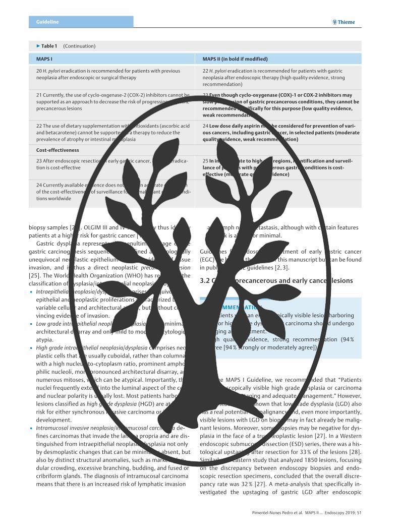
▶ Table 1 (Continuation)
MAPS I MAPS II (in bold if modified)
12 Even though diverse studies assessed age, gender, and H. pylori viru-lence factors as well as host genetic variations, no clinical recommen-dations can be made for targeted management based on these factorswith regard to diagnosis and surveillance
12 Even though diverse studies assessed age, gender, and H. pylori viru-lence factors, as well as host genetic variations, no clinical recommen-dations regarding diagnosis and surveillance can be made for targetedmanagement based on these factors (low quality evidence, weakrecommendation)
Surveillance
13 Patients with low grade dysplasia in the absence of an endoscopicallydefined lesion should receive follow-up within 1 year after diagnosis. Inthe presence of an endoscopically defined lesion, endoscopic resectionshould be considered, to obtain a more accurate histological diagnosis
13 In patients with dysplasia in the absence of an endoscopicallydefined lesion immediate high quality endoscopic reassessmentwith CE (virtual or dye-based) is recommended. If no lesion isdetected in this high quality endoscopy, biopsies for staging ofgastritis (if not previously done) and endoscopic surveillancewithin 6 months (if high grade dysplasia) to 12 months (if low gradedysplasia) are recommended (low quality evidence, strongrecommendation)
14 For patients with high grade dysplasia in the absence of endoscopic-ally defined lesions, immediate endoscopic reassessment with extensivebiopsy sampling and surveillance at 6-month to 1-year intervals isindicated
15 For those patients with mild to moderate atrophy/intestinal meta-plasia restricted to the antrum there is no evidence to recommendsurveillance
14 For patients with mild to moderate atrophy restricted to the antrumthere is no evidence to recommend surveillance (moderate quality evi-dence, strong recommendation)
15 Patients with IM at a single location have a higher risk of gastriccancer. However, this increased risk does not justify surveillance inmost cases, particularly if a high quality endoscopy with biopsieshas excluded advanced stages of atrophic gastritis (moderate quali-ty evidence, strong recommendation)
16 In patients with IM at a single location but with a family historyof gastric cancer, or with incomplete IM, or with persistent H. pylorigastritis, endoscopic surveillance with chromoendoscopy andguided biopsies in 3 years’ time may be considered (low qualityevidence, weak recommendation)
16 Endoscopic surveillance should be offered to patients with extensiveatrophy and/or intestinal metaplasia (i. e., atrophy and/or intestinalmetaplasia in the antrum and corpus)
17 Patients with extensive atrophy and/or intestinal metaplasia shouldreceive follow-up every 3 years after diagnosis
17 Patients with advanced stages of atrophic gastritis (severeatrophic changes or intestinal metaplasia in both antrum andcorpus, OLGA/OLGIM III/IV) should be followed up with a highquality endoscopy every 3 years (low quality evidence, strongrecommendation)
18 Patients with advanced stages of atrophic gastritis and with afamily history of gastric cancer may benefit from a more intensivefollow-up (e. g. every 1–2 years after diagnosis) (low qualityevidence, weak recommendation)
19 Patients with autoimmune gastritis may benefit from endo-scopic follow-up every 3–5 years (low quality evidence, weakrecommendation)
Therapy
18 H. pylori eradication heals nonatrophic chronic gastritis and it maylead to partial regression of atrophic gastritis
20 H. pylori eradication heals nonatrophic chronic gastritis, may leadto regression of atrophic gastritis, and reduces the risk of gastriccancer in patients with nonatrophic and atrophic gastritis, and,therefore, it is recommended in patients with these conditions(high quality evidence, strong recommendation)
19 In patients with intestinal metaplasia, H. pylori eradication does notappear to reverse intestinal metaplasia but it may slow progression toneoplasia, and therefore it is recommended
21 In patients with established IM, H. pylori eradication does not ap-pear to significantly reduce the risk of gastric cancer, at least in theshort term, but reduces inflammation and atrophy and, therefore, itshould be considered (low quality evidence, weak recommendation)
Pimentel-Nunes Pedro et al. MAPS II … Endoscopy 2019; 51
biopsy samples [23]. OLGIM III and IV stages may thus identifypatients at a higher risk for gastric cancer [18, 19].
Gastric dysplasia represents the penultimate stage of thegastric carcinogenesis sequence. It is defined as histologicallyunequivocal neoplastic epithelium without evidence of tissueinvasion, and is thus a direct neoplastic precancerous lesion[25]. The World Health Organization (WHO) has reiterated theclassification of dysplasia/intraepithelial neoplasia [26]:▪ Intraepithelial neoplasia/dysplasia comprises unequivocally
epithelial and neoplastic proliferations characterized byvariable cellular and architectural atypia, but without con-vincing evidence of invasion.
▪ Low grade intraepithelial neoplasia/dysplasia shows minimalarchitectural disarray and only mild to moderate cytologicalatypia.
▪ High grade intraepithelial neoplasia/dysplasia comprises neo-plastic cells that are usually cuboidal, rather than columnar,with a high nucleus-to-cytoplasm ratio, prominent ampho-philic nucleoli, more pronounced architectural disarray, andnumerous mitoses, which can be atypical. Importantly, thenuclei frequently extend into the luminal aspect of the cell,and nuclear polarity is usually lost. Most patients harboringlesions classified as high grade dysplasia (HGD) are at highrisk for either synchronous invasive carcinoma or its rapiddevelopment.
▪ Intramucosal invasive neoplasia/intramucosal carcinoma de-fines carcinomas that invade the lamina propria and are dis-tinguished from intraepithelial neoplasia/dysplasia not onlyby desmoplastic changes that can be minimal or absent, butalso by distinct structural anomalies, such as marked glan-dular crowding, excessive branching, budding, and fused orcribriform glands. The diagnosis of intramucosal carcinomameans that there is an increased risk of lymphatic invasion
and lymph node metastasis, although with certain featuresthis risk is absent or minimal.
Guidelines for endoscopic treatment of early gastric cancer(EGC) are beyond the scope of this manuscript but can be foundin published ESGE guidelines [2, 3].
3.2 Gastric precancerous and early cancer lesions
In the MAPS I Guideline, we recommended that “Patientswith endoscopically visible high grade dysplasia or carcinomashould undergo staging and adequate management.” However,several studies have shown that low grade dysplasia (LGD) alsohas a real potential for malignancy and, even more importantly,visible lesions with LGD on biopsy may in fact already be malig-nant lesions. Moreover, some biopsies may be negative for dys-plasia in the face of a true neoplastic lesion [27]. In a Westernendoscopic submucosal dissection (ESD) series, there was a his-tological upstaging after resection for 33% of the lesions [28].Similarly, an Eastern study that analyzed 1850 lesions, focusingon the discrepancy between endoscopy biopsies and endo-scopic resection specimens, concluded that the overall discre-pancy rate was 32% [27]. A meta-analysis that specifically in-vestigated the upstaging of gastric LGD after endoscopic
▶ Table 1 (Continuation)
MAPS I MAPS II (in bold if modified)
20 H. pylori eradication is recommended for patients with previousneoplasia after endoscopic or surgical therapy
22 H. pylori eradication is recommended for patients with gastricneoplasia after endoscopic therapy (high quality evidence, strongrecommendation)
21 Currently, the use of cyclo-oxgenase-2 (COX-2) inhibitors cannot besupported as an approach to decrease the risk of progression of gastricprecancerous lesions
23 Even though cyclo-oxygenase (COX)-1 or COX-2 inhibitors mayslow progression of gastric precancerous conditions, they cannot berecommended specifically for this purpose (low quality evidence,weak recommendation)
22 The use of dietary supplementation with antioxidants (ascorbic acidand betacarotene) cannot be supported as a therapy to reduce theprevalence of atrophy or intestinal metaplasia
24 Low dose daily aspirin may be considered for prevention of vari-ous cancers, including gastric cancer, in selected patients (moderatequality evidence, weak recommendation)
Cost-effectiveness
23 After endoscopic resection of early gastric cancer, H. pylori eradica-tion is cost-effective
25 In intermediate to high risk regions, identification and surveil-lance of patients with precancerous gastric conditions is cost-effective (moderate quality evidence)
24 Currently available evidence does not allow an accurate estimationof the cost-effectiveness of surveillance for premalignant gastric condi-tions worldwide
RECOMMENDATION
5 Patients with an endoscopically visible lesion harboringlow or high grade dysplasia or carcinoma should undergostaging and treatment.High quality evidence, strong recommendation (94%agree [94% strongly or moderately agree]).
Pimentel-Nunes Pedro et al. MAPS II … Endoscopy 2019; 51
Guideline
resection found that this happens in 25% of lesions, with 7%being upstaged to malignant [29]. Taking all this evidencetogether, we can conclude that endoscopic biopsies are insuffi-cient for correct diagnosis of visible gastric lesions and that anendoscopically visible lesion with any neoplastic change shouldbe considered for treatment.
4 Diagnosis and staging4.1 Endoscopy
neoplastic conditions or lesions with high accuracy [41–45].In a recently published meta-analysis including 10 studies, 699patients, and 902 lesions, the pooled sensitivity, specificity, andarea under the curve (AUC) of dye-CE were 0.90 (95% confi-dence interval [CI] 0.87–0.92), 0.82 (95%CI 0.79–0.86), and0.95, respectively, these results being significantly better thanWLE alone (risk difference of 0.36 for neoplasia and 0.17 forpremalignant conditions) [46]. However, dye-CE is cumber-some and significantly lengthens endoscopic procedures. Thisfavors virtual CE which is available at the touch of a button.
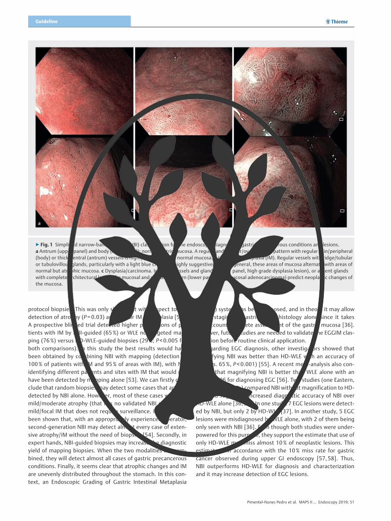
Several studies focused on the role of virtual CE in the diag-nosis of gastric precancerous conditions. A systematic reviewshowed that most studies addressed narrow-band imaging(NBI) (mainly with magnification). The pooled sensitivity andspecificity for the diagnosis of IM were 86% and 77%, and fordysplasia/early cancer these values were 90% and 83%, respec-tively [47]. However, the authors concluded that few studiesaddressed interobserver reliability and that there was no valida-ted classification. Other investigators evaluated all the NBI pat-terns previously described, and created and validated a simpli-fied NBI (without magnification) classification using only repro-ducible NBI features (▶Fig. 1) [48]. The global accuracy for thediagnosis of IM was 84% and for dysplasia it was 95%. However,these results clearly depend on training and are better with ex-perienced endoscopists [48, 49]. External validation of this clas-sification in a prospective multicenter study involving five inter-national Western centers (some using near-focus and second-generation NBI) showed a sensitivity and specificity of 87%and 97% for the diagnosis of IM and 92% and 99% for thediagnosis of dysplasia [36]. The diagnostic accuracy rate wasof 94% (11% higher than HD-WLE), with the greatest advan-tage of NBI over or after WLE being sensitivity for detecting IM(87% vs. 53%, P<0.001) and sensitivity for neoplasia (92% vs.74%) [36]. Altogether these results supported the ESGE Tech-nology Review on advanced imaging which suggested this clas-sification as the one to be used in this context [50].
Other studies comparing HD-WLE to NBI consistentlyshowed better results with NBI for detecting both IM and EGC.A large multicenter prospective randomized study reached thesame conclusions this time in an Eastern population. Again,even though specificities for IM and cancer were the same, thesensitivities for IM (92% vs. 59%) and particularly for cancer(100% vs. 29%) were much higher with second-generation NBIwhen compared to HD-WLE [37]. In an Indian randomized pro-spective crossover study the conclusions were very similar re-garding IM, with the frequency of IM detection by NBI beingsignificantly higher than by WLE (P=0.001) [51].
We can conclude that NBI is better than HD-WLE for the de-tection and diagnosis of IM, but is it better than standard non-targeted biopsy sampling? In a comparative study including119 patients, the overall sensitivity, specificity, and accuracyof WLE nontargeted biopsies taken according to the Sydney-Houston protocol were compared with NBI-guided biopsies.For predicting atrophy, the WLE nontargeted biopsies vs. NBIresults were 86% vs. 62%, 100% vs. 97%, and 93% vs. 80%,respectively; for IM they were 80% vs. 72%, 100% vs. 93%, and90% vs. 82%. These results were slightly better for nontargeted
STATEMENT
6 High definition endoscopy with chromoendoscopy (CE)is better than high definition white-light endoscopy alonefor the diagnosis of gastric precancerous conditions andearly neoplastic lesions.High quality evidence (94% agree [94% strongly or mod-erately agree.
RECOMMENDATION
7 Whenever available and after proper training, virtualCE, with or without magnification, should be used forthe diagnosis of gastric precancerous conditions, by guid-ing biopsy for staging atrophic and metaplastic changesand by helping to target neoplastic lesions.Moderate quality evidence, strong recommendation(94% agree [94% strongly or moderately agree]).
Classical studies of conventional white-light endoscopy (WLE) showed that the correlation between histological and endoscopic findings for the diagnosis of gastric precancerous conditions was poor [30 – 34]. However, recent studies with high definition WLE (HD-WLE) presented promising results. For preneoplastic conditions, a cross-sectional study showed that HD-WLE had a global accuracy of 88 % for the diagnosis of IM with a sensitivity of 75 % and specificity of 94 % [35]. In a real-time multicenter prospective study, the global accuracy of HD-WLE was 83 %, with a specificity of 98 % for IM but with only 53 %sensitivity [36]. These results were confirmed in another multi-center prospective study, that showed a 98 % specificity for IM but again with a low sensitivity of 59 % [37]. For the diagnosis of neoplastic lesions these two studies showed low sensitivities of 74 % and 29 %, respectively, although the specificities were higher than 95 % [36, 37]. HD-WLE with magnification may im-prove these results; however, the data are too scarce to provide definitive conclusions [38 – 40]. So, even though these results for HD-WLE are satisfactory for IM and for early neoplastic le-sions they are far from perfect, particularly regarding the sensi-tivity in the diagnosis of these lesions.
Conventional CE with application of dyes (indigo carmine, methylene blue, acetic acid, or hematoxylin) has consistently been associated with the detection of gastric preneoplastic or
Pimentel-Nunes Pedro et al. MAPS II … Endoscopy 2019; 51
protocol biopsies. This was only significant with respect to thedetection of atrophy (P=0.03) and not for IM or dysplasia [52].A prospective blinded trial detected higher proportions of pa-tients with IM by NBI-guided (65%) or WLE nontargeted map-ping (76%) versus HD-WLE-guided biopsies (29%; P<0.005 forboth comparisons). In this study the best results would havebeen obtained by combining NBI with mapping (detection of100% of patients with IM and 95% of areas with IM), with NBIidentifying different patients and sites with IM that would nothave been detected by mapping alone [53]. We can firstly con-clude that random biopsies may detect some cases that are notdetected by NBI alone. However, most of these cases will havemild/moderate atrophy (that has no validated NBI pattern) ormild/focal IM that does not require surveillance. In fact, it hasbeen shown that, with an appropriately experienced operator,second-generation NBI may detect almost every case of exten-sive atrophy/IM without the need of biopsies [54]. Secondly, inexpert hands, NBI-guided biopsies may increase the diagnosticyield of mapping biopsies. When the two modalities are com-bined, they will detect almost all cases of gastric precancerousconditions. Finally, it seems clear that atrophic changes and IMare unevenly distributed throughout the stomach. In this con-text, an Endoscopic Grading of Gastric Intestinal Metaplasia
(EGGIM) system has been proposed, and in theory it may allowbetter staging of gastritis than histology alone since it takesinto account complete assessment of the gastric mucosa [36].However, future studies are needed to validate the EGGIM clas-sification before routine clinical application.
Regarding EGC diagnosis, other investigators showed thatmagnifying NBI was better than HD-WLE with an accuracy of90% (vs. 65%, P<0.001) [55]. A recent meta-analysis also con-cluded that magnifying NBI is better than WLE alone with anAUC of 0.96 for diagnosing EGC [56]. Two studies (one Eastern,the other Western) compared NBI without magnification to HD-WLE and suggested increased diagnostic accuracy of NBI overHD-WLE alone [36, 37]. In one study, 7 EGC lesions were detect-ed by NBI, but only 2 by HD-WLE [37]. In another study, 5 EGClesions were misdiagnosed by WLE alone, with 2 of them beingonly seen with NBI [36]. Even though both studies were under-powered for this purpose, they support the estimate that use ofonly HD-WLE may miss almost 10% of neoplastic lesions. Thisestimate is in accordance with the 10% miss rate for gastriccancer observed during upper GI endoscopy [57, 58]. Thus,NBI outperforms HD-WLE for diagnosis and characterizationand it may increase detection of EGC lesions.
▶ Fig. 1 Simplified narrow-band imaging (NBI) classification for the endoscopic diagnosis of gastric precancerous conditions and lesions.a Antrum (upper panel) and body (lower panel): normal gastric mucosa. A regular and circular/oval mucosal pattern with regular thin/peripheral(body) or thick/central (antrum) vessels is highly predictive of a normal mucosa. b Intestinal metaplasia (IM). Regular vessels with ridge/tubularor tubulovillous glands, particularly with a light blue crest, are highly suggestive of IM. In general, these areas of mucosa alternate with areas ofnormal but atrophic mucosa. c Dysplasia/carcinoma. Irregular vessels and glands (upper panel, high grade dysplasia lesion), or absent glandswith complete architectural loss of the mucosal and vascular pattern (lower panel, intramucosal adenocarcinoma) predict neoplastic changes ofthe mucosa.
Pimentel-Nunes Pedro et al. MAPS II … Endoscopy 2019; 51
Guideline
There is less evidence to support other methods of virtual CEsuch as i-Scan digital contrast and flexible spectral imaging col-or enhancement (FICE). There are currently insufficient data torecommend routine clinical use of these techniques, eventhough in theory and after proper training they could havesimilar applications [47, 59]. A few prospective studies suggest-ed that blue laser imaging may achieve similar results to NBI[60–62]. Other emerging technologies, such as confocal endo-microscopy, endocytoscopy, Raman spectroscopy, and polari-metry, may have a future role but at this stage cannot be re-commended for routine clinical use [59].
4.2 Biopsy sampling
correct staging is debated. More biopsies will allow better stag-ing. However, in clinical practice more biopsies mean moretime and higher procedure costs. In the MAPS I Guideline, werecommended at least two biopsies from the antrum and twofrom the corpus, and the lack of obligatory biopsy of the inci-sura was a matter of some controversy. In fact, the incisuramay be the anatomical location with the highest incidence andseverity of IM [63–65]. This is used to support an additionalbiopsy of the incisura. The updated Sydney system is the mostwidely accepted protocol for the classification and grading ofgastritis. It recommends at least five biopsies: two from the an-trum (from the greater and lesser curvature, 3 cm from the py-lorus); one from the incisura; and two from the body (from thelesser curvature, 4 cm proximal to the incisura, and from thegreater curvature, middle). This differed from the initial Sydneyprotocol that recommended only two biopsies from the corpusand two from the antrum [20]. However, the need to samplethe incisura was based mostly on the notion that atrophic/me-taplastic changes appear first in the incisura even though therewere no data suggesting a clinical benefit. In this regard a largestudy, published after the MAPS I Guideline, evaluated 400738biopsy sets and found that compliance with the original Sydneysystem (two antrum, two corpus) had the highest yield for thediagnosis of H. pylori infection and IM when compared with allother biopsy strategies [66]. More biopsies or inclusion of an in-cisura biopsy yielded minimal additional diagnostic informationwith more costs. Some studies, published after MAPS I, specifi-cally addressed the benefit of incisura biopsy sampling. The in-clusion of incisura biopsy increased the proportion of patientsclassified with high risk stages (OLGA III/IV or OLGIM III/IV) inthree studies (two European studies including nonselected po-pulations [65, 67] and one Korean study in high risk patients[64]). All found that the incisura biopsy increased the propor-tion of patients with high risk stages. Considering the two Euro-pean studies from nonselected populations together, withoutthe incisura biopsy, there was a downgrading from high riskOLGA stages to low risk OLGA in 14/1048 patients (absolute dif-ference 1.33%) and from high risk OLGIM to low risk OLGIM in13/1048 patients (absolute difference 1.24%). This translatesinto a number needed to treat of 75–80, meaning that one in75–80 patients will not be correctly included in a high riskgroup if incisura biopsy is not performed. Another Europeanstudy evaluated classification systems in a high risk population(first-degree relatives of early onset gastric cancer patients)using OLGA and OLGIM staging systems that were modified byexclusion of the incisura biopsy, and demonstrated an overall15% and 30% downgrade of staging in comparison with the ori-ginal OLGA/OLGIM systems. In high risk stages, the downgradeof staging was less pronounced (5%) for both modified stagingsystems in comparison with the original OLGA system [68].Another study comparing different biopsy protocols reportedthat biopsy of the incisura did not provide additional benefit asthe prevalence of IM in the incisura was similar to that in otherbiopsy sites, although the impact of incisura biopsies in highrisk phenotypes was not assessed [66].
RECOMMENDATION
10 Systems for histopathological staging (e. g. OLGA andOLGIM assessment) can be used to identify patients withadvanced stages of atrophic gastritis. If these systems areused to stratify patients, additional biopsy of the incisurashould be considered.Moderate quality evidence, weak recommendation (88%agree [58% strongly or moderately agree]).
RECOMMENDATION
9 Biopsies of at least two topographic sites (from boththe antrum and the corpus, at the lesser and greater cur-vature of each) should be taken and clearly labelled in twoseparate vials. Additional biopsies of visible neoplasticsuspicious lesions should be taken.Moderate quality evidence, strong recommendation(94% agree [82% strongly or moderately agree]).
RECOMMENDATION
8 For adequate staging of gastric precancerous condi-tions, a first-time diagnostic upper gastrointestinalendoscopy should include gastric biopsies both forH. pylori infection diagnosis and for identification of ad-vanced stages of atrophic gastritisModerate quality evidence, strong recommendation(88% agree [77% strongly or moderately agree]).
Considering that most endoscopists are not yet familiar with advanced imaging patterns, at present we cannot recommend exclusively endoscopic staging of gastritis without biopsies. However, current evidence suggests that CE-targeted biopsies plus mapping biopsies are the best way of detecting most cases of advanced gastritis. For these reasons we recommend that, when available, CE should be used for targeted biopsies.
When CE is not available (or the endoscopist doubts the ad-vanced imaging diagnosis), the number of biopsies needed for
Pimentel-Nunes Pedro et al. MAPS II … Endoscopy 2019; 51
In summary, this small additional yield from an incisurabiopsy needs to be balanced against costs and workload. Wetherefore recommend a minimum of two biopsies from the an-trum and two biopsies from the corpus, noting that adding anincisura biopsy can be considered in order to maximize the de-tection of patients with precancerous conditions, especially incases where CE is not available to target biopsies. Moreover,this additional biopsy will allow more precise evaluation ofOLGA and OLGIM stages, that have been proven to correlatewith risk for cancer progression [69–71].
Regarding the number of vials, even though separate vialsmay not be required among expert pathologists, as antral andcorpus mucosa can be easily distinguished in the absence of se-vere atrophic changes, use of a single vial cannot be recom-mended in all cases. Future studies should evaluate specificscenarios when antrum, incisura, and corpus samples can besent in the same vial.
4.3 Noninvasive assessment
As stated in the MAPS I Guideline, a low pepsinogen I serumlevel, a low pepsinogen I/II ratio, or both, are good indicators ofatrophic changes in the gastric mucosa. A 2004 meta-analysissuggested that pepsinogen I≤50ng/mL and pepsinogen I/IIratio ≤3 were the best cutoff values for dysplasia diagnosis[72]. Several articles published after MAPS I confirm levels ofpepsinogens to be good indicators of extensive atrophic gastri-tis and of gastric cancer [73, 74]. A 2015 meta-analysis on pep-sinogen tests in gastric cancer and atrophic gastritis suggesteda good correlation between decreased pepsinogen serum levelsand atrophy [75]. In this meta-analysis, the summary sensitivityand summary specificity for gastric cancer diagnosis were 0.69(95%CI 0.60–0.76) and 0.73 (95%CI 0.62–0.82), respectively.Corresponding values for atrophic gastritis diagnosis were 0.69(95%CI 0.55–0.80) and 0.88 (95%CI 0.77–0.94), respectively.The AUC for gastric cancer diagnosis was 0.76 (95%CI 0.72–0.80) and for atrophic gastritis it was 0.85 (95%CI 0.82–0.88).A Fagan plot indicated that the use of pepsinogen serum levelscould moderately improve the gastric cancer and atrophy de-tection rate, confirming a moderate efficiency of pepsinogenserum levels for gastric cancer and atrophic gastritis diagnosis.In a subgroup analysis the authors concluded that combininglow pepsinogen I level with the pepsinogen I/II ratio is the bestway of detecting gastric cancer (AUC 0.78) and atrophic gastri-tis (AUC 0.87). However, different cutoff values were used,
although most studies used pepsinogen I < 70ng/mL and pepsi-nogen I/II ratio < 3 as the best cutoff values. In fact, these arewidely accepted cutoff values for gastric cancer screening inJapan [76]. The authors concluded that pepsinogen serum lev-els have a potentially significant role in the identification of po-pulations at high risk for gastric cancer and could be used formass screening. However, they note that there was greatheterogeneity between studies. Moreover, different methodsare used for quantifying levels of pepsinogens and in thismeta-analysis enzyme-linked immunoassay (ELISA) was slightlysuperior to the other methods, with this difference possibly in-ducing heterogeneity [75]. In fact, different methods may beused for pepsinogen quantification and results may differ be-tween tests [77]. Therefore, cutoff values validated for a partic-ular assay should be used, and cannot be generalized to all as-says.
Other serum molecule levels were studied as markers of gas-tric atrophy. A 2017 systematic review and meta-analysis fo-cused on the combination of pepsinogen I/II, gastrin-17, andanti-Helicobacter antibodies for diagnosing atrophic gastritis[78]. However, the design of this meta-analysis does not allowassessment of the individual performance of each marker fordetecting atrophy. Moreover, previously published evidencedemonstrated little yield from adding gastrin-17 to pepsinogenassessment for detecting atrophy [79]. On the other hand, add-ing H. pylori serology to pepsinogen level evaluation may helpto detect patients at higher risk of gastric cancer [80, 81]. In a2014 cohort of 4655 patients followed up for 16 years, therewas a progressive increase in cancer risk, going from thosewith no gastritis to those with chronic H. pylori-positive gastritiswithout extensive atrophy (H. pylori-positive, normal pepsino-gen levels; hazard ratio [HR] 8.9, 95%CI 2.7–54.7), to thosewith extensive chronic atrophic gastritis (defined by pepsino-gen I < 70ng/mL and pepsinogen I/II ratio < 3) with H. pylori-po-sitive serology (HR 17.7, 95%CI 5.4–108), and finally to thosewith atrophic gastritis with H. pylori-negative serology, sugges-tive of extensive IM (HR 69.7, 95%CI 14–503) [81].
Other methods for noninvasive assessment of gastric muco-sal atrophy, including evaluation of decreased serum ghrelin[82–84], trefoil factors [85], a panel of microRNAs [86], andvolatile organic compounds in exhaled air [87], have been sug-gested, with good results. However, the available evidence forthese tests is not sufficient and further studies are required be-fore they can be recommended for clinical application.
In conclusion, pepsinogen serum levels are currently thebest evaluated noninvasive test for detecting patients with ad-vanced atrophic gastritis. Low pepsinogen I serum levels, parti-cularly when associated with H. pylori-negative serologicalstatus, may identify patients at higher risk of gastric cancer towhom endoscopy should be offered.
RECOMMENDATION
11 Low pepsinogen I serum levels or/and a low pepsino-gen I/II ratio identify patients with advanced stages ofatrophic gastritis, and endoscopy is recommended forthese patients, particularly if H. pylori serology isnegative.Moderate quality evidence, strong recommendation(88% agree [76% strongly or moderately agree]).
Pimentel-Nunes Pedro et al. MAPS II … Endoscopy 2019; 51
Guideline
4.4 Additional risk factors 5 Surveillance5.1 Dysplasia
Most routine gastroscopies are performed with standard de-finition WLE. As we have seen, CE (virtual or dye-based) increas-es accuracy for detection of dysplasia. A prospective study thatincluded 20 patients with a diagnosis of HGD or carcinoma,without visible endoscopic lesions in the index endoscopy,showed that immediate endoscopic reassessment with highdefinition endoscopes and virtual CE allowed the identificationof visible lesions and adequate treatment in 18 patients [105].Conventional CE also improves the detection of precancerousconditions and lesions [106]. A systematic review and meta-analysis reported that approximately 10% of the patients witha gastric cancer diagnosis had undergone a recent endoscopyin which the gastric cancer was not diagnosed (because ofboth missed endoscopic lesions or nonmalignant pathology di-agnosis). A recent study also showed that 8.6% of the patientswith EGCs had a simultaneous lesion that was not detected inthe diagnostic endoscopy [57, 107]. The rate of missed lesionstended to be higher in primary care and screening settings thanin secondary and tertiary care. Another study showed that alonger endoscopy time (> 7 minutes) was associated with ahigher likelihood of detecting neoplastic lesions (odds ratio[OR] 3.42, 95%CI 1.25–10.38) [108]. Moreover, a finding ofdysplasia in nontargeted biopsies significantly increases therisk of gastric cancer, which may be as high as 6% per year[109]. A recent Swedish study, that to the best of our knowl-edge is the largest follow-up study to date among patientswith gastric precancerous conditions, suggested a lower risk ofgastric cancer for patients with dysplasia. However, theyexcluded the first 2 years of follow-up and concluded that thismight be the reason for the lower risk of gastric cancer sincemany lesions might have been there already [110].
“Indefinite for dysplasia/neoplasia” should not be viewed in-itially as an innocuous diagnosis although in the majority ofpatients the prognosis is favorable. Indeed, a study found that26.8% of resected lesions that had been characterized as inde-finite for dysplasia/neoplasia in preresection biopsies were infact neoplastic (5.0% adenomas and 21.8% EGCs) [111].Another study found that reassessment of indefinite for dys-plasia biopsies by three expert gastrointestinal pathologists
RECOMMENDATION
13 In patients with dysplasia in the absence of an endo-scopically defined lesion immediate high quality endo-scopic reassessment with CE (virtual or dye-based) is re-commended. If no lesion is detected in this high qualityendoscopy, biopsies for staging of gastritis (if not pre-viously done) and endoscopic surveillance within6 months (if high grade dysplasia) to 12 months (if lowgrade dysplasia) are recommended.Low quality evidence, strong recommendation (88%agree [88% strongly or moderately agree]).
RECOMMENDATION
12 Even though diverse studies assessed age, gender, andH. pylori virulence factors, as well as host genetic varia-tions, no clinical recommendations regarding diagnosisand surveillance can be made for targeted managementbased on these factors.Low quality evidence, weak recommendation (100%agree [88% strongly or moderately agree]).
Assuming the gene-environment interaction for gastric can-cer, multiple risk factors have been linked to the multistep pro-gression from chronic nonatrophic gastritis to atrophic gastri-tis, IM, dysplasia, and finally cancer [10].
H. pylori plays a pivotal role in this progression and was clas-sified as a type 1 carcinogen in 1994 by the WHO [88]. It is believed that the combination of a virulent organism in a genetically susceptible host is associated with more severe chronic inflammation and more rapid progression to gastric cancer, at least for the Lauren intestinal type [89 – 91].
Different strains of H. pylori vary in their carcinogenic poten-tial, with those containing virulence factors, such as the cyto-toxin-associated antigen (cagA) protein and the vacuolating toxin A (vacA), inducing a higher degree of inflammation and increasing the risk for gastric cancer [92 – 97]. Nevertheless, there are no studies addressing the clinical usefulness of geno-typing H. pylori strains with regard to the management and sur-veillance of gastric precancerous conditions/lesions.
An immense number of studies have addressed the implica-tions of genes and genetic host variations for gastric carcino-genesis. The best characterized are those that play a role in the inflammatory response to H. pylori infection and inflamma-tion of the gastric mucosa, leading to mucosal atrophy and pro-gression to cancer. These include host genetic interleukin poly-morphisms of IL-1B, IL1-receptor antagonist (IL-1RN), IL8, IL10, and TNF-α [98 – 104]. However, the heterogeneity of the results makes it difficult to translate them into recommendations for daily clinical practice.
Pimentel-Nunes Pedro et al. MAPS II … Endoscopy 2019; 51
changed the diagnosis to dysplasia in 11 /46 patients (10 LGDand 1 HGD) [112].
All of this suggests that patients with diagnoses from non-targeted biopsies of indefinite for dysplasia, of dysplasia, or ofcarcinoma benefit from a careful endoscopic reassessment incenters with experience in the diagnosis and endoscopic treat-ment of EGC. We recommend that pathology slides should bereviewed by an expert GI pathologist and recommend immedi-ate (as soon as possible) high quality endoscopic reassessmentwith CE. If a lesion is seen and the endoscopic assessment sug-gests dysplasia, we recommend resection without need of fur-ther biopsies. If endoscopic reassessment with CE does not re-veal a visible lesion and repeat nontargeted biopsies do notshow dysplasia/neoplasia, then staging the severity and extentof preneoplastic conditions in such cases can help to define thesurveillance program. A retrospective study of patients with in-definite for dysplasia lesions at enrollment and OLGA staging,with a median follow-up of 31 months, did not detect dysplasiain any patient with OLGA 0 /I/II, while 6 cases of LGD/HGD weredetected in 25 patients with OLGA III/IV during follow-up [113].
With the above considerations in mind, patients with a diag-nosis of indefinite for dysplasia/neoplasia or of dysplasia/intra-mucosal carcinoma in random biopsies (i. e., no clear lesionidentified at endoscopy) should be promptly referred to anexpert endoscopy center and have an endoscopic reassessmentwith high definition endoscopes and CE (dye or virtual). If nolesion is identified in this high quality endoscopy, endoscopicrevaluation is recommended at a 6-month (if previous HGD) to12-month (if previous LGD) interval, with further adjustmentaccording to the severity and extent of precancerous condi-tions (▶Fig. 2).
5.2 Atrophic gastritis/intestinal metaplasiaGastric precancerous conditions are frequent in the general
population (although with wide geographical variability ac-cording to H. pylori infection prevalence). The annual incidenceof gastric cancer has been reported to be 0.1%–0.25% in pa-tients with chronic atrophic gastritis and 0.25% in patientswith IM, and may be as high as 1.36% person-year for any gas-tric neoplasia (including dysplasia and neuroendocrine tumors)[109, 114]. Cumulative incidences of gastric cancer of 2.4% at10 years in patients with IM were reported, and a Swedishstudy reported a cumulative incidence at 20 years of approxi-mately 2% in patients with atrophic gastritis and of 2.5% in pa-tients with IM [110]. A Japanese study found higher cumulativeincidences of gastric cancer at 5 years, reaching 1.9%–10% inpatients with extensive endoscopic atrophy and 5.3% –9.8% inpatients with IM [115].
Surveillance of patients with precancerous conditions allowsthe detection of lesions at early stages (with a significant pro-portion being amenable to endoscopic resection) and was re-commended in the MAPS I Guideline in patients with extensiveatrophy or IM (in both corpus and antrum). The extent of pre-neoplastic changes was identified as a risk factor for progres-sion, as well as family history of gastric cancer and type IIIincomplete IM.
Extent and presence of IM Some recent studies confirmedthe presence and extent of IM as risk factors for gastric cancer.An Italian prospective study found a significantly increased risk
RECOMMENDATION
16 In patients with IM at a single location but with a fam-ily history of gastric cancer, or with incomplete IM, orwith persistent H. pylori gastritis, endoscopic surveillancewith CE and guided biopsies in 3 years’ time may beconsidered.Low quality evidence, weak recommendation (82% agree[76% strongly or moderately agree]).
RECOMMENDATION
18 Patients with advanced stages of atrophic gastritis andwith a family history of gastric cancer may benefit from amore intensive follow-up (e. g. every 1–2 years afterdiagnosis).Low quality evidence, weak recommendation (82% agree[65% strongly or moderately agree]).
RECOMMENDATION
17 Patients with advanced stages of atrophic gastritis(severe atrophic changes or IM in both antrum and cor-pus, OLGA/OLGIM III/IV) should be followed up with ahigh quality endoscopy every 3 years.Low quality evidence, strong recommendation (100%agree [94% strongly or moderately agree]).
RECOMMENDATION
14 For patients with mild to moderate atrophy restrictedto the antrum there is no evidence to recommend surveil-lance.Moderate quality evidence, strong recommendation(100% agree [100% strongly or moderately agree]).
RECOMMENDATION
15 Patients with IM at a single location have a higher riskof gastric cancer. However, this increased risk does notjustify surveillance in most cases, particularly if a highquality endoscopy with biopsies has excluded advancedstages of atrophic gastritis.Moderate quality evidence, strong recommendation(100% agree [82% strongly or moderately agree]).
Pimentel-Nunes Pedro et al. MAPS II … Endoscopy 2019; 51
Guideline
of gastric neoplasia in patients with OLGA and OLGIM stages III/IV at baseline, while extensive atrophy (antrum and corpus) wasalso associated with a trend to higher risk of progression al-though this was not statistically significant on multivariable a-nalysis (HR 7.2, 95%CI 0.7–6.84) [114]. Extensive IM was alsofound to be associated with a higher risk of progression in a USstudy [116]. A Japanese study also found that IM in the corpus(isolated or antrum and corpus) and extensive endoscopic atro-phy at baseline were independent predictors of gastric cancerat follow-up [115]. A case-control study found that OLGIM II-IV(but not OLGA II-IV) and corpus-predominant gastritis were sig-nificantly more frequent in gastric cancer patients than in con-trols [117]. Another case-control study found that OLGA III/IV,OLGIM III/IV, and endoscopically classified moderate-to-severeatrophy were significantly more frequent in gastric cancer pa-tients [118]. Another study using endoscopic grading of atro-phy found that gastric cancer risk was increased in patientswith extensive atrophy (5.33% in patients with atrophy presentin the entire stomach vs. 0% and 0.25% in patients with atrophylimited to the gastric antrum and atrophy in the incisura or low-er corpus, respectively) [119]. A Korean study reported thatOLGA III/IV and OLGIM I– IV were independent risk factors forgastric cancer, especially the intestinal type, showing thateven nonextensive IM may significantly increase the risk of gas-tric cancer [120]. The adjusted odds ratios for the differentstages were: OLGA III 2.09, OLGA IV 2.04; OLGIM I 2.38, OLGIMII 2.97, OLGIM III 7.89, OLGIM IV 13.20 (all statistically signifi-cant). OLGA IV, histological IM, and a higher classification ofendoscopic atrophy were also identified as independent riskfactors in a prospective Korean study with follow-up >3 years[121]. These studies suggest that the presence of IM (as a sur-rogate of advanced gastritis) may be of equal or more impor-tance than the extent of atrophy without IM, since the risk ofgastric cancer was higher with OLGIM I/II than with OLGA III/IV. This accords with other previous studies that evaluated therisk of gastric cancer in patients with only atrophy or IM (inde-pendently of extent), and which showed that the risk of gastriccancer is higher in IM patients (not considering extent) than inpatients with atrophy [109]. In agreement, a recent study inSweden that analyzed more than 400000 patients concludedthat IM (independently of extent) significantly increases therisk of gastric cancer. Interestingly, it showed that a secondendoscopic surveillance with biopsies can have significantprognostic value, since downgrading of gastritis (to no IM de-tected) is associated with less risk of progression to cancer(and then these patients may not benefit from follow-up) [110].
Nevertheless, the prevalence of focal IM in the populationmay be as high as 25% and it seems unreasonable to follow upall of these patients [122]. Moreover, even though the presentauthors recognize that focal IM may increase the risk of gastriccancer compared to no IM or even to only atrophy, this risk ap-pears too small to justify surveillance [19]. On the other hand,extensive IM significantly increases the risk of gastric cancercompared to focal IM, and in this scenario, surveillance is re-commended.
Other factors may influence the risk for cancer:
Incomplete IM A Spanish prospective multicenter studywith a mean follow-up of 12 years found that incomplete IMwas associated with a significantly higher risk of gastric cancerwhen compared with complete IM (HR 2.57, 95%CI 1.06–6.26)[123]. A systematic review from the same authors also reportedthat in 10 follow-up studies, incomplete type III IM was asso-ciated with significantly higher risk of gastric cancer in 6 stud-ies, with a 6–11-fold higher risk [124]. A recent study with afollow-up of 16 years also showed that incomplete-type IMwas associated with a higher risk of progression to cancer thanthe complete type (OR 11.3, 95%CI 1.4–91.4) [19]. These find-ings suggest that incomplete IM is associated with a risk of pro-gression similar to that attributed to extensive atrophy or fam-ily history of gastric cancer. For these reasons, when reported,this information can have prognostic value and can aid in theselection of patients for surveillance. However, incomplete IMis not always found in the gastrectomy specimens of gastriccancer patients [125–127]. Additional studies are required be-fore subtyping can be routinely recommended.
Family history Although most gastric cancers are sporadic,some kind of familial aggregation occurs in 10% of cases [128].Having a first-degree relative with gastric cancer is a consistentrisk factor for gastric cancer, with an odds ratio varying from 2to 10 in relation to geographic region and ethnicity [129]. Im-portantly, adjustment for environmental factors does not alterthis risk. Having a second-degree relative with gastric canceralso confers a higher risk of development of the disease, but toa lesser extent [130]. It is believed that this familial clustering ofgastric cancer is due to an inherited genetic susceptibility,shared environmental or lifestyle factors, shared susceptibilityto H. pylori, sharing the same cytotoxic H. pylori strain, or a com-bination of these factors. Accordingly, a meta-analysis showedthat first-degree relatives of gastric cancer patients have an in-creased prevalence of H. pylori infection (OR 1.93), gastric atro-phy (OR 2.2) and IM (OR 1.98) [131]. Also, first-degree relativesof early-onset gastric cancer patients have increased prevalen-ces of high stage gastritis (OLGA stage III/IV) and dysplasia thatseem to be associated with high virulence H. pylori strains andpro-inflammatory host genotypes [68, 132].
Thus, these data show that first-degree relatives of gastriccancer patients have an increased prevalence of H. pylori infec-tion and precancerous conditions/lesions, as well as an in-creased risk for gastric cancer.
Regarding progression of precancerous conditions, a USstudy found an increased risk for progression in patients withIM and a family history of gastric cancer (P=0.002) [116]. In anItalian cohort, family history was also associated with a higherrisk for progression in patients with gastric atrophy althoughthis was not statistically significant [114]. Although there isonly scarce evidence that precancerous conditions in relativesof a gastric cancer patient progress more rapidly through thecarcinogenic cascade to cancer than similar conditions in mat-ched controls in a general population, it seems reasonable torecommend a more intensive follow-up in patients with exten-sive atrophy/IM and a first-degree family history of gastriccancer.
Pimentel-Nunes Pedro et al. MAPS II … Endoscopy 2019; 51
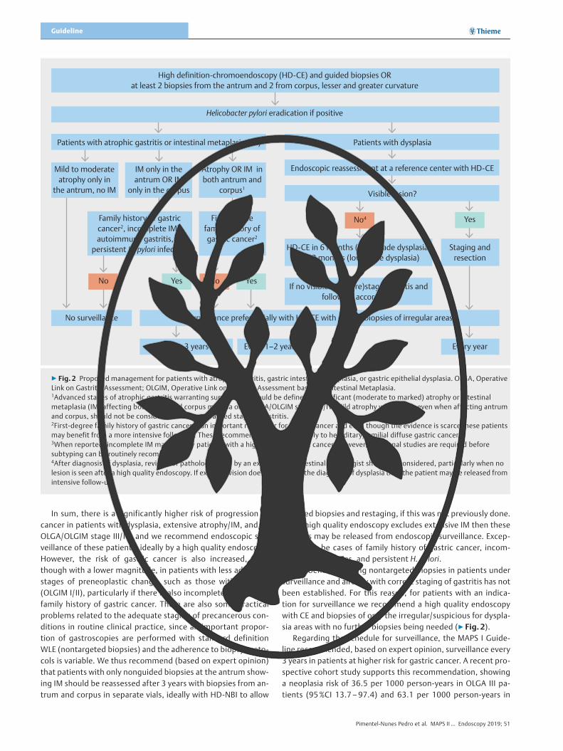
In sum, there is a significantly higher risk of progression tocancer in patients with dysplasia, extensive atrophy/IM, and/orOLGA/OLGIM stage III/IV, and we recommend endoscopic sur-veillance of these patients, ideally by a high quality endoscopy.However, the risk of gastric cancer is also increased, eventhough with a lower magnitude, in patients with less advancedstages of preneoplastic change, such as those with focal IM(OLGIM I/II), particularly if there is also incomplete IM and/or afamily history of gastric cancer. There are also some practicalproblems related to the adequate staging of precancerous con-ditions in routine clinical practice, since an important propor-tion of gastroscopies are performed with standard definitionWLE (nontargeted biopsies) and the adherence to biopsy proto-cols is variable. We thus recommend (based on expert opinion)that patients with only nonguided biopsies at the antrum show-ing IM should be reassessed after 3 years with biopsies from an-trum and corpus in separate vials, ideally with HD-NBI to allow
targeted biopsies and restaging, if this was not previously done.If this high quality endoscopy excludes extensive IM then thesepatients may be released from endoscopic surveillance. Excep-tions may be cases of family history of gastric cancer, incom-plete IM in biopsies, and persistent H. pylori.
The benefit of doing nontargeted biopsies in patients undersurveillance and already with correct staging of gastritis has notbeen established. For this reason, for patients with an indica-tion for surveillance we recommend a high quality endoscopywith CE and biopsies of only the irregular/suspicious for dyspla-sia areas with no further biopsies being needed (▶Fig. 2).
Regarding the schedule for surveillance, the MAPS I Guide-line recommended, based on expert opinion, surveillance every3 years in patients at higher risk for gastric cancer. A recent pro-spective cohort study supports this recommendation, showinga neoplasia risk of 36.5 per 1000 person-years in OLGA III pa-tients (95%CI 13.7–97.4) and 63.1 per 1000 person-years in
High definition-chromoendoscopy (HD-CE) and guided biopsies OR at least 2 biopsies from the antrum and 2 from corpus, lesser and greater curvature
Helicobacter pylori eradication if positive
Surveillance preferentially with HD-CE with guided biopsies of irregular areasNo surveillance
Patients with atrophic gastritis or intestinal metaplasia (IM) Patients with dysplasia
Endoscopic reassessment at a reference center with HD-CE
Yes
Yes
No
No4
YesNo
Visible lesion?
HD-CE in 6�months (high grade dysplasia) to 12�months (low grade dysplasia)
If no visible lesion (re)stage gastritis and follow up accordingly
Staging and resection
Every yearEvery 1–2�yearsEvery 3�years
Mild to moderate atrophy only in
the antrum, no IM
IM only in the antrum OR IM
only in the corpus
Atrophy OR IM in both antrum and
corpus1
Family history of gastric cancer2, incomplete IM3, autoimmune gastritis, or
persistent H. pylori infection
First-degree family history of gastric cancer2
▶ Fig. 2 Proposed management for patients with atrophic gastritis, gastric intestinal metaplasia, or gastric epithelial dysplasia. OLGA, OperativeLink on Gastritis Assessment; OLGIM, Operative Link on Gastritis Assessment based on Intestinal Metaplasia.1Advanced stages of atrophic gastritis warranting surveillance should be defined as significant (moderate to marked) atrophy or intestinalmetaplasia (IM) affecting both antral and corpus mucosa or as OLGA/OLGIM stages III/IV. Mild atrophy without IM, even when affecting antrumand corpus, should not be considered to be an advanced stage of gastritis.2First-degree family history of gastric cancer is an important risk factor for gastric cancer and even though the evidence is scarce these patientsmay benefit from a more intensive follow-up. These recommendations do not apply to hereditary/familial diffuse gastric cancer.3When reported, incomplete IM may identify patients with a higher risk of gastric cancer. However, additional studies are required beforesubtyping can be routinely recommended.4After diagnosis of dysplasia, revision of pathology slides by an expert gastrointestinal pathologist should be considered, particularly when nolesion is seen after a high quality endoscopy. If expert revision does not confirm the diagnosis of dysplasia then the patient may be released fromintensive follow-up.
Pimentel-Nunes Pedro et al. MAPS II … Endoscopy 2019; 51
Guideline
OLGA IV patients (95%CI 20.3–195.6) [133]. The authors sug-gested that the best follow-up surveillance interval would be 2instead of 3 years. However, the cost-effectiveness of a 2-yearinterval for every patient may not be ideal and the evidence isnot strong enough to change the recommended 3-year surveil-lance interval. Nevertheless, the present authors recognize thatpatients with extensive IM, and also with at least one of persist-ent H. pylori infection, incomplete IM, or, particularly, a first-de-gree family history of gastric cancer, may benefit from a tighterendoscopic surveillance schedule (e. g. every 1 –2 years). Theserecommendations do not apply to hereditary/familial diffusegastric cancer, for which there are specific guidelines [134].
5.3 Autoimmune gastritis
cancer relative risk in pernicious anemia was 6.8 (95%CI 2.6–18.1) [146]. The drawback of this meta-analysis is again thatmany patients included in these studies may have had low vita-min B12 serum levels because of conditions other than auto-immune gastritis.
Therefore, there is some evidence suggesting that auto-immune gastritis is a precancerous condition that may justifyendoscopic monitoring. Nevertheless, there is no recommen-ded follow-up interval to date.
Since the largest excess risk of gastric cancer incidenceamong patients with pernicious anemia has been found duringthe first year of follow-up [141, 143], there is evidence to re-commend endoscopic screening to all patients at the time ofthe diagnosis.
Several cohort studies prospectively evaluated the risk ofgastric cancer in patients with pernicious anemia, with varyingfollow-up intervals from 3 to 7 years [114, 139, 140, 142, 147–149]. One study [147] performed follow-up gastroscopies3 years after primary screening examination of 56 patients andidentified on follow-up 2 patients with gastric adenocarcinoma,no patient with HGD, and 49 patients with IM. Another study[142] followed up a group of 27 patients for 6 to 7 years afterinitial investigation. None of the patients had developed gastriccancer since the initial endoscopy and the distribution of dys-plasia was virtually unchanged. The only randomized controlledtrial (RCT) to determine the most effective time interval for thefirst follow-up endoscopy after diagnosis of corpus-predomi-nant atrophic gastritis randomly assigned 24 patients to a 24-or 48-month follow-up interval [148]. No gastric cancer wasfound in either group, but a patient from the 48-month groupdeveloped a neuroendocrine tumor. The authors concludedthat the first follow-up need not be earlier than 4 years afterdiagnosis, with this interval being satisfactory for detection ofpotential neoplastic lesions. Considering the heterogeneity ofthe described cohorts and the absence of larger RCTs with long-er follow-up, we recommend follow-up endoscopy at 3- to 5-year intervals in patients with autoimmune gastritis.
6 Therapy6.1 Helicobacter pylori eradication
RECOMMENDATION
19 Patients with autoimmune gastritis may benefit fromendoscopic follow-up every 3–5 years.Low quality evidence, weak recommendation (82% agree[76% strongly or moderately agree]).
RECOMMENDATION
20 H. pylori eradication heals nonatrophic chronic gastri-tis, may lead to regression of atrophic gastritis, and re-duces the risk of gastric cancer in patients withnonatrophic and atrophic gastritis, and, therefore, it isrecommended in patients with these conditions.High quality evidence, strong recommendation (87%agree [87% strongly or moderately agree]).
Autoimmune gastritis is a chronic progressive inflammatory condition that results in the replacement of the parietal cell mass by atrophic and metaplastic mucosa, leading to a corpus-predominant atrophic gastritis, reduced or absent acid produc-tion, and loss of intrinsic factor which may progress to a severe form of vitamin B12-deficiency anemia known as pernicious anemia. Both gastric carcinoma and neuroendocrine tumors are the most dreaded long-standing complications of perni-cious anemia.
Most of the evidence on the risk of gastric cancer associated with pernicious anemia comes from case-control [135, 136] and cohort studies [114, 137 – 144]. One study based on the Surveillance, Epidemiology, and End Results (SEER) database compared 1 138 390 pernicious anemia cases to 100 000 mat-ched individuals [135]. Individuals with pernicious anemia were at increased risk for noncardia gastric adenocarcinoma (OR 2.18, 95 %CI 1.94 – 2.45) and gastric carcinoid tumors (OR 11.43, 95 %CI 8.90 – 14.69). However, the diagnosis of auto-immune gastritis in this study was rather flawed as it was solely based on low levels of vitamin B12 [145]. Therefore, many of the patients supposedly with autoimmune gastritis probably had other causes of low serum vitamin B12, and the risk of can-cer for genuine autoimmune gastritis patients was likely under-estimated. A Swedish study followed 21 265 patients with per-nicious anemia for an average of 7.1 years [138]. These patients had a significant excess risk for gastric cancer distal to the car-dia (standardized incidence ratio [SIR] 2.4, 95 %CI 2.1 – 2.7). The excess risks increased with increasing follow-up duration. Among distal gastric cancers, the most conspicuous excess risk was for carcinoid tumors (SIR 26.4, 95 %CI 14.8 – 43.5). The abovementioned criticism with respect to the diagnosis of autoimmune gastritis also pertained to this study.
A recent meta-analysis with 27 studies and a total of 22 417 patients showed that the calculated pooled gastric cancer inci-dence rate was 0.27 % per person-year and the overall gastric
Pimentel-Nunes Pedro et al. MAPS II … Endoscopy 2019; 51
Since publication of the MAPS I Guideline, three meta-analyses have been performed regarding the effect of H. pylorieradication on chronic gastritis and risk of gastric cancer [150–152]. The first meta-analysis included only prospective trialsand RCTs on H. pylori eradication with a focus on histology (be-fore and after treatment) and not on the risk of gastric cancer[151]. The authors concluded that IM in the antrum and atroph-ic gastritis in both the antrum and corpus regressed after eradi-cation of H. pylori, although this effect was not seen for IM inthe corpus. This meta-analysis was statistically more powerfulthan previous ones on this subject and strongly suggests thatH. pylori eradication halts progression of precancerous condi-tions even after IM has appeared. In fact, when studies with alonger follow-up (> 5 years) and with larger groups are ana-lyzed, they do show a statistical improvement for IM both atthe antrum and at the corpus after H. pylori eradication therapyhas been received [19, 153–155]. One study even showed nostatistically significant difference with regard to IM in compari-son to an H. pylori-negative group, for the corpus 3 years afterH. pylori eradication and for the antrum 5 years after H. pylorieradication [153]. Despite the possibility of sampling error, itappears logical that for a lesion that occurs after decades of in-fection, reversion also only occurs after a very long period andthe risk of gastric cancer may also only decrease in the long butnot the short term.
Both meta-analyses that focused on the risk of gastric cancerafter H. pylori eradication concluded that H. pylori eradicationsignificantly decreases the risk of gastric cancer in patientswith chronic atrophic or nonatrophic gastritis (pooled relativerisk [RR] 0.64, 95%CI 0.48–0.85) but not in patients with IMor dysplasia (RR 0.88, 95%CI 0.59–1.31) [150, 152]. However,only a few of the studies included in these meta-analyses had along follow-up period (more than 10 years).
In conclusion, there is strong evidence suggesting that H. py-lori eradication is highly beneficial in patients with chronic non-atrophic and atrophic gastritis, both histologically and in redu-cing gastric cancer risk. At later stages of gastritis (establishedIM) weaker evidence suggests that H. pylori eradication hasbeneficial histological effects, with no conclusive effect,
however, on gastric cancer risk reduction. Nevertheless, nostudy suggested that H. pylori eradication has negative effectson patients with IM and, so, considering the positive histologi-cal effects of H. pylori eradication, it is the opinion of the pres-ent authors that H. pylori eradication should also be offered topatients with IM. It is also important to note that H. pylori infec-tion is now considered an infectious disease and eradication isrecommended in most cases, regardless of the presence of pre-cancerous conditions [156].
There is also controversy about H. pylori eradication therapyafter endoscopic removal of gastric superficial neoplasia. Afterpublication of the MAPS I Guideline, a multicenter retrospectivestudy including six Japanese centers and 268 patients contra-dicted its recommendation in favor of such treatment [157]. Inthat study, even though metachronous gastric cancer devel-oped in 14.3% versus 8.5% of the patients of the H. pylori-per-sistent vs. the H. pylori-eradicated group, the baseline severityof mucosal atrophy and a follow-up of more than 5 years werethe only independent risk factors for metachronous neoplasia[157]. A prospective RCT that included 901 patients failed toshow that H. pylori eradication reduced the risk of metachro-nous lesions (2.2% treated vs. 3.7% nontreated, P=0.15)[158]. This trial contradicted another previously published RCTon this subject that showed a significant reduction of gastriccancer in H. pylori-eradicated patients [159]. Moreover, inanother retrospective study that included 2089 patients whounderwent endoscopic resection of a superficial lesion, the in-cidence of metachronous gastric cancer was 10.9 cases per1000 person-years in the H. pylori-negative group, 14.7 casesper 1000 person-years in the H. pylori-eradicated group, and29.7 cases per 1000 person-years in the group without H. pylorieradication (HR 1.9 when compared to H. pylori-eradicatedgroup, P=0.02) [160]. Two meta-analyses on this subject in-cluded the same 10 studies (8 nonrandomized, 2 randomized),but with 5914 and 5881 patients because of different inclusioncriteria. They reached the identical conclusion that H. pylori era-dication reduces the risk of metachronous lesions with a riskratio of 0.467 (95%CI 0.362–0.602; P <0.001) [161, 162]. Final-ly, a 2018 double-blind, placebo-controlled RCT that included396 patients in the intention-to-treat analysis conclusivelyshowed that H. pylori eradication in this group of patients re-duced the risk of metachronous lesions to almost half (7% vs.13%; HR 0.5, 95%CI 0.26–0.94) [163].
In summary, H. pylori eradication has the largest impact ongastric cancer risk in patients with nonatrophic gastritis andearly stages of atrophy. Nevertheless, a small benefit of eradica-tion is still seen at later stages of gastritis and even after resec-tion of a lesion. For this reason, we recommend that H. pylorieradication should always be considered.
RECOMMENDATION
21 In patients with established IM, H. pylori eradicationdoes not appear to significantly reduce the risk of gastriccancer, at least in the short term, but reduces inflamma-tion and atrophy and, therefore, it should be considered.Low quality evidence, weak recommendation (87% agree[75% strongly or moderately agree]).
RECOMMENDATION
22 H. pylori eradication is recommended for patients withgastric neoplasia after endoscopic therapy.High quality evidence, strong recommendation (100%agree [100% strongly or moderately agree]).
Pimentel-Nunes Pedro et al. MAPS II … Endoscopy 2019; 51
Guideline
6.2 Other therapies considered, the effect of aspirin was nonsignificant (RR 0.81,95%CI 0.52–1.26). In sum, evidence suggests that NSAIDsmay slow down the progression of gastric precancerous condi-tions. However, this effect is small and eventually nonsignifi-cant after H. pylori eradication or in more advanced lesions.Considering that NSAIDs have a potential for serious adverseevents it is the opinion of the present authors that they cannotbe recommended specifically for this purpose. The exceptionmay be low dose aspirin since it has a better safety profile andits beneficial effects are more generalized, reducing also cardi-ovascular death risk and the risk of development of other can-cers, and therefore it could be considered in selected patients.
Rebamipide and moluodan The effects of two other drugson pathological findings has been investigated; the drugs wereadministered for 6–12 months following H. pylori eradicationtherapy if required. Rebamipide, a free-radical scavenger, re-duced inflammation, IM, and LGD in one RCT [174], and re-duced chronic inflammation but not IM in another RCT [175];the RCTs included a total of 280 patients. Moluodan, a prepara-tion of Chinese medicine herbs, was associated with a decreasein the dysplasia score, with dysplasia disappearance reported in24.6% of patients in an RCT (196 patients) [176]. Future studiesshould confirm these results before any recommendation canbe made regarding these therapies.
Antioxidant vitamins With respect to antioxidant vitaminsupplementation, no new studies evaluating their effects ongastric precancerous conditions were identified. In the generalpopulation, intake of some vitamins may decrease the risk ofgastric cancer (RR 0.77, 95%CI 0.71–0.83) according to ameta-analysis of 47 studies including 1221392 participants[177]. A significant risk reduction of approximately one thirdwas found for vitamins A, C, and E at daily doses of 1.5mg,100mg, and 10mg, respectively. The risk reduction was notedonly in studies where low dose dietary vitamins were used, notwhen vitamins were administered at high dose or as a drug sup-plement. Some authors have suggested that an efficient inter-vention would aim at nutrient repletion (i. e., with physiologicalas opposed to pharmacological doses) in high risk populationswith poor nutrition [178]. Regarding potential harm, a meta-analysis (53 RCTs, 241883 participants) found that supplemen-tation with vitamins A and E in doses higher than the recom-mended daily allowances (as used in some of the studies includ-ed in the abovementioned meta-analysis [177]) were associat-ed with increased mortality [179].
In line with this, two meta-analyses found a twofold higherrisk of gastric cancer in individuals consuming low versus highamounts of allium vegetables [180], and in those consuming a“Western/unhealthy” diet, rich in starchy foods, meat, and fats,versus a “prudent/healthy” diet rich in fruits and vegetables[181].
RECOMMENDATION
24 Low dose daily aspirin may be considered for preven-tion of various cancers, including gastric cancer, in select-ed patients.Moderate quality evidence, weak recommendation (94%agree [47% strongly or moderately agree]).
RECOMMENDATION
23 Even though cyclo-oxygenase (COX)-1 or COX-2 inhi-bitors may slow progression of gastric precancerous con-ditions, they cannot be recommended specifically for thispurpose.Low quality evidence, weak recommendation (100%agree [94% strongly or moderately agree]).
COX inhibitors Earlier meta-analyses (2003, 2010) had sug-gested a lower risk of gastric cancer in users of COX-inhibitors [164, 165]. Since the publication of MAPS I, few studies have specifically addressed this issue but all added evidence sup-porting this position. In 2013, a prospective nonrandomized study reported on the role of selective COX-2 inhibitor treat-ment in patients with precancerous gastric conditions: after 1 year of treatment with celecoxib following H. pylori eradica-tion, IM regression was more frequent in the treatment versus the control group (44.3 % vs. 14.3 %, total 140 patients) [166]. Other studies suggest that inhibition of COX may slow progres-sion of gastric precancerous conditions and in theory may de-crease the risk of gastric cancer development. A double-blind RCT, including 1024 participants who received H. pylori era-dication treatment or placebo followed by celecoxib or placebo (i. e., four different groups studied), showed that regression of gastric precancerous conditions significantly increased both in the eradication group (59 % vs. 41 % placebo) and in the cele-coxib group (53 % vs. 41 % placebo) with an OR of 1.72 (95 %CI 1.07 – 2.76) for celecoxib and 2.19 (95 %CI 1.32 – 3.64) for H. py-lori eradication [167]. However, in this study no statistically sig-nificant benefit was observed for celecoxib after H. pylori eradi-cation. Moreover, 9 cancers developed in this study but it was underpowered to show a therapeutic benefit of any strategy.
Nonsteroidal anti-inflammatory drugs (NSAIDs) Several meta-analyses investigating the role of aspirin and other NSAIDs on the risk of gastric cancer have accumulated, all of them showing a favorable effect [168 – 173]. In the most recent meta-analysis, which included 24 studies, both nonaspirin NSAIDs (RR 0.86, 95 %CI 0.80 – 0.94) and aspirin (RR 0.70, 95 %CI 0.62 – 0.80) significantly reduced noncardia gastric cancer risk [168]. However, the vast majority of original studies did not include patients with precancerous gastric conditions. Moreover, the protective effect of aspirin/NSAIDs was more marked for noncardia gastric cancer and in H. pylori-positive in-dividuals. In fact, when only H. pylori-negative patients were
Pimentel-Nunes Pedro et al. MAPS II … Endoscopy 2019; 51
7 Cost-effectiveness of surveillance andscreening
Surveillance of precancerous conditions In 2012 whenthe MAPS I Guideline was issued, three studies had been pub-lished regarding the cost-effectiveness of surveillance of pre-cancerous conditions. They had provided conflicting results,mainly because of different estimates for progression to dys-plasia or cancer. Since then only three further studies havebeen published, all in intermediate risk countries.
Areia et al. used a Markov model for Portugal, comparing thecost-utility of three different endoscopic surveillance strate-gies, every 3, 5, or 10 years, for patients with extensive precan-cerous conditions aged 50–75 years [182]. It showed thatendoscopic surveillance of patients with extensive precancer-ous conditions every 3 years was cost-effective compared tono surveillance and was better than the 5- and 10-year strate-gies.
Zhou et al. also applied a Markov model in Singapore andcompared the cost-utility of several endoscopic surveillance orscreening strategies, every 1 or 2 years, for patients aged 50–69 [183]. It showed that endoscopic surveillance of patientswith precancerous conditions every 2 years was the most cost-effective strategy while screening strategies were extendedlydominated (i. e., had a lower incremental cost-effectiveness orwere cost-ineffective).
A third cost-utility model from Wu et al., also for Singapore,compared endoscopic surveillance every 1 year versus endo-scopic screening every 2 years versus nothing, for patientsaged 50–69, and concluded that annual endoscopic surveil-lance was cost-effective for patients with precancerous condi-tions [184].
All three models demonstrated that endoscopic surveillanceof patients with precancerous conditions in countries with anintermediate risk for gastric cancer was cost-effective, as sug-gested by the MAPS I Guideline. The recommended 3-year in-terval was specifically modelled only in the study from Portugaland proved to be better than longer 5- or 10-year intervals,while the studies from Singapore showed that a 1- or 2-year in-terval was most suitable but did not model the 3-year option.
In conclusion, endoscopic surveillance every 3 years of pa-tients with precancerous conditions in countries with an inter-mediate risk for gastric cancer is cost-effective, but further eco-nomic studies would be welcome to further define the optimalinterval for endoscopy.
Endoscopic screening for gastric cancer. Regarding endo-scopic screening for gastric cancer in the general population, at
present it is applied only in high risk populations, such as thoseof Japan and Korea, and several economic studies have alreadybeen published proving its cost-effectiveness [185].
Since 2012, two further studies have been published for theKorean population, again concluding that endoscopic screen-ing for this high risk population is cost-effective. Chang et al.,using a cost-utility model from a societal perspective for thepopulation aged 50–80 years, concluded that endoscopicscreening annually for men and biennially for women was theoptimal cost-effective option among 12 different strategies[186]. Another cost-effectiveness model, also from a societalperspective in patients older than 40 years, demonstrated thatannual endoscopic screening was better than X-ray screeningor no screening [187].
Regarding the cost-effectiveness of endoscopic screeningfor gastric cancer in intermediate or low incidence countries,three models have been published since preparation of theMAPS I Guideline. Yeh et al. investigated a one-time gastriccancer screening strategy for an American population at age50, with a cost-utility analysis from a societal perspective, com-paring serum pepsinogen test screening followed by endoscopyif results were positive versus endoscopic screening; they con-cluded that for this low risk population neither option was cost-effective [188]. Others also used a Markov cost-utility model forthe US population, and analyzed the option of adding a one-time screening upper endoscopy at the time of screening colo-noscopy in 50-year-old patients. They concluded that this op-tion was not cost-effective for this low risk population despitethe reduced endoscopy costs [189]. Finally, Areia et al. model-led the option of adding a screening upper endoscopy at thetime of screening colonoscopy, in Portugal, an intermediate tohigh risk country for gastric cancer [190]. Using a Markov cost-utility analysis they compared three screening strategies:stand-alone upper endoscopy, endoscopy combined with acolorectal cancer screening colonoscopy after a positive fecaloccult blood test, or pepsinogen serology screening. The con-clusion was that endoscopic gastric cancer screening was cost-effective if combined with a screening colonoscopy in Portugal.Furthermore, this strategy might prove to be cost-effective inother European countries with a gastric cancer risk ≥10 per100000 inhabitants, depending on further specific economicstudies for each local setting.
8 Research agendaDuring the updating of this Guideline, gaps in evidence re-mained noticeable. Further research is needed, including:▪ Large randomized trials with different surveillance
schedules, that would elucidate the natural history of eachstage of gastric changes (and identify further variables forpredicting progression), and also clarify the benefit of eachsurveillance interval;
▪ Adequately designed trials to assess the effect of interven-tions such as H. pylori eradication and others regarding thepremalignant stomach;
▪ Studies to address methodologies and target populations forscreening these lesions in Western countries.
RECOMMENDATION
25 In intermediate to high risk regions, identification andsurveillance of patients with precancerous gastric condi-tions is cost-effective.Moderate quality evidence) (100% agree [94% strongly ormoderately agree]).
Pimentel-Nunes Pedro et al. MAPS II … Endoscopy 2019; 51
Guideline
DisclaimerThe legal disclaimer for ESGE guidelines [191] applies to thecurrent Guideline.
AcknowledgmentsThe authors gratefully thank Dr. Cesare Hassan, Nuovo ReginaMargherita Hospital, Rome, Italy, and Professor KonstantinosTriantafyllou, Hepatogastroenterology Unit, Attikon UniversityGeneral Hospital, Athens, Greece, for their critical review ofthe Guideline.
Competing interests
M. Leja has shares in and receives a salary from the Digestive DiseasesCentre GASTRO, SIA (from approximately 2000 to present); his de-partment receives research support with a special offer for reagents(including for pepsinogen detection) from Eiken Chemical (2013 topresent); he is a Board member of the Latvian Association of Gastro-enterology (from approximately 2000 to present), F. Megraud’s de-partment has received a grant from Allergan (2014 to February2019). J. E. van Hooft has received lecture fees from Medtronic(2014–2015) and consultancy fees from Boston Scientific (2014–2016); her department has received research grants from Cook Med-ical (2014–2018) and Abbott (2014–2017). B. Annibale, M. Areia,R. Barros, F. Carneiro, M. Dinis-Ribeiro, J.-M. Dumonceau, G. Esposito,J.-F. Fléjou, M. Garrido, I. Kikuste, E. J. Kuipers, D. Libânio, R. Marcos-Pinto, T. Matysiak-Budnik, and P. Pimentel-Nunes have no competinginterests.
References
[1] Bray F, Ferlay J, Soerjomataram I et al. Global cancer statistics 2018:GLOBOCAN estimates of incidence and mortality worldwide for 36cancers in 185 countries. CA Cancer J Clin 2018; 68: 393–424
[2] Pimentel-Nunes P, Dinis-Ribeiro M. Endoscopic submucosal dissec-tion in the treatment of gastrointestinal superficial lesions: Follow theguidelines! GE Port J Gastroenterol 2015; 22: 184–186
[3] Pimentel-Nunes P, Dinis-Ribeiro M, Ponchon T et al. Endoscopic sub-mucosal dissection: European Society of Gastrointestinal Endoscopy(ESGE) Guideline. Endoscopy 2015; 47: 829–854
[4] Dinis-Ribeiro M, Areia M, de Vries AC et al. Management of precan-cerous conditions and lesions in the stomach (MAPS): guideline fromthe European Society of Gastrointestinal Endoscopy (ESGE), EuropeanHelicobacter Study Group (EHSG), European Society of Pathology(ESP), and the Sociedade Portuguesa de Endoscopia Digestiva (SPED).Endoscopy 2012; 44: 74–94
[5] Dinis-Ribeiro M, Areia M, de Vries AC et al. Management of precan-cerous conditions and lesions in the stomach (MAPS): guideline fromthe European Society of Gastrointestinal Endoscopy (ESGE), EuropeanHelicobacter Study Group (EHSG), European Society of Pathology(ESP), and the Sociedade Portuguesa de Endoscopia Digestiva (SPED).Virchows Arch 2012; 460: 19–46
[6] Bisschops R, Areia M, Coron E et al. Performance measures for uppergastrointestinal endoscopy: a European Society of GastrointestinalEndoscopy (ESGE) Quality Improvement Initiative. Endoscopy 2016;48: 843–864
[7] AGREE Collaboration. Development and validation of an internationalappraisal instrument for assessing the quality of clinical practiceguidelines: the AGREE project. Qual Saf Health Care 2003; 12: 18–23
[8] Atkins D, Eccles M, Flottorp S et al. Systems for grading the quality ofevidence and the strength of recommendations I: critical appraisal ofexisting approaches The GRADE Working Group. BMC Health Serv Res2004; 4: 38
[9] Guyatt GH, Oxman AD, Vist GE et al. GRADE: an emerging consensuson rating quality of evidence and strength of recommendations. BMJ2008; 336: 924–926
[10] Correa P. Human gastric carcinogenesis: a multistep and multifactor-ial process – First American Cancer Society Award Lecture on CancerEpidemiology and Prevention. Cancer Res 1992; 52: 6735–6740
[11] Kapadia CR. Gastric atrophy, metaplasia and dysplasia: a clinical per-spective. J Clin Gastroenterol 2003; 36 : S29–36
[12] Carneiro F, Machado JC, David L et al. Current thoughts on the histo-pathogenesis of gastric cancer. Eur J Cancer Prev 2001; 10: 101–102
[13] Ihamaki T, Sankkonen M, Siurala M. Long-term observation of sub-jects with normal mucosa and with superficial gastritis: results of 23–27 years’ follow-up examinations. Scand J Gastroenterol 1978; 13:771–775
[14] Ormiston MC, Gear MW, Codling BW. Five year follow-up study ofgastritis. J Clin Pathol 1982; 35: 757–760
[15] Laurèn P. The two histological main types of gastric carcinoma: dif-fuse and so-called intestinal-type carcinoma. Acta Pathol MicrobiolScand 1965; 64: 31–49
[16] Fukao A, Hisamichi S, Ohsato N et al. Correlation between the preva-lence of gastritis and gastric cancer in Japan. Cancer Causes Control1993; 4: 17.20
[17] Genta RM. Gastric atrophy and atrophic gastritis – nebulous conceptsin search of a definition. Aliment Pharmacol Ther 1998; 12: 17–23
[18] Yue H, Shan L, Bin L. The significance of OLGA and OLGIM stagingsystems in the risk assessment of gastric cancer: a systematic reviewand meta-analysis. Gastric Cancer 2018; 21: 579–587, Epub 2018 Feb19
[19] Mera RM, Bravo LE, Camargo MC et al. Dynamics of Helicobacter py-lori infection as a determinant of progression of gastric precancerouslesions: 16-year follow-up of an eradication trial. Gut 2018; 67:1239–1246, Epub 2017 Jun 24
[20] Dixon MF, Genta RM, Yardley JH et al. Classification and grading ofgastritis. The updated Sydney System. International workshop on thehistopathology of gastritis, Houston 1994. Am J Surg Pathol 1996; 20:1161–1181
[21] Misiewicz JJ. The Sydney System: a new classification of gastritis.Introduction. J Gastroenterol Hepatol 1991; 6: 207–208
[22] Plummer M, Buiatti E, Lopez G et al. Histological diagnosis of precan-cerous lesions of the stomach: a reliability study. Int J Epidemiol 1997;26: 716–720
[23] Cotruta B, Gheorghe C, Iacob R et al. The orientation of gastric biopsysamples improves the inter-observer agreement of the OLGA stagingsystem. J Gastrointestin Liver Dis 2017; 26: 351–356
[24] Isajevs S, Liepniece-Karele I, Janciauskas D et al. Gastritis staging:interobserver agreement by applying OLGA and OLGIM systems.Virchows Arch 2014; 464: 403–407
[25] Correa P. Clinical implications of recent developments in gastric can-cer pathology and epidemiology. Semin Oncol 1985; 12: 2–10
[26] Lauwers G, Carneiro F, Graham D et al. Gastric carcinoma. In:Theise N, ed. WHO Classification of tumours of the digestive system.Lyon: IARC Press; 2010: 48–58
[27] Lim H, Jung HY, Park YS et al. Discrepancy between endoscopic for-ceps biopsy and endoscopic resection in gastric epithelial neoplasia.Surg Endosc 2014; 28: 1256–1262
[28] Pimentel-Nunes P, Mourao F, Veloso N et al. Long-term follow-up afterendoscopic resection of gastric superficial neoplastic lesions in Por-tugal. Endoscopy 2014; 46: 933–940
Pimentel-Nunes Pedro et al. MAPS II … Endoscopy 2019; 51
[29] Zhao G, Xue M, Hu Y et al. How commonly is the diagnosis of gastriclow grade dysplasia upgraded following endoscopic resection? Ameta-analysis PLoS One 2015; 10: e0132699
[30] Atkins L, Benedict EB. Correlation of gross gastroscopic findings withgastroscopic biopsy in gastritis. N Engl J Med 1956; 254: 641–644
[31] Carpenter HA, Talley NJ. Gastroscopy is incomplete without biopsy:clinical relevance of distinguishing gastropathy from gastritis.Gastroenterology 1995; 108: 917–924
[32] Eshmuratov A, Nah JC, Kim N et al. The correlation of endoscopic andhistological diagnosis of gastric atrophy. Dig Dis Sci 2010; 55: 1364–1375
[33] Redéen S, Petersson F, Jönsson KA et al. Relationship of gastroscopicfeatures to histological findings in gastritis and Helicobacter pyloriinfection in a general population sample. Endoscopy 2003; 35:946–950
[34] Stathopoulos G, Goldberg RD, Blackstone MO. Endoscopic diagnosisof intestinal metaplasia. Gastrointest Endosc 1990; 36: 544–545
[35] Panteris V, Nikolopoulou S, Lountou A et al. Diagnostic capabilities ofhigh-definition white light endoscopy for the diagnosis of gastricintestinal metaplasia and correlation with histologic and clinical data.Eur J Gastroenterol Hepatol 2014; 26: 594–601
[36] Pimentel-Nunes P, Libanio D, Lage J et al. A multicenter prospectivestudy of the real-time use of narrow-band imaging in the diagnosis ofpremalignant gastric conditions and lesions. Endoscopy 2016; 48:723–730
[37] Ang TL, Pittayanon R, Lau JY et al. A multicenter randomized com-parison between high-definition white light endoscopy and narrowband imaging for detection of gastric lesions. Eur J GastroenterolHepatol 2015; 27: 1473–1478
[38] Anagnostopoulos GK, Yao K, Kaye P et al. High-resolution magnifica-tion endoscopy can reliably identify normal gastric mucosa, Helico-bacter pylori-associated gastritis, and gastric atrophy. Endoscopy2007; 39: 202–207
[39] Gonen C, Simsek I, Sarioglu S et al. Comparison of high resolutionmagnifying endoscopy and standard videoendoscopy for the diagno-sis of Helicobacter pylori gastritis in routine clinical practice: a pro-spective study. Helicobacter 2009; 14: 12–21
[40] Tahara T, Shibata T, Nakamura M et al. Gastric mucosal pattern byusing magnifying narrow-band imaging endoscopy clearly distingui-shes histological and serological severity of chronic gastritis. Gastro-intest Endosc 2009; 70: 246–253
[41] Areia M, Amaro P, Dinis-Ribeiro M et al. External validation of a classi-fication for methylene blue magnification chromoendoscopy in pre-malignant gastric lesions. Gastrointest Endosc 2008; 67: 1011–1018
[42] Dinis-Ribeiro M, da Costa-Pereira A, Lopes C et al. Magnificationchromoendoscopy for the diagnosis of gastric intestinal metaplasiaand dysplasia. Gastrointest Endosc 2003; 57: 498–504
[43] Kono Y, Takenaka R, Kawahara Y et al. Chromoendoscopy of gastricadenoma using an acetic acid indigocarmine mixture. World J Gas-troenterol 2014; 20: 5092–5097
[44] Mouzyka S, Fedoseeva A. Chromoendoscopy with hematoxylin in theclassification of gastric lesions. Gastric Cancer 2008; 11: 15–21; dis-cussion 21–12
[45] Tanaka K, Toyoda H, Kadowaki S et al. Surface pattern classification byenhanced-magnification endoscopy for identifying early gastric can-cers. Gastrointest Endosc 2008; 67: 430–437
[46] Zhao Z, Yin Z, Wang S et al. Meta-analysis: The diagnostic efficacy ofchromoendoscopy for early gastric cancer and premalignant gastriclesions. J Gastroenterol Hepatol 2016; 31: 1539–1545
[47] Kikuste I, Marques-Pereira R, Monteiro-Soares M et al. Systematic re-view of the diagnosis of gastric premalignant conditions and neopla-sia with high-resolution endoscopic technologies. Scand J Gastroen-terol 2013; 48: 1108–1117
[48] Pimentel-Nunes P, Dinis-Ribeiro M, Soares JB et al. A multicenter vali-dation of an endoscopic classification with narrow band imaging forgastric precancerous and cancerous lesions. Endoscopy 2012; 44:236–246
[49] Dias-Silva D, Pimentel-Nunes P, Magalhaes J et al. The learning curvefor narrow-band imaging in the diagnosis of precancerous gastric le-sions by using web-based video. Gastrointest Endosc 2014; 79:910–920; quiz 983-e911, 983 e914
[50] East JE, Vleugels JL, Roelandt P et al. Advanced endoscopic imaging:European Society of Gastrointestinal Endoscopy (ESGE) TechnologyReview. Endoscopy 2016; 48: 1029–1045
[51] Dutta AK, Sajith KG, Pulimood AB et al. Narrow band imaging versuswhite light gastroscopy in detecting potentially premalignant gastriclesions: a randomized prospective crossover study. Indian J Gastroen-terol 2013; 32: 37–42
[52] Xirouchakis E, Laoudi F, Tsartsali L et al. Screening for gastric prema-lignant lesions with narrow band imaging, white light and updatedSydney protocol or both? Dig Dis Sci 2013; 58: 1084–1090
[53] Buxbaum JL, Hormozdi D, Dinis-Ribeiro M et al. Narrow-band imagingversus white light versus mapping biopsy for gastric intestinal meta-plasia: a prospective blinded trial. Gastrointest Endosc 2017; 86:857–865
[54] Lage J, Pimentel-Nunes P, Figueiredo PC et al. Light-NBI to identifyhigh-risk phenotypes for gastric adenocarcinoma: do we still needbiopsies? Scand J Gastroenterol 2016; 51: 501–506
[55] Ezoe Y, Muto M, Uedo N et al. Magnifying narrowband imaging ismore accurate than conventional white-light imaging in diagnosis ofgastric mucosal cancer. Gastroenterology 2011; 141: 2017–2025e2013
[56] Zhang Q, Wang F, Chen ZY et al. Comparison of the diagnostic effica-cy of white light endoscopy and magnifying endoscopy with narrowband imaging for early gastric cancer: a meta-analysis. Gastric Cancer2016; 19: 543–552
[57] Pimenta-Melo AR, Monteiro-Soares M, Libanio D et al. Missing rate forgastric cancer during upper gastrointestinal endoscopy: a systematicreview and meta-analysis. Eur J Gastroenterol Hepatol 2016; 28:1041–1049
[58] Pimentel-Nunes P, Libanio D, Dinis-Ribeiro M. Evaluation and man-agement of gastric superficial neoplastic lesions. GE Port J Gastroen-terol 2017; 24: 8–21
[59] Lee JW, Lim LG, Yeoh KG. Advanced endoscopic imaging in gastricneoplasia and preneoplasia. BMJ Open Gastroenterol 2017; 4:e000105
[60] Dohi O, Yagi N, Majima A et al. Diagnostic ability of magnifyingendoscopy with blue laser imaging for early gastric cancer: a pro-spective study. Gastric Cancer 2017; 20: 297–303
[61] Dohi O, Yagi N, Yoshida S et al. Magnifying blue laser imaging versusmagnifying narrow-band imaging for the diagnosis of early gastriccancer: a prospective, multicenter, comparative study. Digestion2017; 96: 127–134
[62] Kaneko K, Oono Y, Yano T et al. Effect of novel bright image enhancedendoscopy using blue laser imaging (BLI). Endosc Int Open 2014; 2:E212–219
[63] Mao XY, Xu SF, Liu Q et al. Anatomical predilection of intestinalmetaplasia based on 78,335 endoscopic cases. Saudi J Gastroenterol2016; 22: 154–160
[64] Kim YI, Kook MC, Cho SJ et al. Effect of biopsy site on detection ofgastric cancer high-risk groups by OLGA and OLGIM stages. Helico-bacter 2017: doi:10.1111/hel.12442
[65] Isajevs S, Liepniece-Karele I, Janciauskas D et al. The effect of incisuraangularis biopsy sampling on the assessment of gastritis stage. Eur JGastroenterol Hepatol 2014; 26: 510–513
Pimentel-Nunes Pedro et al. MAPS II … Endoscopy 2019; 51
Guideline
[66] Lash JG, Genta RM. Adherence to the Sydney System guidelines in-creases the detection of Helicobacter gastritis and intestinal meta-plasia in 400738 sets of gastric biopsies. Aliment Pharmacol Ther2013; 38: 424–431
[67] Varbanova M, Wex T, Jechorek D et al. Impact of the angulus biopsyfor the detection of gastric preneoplastic conditions and gastric can-cer risk assessment. J Clin Pathol 2016; 69: 19–25
[68] Marcos-Pinto R, Carneiro F, Dinis-Ribeiro M et al. First-degree rela-tives of patients with early-onset gastric carcinoma show even atyoung ages a high prevalence of advanced OLGA/OLGIM stages anddysplasia. Aliment Pharmacol Ther 2012; 35: 1451–1459
[69] Rugge M, Meggio A, Pennelli G et al. Gastritis staging in clinical prac-tice: the OLGA staging system. Gut 2007; 56: 631–636
[70] Rugge M, de Boni M, Pennelli G et al. Gastritis OLGA-staging and gas-tric cancer risk: a twelve-year clinico-pathological follow-up study.Aliment Pharmacol Ther 2010; 31: 1104–1111
[71] Capelle LG, de Vries AC, Haringsma J et al. The staging of gastritis withthe OLGA system by using intestinal metaplasia as an accurate alter-native for atrophic gastritis. Gastrointest Endosc 2010; 71:1150–1158
[72] Dinis-Ribeiro M, Yamaki G, Miki K et al. Meta-analysis on the validity ofpepsinogen test for gastric carcinoma, dysplasia or chronic atrophicgastritis screening. J Med Screen 2004; 11: 141–147
[73] Cho JH, Jeon SR, Kim HG et al. The serum pepsinogen levels for riskassessment of gastric neoplasms: New proposal from a case-controlstudy in Korea. Medicine (Baltimore) 2017; 96: e7603
[74] Tong Y, Wu Y, Song Z et al. The potential value of serum pepsinogenfor the diagnosis of atrophic gastritis among the health check-uppopulations in China: a diagnostic clinical research. BMC Gastroen-terol 2017; 17: 88
[75] Huang YK, Yu JC, Kang WM et al. Significance of serum pepsinogensas a biomarker for gastric cancer and atrophic gastritis screening: asystematic review and meta-analysis. PLoS One 2015; 10: e0142080
[76] Kitahara F, Kobayashi K, Sato T et al. Accuracy of screening for gastriccancer using serum pepsinogen concentrations. Gut 1999; 44:693–697
[77] Leja M, Camargo MC, Polaka I et al. Detection of gastric atrophy bycirculating pepsinogens: A comparison of three assays. Helicobacter2017: doi:10.1111/hel.12393
[78] Zagari RM, Rabitti S, Greenwood DC et al. Systematic review withmeta-analysis: diagnostic performance of the combination of pepsi-nogen, gastrin-17 and anti-Helicobacter pylori antibodies serum as-says for the diagnosis of atrophic gastritis. Aliment Pharmacol Ther2017; 46: 657–667
[79] Leja M, Kupcinskas L, Funka K et al. Value of gastrin-17 in detectingantral atrophy. Adv Med Sci 2011; 56: 145–150
[80] Watabe H, Mitsushima T, Yamaji Y et al. Predicting the developmentof gastric cancer from combining Helicobacter pylori antibodies andserum pepsinogen status: a prospective endoscopic cohort study. Gut2005; 54: 764–768
[81] Yoshida T, Kato J, Inoue I et al. Cancer development based on chronicactive gastritis and resulting gastric atrophy as assessed by serumlevels of pepsinogen and Helicobacter pylori antibody titer. Int J Can-cer 2014; 134: 1445–1457
[82] Kawashima J, Ohno S, Sakurada T et al. Circulating acylated ghrelinlevel decreases in accordance with the extent of atrophic gastritis. JGastroenterol 2009; 44: 1046–1054
[83] Eun Bae S, Hoon Lee J, Soo Park Y et al. Decrease of serum total ghrelinin extensive atrophic gastritis: comparison with pepsinogens in his-tological reference. Scand J Gastroenterol 2016; 51: 137–144
[84] Kasai C, Sugimoto K, Moritani I et al. Changes in plasma ghrelin andleptin levels in patients with peptic ulcer and gastritis following era-
dication of Helicobacter pylori infection. BMC Gastroenterol 2016;16: 119
[85] Shi SQ, Cai JT, Yang JM. Expression of trefoil factors 1 and 2 in pre-cancerous condition and gastric cancer. World J Gastroenterol 2006;12: 3119–3122
[86] Link A, Schirrmeister W, Langner C et al. Differential expression ofmicroRNAs in preneoplastic gastric mucosa. Scientific reports 2015;5: 8270
[87] Amal H, Leja M, Funka K et al. Detection of precancerous gastric le-sions and gastric cancer through exhaled breath. Gut 2016; 65:400–407
[88] [Anonymous] Schistosomes, liver flukes and Helicobacter pylori.IARC Working Group on the evaluation of carcinogenic risks to hu-mans. Lyon, 7–14 June 1994. IARC Monogr Eval Carcinog Risks Hum1994; 61: 1–241
[89] Figueiredo C, Machado JC, Pharoah P et al. Helicobacter pylori andinterleukin 1 genotyping: an opportunity to identify high-risk indi-viduals for gastric carcinoma. J Natl Cancer Inst 2002; 94: 1680–1687
[90] Amieva MR, El-Omar EM. Host-bacterial interactions in Helicobacterpylori infection. Gastroenterology 2008; 134: 306–323
[91] Machado JC, Figueiredo C, Canedo P et al. A proinflammatory geneticprofile increases the risk for chronic atrophic gastritis and gastriccarcinoma. Gastroenterology 2003; 125: 364–371
[92] Huang JQ, Zheng GF, Sumanac K et al. Meta-analysis of the relation-ship between cagA seropositivity and gastric cancer. Gastroenterolo-gy 2003; 125: 1636–1644
[93] Basso D, Zambon CF, Letley DP et al. Clinical relevance of Helicobacterpylori cagA and vacA gene polymorphisms. Gastroenterology 2008;135: 91–99
[94] Rizzato C, Kato I, Plummer M et al. Risk of advanced gastric precan-cerous lesions in Helicobacter pylori infected subjects is influenced byABO blood group and cagA status. Int J Cancer 2013; 133: 315–322
[95] Palli D, Masala G, Del Giudice G et al. CagA+ Helicobacter pylori in-fection and gastric cancer risk in the EPIC-EURGAST study. Int J Cancer2007; 120: 859–867
[96] Winter JA, Letley DP, Cook KW et al. A role for the vacuolating cyto-toxin, VacA, in colonization and Helicobacter pylori-induced meta-plasia in the stomach. J Infect Dis 2014; 210: 954–963
[97] Cover TL. Helicobacter pylori diversity and gastric cancer risk. MBio2016; 7: e01869–01815
[98] El-Omar EM, Carrington M, Chow WH et al. Interleukin-1 polymorph-isms associated with increased risk of gastric cancer. Nature 2000;404: 398–402
[99] Persson C, Canedo P, Machado JC et al. Polymorphisms in inflamma-tory response genes and their association with gastric cancer: a hugesystematic review and meta-analyses. Am J Epidemiol 2011; 173:259–270
[100] Wang P, Xia HH-X, Zhang J-Y et al. Association of interleukin-1 genepolymorphisms with gastric cancer: A meta-analysis. Int J Cancer2007; 120: 552–562
[101] Camargo MC, Mera R, Correa P et al. Interleukin-1beta and interleu-kin-1 receptor antagonist gene polymorphisms and gastric cancer: ameta-analysis. Cancer Epidemiol Biomarkers Prev 2006; 15:1674–1687
[102] Xue H, Lin B, Ni P et al. Interleukin-1B and interleukin-1 RN poly-morphisms and gastric carcinoma risk: a meta-analysis. J Gastroen-terol Hepatol 2010; 25: 1604–1617
[103] Loh M, Koh KX, Yeo BH et al. Meta-analysis of genetic polymorph-isms and gastric cancer risk: variability in associations according torace. Eur J Cancer 2009; 45: 2562–2568
[104] Hnatyszyn A, Wielgus K, Kaczmarek-Rys M et al. Interleukin-1 genepolymorphisms in chronic gastritis patients infected with Helico-
Pimentel-Nunes Pedro et al. MAPS II … Endoscopy 2019; 51
bacter pylori as risk factors of gastric cancer development. Arch Im-munol Ther Exp (Warsz) 2013; 61: 503–512
[105] Simone A, Casadei A, De Vergori E et al. Rescue endoscopy to iden-tify site of gastric dysplasia or carcinoma found at random biopsies.Dig Liver Dis 2011; 43: 721–725
[106] Yep-Gamarra V, Diaz-Velez C, Araujo I et al. [Usefulness of systema-tic chromoendoscopy with a double dye staining technique for thedetection of dysplasia in patients with premalignant gastric lesions].Gastroenterol Hepatol 2016; 39: 49–54
[107] Gong EJ, Lee JH, Jung K et al. Characteristics of missed simultaneousgastric lesions based on double-check analysis of the endoscopicimage. Clin Endosc 2017; 50: 261–269
[108] Teh JL, Tan JR, Lau LJ et al. Longer examination time improves de-tection of gastric cancer during diagnostic upper gastrointestinalendoscopy. Clin Gastroenterol Hepatol 2015; 13: 480–487.e2
[109] de Vries AC, van Grieken NC, Looman CW et al. Gastric cancer risk inpatients with premalignant gastric lesions: a nationwide cohortstudy in the Netherlands. Gastroenterology 2008; 134: 945–952
[110] Song H, Ekheden IG, Zheng Z et al. Incidence of gastric canceramong patients with gastric precancerous lesions: observationalcohort study in a low risk Western population. BMJ 2015; 351: h3867
[111] Goo JJ, Choi CW, Kang DH et al. Risk factors associated with diag-nostic discrepancy of gastric indefinite neoplasia: Who need en blocresection? Surg Endosc 2015; 29: 3761–3767
[112] Raftopoulos SC, Kumarasinghe P, de Boer B et al. Gastric intraepi-thelial neoplasia in a Western population. Eur J Gastroenterol Hepa-tol 2012; 24: 48–54
[113] Fassan M, Pizzi M, Farinati F et al. Lesions indefinite for intraepithelialneoplasia and OLGA staging for gastric atrophy. Am J Clin Pathol2012; 137: 727–732
[114] Lahner E, Esposito G, Pilozzi E et al. Occurrence of gastric cancer andcarcinoids in atrophic gastritis during prospective long-term followup. Scand J Gastroenterol 2015; 50: 856–865
[115] Shichijo S, Hirata Y, Niikura R et al. Histologic intestinal metaplasiaand endoscopic atrophy are predictors of gastric cancer develop-ment after Helicobacter pylori eradication. Gastrointest Endosc2016; 84: 618–624
[116] Reddy KM, Chang JI, Shi JM et al. Risk of gastric cancer among pa-tients with intestinal metaplasia of the stomach in a US integratedhealth care system. Clin Gastroenterol Hepatol 2016; 14:1420–1425
[117] Tsai YC, Hsiao WH, Yang HB et al. The corpus-predominant gastritisindex may serve as an early marker of Helicobacter pylori-infectedpatients at risk of gastric cancer. Aliment Pharmacol Ther 2013; 37:969–978
[118] Zhou Y, Li HY, Zhang JJ et al. Operative link on gastritis assessmentstage is an appropriate predictor of early gastric cancer. World JGastroenterol 2016; 22: 3670–3678
[119] Masuyama H, Yoshitake N, Sasai T et al. Relationship between thedegree of endoscopic atrophy of the gastric mucosa and carcino-genic risk. Digestion 2015; 91: 30–36
[120] Cho SJ, Choi IJ, Kook MC et al. Staging of intestinal- and diffuse-typegastric cancers with the OLGA and OLGIM staging systems. AlimentPharmacol Ther 2013; 38: 1292–1302
[121] Kodama M, Murakami K, Okimoto T et al. Histological characteristicsof gastric mucosa prior to Helicobacter pylori eradication may pre-dict gastric cancer. Scand J Gastroenterol 2013; 48: 1249–1256
[122] Marques-Silva L, Areia M, Elvas L et al. Prevalence of gastric precan-cerous conditions: a systematic review and meta-analysis. Eur J Gas-troenterol Hepatol 2014; 26: 378–387
[123] Gonzalez CA, Sanz-Anquela JM, Companioni O et al. Incomplete typeof intestinal metaplasia has the highest risk to progress to gastric
cancer: results of the Spanish follow-up multicenter study. J Gastro-enterol Hepatol 2016; 31: 953–958
[124] Gonzalez CA, Sanz-Anquela JM, Gisbert JP et al. Utility of subtypingintestinal metaplasia as marker of gastric cancer risk. A review of theevidence. Int J Cancer 2013; 133: 1023–1032
[125] Conchillo JM, Houben G, de Bruine A et al. Is type III intestinal meta-plasia an obligatory precancerous lesion in intestinal-type gastriccarcinoma? Eur J Cancer Prev 2001; 10: 307–312
[126] Matsukura N, Onda M, Tokunaga A et al. Mucosal IgA antibodyagainst Helicobacter pylori in chronic gastritis and intestinal meta-plasia detected by the Tes-Tape method in resection specimensafter gastrectomy for gastric cancer. Cancer 1995; 75: 1472–1477
[127] Silva S, Filipe MI. Intestinal metaplasia and its variants in the gastricmucosa of Portuguese subjects: a comparative analysis of biopsyand gastrectomy material. Hum Pathol 1986; 17: 988–995
[128] Oliveira C, Pinheiro H, Figueiredo J et al. Familial gastric cancer: ge-netic susceptibility, pathology, and implications for management.Lancet Oncol 2015; 16: e60– e70
[129] Yaghoobi M, Bijarchi R, Narod SA. Family history and the risk of gas-tric cancer. Br J Cancer 2010; 102: 237–242
[130] Safaee A, Moghimi-Dehkordi B, Fatemi SR et al. Family history ofcancer and risk of gastric cancer in Iran. Asian Pac J Cancer Prev2011; 12: 3117–3120
[131] Rokkas T, Sechopoulos P, Pistiolas D et al. Helicobacter pylori infec-tion and gastric histology in first-degree relatives of gastric cancerpatients: a meta-analysis. Eur J Gastroenterol Hepatol 2010; 22:1128–1133
[132] Marcos-Pinto R, Dinis-Ribeiro M, Carneiro F et al. First-degree rela-tives of early-onset gastric cancer patients show a high risk for gas-tric cancer: phenotype and genotype profile. Virchows Arch 2013;463: 391–399
[133] Rugge M, Meggio A, Pravadelli C et al. Gastritis staging in the endo-scopic follow-up for the secondary prevention of gastric cancer: a5-year prospective study of 1755 patients. Gut 2019; 68: 11–17
[134] van der Post RS, Vogelaar IP, Carneiro F et al. Hereditary diffuse gas-tric cancer: updated clinical guidelines with an emphasis on germ-line CDH1 mutation carriers. J Med Genet 2015; 52: 361–374
[135] Murphy G, Dawsey SM, Engels EA et al. Cancer risk after perniciousanemia in the US elderly population. Clin Gastroenterol Hepatol2015; 13: 2282–2289, e2281–2284
[136] Landgren AM, Landgren O, Gridley G et al. Autoimmune disease andsubsequent risk of developing alimentary tract cancers among 4.5million US male veterans. Cancer 2011; 117: 1163–1171
[137] Hemminki K, Liu X, Ji J et al. Autoimmune disease and subsequentdigestive tract cancer by histology. Ann Oncol 2012; 23: 927–933
[138] Ye W, Nyren O. Risk of cancers of the oesophagus and stomach byhistology or subsite in patients hospitalised for pernicious anaemia.Gut 2003; 52: 938–941
[139] Kokkola A, Sjoblom SM, Haapiainen R et al. The risk of gastric carci-noma and carcinoid tumours in patients with pernicious anaemia. Aprospective follow-up study. Scand J Gastroenterol 1998; 33: 88–92
[140] Mellemkjoer L, Gridley G, Moller H et al. [Pernicious anemia and riskof cancer in Denmark]. Ugeskr Laeger 1997; 159: 4850–4853
[141] Hsing AW, Hansson LE, McLaughlin JK et al. Pernicious anemia andsubsequent cancer. A population-based cohort study. Cancer 1993;71: 745–750
[142] Armbrecht U, Stockbrugger RW, Rode J et al. Development of gastricdysplasia in pernicious anaemia: a clinical and endoscopic follow upstudy of 80 patients. Gut 1990; 31: 1105–1109
[143] Brinton LA, Gridley G, Hrubec Z et al. Cancer risk following perni-cious anaemia. Br J Cancer 1989; 59: 810–813
Pimentel-Nunes Pedro et al. MAPS II … Endoscopy 2019; 51
Guideline
[144] Svendsen JH, Dahl C, Svendsen LB et al. Gastric cancer risk in achlor-hydric patients. A long-term follow-up study. Scand J Gastroenterol1986; 21: 16–20
[145] Kuipers EJ. Pernicious anemia, atrophic gastritis, and the risk of can-cer. Clin Gastroenterol Hepatol 2015; 13: 2290–2292
[146] Vannella L, Lahner E, Osborn J et al. Systematic review: gastric cancerincidence in pernicious anaemia. Aliment Pharmacol Ther 2013; 37:375–382
[147] Sjoblom SM, Sipponen P, Jarvinen H. Gastroscopic follow up of per-nicious anaemia patients. Gut 1993; 34: 28–32
[148] Lahner E, Caruana P, D'Ambra G et al. First endoscopic-histologicfollow-up in patients with body-predominant atrophic gastritis:when should it be done? Gastrointest Endosc 2001; 53: 443–448
[149] Sjoblom SM, Sipponen P, Miettinen M et al. Gastroscopic screeningfor gastric carcinoids and carcinoma in pernicious anemia. Endos-copy 1988; 20: 52–56
[150] Chen HN, Wang Z, Li X et al. Helicobacter pylori eradication cannotreduce the risk of gastric cancer in patients with intestinal metapla-sia and dysplasia: evidence from a meta-analysis. Gastric Cancer2016; 19: 166–175
[151] Kong YJ, Yi HG, Dai JC et al. Histological changes of gastric mucosaafter Helicobacter pylori eradication: a systematic review and meta-analysis. World J Gastroenterol 2014; 20: 5903–5911
[152] Rokkas T, Rokka A, Portincasa P. A systematic review and meta-a-nalysis of the role of Helicobacter pylori eradication in preventinggastric cancer. Ann Gastroenterol 2017; 30: 414–423
[153] Hwang YJ, Kim N, Lee HS et al. Reversibility of atrophic gastritis andintestinal metaplasia after Helicobacter pylori eradication – a pro-spective study for up to 10 years. Aliment Pharmacol Ther 2018; 47:380–390
[154] Kodama M, Murakami K, Okimoto T et al. Helicobacter pylori eradi-cation improves gastric atrophy and intestinal metaplasia in long-term observation. Digestion 2012; 85: 126–130
[155] Kodama M, Murakami K, Okimoto T et al. Ten-year prospective fol-low-up of histological changes at five points on the gastric mucosaas recommended by the updated Sydney system after Helicobacterpylori eradication. J Gastroenterol 2012; 47: 394–403
[156] Malfertheiner P, Megraud F, O'Morain CA et al. Management of Heli-cobacter pylori infection – the Maastricht V/Florence Consensus Re-port. Gut 2017; 66: 6–30
[157] Maehata Y, Nakamura S, Fujisawa K et al. Long-term effect of Heli-cobacter pylori eradication on the development of metachronousgastric cancer after endoscopic resection of early gastric cancer.Gastrointest Endosc 2012; 75: 39–46
[158] Choi J, Kim SG, Yoon H et al. Eradication of Helicobacter pylori afterendoscopic resection of gastric tumors does not reduce incidence ofmetachronous gastric carcinoma. Clin Gastroenterol Hepatol 2014;12: 793–800 e791
[159] Fukase K, Kato M, Kikuchi S et al. Effect of eradication of Helicobac-ter pylori on incidence of metachronous gastric carcinoma afterendoscopic resection of early gastric cancer: an open-label, rando-mised controlled trial. Lancet 2008; 372: 392–397
[160] Bae SE, Jung HY, Kang J et al. Effect of Helicobacter pylori eradicationon metachronous recurrence after endoscopic resection of gastricneoplasm. Am J Gastroenterol 2014; 109: 60–67
[161] Bang CS, Baik GH, Shin IS et al. Helicobacter pylori eradication forprevention of metachronous recurrence after endoscopic resectionof early gastric cancer. J Korean Med Sci 2015; 30: 749–756
[162] Jung DH, Kim JH, Chung HS et al. Helicobacter pylori eradication onthe prevention of metachronous lesions after endoscopic resectionof gastric neoplasm: a meta-analysis. PLoS One 2015; 10: e0124725
[163] Choi IJ, Kook MC, Kim YI et al. Helicobacter pylori therapy for theprevention of metachronous gastric cancer. N Engl J Med 2018; 378:1085–1095
[164] Tian W, Zhao Y, Liu S et al. Meta-analysis on the relationship be-tween nonsteroidal anti-inflammatory drug use and gastric cancer.Eur J Cancer Prev 2010; 19: 288–298
[165] Wang WH, Huang JQ, Zheng GF et al. Non-steroidal anti-inflamma-tory drug use and the risk of gastric cancer: a systematic review andmeta-analysis. J Natl Cancer Inst 2003; 95: 1784–1791
[166] Sheu BS, Tsai YC, Wu CT et al. Long‐term celecoxib can prevent theprogression of persistent gastric intestinal metaplasia after H. pylorieradication. Helicobacter 2013; 18: 117–123
[167] Wong BC, Zhang L, Ma JL et al. Effects of selective COX-2 inhibitorand Helicobacter pylori eradication on precancerous gastric lesions.Gut 2012; 61: 812–818
[168] Huang X-Z, Chen Y, Wu J et al. Aspirin and non-steroidal anti-in-flammatory drugs use reduce gastric cancer risk: A dose-responsemeta-analysis. Oncotarget 2017; 8: 4781–4795
[169] Kong P, Wu R, Liu X et al. The effects of anti-inflammatory drugtreatment in gastric cancer prevention: an update of a meta-analy-sis. J Cancer 2016; 7: 2247–2257
[170] Ye X, Fu J, Yang Y et al. Frequency-risk and duration-risk relationshipsbetween aspirin use and gastric cancer: a systematic review andmeta-analysis. PLoS ONE 2013; 8: e71522
[171] Bosetti C, Rosato V, Gallus S et al. Aspirin and cancer risk: a quanti-tative review to 2011. Ann Oncol 2012; 23: 1403–1415
[172] Tian W, Zhao Y, Liu S et al. Meta-analysis on the relationship be-tween nonsteroidal anti-inflammatory drug use and gastric cancer.Eur J Cancer Prev 2010; 19: 288–298
[173] Abnet CC, Freedman ND, Kamangar F et al. Non-steroidal anti-in-flammatory drugs and risk of gastric and oesophageal adenocarci-nomas: results from a cohort study and a meta-analysis. Br J Cancer2009; 100: 551–557
[174] Han X, Jiang K, Wang B et al. Effect of rebamipide on the premalig-nant progression of chronic gastritis: a randomized controlled study.Clin Drug Investig 2015; 35: 665–673
[175] Kamada T, Sato M, Tokutomi T et al. Rebamipide improves chronicinflammation in the lesser curvature of the corpus after Helicobac-ter pylori eradication: a multicenter study. Biomed Res Int 2015;2015: 865146–865148
[176] Tang XD, Zhou LY, Zhang ST et al. Randomized double-blind clinicaltrial of Moluodan () for the treatment of chronic atrophic gastritiswith dysplasia. Chin J Integr Med 2016; 22: 9–18
[177] Kong P, Cai Q, Geng Q et al. Vitamin intake reduce the risk of gastriccancer: meta-analysis and systematic review of randomized and ob-servational studies. PLoS One 2014; 9: e116060
[178] Taylor PR. Prevention of gastric cancer: a miss. J Natl Cancer Inst2007; 99: 101–103
[179] Bjelakovic G, Nikolova D, Gluud C. Meta-regression analyses, meta-analyses, and trial sequential analyses of the effects of supplemen-tation with beta-carotene, vitamin A, and vitamin E singly or in dif-ferent combinations on all-cause mortality: do we have evidence forlack of harm? PLoS One 2013; 8: e74558
[180] Zhou Y, Zhuang W, Hu W et al. Consumption of large amounts ofallium vegetables reduces risk for gastric cancer in a meta-analysis.Gastroenterology 2011; 141: 80–89
[181] Bertuccio P, Rosato V, Andreano A et al. Dietary patterns and gastriccancer risk: a systematic review and meta-analysis. Ann Oncol 2013;24: 1450–1458
[182] Areia M, Dinis-Ribeiro M, Rocha Goncalves F. Cost-utility analysis ofendoscopic surveillance of patients with gastric premalignant con-ditions. Helicobacter 2014; 19: 425–436
Pimentel-Nunes Pedro et al. MAPS II … Endoscopy 2019; 51
[183] Zhou HJ, Dan YY, Naidoo N et al. A cost-effectiveness analysis eval-uating endoscopic surveillance for gastric cancer for populationswith low to intermediate risk. PLoS One 2013; 8: e83959
[184] Wu JT, Zhou J, Naidoo N et al. Determining the cost-effectiveness ofendoscopic surveillance for gastric cancer in patients with precan-cerous lesions. Asia Pac J Clin Oncol 2016; 12: 359–368
[185] Areia M, Carvalho R, Cadime AT et al. Screening for gastric cancerand surveillance of premalignant lesions: a systematic review ofcost-effectiveness studies. Helicobacter 2013; 18: 325–337
[186] Chang HS, Park EC, Chung W et al. Comparing endoscopy and uppergastrointestinal X-ray for gastric cancer screening in South Korea: acost-utility analysis. Asian Pac J Cancer Prev 2012; 13: 2721–2728
[187] Cho E, Kang MH, Choi KS et al. Cost-effectiveness outcomes of thenational gastric cancer screening program in South Korea. Asian PacJ Cancer Prev 2013; 14: 2533–2540
[188] Yeh JM, Hur C, Ward Z et al. Gastric adenocarcinoma screening andprevention in the era of new biomarker and endoscopic technolo-gies: a cost-effectiveness analysis. Gut 2016; 65: 563–574
[189] Gupta N, Bansal A, Wani SB et al. Endoscopy for upper GI cancerscreening in the general population: a cost-utility analysis. Gastro-intest Endosc 2011; 74: 610–624 e612
[190] Areia M, Spaander MC, Kuipers EJ et al. Endoscopic screening forgastric cancer: A cost-utility analysis for countries with an inter-mediate gastric cancer risk. United European Gastroenterol J 2018;6: 192–202
[191] Dumonceau JM, Hassan C, Riphaus A et al. European Society of Gas-trointestinal Endoscopy (ESGE) Guideline Development Policy.Endoscopy 2012; 44: 626–629
Pimentel-Nunes Pedro et al. MAPS II … Endoscopy 2019; 51
Guideline