Embed Size (px)
Citation preview
RESEARCH ARTICLE Open Access
Acetic acid-indigo carmine chromoendoscopy fordelineating early gastric cancers: its usefulnessaccording to histological typeBong Eun Lee1, Gwang Ha Kim1*, Do Youn Park2, Dae Hwan Kim3, Tae Yong Jeon3, Su Bum Park1,Hyun Seok You1, Dong Yup Ryu1, Dong Uk Kim1, Geun Am Song1
Abstract
Background: Endoscopic treatments, such as endoscopic submucosal dissection (ESD) and laparoscopicgastrectomy, are increasingly used to treat a subset of patients with early gastric cancer (EGC). To achievesuccessful outcomes, it is very important to accurately determine the lateral extent of the tumor. Therefore, weinvestigated the diagnostic performance of chromoendoscopy using indigo carmine dye added to acetic acid (AIchromoendoscopy) in delineating differentiated or undifferentiated adenocarcinomas in patients with EGC.
Methods: We prospectively included 151 lesions of 141 patients that had an endoscopic diagnosis of EGC. All thelesions were examined by conventional endoscopy and AI chromoendoscopy before ESD or laparoscopicgastrectomy. The border clarification between the lesion and the normal mucosa was classified as distinct orindistinct before and after AI chromoendoscopy.
Results: The borders of the lesions were distinct in 66.9% (101/151) with conventional endoscopy and in 84.1%(127/151) with AI chromoendoscopy (P < 0.001). Compared with conventional endoscopy, AI chromoendoscopyclarified the border in a significantly higher percentage of differentiated adenocarcinomas (74/108 [68.5%] vs 97/108 [89.8%], respectively, P < 0.001). However, the border clarification rate for undifferentiated adenocarcinomas didnot differ between conventional endoscopy and AI chromoendoscopy (27/43 [62.8%] vs 30/43 [70.0%], respectively,P = 0.494).
Conclusions: AI chromoendoscopy is useful in determining the lateral extent of EGCs. However, its usefulness isreduced in undifferentiated adenocarcinomas.
BackgroundThere has been a reduced incidence of gastric cancer inwestern countries over the past few decades. However,gastric cancer is still the second leading cause of cancerdeaths in the world, and it is the most prevalent malig-nancy in Korea [1,2]. Early gastric cancer (EGC) isdefined as a gastric cancer that is confined to themucosa or submucosa, regardless of the presence orabsence of lymph node metastasis [3]. The proportionof EGC cases is increasing in Korea because endoscopicscreening for gastric cancer has been adopted [4]. As a
result, endoscopic treatments such as endoscopic sub-mucosal dissection (ESD) and laparoscopic gastrectomyare increasingly used to treat a subset of patients withEGC in both Korea and Japan [5-8].To achieve a successful outcome, it is very important
to accurately determine the lateral extent of the tumor.This has traditionally been done with conventionalendoscopy and chromoendoscopy using indigo carminedye [9,10]. However, it is sometimes difficult to identifythe margins of the tumors, especially those of superficialor flat-type tumors. Magnifying endoscopes have report-edly been useful in overcoming this difficulty [11,12] buttheir use is limited by the technical difficulties in manip-ulating the scopes. Therefore, easier methods arerequired that make it possible to accurately determine
* Correspondence: [email protected] of Internal Medicine, Pusan National University School ofMedicine and Medical Research Institute, Pusan National University Hospital,Busan, KoreaFull list of author information is available at the end of the article
Lee et al. BMC Gastroenterology 2010, 10:97http://www.biomedcentral.com/1471-230X/10/97
© 2010 Lee et al; licensee BioMed Central Ltd. This is an Open Access article distributed under the terms of the Creative CommonsAttribution License (http://creativecommons.org/licenses/by/2.0), which permits unrestricted use, distribution, and reproduction inany medium, provided the original work is properly cited.

the lateral extent of these tumors. Chromoendoscopywith indigo carmine dye added to acetic acid (AI chro-moendoscopy) has recently been reported to improvethe diagnostic yield in terms of recognizing the tumorborders in patients with EGC [13,14]. However, themajority of subjects included in these studies had differ-entiated adenocarcinomas. Therefore, the current studywas performed to prospectively investigate the diagnos-tic performance of AI chromoendoscopy in delineatingdifferentiated or undifferentiated adenocarcinomas inpatients with EGC.
MethodsFrom January 2007 to May 2009, a total of 151 lesionsin 141 patients (85 men and 56 women; age range, 35-81 years; mean age 60 years) with an endoscopic diagno-sis of EGC were enrolled prospectively. These patientshad previously undergone endoscopic ultrasonographyand computed tomography assessments, and werescheduled to undergo ESD or surgery.This study was approved by the Institutional Review
Board at Pusan National University Hospital andinformed consent was obtained from all the patientsbefore their examination.
Diagnostic proceduresAll the lesions detected by high-definition video endo-scopy (EVIS LUCERA GIF-H260; Olympus Optical Co.,Ltd, Tokyo, Japan) were examined by a single experi-enced endoscopist (G.H. Kim) as follows: step 1, mucusadhering to the mucosa was washed away as thoroughlyas possible before the examination of the lesion; step 2,10-20 mL of 1.5% acetic acid was sprinkled evenly overand around the lesion using a washing pipe (PW-5L-1;Olympus); step 3, 10-20 mL of 0.2% indigo carmine dyewas similarly sprinkled 30-60 seconds later using awashing pipe; step 4, the area was washed with cleanwater 20-30 seconds later for the final view (Figure 1, 2).Before and after AI chromoendoscopy, the border
clarification between the lesion and the normal mucosawas classified as distinct or indistinct by observationwith the naked eye. The clarity of the endoscopic imageafter AI chromoendoscopy was also classified as clear,mottled, or unclear.
Clinicopathological reviewESD or laparoscopic gastrectomy was performed withinone week of AI chromoendoscopy. The resected speci-mens were fixed in 10% buffered formalin. Carcinomaswith adjacent non-neoplastic mucosa were serially cutinto 2 mm parallel slices and embedded in paraffin, andthen sectioned and stained with hematoxylin-eosin forhistological examination. The clinicopathological find-ings, such as age, sex, macroscopic shape, tumor site,
tumor size, histological type and depth of invasion, werereviewed according to the Japanese Classification ofGastric Carcinomas [15]. The depth of tumor invasionwas classified as mucosal, submucosal, or advanced (thetumor had invaded the muscularis propria or deeper).
Statistical analysisThe difference in the border clarification when conven-tional endoscopy or AI chromoendoscopy was used andthe differences in the clinicopathological characteristicsaccording to the border clarification and the clarity ofthe endoscopic image were assessed using c2 test orFisher’s exact test. Multivariate logistic regression analy-sis was used to identify variables predicting the borderclarification after AI chromoendoscopy. Influencing fac-tors/covariates for the border clarification were tumorsize, macroscopic type, histopathological diagnosis anddepth of tumor invasion. A P value < 0.05 was consid-ered statistically significant. The statistical calculationswere performed with the SPSS version 12.0 for Win-dows software (SPSS Inc., Chicago, IL, USA).
ResultsThe clinicopathological characteristics of the patientsenrolled in this study are summarized in Table 1. Sixtylesions were treated by ESD and 91 lesions were treatedby laparoscopic gastrectomy. The lesions were locatedin the upper third of the stomach for 7.3% (11/151), inthe middle third for 39.7% (60/151) and in the lowerthird for 53.0% (80/151). Macroscopically, the lesionswere classified as flat/elevated (64/151, 42.4%),depressed (64/151, 42.4%), or excavated (23/151, 15.2%).Histopathologically, the lesions were diagnosed as differ-entiated adenocarcinomas in 71.5% (108/151) and undif-ferentiated adenocarcinomas in 28.5% (43/151). Thedepth of tumor invasion was mucosal in 71.5% (108/151), submucosal in 25.8% (39/151), and advanced in2.7% (4/151).The border of the lesion was distinct in 66.9% (101/
151) with conventional endoscopy and in 84.1% (127/151) with AI chromoendoscopy (P < 0.001) (Table 2)(Figure 3). Compared with conventional endoscopy, AIchromoendoscopy clarified the border in a significantlyhigher percentage of differentiated adenocarcinomas(74/108 [68.5%] vs 97/108 [89.8%], respectively, P <0.001). However, the border clarification rate for undif-ferentiated adenocarcinomas did not differ betweenconventional endoscopy and AI chromoendoscopy (27/43 [62.8%] vs 30/43 [70.0%], respectively, P = 0.494).Of the 50 lesions with indistinct borders on conven-tional endoscopy, AI chromoendoscopy clarified theborders in 66.0% (33/50) (27 of 34 differentiated ade-nocarcinomas and 6 of 16 undifferentiatedadenocarcinomas).
Lee et al. BMC Gastroenterology 2010, 10:97http://www.biomedcentral.com/1471-230X/10/97
Page 2 of 8
When the indistinct border group was comparedwith the distinct border group after AI chromoendo-scopy, there were no differences in the tumor size,location, or depth (Table 3). The lesions with anexcavated morphology showed a higher frequency ofindistinct borders than those with a flat/elevated ordepressed morphology (P = 0.034). Undifferentiatedcarcinomas had a higher frequency of indistinct bor-ders than differentiated carcinomas (13/43 [30.2%] vs11/108 [10.2%], respectively, P = 0.002). On multi-variate regression analysis, only the histopathologicaltype was associated with the border clarification(Table 4).
The clarity of the lesions after AI chromoendoscopydid not differ according to the tumor size, location, ordepth (Table 5). The lesions with a flat/elevated mor-phology showed a higher frequency of clear images thanthose with a depressed or excavated morphology (P =0.009). Undifferentiated adenocarcinomas showed ahigher frequency of mottled appearance than differen-tiated adenocarcinomas (30/43 [69.8%] vs 2/108 [1.9%],respectively, P = 0.002).
DiscussionIn this study, the diagnostic performance of conven-tional endoscopy in recognizing tumor borders was
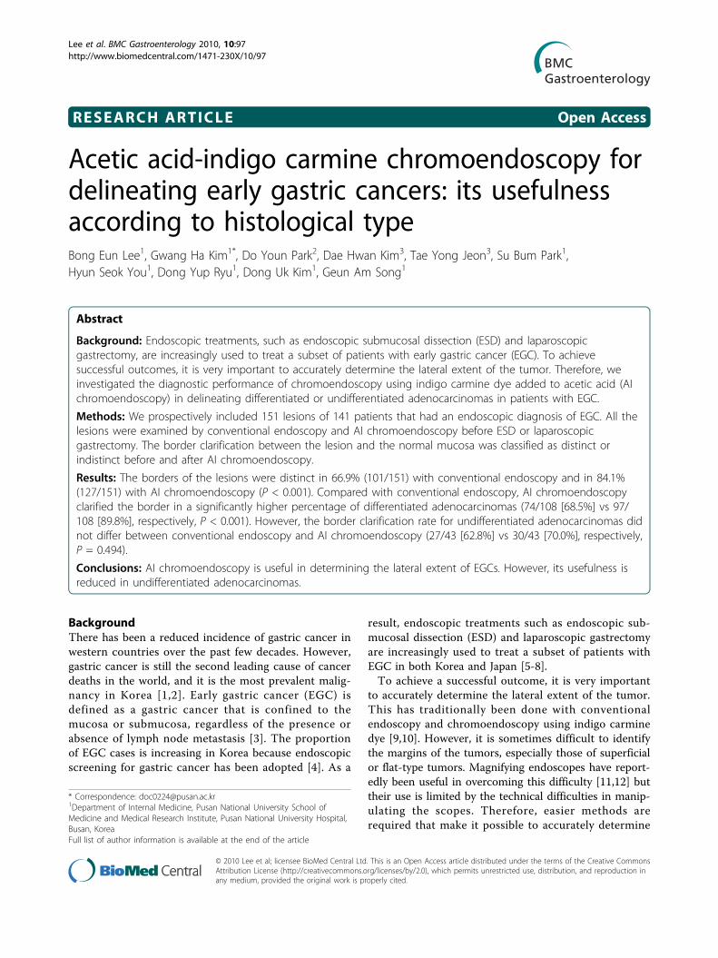
Figure 1 Chromoendoscopy of a differentiated adenocarcinoma. (A) A combined flat and elevated lesion with an unclear border at thelower body of the stomach is shown. (B) Endoscopic view after acetic acid was sprinkled. (C) Endoscopic view after indigo carmine wasadditionally sprinkled. (D) Endoscopic view after the lesion was washed with clean water. After chromoendoscopy with indigo carmine dyeadded to acetic acid, the lesion’s borders became distinct and the clarity of the image is high. The lesion was resected by endoscopicsubmucosal dissection and was shown to be a differentiated adenocarcinoma.
Lee et al. BMC Gastroenterology 2010, 10:97http://www.biomedcentral.com/1471-230X/10/97
Page 3 of 8
inadequate (only 66.9%) in patients with an endoscopicdiagnosis of EGC. AI chromoendoscopy increased therecognition rate of tumor borders to 84.1%, especially indifferentiated adenocarcinomas.The accurate determination of the pre-treatment lat-
eral extent of a tumor is critical for successful endo-scopic resection and laparoscopic gastrectomy inpatients with EGC. Inadequate determination of the lat-eral extent may result in an incomplete resection, whichwould increase the rate of local relapse. During endo-scopic resection such as ESD, the entire border betweenthe tumor and the normal mucosa is electrosurgicallymarked, approximately 5 mm from the lesion, and the
procedure is then performed [16]. During a surgicaloperation such as a laparoscopic gastrectomy, EGClesions cannot be identified by inspecting the serosalsurface and are usually impossible to palpate manuallybecause the depth of the invasion is shallow. Therefore,the day before surgery, two or three endoscopic clipsare usually placed at the mucosa approximately 1-2 cmfrom the proximal margin of the lesion in the oral direc-tion [17]. In this study, we used the same localizationtechnique for endoscopic resection and laparoscopicgastrectomy.The diagnostic performance for determining the lateral
extent of a tumor with conventional endoscopy is
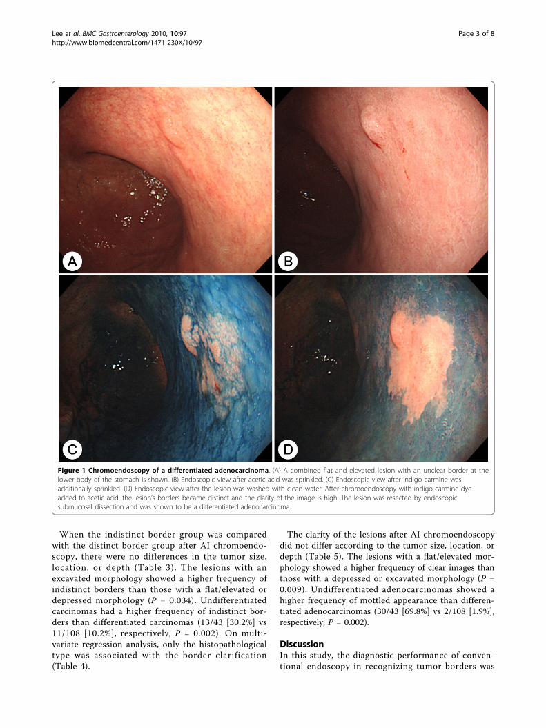
Figure 2 Chromoendoscopy of an undifferentiated adenocarcinoma. (A) A flat discolored lesion with an unclear border at the lower bodyof the stomach is shown. (B) Endoscopic view after acetic acid was sprinkled. (C) Endoscopic view after indigo carmine was additionallysprinkled. (D) Endoscopic view after the lesion was washed with clean water. After chromoendoscopy with indigo carmine dye added to aceticacid, the lesion’s border was still indistinct and the image was mottled. The lesion was resected by laparoscopic gastrectomy and was shown tobe an undifferentiated adenocarcinoma.
Lee et al. BMC Gastroenterology 2010, 10:97http://www.biomedcentral.com/1471-230X/10/97
Page 4 of 8
inadequate because the tumor rims are often almost thesame height and color as the surrounding normal mucosa[13]. Chromoendoscopy with indigo carmine dye, whichis not absorbed by the mucosa but pools in crevices andvalleys, thus defining the irregularities in the mucosalarchitecture, has been used for over 30 years and is still astrong modality for identifying gastric lesions [9,10,13].However, the accurate delineation of the tumor area isoften difficult because the dye simply contrasts thesurface irregularity of the tumor [18].Magnifying endoscopy has recently been reported as
useful in determining the lateral spread of gastric can-cers [11,12]. However, magnifying endoscopy is not pop-ular and there is no generally accepted standard foridentifying the patterns of tumors, which limits the role
of magnifying endoscopy in determining the lateralextent of a tumor.The technique based on the application of acetic acid
during the endoscopy was first used to observe the spe-cialized columnar epithelium of Barrett’s esophagus [19].This technique was then adopted for the assessment ofgastric neoplasms [11,20]. The transient whitish colori-zation of the epithelial surface, which occurs after thespraying of acetic acid, is a consequence of the increasedopacity. This corresponds to a reversible alternation ofthe three-dimensional structures of the cytoplasmic pro-teins [13]. However, the lateral margins were success-fully identified with acetic acid in only 42-53% of gastricneoplasms [13].Based on chromoendoscopy with acetic acid, Yama-
shita et al. recently described the use of an indigo car-mine and acetic acid mixture to accurately identify themargins of gastric cancers in 27 cases, which was evenpossible with low-resolution endoscopy [18]. The speci-ficity and sensitivity were 98.0% and 100%, respectively,based on biopsy samples from the demarcated areas orjust outside the areas. In a prospective study of 53 neo-plasms, which compared AI chromoendoscopy withconventional chromoendoscopy using indigo carmine oracetic acid alone, the diagnostic performance of AIchromoendoscopy (94.3%) was significantly better thanthat of the other modalities [13]. Of the 53 lesions, 49were differentiated adenocarcinomas, 3 were adenomasand only one lesion was an undifferentiated adenocarci-noma. Similarly, in our present study, AI chromoendo-scopy clarified the borders in 89.8% (97/108) of thedifferentiated adenocarcinomas.However, there has been no report on the perfor-
mance of AI chromoendoscopy in the assessment ofundifferentiated adenocarcinomas. In the present study,the borders of undifferentiated adenocarcinomas weredistinct in 62.8% with conventional endoscopy and in70.0% with AI chromoendoscopy. Of the 16 undifferen-tiated adenocarcinomas with an indistinct border duringconventional endoscopy, AI chromoendoscopy clarifiedthe borders in only 6 lesions. Therefore, the diagnosticperformance of AI chromoendoscopy in assessing
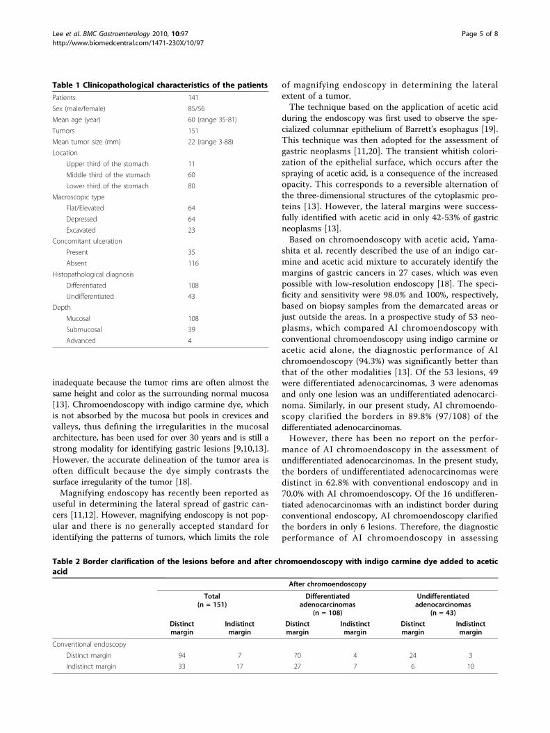
Table 1 Clinicopathological characteristics of the patients
Patients 141
Sex (male/female) 85/56
Mean age (year) 60 (range 35-81)
Tumors 151
Mean tumor size (mm) 22 (range 3-88)
Location
Upper third of the stomach 11
Middle third of the stomach 60
Lower third of the stomach 80
Macroscopic type
Flat/Elevated 64
Depressed 64
Excavated 23
Concomitant ulceration
Present 35
Absent 116
Histopathological diagnosis
Differentiated 108
Undifferentiated 43
Depth
Mucosal 108
Submucosal 39
Advanced 4
Table 2 Border clarification of the lesions before and after chromoendoscopy with indigo carmine dye added to aceticacid
After chromoendoscopy
Total(n = 151)
Differentiatedadenocarcinomas
(n = 108)
Undifferentiatedadenocarcinomas
(n = 43)
Distinctmargin
Indistinctmargin
Distinctmargin
Indistinctmargin
Distinctmargin
Indistinctmargin
Conventional endoscopy
Distinct margin 94 7 70 4 24 3
Indistinct margin 33 17 27 7 6 10
Lee et al. BMC Gastroenterology 2010, 10:97http://www.biomedcentral.com/1471-230X/10/97
Page 5 of 8

undifferentiated adenocarcinomas seems to be unsatis-factory compared with the diagnostic performanceof AI chromoendoscopy in assessing differentiatedadenocarcinomas.We also investigated the differences in the clinico-
pathological characteristics of the lesions in the distinctborder group and the indistinct border group after AIchromoendoscopy. The frequency of an indistinct bor-der was higher for lesions with an excavated morphol-ogy as well as with undifferentiated adenocarcinoma.During this study, we discovered that there was a
difference between differentiated and undifferentiatedadenocarcinomas in the clarity of the lesions after AIchromoendoscopy. A mottled appearance was morecommon in undifferentiated adenocarcinomas than indifferentiated adenocarcinomas. These results can beexplained by the fact that undifferentiated adenocarcino-mas infiltrate diffusely among the normal gastric gland-ular cells.The exact mechanism of AI chromoendoscopy is still
unclear, but we propose a possible mechanism. Whenacetic acid is sprinkled, the surrounding non-cancerous
Figure 3 The rates of border clarification by conventional endoscopy and chromoendoscopy with indigo carmine dye added to aceticacid (AI chromoendoscopy) according to the histological type of the lesion.
Table 3 Clinicopathological characteristics of the lesions of the distinct border group and the indistinct border groupafter chromoendoscopy with indigo carmine dye added to acetic acid
Distinct border group(n = 127)
Indistinct border group(n = 24)
P value
Tumor size 0.232
≤ 2 cm 80 12
> 2 cm 47 12
Location 0.788
Upper third of the stomach 10 1
Middle third of the stomach 51 9
Lower third of the stomach 66 14
Macroscopic type 0.034
Flat/Elevated 59 5
Depressed 52 12
Excavated 16 7
Histopathological diagnosis 0.002
Differentiated 97 11
Undifferentiated 30 13
Depth 0.273
Mucosal 93 15
Submucosal 30 9
Advanced 4 0
Lee et al. BMC Gastroenterology 2010, 10:97http://www.biomedcentral.com/1471-230X/10/97
Page 6 of 8
mucosa whitens, but the cancerous mucosa does not,which produces good contrast between the pinkishcancer lesion and the surrounding non-cancerous tis-sue. If indigo carmine is additionally sprinkled, thenthe surrounding whitish non-cancerous mucosa isstained blue and the pinkish cancer is not stained.This color difference is made clearer by washing withclean water.However, in seven of the 101 lesions with clear bor-
ders on conventional endoscopy, the borders becameless clear after AI chromoendoscopy. This problemwould be attributed to the increased secretion of mucusfrom the gastric mucosa after the acetic acid wassprinkled on it, which resulted in the adhesion of themucus to the surface of the lesion, reducing the contrastbetween the lesion and the normal mucosa [14].
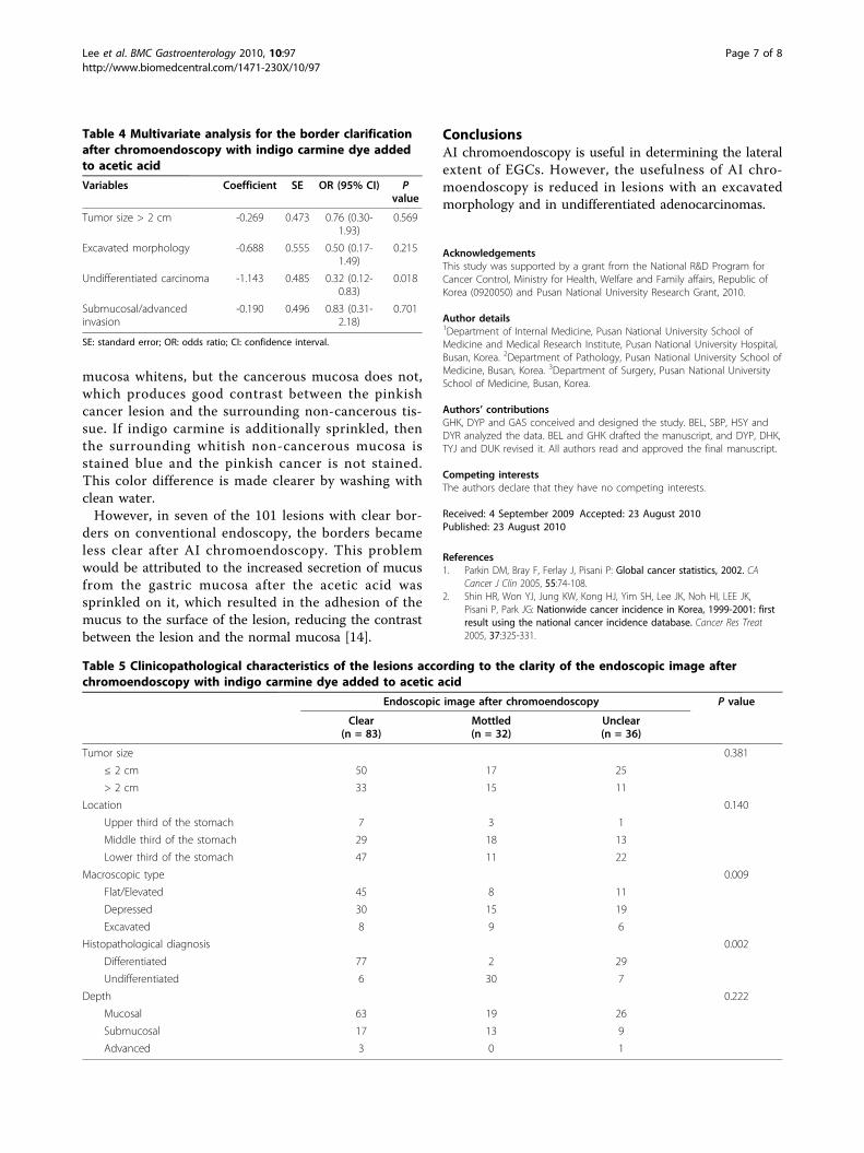
ConclusionsAI chromoendoscopy is useful in determining the lateralextent of EGCs. However, the usefulness of AI chro-moendoscopy is reduced in lesions with an excavatedmorphology and in undifferentiated adenocarcinomas.
AcknowledgementsThis study was supported by a grant from the National R&D Program forCancer Control, Ministry for Health, Welfare and Family affairs, Republic ofKorea (0920050) and Pusan National University Research Grant, 2010.
Author details1Department of Internal Medicine, Pusan National University School ofMedicine and Medical Research Institute, Pusan National University Hospital,Busan, Korea. 2Department of Pathology, Pusan National University School ofMedicine, Busan, Korea. 3Department of Surgery, Pusan National UniversitySchool of Medicine, Busan, Korea.
Authors’ contributionsGHK, DYP and GAS conceived and designed the study. BEL, SBP, HSY andDYR analyzed the data. BEL and GHK drafted the manuscript, and DYP, DHK,TYJ and DUK revised it. All authors read and approved the final manuscript.
Competing interestsThe authors declare that they have no competing interests.
Received: 4 September 2009 Accepted: 23 August 2010Published: 23 August 2010
References1. Parkin DM, Bray F, Ferlay J, Pisani P: Global cancer statistics, 2002. CA
Cancer J Clin 2005, 55:74-108.2. Shin HR, Won YJ, Jung KW, Kong HJ, Yim SH, Lee JK, Noh HI, LEE JK,
Pisani P, Park JG: Nationwide cancer incidence in Korea, 1999-2001: firstresult using the national cancer incidence database. Cancer Res Treat2005, 37:325-331.
Table 4 Multivariate analysis for the border clarificationafter chromoendoscopy with indigo carmine dye addedto acetic acid
Variables Coefficient SE OR (95% CI) Pvalue
Tumor size > 2 cm -0.269 0.473 0.76 (0.30-1.93)
0.569
Excavated morphology -0.688 0.555 0.50 (0.17-1.49)
0.215
Undifferentiated carcinoma -1.143 0.485 0.32 (0.12-0.83)
0.018
Submucosal/advancedinvasion
-0.190 0.496 0.83 (0.31-2.18)
0.701
SE: standard error; OR: odds ratio; CI: confidence interval.
Table 5 Clinicopathological characteristics of the lesions according to the clarity of the endoscopic image afterchromoendoscopy with indigo carmine dye added to acetic acid
Endoscopic image after chromoendoscopy P value
Clear(n = 83)
Mottled(n = 32)
Unclear(n = 36)
Tumor size 0.381
≤ 2 cm 50 17 25
> 2 cm 33 15 11
Location 0.140
Upper third of the stomach 7 3 1
Middle third of the stomach 29 18 13
Lower third of the stomach 47 11 22
Macroscopic type 0.009
Flat/Elevated 45 8 11
Depressed 30 15 19
Excavated 8 9 6
Histopathological diagnosis 0.002
Differentiated 77 2 29
Undifferentiated 6 30 7
Depth 0.222
Mucosal 63 19 26
Submucosal 17 13 9
Advanced 3 0 1
Lee et al. BMC Gastroenterology 2010, 10:97http://www.biomedcentral.com/1471-230X/10/97
Page 7 of 8

3. Rubin E, Palazzo J: The gastrointestinal tract. In Rubin’s pathology. Editedby: Rubin E, Gorstein F, Rubin F, Schwarting R, Strayer D. Philadelphia:Lippincott Williams 2005:660-739.
4. Lee HJ, Yang HK, Ahn YO: Gastric cancer in Korea. Gastric Cancer 2002,5:177-182.
5. Kim JJ, Lee JH, Jung HY, Lee GH, Cho JY, Ryu CB, Chun HJ, Park JJ, Lee WS,Kim HS, Chung MG, Moon JS, Choi SR, Song GA, Jeong HY, Jee SR, Seol SY,Yoon YB: EMR for early gastric cancer in Korea: a multicenterretrospective study. Gastrointest Endosc 2007, 66:693-700.
6. Min BH, Lee JH, Kim JJ, Shim SG, Chang DK, Kim YH, Rhee PL, Kim KM,Park CK, Rhee JC: Clinical outcomes of endoscopic submucosal dissection(ESD) for treating early gastric cancer: Comparison with endoscopicmucosal resection after circumferential precutting (EMR-P). Dig Liver Dis2008, 41:201-209.
7. Ryu KW, Kim YW, Lee JH, Nam BH, Kook MC, Choi IJ, Bae JM: Surgicalcomplications and the risk factors of laparoscopy-assisted distalgastrectomy in early gastric cancer. Ann Surg Oncol 2008, 15:1625-1631.
8. Lee JH, Ryu KW, Doh YW, Bae JS, Kim YW, Bae JM: Liver lift: A simplesuture technique for liver retraction during laparoscopic gastric surgery.J Surg Oncol 2007, 95:83-85.
9. Demirci S, Gohchi A: A comparative study for fiberoptic and videoendoscopic determination of the extent in minimal changes of gastricmucosa using indigo dye spraying. Surg Endosc 1990, 4:80-82.
10. Okabayashi T, Gotoda T, Kondo H, Ono H, Oda I, Fujishiro M, Yachida S:Usefulness of indigo carmine chromoendoscopy and endoscopicclipping for accurate preoperative assessment of proximal gastriccancer. Endoscopy 2000, 32:S62.
11. Tanaka K, Toyoda H, Kadowaki S, Kosaka R, Shiraishi T, Imoto I, Shiku H,Adachi Y: Features of early gastric cancer and gastric adenoma byenhanced-magnification endoscopy. J Gastroenterol 2006, 41:332-338.
12. Otsuka Y, Niwa Y, Ohmiya N, Ando N, Ohashi A, Hirooka Y, Goto H:Usefulness of magnifying endoscopy in the diagnosis of early gastriccancer. Endoscopy 2004, 36:165-169.
13. Sakai Y, Eto R, Kasanuki J, Kondo F, Kato K, Arai M, Suzuki T, Kobayashi M,Matsumura T, Bekku D, Ito K, Nakamoto S, Tanaka T, Yokosuka O:Chromoendoscopy with indigo carmine dye added to acetic acid in thediagnosis of gastric neoplasia: a prospective comparative study.Gastrointest Endosc 2008, 68:635-641.
14. Iizuka T, Kikuchi D, Hoteya S, Yahagi N: The acetic acid + indigocarminemethod in the delineation of gastric cancer. J Gastroenterol Hepatol 2008,23:1358-1361.
15. Japanese Gastric Cancer A: Japanese Classification of Gastric Carcinoma -2nd English Edition. Gastric Cancer 1998, 1:10-24.
16. Gotoda T, Kondo H, Ono H, Saito D, Shimoda T: Result of an endoscopicmucosal resection demonstrated at the International Gastric CancerCongress in New York. Gastric Cancer 2002, 5:183-184.
17. Ryu KW, Lee JH, Choi IJ, Bae JM: Preoperative endoscopic clipping:localizing technique of early gastric cancer. J Surg Oncol 2003, 82:75-77.
18. Yamashita H, Kitayama J, Ishigami H, Yamada J, Miyato H, Kaisaki S,Nagawa H: Endoscopic instillation of indigo carmine dye with acetic acidenables the visualization of distinct margin of superficial gastric lesion;Usefulness in endoscopic treatment and diagnosis of gastric cancer. DigLiver Dis 2007, 39:389-391.
19. Guelrud M, Herrera I: Acetic acid improves identification of remnantislands of Barrett’s epithelium after endoscopic therapy. GastrointestEndosc 1998, 47:512-515.
20. Yagi K, Aruga Y, Nakamura A, Sekine A, Umezu H: The study of dynamicchemical magnifying endoscopy in gastric neoplasia. Gastrointest Endosc2005, 62:963-969.
Pre-publication historyThe pre-publication history for this paper can be accessed here:http://www.biomedcentral.com/1471-230X/10/97/prepub
doi:10.1186/1471-230X-10-97Cite this article as: Lee et al.: Acetic acid-indigo carminechromoendoscopy for delineating early gastric cancers: its usefulnessaccording to histological type. BMC Gastroenterology 2010 10:97.
Submit your next manuscript to BioMed Centraland take full advantage of:
• Convenient online submission
• Thorough peer review
• No space constraints or color figure charges
• Immediate publication on acceptance
• Inclusion in PubMed, CAS, Scopus and Google Scholar
• Research which is freely available for redistribution
Submit your manuscript at www.biomedcentral.com/submit
Lee et al. BMC Gastroenterology 2010, 10:97http://www.biomedcentral.com/1471-230X/10/97
Page 8 of 8