Embed Size (px)
Citation preview
Department of Medical GeneticsUniversity of HelsinkiFinland
MOLECULAR GENETICS OF NON-SYNDROMIC CLEFT PALATE
AND VAN DER WOUDE SYNDROME
Hannele Koillinen
Helsinki University Biomedical Dissertation No. 41
Academic Dissertation
To be publicly discussed with the permission of the Faculty of Medicine, University of
Helsinki, in the small lecture hall of Haartman Institute,
Haartmaninkatu 3, Helsinki, on December 12th2003, at 12 noon.
Helsinki 2003
1
Supervised by
Juha Kere, M.D., ProfessorDepartment of Biosciences at Novumand Clinical Research CenterKarolinska InstituteStockholm, SwedenandDepartment of Medical GeneticsUniversity of HelsinkiHelsinki, Finland
Reviewed by
Mirja Somer, M.D., docentDepartment of Medical GeneticsFamily Federation of FinlandHelsinki, Finland
Markus Perola, M.D., docentDepartment of Human Molecular GeneticsNational Public Health InstituteUniversity of HelsinkiHelsinki, Finland
Official opponent
Jaakko Ignatius, M.D., ProfessorDepartment of Medical GeneticsUniversity of OuluOulu, Finland
ISBN 952-10-1482-2ISBN 952-10-1483-0 (pdfversion: http://ethesis.helsinki.fi)ISSN 1457-8433YliopistopainoHelsinki 2003
2
CONTENTS
LIST OF ORIGINAL PUBLICATIONS ....................................................................................4ABBREVIATIONS ....................................................................................................................5ABSTRACT ...............................................................................................................................6INTRODUCTION ......................................................................................................................7REVIEW OF THE LITERATURE ............................................................................................81 CLEFT PALATE.................................................................................................................8
1.1 Embryology.................................................................................................................81.1.1 Normal and abnormal development of the palate..................................................81.1.2 Animal studies .......................................................................................................91.1.3 Extrinsic factors in humans .................................................................................10
1.2 Classification and epidemiology..............................................................................111.2.1 Cleft types............................................................................................................111.2.2 Epidemiology of cleft palate only........................................................................12
1.3 Genetics......................................................................................................................121.3.1 Model of inheritance and estimation of numbers of loci.....................................121.3.2 Previous molecular and chromosomal studies.....................................................131.3.3 Associated syndromes .........................................................................................15
2 GENE MAPPING..............................................................................................................192.1 Gene mapping in complex diseases.........................................................................192.1.1 Power estimations................................................................................................192.1.2 Genetic markers ...................................................................................................192.1.3 Linkage analysis ..................................................................................................19
2.2 Finland - the northern isolation ..............................................................................212.2.1 Short review of the history of the Finnish population .........................................212.2.2 The Finnish disease heritage................................................................................212.2.2.1 Single-gene disorders....................................................................................212.2.2.2 Genetically complex diseases .......................................................................22
AIMS OF THE STUDY ...........................................................................................................23SUBJECTS AND METHODS.................................................................................................24
3.1 Ethical issues.............................................................................................................243.2 Patients......................................................................................................................243.3 Family history and genealogical studies.................................................................253.4 DNA samples.............................................................................................................253.5 Microsatellite markers .............................................................................................253.6 Linkage and LD analysis..........................................................................................273.7 Power estimations.....................................................................................................273.8 COL2A1, COL11A1 and COL11A2 sequencing...................................................28
RESULTS.................................................................................................................................294.1 Power estimations.....................................................................................................294.2 Families with Van der Woude syndrome...............................................................294.3 Families with non-syndromic cleft palate...............................................................304.3.1 Candidate regions ................................................................................................304.3.2 Genome-wide screen ...........................................................................................30
3
4.4 COL2A1, COL11A1 and COL11A2 sequence variations in patients with Robinsequence, non-syndromic micrognathia and CPO......................................................31
DISCUSSION...........................................................................................................................325.1 About the aim of the study.......................................................................................325.2 Methodological considerations................................................................................325.3 VWS...........................................................................................................................335.4 Non-syndromic cleft palate......................................................................................345.5 Robin sequence.........................................................................................................34
CONCLUSIONS ......................................................................................................................36ACKNOWLEDGMENTS ........................................................................................................37REFERENCES .........................................................................................................................46
4
LIST OF ORIGINAL PUBLICATIONS
I
Wong FK, Koillinen H, Rautio J, Teh BT, Ranta R, Karsten A, Larson O, Linder-AronsonS, Huggare J, Larsson C, Kere J (2001) Genetic heterogeneity and exclusion of a modifyinglocus at 17p11.2-p11.1 in Finnish families with van der Woude syndrome. Journal ofMedical Genetics 38 (3):198-202
II
Koillinen H, Wong FK, Rautio J, Ollikainen V, Karsten A, Larson O, Teh BT, Huggare J,Lahermo P, Larsson C, Kere J (2001) Mapping of the second locus for the Van der Woudesyndrome to chromosome 1p34. European Journal of Human Genetics 9(10):747-752
III
Melkoniemi M, Koillinen H, Männikkö M, Warman ML, Pihlajamaa T, Kääriäinen H, RautioJ, Hukki J, Stofko JA, Cisneros GJ, Krakow D, Cohn DH, Kere J, Ala-Kokko L (2003)Collagen XI sequence variations in nonsyndromic cleft palate, Robin sequence andmicrognathia. European Journal of Human Genetics 11(3):265-271
IV
Koillinen H, Ollikainen V, Rautio J, Hukki J, Kere J (2003) Linkage and linkagedisequilibrium searched between non-syndromic cleft palate and four candidate loci. Journalof Medical Genetics 40(6):464-468
V
Koillinen H, Lahermo P, Rautio J, Hukki J, Kere J. A genomewide scan of non-syndromiccleft palate only in Finnish multiplex families. Submitted.
5
ABBREVIATIONS
bp base pair
CATCH cardiac-abnormal facies-thymic hypoplasia-cleft palate-hypocalcemia
cM centiMorgan
CL cleft lip
CP cleft palate
CPH cleft of the hard palate
CPS cleft of the soft palate
CPO cleft palate only
CPSM submucous cleft palate
CL/P cleft lip with or without cleft palate
del deletion
dup duplication
DNA deoxyribonucleic acid
IBD identical by descent
IBS identical by state
IRF6 interferon regulatory factor 6
LD linkage disequilibrium
LOD logarithm of odds
Mb megabase
MZ monozygous
NPL non-parametric linkage analysis
PCR polymerase chain reaction
RS Robin sequence
SNP single nucleotide polymorphism
TDT transmission disequilibrium test
TGFβ3 transforming growth factor beta3
VWS Van der Woude syndrome
6
ABSTRACT
Background Cleft palate is one of the most common congenital malformations. Theincidence of non-syndromic cleft palate only is ~1/1000 live births in Finland. Theetiopathogenesis of clefts has been widely studied but is still poorly understood. It has beenestimated that about half of the cases are nonsyndromic. Nonsyndromic cleft palate isconsidered to be a genetically complex, multifactorial disease. The aim of our research was tostudy genetic component influencing on non-syndromic CPO and Van der Woude syndrome(VWS), which is one of the most common cleft syndromes. We also wanted to study the roleof collagens in Robin sequence (RS), which is a triad of cleft palate, micrognathia andglossoptosis.
MethodsPatients and their families were recruited from the Cleft Center, Helsinki UniversityHospital. In addition, a few patients were from the USA. Genotyping was done usingpolymorphic microsatellite markers. Linkage and linkage disequilibrium (LD) between non-syndromic CPO and candidate regions/genes 1p34, 2q32, 22q11, MSX1 and TGFβ3 wereanalysed using 24 multiplex families. A genome-wide scan was performed in nine of thelargest families with non-syndromic CPO and in one large VWS family unlinked to thepreviously reported VWS locus in 1q32-q41. COL2A1, COL11A1 and COL11A2 weresequenced in 24 RS patients, 17 CPO patients and 21 patients with micrognathia.
ResultsWe found a second locus for VWS in 1p34 that has not previously been reported.Candidate regions/genes did not show any evidence of linkage or LD with non-syndromicCPO. In the genome-wide scan, no significant linkage could be detected, but severalinteresting regions were found. Two disease-associated mutations were found in COL11A1and COL11A2 in RS patients. Moreover, two putatively disease-associated mutations werefound.
ConclusionsCandidate regions/genes 2q32, 22q11, MSX1 and TGFβ3 do not play majorroles in cleft palate formation in Finnish multiplex families. Failure to detect significantlinkage in the genome-wide scan suggests that there might be multiple genes involved in non-syndromic CPO in Finland. Narrowing down the critical region in 1p34 will be essential instudying the second VWS locus. COL11A1 and COL11A2 have some impact on the Robinsequence but further studies are needed.
Key words cleft palate, Van der Woude syndrome, Robin sequence, genome-wide scan, 1p34,22q11, 2q32, MSX1, TGFβ3, linkage

7
INTRODUCTION
Cleft palate is one of the most common congenital malformations worldwide. It can appear asa part of a syndrome, with associated malformations or as isolated, non-syndromic cleft palateonly (MIM 119540). It has been estimated that about half of the cases are non-syndromic(Murray 2002). The incidence of non-syndromic CPO is approx. 1/1000 live births in Finlandand this is one of the highest seen among white people (Lilius 1992). The autosomaldominantly inherited Van der Woude syndrome (VWS) (MIM 119300) is one of the mostcommon cleft syndromes. The incidence of VWS has been estimated to be 1/34 000 livebirths(Rintala et al. 1985). The Robin sequence (RS) (MIM 216800) denotes a triad of cleft palate,micrognathia and glossoptosis. The Robin sequence is the most common recurrence patternrecognised in syndromic cleft palate patients in Finland (Lilius 1992). It can appear inisolation but it is also seen as a part of another syndrome, most commonly the CATCH andStickler syndromes (Sheffield et al. 1987, Jones 1997, Holder-Espinasse et al. 2001, van denElzen et al. 2001).
The etiology and pathogenesis of cleft formation have been extensively studied but it is stillpoorly understood. On the basis of mouse studies, cleft palate seems to be either a growth or afusion failure of the secondary palate. In humans, some families with non-syndromic CPOshow an autosomal dominant model of inheritance but, in most cases, the model of inheritanceis not clearly mendelian. It has been widely accepted that the risk of recurrence is∼ 2 % if onechild already has CPO,∼ 6 % if one parent has it and∼ 15 % if one child and one parent have it(Curtis et al. 1961). For a monozygous twin the risk is 50-60 % (Murray 2002). These factsclearly show that CPO has a strong genetic component. Numerous previous studies havesuggested that many extrinsic factors might influence cleft formation. Thus, non-syndromicCPO and RS are considered to be genetically complex, multifactorial diseases (Murray 1995,Wyszynski et al. 1996, Schutte and Murray 1999, Murray 2002).
On the basis of studies with knockout mice, cytogenetic rearrangements in humans with clefts,identified mutations behind cleft syndromes, and genetic studies on cleft lip and cleft lip withor without cleft palate, several candidate genes and candidate chromosomal regions for CPOexist. No convincing linkage to non-syndromic CPO or non-syndromic RS has beenestablished and, thus, no genes causing non-syndromic CPO or non-syndromic RS have beenidentified. VWS linked to 1q32-q41 has been found to be caused by mutations in IRF6 gene.
Before and during our study, several association, linkage and mutation studies on CPO havebeen carried out. None of them has been performed with patients from isolated populationslike Finland. No genome scans on CPO have been performed. Also, no genetic heterogeneityin VWS has been reported.
The aim of our study was to map a gene responsible for non-syndromic CPO with the help ofFinnish multiplex families. In the beginning, the other aim was to narrow down the VWSregion in 1q32-q41. We also wanted to study the roles of collagen genes in RS.
8
REVIEW OF THE LITERATURE
1 CLEFT PALATE
1.1 Embryology
1.1.1 Normal and abnormal development of the palate
The palate is phylogenetically an old structure. The beginning of the secondary palate is seenin the most primitive reptiles. The development of the secondary palate in mammals has beenan important step in the evolution because the palate is a necessary aid in the maintenance ofbreathing, while the mouth is functioning in eating.
Cleft palate is a common congenital malformation due to unknown etiological mechanisms.Normally the mouth is roofed by the hard and soft palate, which separate the oral cavity fromthe nasal cavities. The hard palate can be divided into the primary and the secondary palate. Inhumans the primary palate is anterior to and the secondary palate posterior to the foramenincisivum. The primary palate and the upper lip are formed from the medial nasal process bythe end of the seventh developmental week (Sariola 2003). At the same time, two palatalshelves are derived from the maxillar processes (Ferguson 1988). These are composed ofmesenchymal cells surrounded by undifferentiated epithelial cells and the extracellular matrix.Unsulphated glycosaminoglycans, collagen and other glycoproteins are the main componentsof the palatal extracellular matrix (Brown et al. 2002).
Cells in mesenchymal maxillary processi are derived from the neural crest. The neural crest isa temporary organ, which is obviously induced already in the gastrulation phase. Mammalianorgans like spinal ganglia and part of the autonomic nervous system are derived from theneural crest. The origins of Schwann cells, glial cells and pigment cells are in the neural crest.Neural crest -derived cells in craniofacial regions differentiate into cartilage, bone, muscle,dental papilla ectomesenchyme, dental follicle ectomesenchyme, sensory and motor gangliaand numerous connective tissue components. Migration of neural crest cells through thecomplex extracellular matrix to the final locations is a sensitive process.
At first the palatal shelves are vertically positioned on both sides of the tongue in the primaryoral cavity. Yet unknown mechanisms make the palatal shelves turn to a horisontal position.Intrinsic tissue pressure caused by hydration of hyaluronic acid may have some impact onshelf elevation (Brown et al. 2002). During developmental weeks seven and eight the shelvesfuse to each other, to the primary palate and to the nasal septum. The adhesion takes placebetween opposite medial edge epithelial (MEE) cells. The loss of the epithelial seam issuggested to be caused by apoptosis, by migration of the MEE cells, or by transformation ofMEE cells to mesenchyme (Brown et al. 2002). The fusion is completed in the 10thdevelopmental week. Thus, mainly the formations of primary and secondary palates take placein different developmental weeks (Sariola 2003).
Neither mechanisms of palatal closure, nor the failure of the closure, have been totallyresolved yet. The fusion failure was evident in at least two animal studies, because the normalelevations of palatal shelves were seen (Satokata et al. 1994, Kaartinen et al. 1995). Alsodefective shelf growth, failed elevation or post-fusion rupture of the shelves, have beensuggested as a possible mechanism (Ferguson 1988). Decreased motility of the mandibula due
9
to lack of enough space or to muscle diseases has been proposed to cause cleft palate.Microarray techniques have shown changes in the expressions of numerous genes duringmurine palatogenesis (Brown et al. 2003) The etiology leading to this disrupted palataldevelopment is considered to include multiple genetic and environmental factors (Schutte etal. 1999, Murray 2002, Carinci et al. 2003).
1.1.2 Animal studies
Prenatal exposure to corticosteroids was first reported to cause CP in rodents. These resultshave frequently been confirmed (Iida et al. 1988, Marazita et al. 1988, Fawcett et al. 1996,Montenegro et al. 1998). Folate-deficient mice showed delay in palate development (Burgoonet al. 2002). Prenatal folate administration did not reduce the incidence of CP in procarpazine-treated dams, but less severe types of clefts were seen (Bienengraber et al 2001). Prenatalexposure to irradiation increased the incidence of CP in mice (Hiranuma et al. 2000).
Either a deficiency or an excess of vitamin A (retinol) during pregnancy has been repeatedlyfound to cause CP among other malformations in pigs and rats (Tyan et al. 1987). Prenatalexposure to retinoic acid (RA, oxidized form of retinalaldehyde) produces cleft palate andlimb defects in mice (Abbott et al. 1989). Retinoic acid receptorα (RXRα) is involved in theformation of cleft palate induced by RA (Nugent et al. 1999). RA exposure was shown to alterthe expression of TGFα, TGFβ1, TGFβ2 and TGFβ3 in embryonic palatal shelves (Abbott etal. 1990, Nugent et al. 1998). RA also inhibits Msx1 mRNA expression in palatemesenchymal cells (Nugent et al. 1998). It should be emphasized that different strains of miceshow varying susceptibility to cleft palate induced by drugs (Brown et al. 2002).
Mutations in a very distinct type of genes can lead to cleft palate in mice. These genes encodegrowth factors, receptors, transcription regulators and enzymes for signalling moleculesynthesis. In most of these studies the penetrance is incomplete. In The Transgenic/TargetedMutation Database (http://tbase.jax.org/) nearly 70 mutated mice are reported to exhibit cleftpalate among other malformations.
Msx1 (Hox7) is a member of homeobox containing genes which play important roles duringthe early development of vertebrates. Murine and human HOX7 genes are structurally veryclose to each other (Hewitt et al. 1991). Cleft palate and tooth anomalies are seen in Msx1-deficient mice (Satokata et al. 1994). However, Msx1 is not highly expressed in palatal tissue(Nugent et al. 1998). On the other hand, the penetrance of CP in Msx-1 knockouts is 100%(Satokata et al. 1994). Other homeobox genes are also involved in palatogenesis. Non-syndromic cleft palate is seen in Lhx8-deficient mice (Zhao et al. 2000). Lhx8 is a member ofthe LIM homeobox gene family. Pax9- (Peters et al. 1998), Hoxa-2- (Gendron-Maguire et al.1993), Mhox- (Martin et al. 1995), Dlx1- (Qiu et al. 1997) and Dlx2- (Qiu et al. 1997)deficient mice exhibit cleft palate and other malformations.
Cleft palate, in addition to small mandible, is seen in Egfr (epidermal growth-factor receptor)-knockouts (Miettinen et al.1999). Abnormal lung development and cleft palate is seen in Tgf(transforming growth-factor)β3-deficient mice (Kaartinen et al. 1995, Proetzel et al. 1995).TGFβ3 regulates the expression of chondroitin sulphate proteoglygan on the surface of medialedge epithelial cells, which have an important role in the fusion of palatal shelves (Gato et al.2002, Tudela et al. 2002). The expression of TGFβ3 mRNA is mainly seen in mesenchymal-originated cells (Debrynck et al. 1988). Like other transforming growth factors, TGFβ3
10
controls the proliferation and differentiation of multiple cell types. TGFβ3 maps to 14q24 inhumans (ten Dijke et al. 1988).
Cleft palate, among numerous other malformations, is seen in mice lacking Jagged-2 (Jiang etal. 1998), beta-3 GABAA receptor subunit (GABRB3) (Ciuliat et al. 1995, Condie et al. 1997,Homanics et al. 1997), and Sek4 and Nuk receptors (Orioli et al. 1996). Titf2-null mutantmice exhibit cleft palate, in addition to a sublingual or completely absent thyroid gland(DeFelice et al. 1998).
Homozygous mutations in Col2A1 and in Col11A1 cause cleft palate among othermalformations in mouse (Seegmiller et al. 1968, Brown et al. 1981). Transgenic mice carryinga dominant mutation in Col10A1 develop craniofacial abnormalities (Chung et al. 1997) Micedeleted for 22q11 region have deficits in sensorimotor gating, and in learning and memory(Paylor et al. 2001)
1.1.3 Extrinsic factors in humans
Epidemiological studies have revealed extrinsic factors which seem to increase the risk ofCPO. Overall medicine intake during pregnancy increases the risk (Saxen 1975). Especiallythe intake of benzodiazepins during the 1st trimester was found to be associated with anincreased risk for non-syndromic cleft palate (Saxen 1975). Also antipyretic analgesics otherthan salicylates and opiates during the 1st, but not during the 2nd or 3rd, trimester, seemed toincrease the risk (Saxen 1975). In a recent study, nonsteroidal anti-inflammatory drugs used inearly pregnancy were found to increase the risk of CL/P but not of CPO (Ericson et al. 2001).Prenatal exposure to isotretinoin (a synthetic form of retinoic acid) has been reported innewborn affected by cleft palate and other malformations (Benke 1984, Lammer et al. 1985).There have been several reports of newborns who were prenatally exposed to corticosteroidsand who suffered from CPO (Doig et al. 1956, Harris et al. 1956). The use of corticosteroidsduring the 1st trimester was shown to increase the risk of CL/P (Rodriguez-Pinilla et al. 1998)but, unfortunately, no epidemiological studies on corticosteroids and CPO can be found.
Paternal age over 30 years and maternal pelvic X-ray examination prior to pregnancy werealso found to be associated with increased risk (Saxen 1975). However, these results have notbeen replicated. Maternal age was not found to associate with the risk of CPO (Vieira et al.2002). High birth order seemed to correlate with the risk of both syndromic and nonsyndromicoral clefts (Vieira et al. 2002), but conflicting results were found in an Australian study(Edwards et al. 2003).
Maternal smoking appears to increase the risk more if the mother is a carrier of the rarer allele(Taq1 RFLP) in the TGFα locus (Hwang et al. 1995, Shaw et al. 1996, Shaw et al. 1998).Other studies have not been able to confirm these results (Beaty et al. 1997, Christensen et al.1998). Maternal smoking only was excluded as a causative factor (Shiono et al. 1986, Khouryet al. 1987, Khoury et al. 1989. Werler et al 1990, Lieff et al. 1999, Mitchell et al. 2001), butconflicting results have also been found (Ericson et al. 1979, Romitti et al. 1999, Beaty et al.2001). Wyszynski et al. concluded in a meta-analysis that maternal smoking during the firsttrimester is associated with a higher risk of CPO (Wyszynski et al. 1997). Maternal alcoholconsumption during the 1st trimester was also found to be a predisposing factor (Khoury et al.1989, Lorente et al. 2000) although some others studies did not find any association (Werler etal. 1991, Munger et al. 1996, Shaw et al. 1999, Beaty et al. 2001, Mitchell et al. 2001). Cleft
11
palate is also seen in patients with fetal alcohol syndrome although cleft lip with or withoutcleft palate (CL/P) is considered to be more common in FAS (Johnson et al. 1996, Munger etal. 1996)
A positive association between maternal epilepsy and clefts of offspring has been widelydiscussed. Some studies have shown that maternal epilepsy itself increases the risk of CL/Pbut not of CP (Abrishamchian et al. 1994), but others have failed to confirm this association(Owens et al. 1985, Friis et al. 1986). The prevalence of epilepsy in fathers, siblings and 2nd
degree relatives of CPO patients is not increased compared with the general population (Friis1989, Hecht et al. 1989, Hecht et al. 1990). On the basis of knock-out mice studies, it has beenhypothesised that a decrease in GABAergic transmission might have an impact not only onepilepsy but also on cleft palate formation (Brown et al. 2002). Anticonvulsant drug therapyduring pregnancy increases the risk of cleft palate, and this causal relationship has beenwidely accepted. The use of valproate (Clayton-Smith et al. 1995) or phenytoin (Beghi et al.2001) during pregnancy is associated wit an increased risk for cleft palate (and also for othermalformations). Both phenytoin and valproate decrease serum folate concentration (Berg et al.1988, Wegner et al. 1992) and sparse intake of folic acid during the first trimester has beensuggested to increase the risk of cleft palate (Shaw et al. 1995, Czeizel et al. 1999).Conflicting results have also been published (Hayes et al. 1996).
The methylenetetrahydrofolate reductase (MTFHR) catalyzes the conversion of 5,10-methylenetetrahydrofolate to 5-methyltetrahydrofolate, which is a cosubstrate forremethylation of homocysteine to methionine. Methylenetetrahydrofolate reductase deficiencyleads to homocystinuria (MIM 236250). Significantly higher plasma homocysteine levelswere detected in women carrying fetuses affected with neural tube defects (Mills et al. 1996).C677T mutation reduces the enzyme activity and increases the thermolability of the enzyme,leading to elevated plasma homocysteine levels (Frosst el al. 1995).
The thermolabile MTHFR variant was found to be more common in patients with CPO thanin controls (Mills et al. 1999). Children with C677T variant in MTHFR seem to have anapprox. two-fold risk of CPO (Jugessur et al. 2003). Maternal multivitamin use in earlypregnancy does not clearly decrease the risk of CPO (Itikala et al. 2001, Beaty et al. 2001).
Wyszynski et al. reviewed numerous studies on other potential teratogens in non-syndromicoral clefts (Wyszynski and Beaty 1996). Results of separate studies are often conflicting,which can possibly be explained by variations in genetic and environmental backgrounds(Mitchell et al. 2002).
1.2 Classification and epidemiology
1.2.1 Cleft types
Clefts of the secondary palate can be divided into five subtypes. The most severe form is acleft of both hard and soft palates (CPH complete). In an incomplete cleft palate (CPHincomplete), a part of the hard palate is closed. The soft palate only can be affected (CPS) anduvula bifida (UB) can be considered as a separate entity or as a subtype of CPS. Sometimesthe cleft is covered with mucosa (submucous cleft palate, CPSM). In Finland 13.8 % of cleftpalates are submucous (Rintala et al. 1982) Submucous cleft palate might be difficult to
12
diagnose and, according to McWilliams, only 36 % were diagnosed before the age of 4 years(McWilliams 1991).
1.2.2 Epidemiology of cleft palate only
The worldwide overall incidence of clefts is estimated to be 1/700 with wide variabilityamong races and regions (Murray 2002). Low incidences are seen among black people whilethe highest incidences are seen among American Indians, Japanese and Chinese (Vanderas1987). Usually, the incidence of CL/P is higher than the incidence of CPO (Vanderas 1987),but the opposite result has been found in few studies (Vanderas 1987, Saxen 1975, Lilius1992, FitzPatrick et al. 1994, Jakobsen et al. 2003). Usually, the incidence of cleft lip only islower than that of CL/P or CPO (Vanderas 1987). In Finland, the incidence of CPO is higherthan average, whereas the incidence of CL/P [0.73 per 1000 live births (Lilius 1992)] is lowerthan average. The ratio of CPO / CL/P (%) in Finland is 59/41 (Lilius 1992). The incidence ofnon-syndromic cleft palate in Finland was previously reported to be 1.01 per 1000 live births(Lilius 1992). During the years 1993-2001, the incidence of non-syndromic CPO in Finlandwas reported be 10.6 / 10 000, live- and still births included (Ritvanen, unpublished data).High prevalences of CPO, syndromic forms included, are also seen in the Faroe Islands andGreenland, 1.5 and 1.1 per 1000 live births, respectively (Jakobsen et al. 2003). In north-eastern France, the incidence of non-syndromic CPO was reported to be 0.41 per 1000 livebirths (Stoll 1991). In Italy, the incidence of non-syndromic CPO was found to be 0.34 per1000 (Milan et al. 1994). A low prevalence (0.24/1000) of CPO, syndromic forms included,was detected in an Israeli-Arab community (Jaber et al. 2002).
The distribution of probands is not even in Finland; high incidences are seen in regions nearOulu and in central Finland (Lilius 1992). The regional differences are more striking when thebirthplaces of grandparents of probands are compared; the Oulu region is heavilyoverrepresented (Lilius 1992), but, unfortunately, in this study the syndromic forms are alsoincluded.
In Finland, 40 % of CP patients are male and 60 % are female but these figures also includesyndromic forms (Lilius et al. 1992). Female preponderance, including non-syndromic forms,has also been seen in other studies (Bonaiti et al. 1982, FitzPatrick et al 1994, Milan etal.1994, Robert et al. 1996, Shapira et al. 1999).
1.3 Genetics
1.3.1 Model of inheritance and estimation of numbers of loci
Fogh-Andersen noticed, as early as 1942, that the frequency of CL/P in relatives of a probandwith CPO (and vice versa) was not greater than the frequency in the general population, butthat the frequency of CPO in first-degree relatives of a proband with CPO was higher than thefrequency in the general population (Fogh-Andersen 1942). This result has also beenconfirmed many times although a few studies have reported opposite results (Vanderas et al.1987). In a Danish registry study, the sibling risk of non-syndromic CPO was 2.89(confidence limit 2.01-3.13), while the risk in the general population was 0.058, giving alambda value 49.8 (Christensen et al. 1996). Heritability denotes the degree to which a giventrait or disease is controlled by inheritance. Heritability H can be calculated as (CMZ–
13
CDZ)/(100-CDZ), whereC means concordance, MZ monozygous twins and DZ dizygoustwins. In Finland, the heritability of CPO was estimated to be 49 % (Nordström et al. 1996).The risk of CPO for MZ twins is greater than 50%, on the basis of literature review (Murray2002). Wyszynski estimated the concordance rate to be 22 % (Wyszynski et al. 1996). It hasbeen estimated that the risk of recurrence is∼ 2 % if one child already has CPO,∼ 6 % if oneparent has it and∼ 15 % if one child and one parent have it (Curtis et al. 1961).
Different models of inheritance for non-syndromic CPO have been proposed. Fogh-Andersenproposed, in 1942, an autosomal dominant model with greatly reduced penetrance. Shields etal. analysed Danish CPO pedigrees and proposed the existence of two classes of non-syndromic cleft palate: familial autosomal dominantly inherited CPO and non-familial CPOcaused by extrinsic factors like maternal age (Shields et al. 1981). Also, according to Carter etal., some families show an autosomal dominant model of inheritance, while the rest of thefamilies have heterogeneous factors causing CPO (Carter et al. 1982). Fitzpatrick and Farrallproposed an oligogenic model with six loci of equal effect. Demenais et al. could not showany difference between monogenic and polygenic inheritance with a high proportion ofsporadic cases (Demenais et al. 1984). Clementi et al. found evidence of a major autosomalrecessive locus but only when the penetrance was low and the analysis was limited to CPH(cleft of the hard palate) with no single associated anomaly (Clementi et al. 1997). They didnot include CPSM (submucous cleft) in their analysis. The decision to select only CPH for theanalysis can be based on the observation made by Christensen and Fogh-Andersen thatdifferent subtypes do not segregate within pedigrees (Christensen et al. 1994). The sameobservation was made by Clementi et al., but there were so few relative-pairs in both studiesthat statistical significance could not be reached. A single, autosomal recessive locus wasfound to fit best the CPO data from Latin America (Vieira et al. 2003).
Multiplex CPO families with an autosomal dominant and X-linked recessive model ofinheritance have been reported (Jenkins et al. 1980, Shields et al. 1981, Carter et al. 1982,Rollnick et al. 1986). However, non-syndromic cleft palate is commonly considered to be amultifactorial disease with a strong genetic background combined wit a variety of possibleextrinsic factors (Murray 1995, Wyszynski et al. 1996, Schutte and Murray 1999, Murray2002).
1.3.2 Previous molecular and chromosomal studies
Several association, linkage and linkage disequilibrium studies have been performed (Table1). No indisputable linkage has been reported. Two genome scans for cleft lip with or withoutcleft palate have been published but no convincing linkage was detected (Prescott et al. 1998,2000, Marazita et al. 2002). No genome scans for CPO have been published.
Brewer et al. reported two patients with CPO, mild facial dysmorphia and mild learningdisability. Both the patients hadde novocytogenetic rearrangements involving the sameregion of chromosome 2q32 (Brewer et al. 1999). Cleft palate is frequently seen in patientswith del 4q, dup 3q and dup 10q syndromes. It is occasionally seen in patients with trisomy 8,trisomy 13, trisomy 18, del 3p, del 4p, del 5p, del 9p, del 18p and del 18q syndromes (Jones1997). Deletions in 4p16-14 and in 4q31-35 are highly significantly associated with cleftpalate (Brewer et al. 1998). Duplications in bands 3p24-23, 3p26, 3q23-25, 7q22-32, 8q21,10p15-11, 14q11-21, 16p12-13 and 22q12-13 are significantly associated with cleft palate(Brewer et al. 1999).
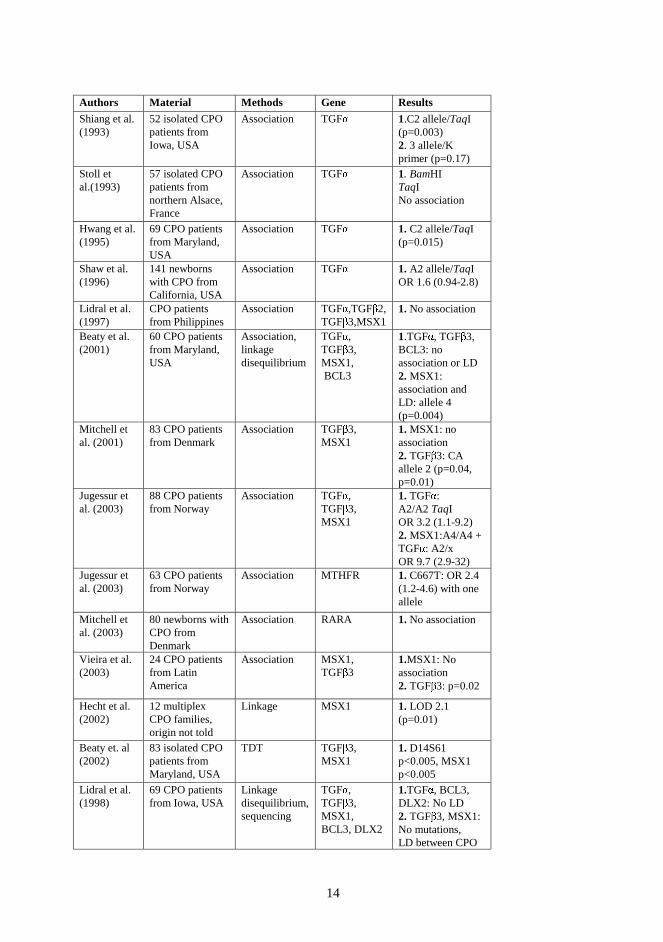
14
Authors Material Methods Gene Results
Shiang et al.(1993)
52 isolated CPOpatients fromIowa, USA
Association TGF� 1.C2 allele/TaqI(p=0.003)2. 3 allele/Kprimer (p=0.17)
Stoll etal.(1993)
57 isolated CPOpatients fromnorthern Alsace,France
Association TGF� 1. BamHITaqINo association
Hwang et al.(1995)
69 CPO patientsfrom Maryland,USA
Association TGF� 1.C2 allele/TaqI(p=0.015)
Shaw et al.(1996)
141 newbornswith CPO fromCalifornia, USA
Association TGF� 1.A2 allele/TaqIOR 1.6 (0.94-2.8)
Lidral et al.(1997)
CPO patientsfrom Philippines
Association TGF� ,TGF�2,
TGF�3,MSX1
1.No association
Beaty et al.(2001)
60 CPO patientsfrom Maryland,USA
Association,linkagedisequilibrium
TGF� ,TGF
�3,
MSX1,BCL3
1.TGF� , TGF�3,
BCL3: noassociation or LD2.MSX1:association andLD: allele 4(p=0.004)
Mitchell etal. (2001)
83 CPO patientsfrom Denmark
Association TGF�3,
MSX11.MSX1: noassociation2.TGF
�3: CA
allele 2 (p=0.04,p=0.01)
Jugessur etal. (2003)
88 CPO patientsfrom Norway
Association TGF� ,TGF
�3,
MSX1
1.TGF� :A2/A2 TaqIOR 3.2 (1.1-9.2)2.MSX1:A4/A4 +TGF� : A2/xOR 9.7 (2.9-32)
Jugessur etal. (2003)
63 CPO patientsfrom Norway
Association MTHFR 1.C667T: OR 2.4(1.2-4.6) with oneallele
Mitchell etal. (2003)
80 newborns withCPO fromDenmark
Association RARA 1.No association
Vieira et al.(2003)
24 CPO patientsfrom LatinAmerica
Association MSX1,TGF
�3
1.MSX1: Noassociation2.TGF
�3: p=0.02
Hecht et al.(2002)
12 multiplexCPO families,origin not told
Linkage MSX1 1. LOD 2.1(p=0.01)
Beaty et. al(2002)
83 isolated CPOpatients fromMaryland, USA
TDT TGF�3,
MSX11.D14S61p<0.005, MSX1p<0.005
Lidral et al.(1998)
69 CPO patientsfrom Iowa, USA
Linkagedisequilibrium,sequencing
TGF� ,TGF
�3,
MSX1,BCL3, DLX2
1.TGF� , BCL3,DLX2: No LD2.TGF
�3, MSX1:
No mutations,LD between CPO
15
and MSX1 CAallele 4
Machida etal. (1999)
89 CPO patientsfrom Iowa, USA
Sequencing TGF� 1.No mutations
Barrow et al.(2002)
2 CPO patientsfrom Iowa, USA
Sequencing p63 1.No mutations
Jezewski etal. 2003
118 CPO patientsfrom Iowa (USA)Japan, Denmark,South America,Philippines,Vietnam
Sequencing MSX1 1. 3/118:272G>A451+887G>T451+1046C>T
FitzPatricket al. 2003
23 RS patientsand 57 CPOpatients
Sequencing SATB2 1.No mutations
Table1. Previously reported molecular studies on non-syndromic cleft palate.
1.3.3 Associated syndromes
Cleft palate can be a sign of a syndrome. In Finland, a recognisable syndrome was found in aretrospective study in14.2 % of patients with cleft palate (Lilius 1992). Shprintzen et al. founda higher prevalence for syndromes in cleft palate patients (Shprintzen et al. 1985). It has beensuggested that approx. 50% of CP cases are non-syndromic (Jones 1988, Murray 2002). Over300 syndromes are found with the key word “cleft palate” in Online Mendelian Inheritance inMan (OMIM) database (http://www.ncbi.nlm.nih.gov/Omim/). The three most commonsyndromes in Finland, according to Lilius, were Robin sequence (3.1 % of all CP patients),van der Woude syndrome (2.3 %) and diastrophic dysplasia (0.6 %). Altogether 39 differentsyndromes were detected. Twenty-five patients had cleft lip with or without cleft palate but108 patients had cleft palate only.
Cleft palate associates with some anomalies without any known syndrome. In Finland, 23.1 %of 938 patients with CP (including recognised syndromes) had associated anomalies (Lilius1992). Cardiovascular anomalies were most common (16 % of all anomalies) and they werestrikingly often associated with the submucous type of cleft palate (52 % of all cardiacanomalies in CP patients were detected in patients with CPSM). Anomalies of the lowerextremities were the second most common (15.7 % of all anomalies), with club foot being themost common. Anomalies of the central nervous system were the third most common (14.0 %of all anomalies). In northeastern France, 46.7% of patients with cleft palate had associatedanomalies (Stoll et al. 2000).
Mutations have been found out in many cleft syndromes. A heterozygous stop mutation in thehomeodomain of MSX1 causes Witkop syndrome (MIM 189500), which is a rare syndromeaffecting also teeth and nails (Witkop 1965, Jumlongras et al. 2001). A point mutation inMSX1 was found to be cosegregating with dominantly inherited tooth agenesis in a largefamily (Vastardis et al. 1996). Hemizygous deletions of MSX1 have been demonstrated insome patients with Wolf-Hirschhorn syndrome (MIM 194190), which is considered a
16
contiguous gene syndrome due to a deletion or a microdeletion in 4p16 (Campbell et al. 1989,Zollino et al. 2003). MSX1 maps to 4p16.1 (Campbell et al. 1989, Padanilam et al. 1992)
Mutations in fibroblast growth-factor receptor 2 (FGFR2) cause Apert syndrome (MIM101200) (Wilkie et al.1995). Treacher Collins syndrome (MIM 154500) is caused bymutations in TCOF1, which encodes treacle (Treacher Collins Syndrome Collaborative Group1996). Treacle is a nucleolar protein, but the pathway from mutations to disease has not yetbeen characterised (Isaac et al. 2000). A sulfate transporter encoding DTDST maps∼ 900 kbproximal to TCOF1 and is involved in diastrophic dysplasia (Hästbacka et al.1994). Mutationsin the T-box transcription factor gene (TBX22) were found to cause X-linked cleft palate(CPX and ankyloglossia) (MIM 303400) (Baybrook et al. 2001). EEC (ectrodactyly-ectodermal dysplasia-cleft lip/ palate, MIM 604292) syndrome is caused by mutations in thep63 gene, which is a homologue of the tumour suppressor genep53 (Celli et al. 1999).Mutations in thyroid transcription factor (TTF-2) cause cleft palate, thyroid dysgenesis andchoana atresia (Clifton-Bligh et al. 1998, Castanet et al. 2002). Lymphoedema-distichiasissyndrome (MIM 153400) is caused by mutations a forkhead transcription factor gene(FOXC2) (Fang et al. 2000).
Autosomal recessive ectodermal dysplasia type 4 (MIM 225060) is characterized by CL/P,hypotrichosis and syndactylies. It is caused by mutations in PVRL1 (poliovirus receptor-like1) (Suzuki et al. 2000). Interestingly, it was found that heterozygosity of one these mutationsstrongly associates with nonsyndromic CL/P in Venezuela (Sözen et al. 2001). This findingshould encourage researchers to study the role of syndromic disorders causing mutations inmore common nonsyndromic forms (in any complex disease) (Murray 2001).
Van der Woude syndrome
Van der Woude syndrome (VWS) (MIM 119300) is a dominantly inherited developmentaldisorder, which was first described by Anne Van der Woude in 1954. The hallmarks of thisrare syndrome are pits and/or sinuses of the lower lip, cleft lip and/or cleft palate. Thepenetrance is estimated to be∼ 90% (Burdick et al. 1985; Murray et al. 1990; Onofre et al.1997), and both sexes are equally affected (Burdick et al.1985). Lip pits are present in∼ 80%,clefts in ∼ 50%, and hypodontia in∼ 25% of gene carriers (Van der Woude 1954; Rintala et al.1981, Burdick et al. 1986, Schinzel et al.1986, Kläusler et al. 1987). The incidence of thesyndrome is estimated to be 1/34 000 live births (Rintala et al. 1985). VWS was found in∼ 2% of Finnish cleft patients (Rintala et al. 1985, Lilius 1992). The mutation rate is estimatedto be∼ 1.8 x 10⁻⁵ (Burdick et al. 1985).
In 1987, Bocian et al. reported a patient with lip pits and a deletion in 1q32-41. Murray et al.found a linkage between VWS and chromosome 1 q in 1990. In 1995, the region wasnarrowed down to an interval of 4.1 cM on 1q32-41 (Sander et al. 1995), and further to 1.6cM in 1996 (Schutte et al. 1996). Microdeletions in 1q32-41 have also been reported infamilies with VWS (Sander et al. 1994, Schutte et al. 1999). A possible modifying locus at17p11.2-11.1 was reported when a large Brazilian family was analysed (Sertie et al. 1999). Anallele in this locus would enhance the probability of CP in an individual also carrying a defectin the VWS locus. Popliteal pterygium syndrome (MIM 119500) was found to be linked toVWS locus (Lees et al. 1999). A nonsense mutation in exon 4 in interferon regulatory factor 6(IFR6) was found in a twin affected by VWS (Kondo et al. 2002) Subsequently, mutations inIRF6 in 45 unrelated VWS families and distinct mutations in 13 families affected with
17
popliteal pterygium syndrome were found (Kondo et al. 2002). They also showed that familymembers shared the same 18-bp deletion in the IRF6 gene regardless of the differentphenotype.
Van der Woude syndrome is of special interest because the phenotype so closely resemblesnon-syndromic forms of both cleft lip and palate (Schutte et al. 1999).
Robin sequence
A combination of micrognathia, glossoptosis (an abnormal backward and downward fall ofthe base of the tongue) and an associated cleft of the soft palate is commonly recognised asRobin sequence (MIM 216800) (Gorlin 1990). The sequence was first described as early as1822 but it bears the name of French stomatologist Pierre Robin who published hisobservations in 1923. Typically, the cleft is U-shaped in Robin patients, while in non-syndromic cleft patients the cleft is usually V-shaped (Larson et al. 1998, Marques et al.1998). Increased incidence of twins among Robin patients has been noted (Holder-Espinasseet al 2001, Knottnerus et al. 2001).
Robin sequence is the most common recurrence pattern recognised in syndromic cleft palatepatients in Finland (Lilius 1992). Robin sequence can appear in isolated form but it is alsoseen as a part of another syndrome, the most common being CATCH and Stickler syndrome(Jones 1997, Sheffield et al. 1987, Holder-Espinasse et al. 2001, van den Elzen 2001). In afollow-up study of Robin patients, 7 out of 24 were found to have Stickler syndrome(Sheffield et al. 1987). Van den Elzen found that 63.5% could be categorised as isolated RS,and the remaining 36.8% could be considered syndromic. Holder-Espinasse classified 48 % asnon-syndromic. Recently, Houdayer et al. described a patient with RS and interstitial deletionin 2q32.3-q33.2, which is the same CPO-associated region reported by Brewer et al. (Breweret al. 1999, Houdayer et al. 2001).
Stickler syndrome
Stickler syndrome(hereditary arthro-ophthalmopathy) is considered to be the most commonautosomal dominant connective tissue disease. The major features are premature degenerativearthropathy, severe progressive myopia with occasional retinal detachment, sensorineuralhearing deficit and typical facies usually with maxillary hypoplasia (Stickler et al. 1965,Stickler et al. 2001). Facial dysmorphia, flat face, small mandible, cleft palate are present in84% of patients (Stickler et al. 2001). The phenotype varies between and within families(Liberfarb et al. 2003). So far, mutations in three different collagen genes have been found tocause Stickler syndrome.
Collagen II is found in cartilage. It is composed of three identicalα(II) chains. Stickler sdrtype 1 (MIM 108300) is caused by mutations resulting in a premature termination codon in theCOL2A1 gene (Ahmad et al. 1991, Brown et al. 1992, Ahmad et al. 1993, Ritvaniemi et al.1993, Annunen et al. 1999). In addition to Stickler syndrome, defects in COL2A1 causenumerous other diseases (Kuivaniemi et al 1997). COL2A1 maps to12q13.1-q13.2(Francomano et al. 1987).
18
Collagen XI is composed of three differentα chains encoded by COL2A1, COL11A1 andCOL11A2 (Eyre et al. 1987, Ala-Kokko et al. 1995, Vuoristo et al. 1995). Collagen XIbelongs to the fibrillar class of collagens and it is expressed in cartilage and the inner ear.
Defects in COL11A1 are the cause of Stickler syndrome type 2 (MIM 604841) (Richards et. al1996, Sirko-Osadsa et al. 1998). This disorder is characterized by progressive myopiabeginning in the first decade of life, vitreo-retinal degeneration, retinal detachment,sensorineural hearing loss, cleft palate, midfacial hypoplasia and osteoarthritis. Marshallsyndrome is also caused by mutations in COL11A1 (Annunen et al. 1999). COL11A1 maps1p21 (Richards et al. 1996).
Defects in COL11A2 cause a form of Stickler sdr (type 3, MIM 184840) which ischaracterized by midfacial hypoplasia, cleft palate, osteoarthritis, and sensorineural hearingloss, but lacks ocular involvement (Sirko-Osadsa et al. 1998). The lack of ocular involvementis due to the replacement ofα2(XI) by α2(V) in the vitreous of the eye (Mayne et al. 1993).COL11A2 maps to 6p21.3 (Brunner et al. 1994). Mutations in COL11A2 also cause non-syndromic hearing loss and, in addition to Stickler syndrome, they are also associated withother autosomal dominant and recessive osteochondrodysplasias (Vikkula et al. 1995,McGuirt et al. 1999, Melkoniemi et al. 2000).
CATCH 22
The incidence of hemizygous 22q11 deletion has been estimated to be 1:4000-6000 live births(Wilson et al. 1994, Botto et al. 2003). Velocardiofacial syndrome (MIM 192430) andDiGeorge syndrome (MIM 188400) are overlapping phenotypes commonly found in patientswith 22q11 deletion (de la Chapelle et al. 1981, Goldberg et al 1985, Goldberg et al. 1993,Stevens et al. 1990). Nine percent of these CATCH patients manifest cleft palate (Ryan et al.1997). Reish et al (2003) found cleft palate in nine out of 38 patients. Patients also have othersigns such as velopharyngeal insufficiency, hypocalcaemia, thymic hypoplasia, cardiacproblems, renal anomalies, abnormal facies, delayed speech and learning difficulties (Ryan etal. 1997, Somer et al. 1997, Digilio et al. 2003). Of the patients with velopharyngealincompetence but without overt clefting, 12.5% have the 22q11 deletion (Boorman et al.2001). It has been estimated that 22q11 deletions may be involved in∼ 5 % of congenital heartdiseases (Wilson 1994). Monozygous twins have been described to exhibit differentphenotypes (Singh et al. 2002). The deletions were found to be of maternal origin in 72 % ofinherited cases (Demczuk et al. 1995). No studies on sizes of deletions in different tissueshave been published. The size of the commonly deleted region is∼ 3 Mb (Carlson et al. 1997).Polymorphic markers in loci D22S944 and D22S941 are most commonly deleted (Morrow etal. 1995). So far the smallest deletion found has been 20 kb (Yamagishi et al. 1999). Thedeletion of 20 kilobases removed exons 1 to 3 of the UFDL1 gene, and the patient had typicalfeatures of 22q11 deletion (Yamagishi et al. 1999). On the other hand, CATCH is consideredto be a “contiguous gene syndrome” (Glover 1995). No hemizygosity of 22q11 was detectedin patients with isolated cleft palate (Mingarelli et al. 1996). Routine screening of 22q11deletion in CPO patients is not recommended (Reish et al. 2003, Ruiter et al. 2003).
19
2 GENE MAPPING
2.1 Gene mapping in complex diseases
2.1.1 Power estimations
Having enough power to detect an existing linkage is an essential question in gene mapping.The SLINK simulation package is widely used when estimating the power of pedigrees todetect traditional parametric linkage (Ott 1989, Weeks et al. 1990). Simulation methods forpopulation data are used when mapping genetically complex diseases. Population size,“bottleneck” size, genetic drift, number of founders and number of generations are variableparameters. POPSIM and EASYPOP are packages for population simulation (Hampe et al.1998, Balloux 2001). Recently, methods and a software package for simulations of humangenetic data in isolated populations were developed (Ollikainen 2002). These populationsimulation programs may help the researcher to estimate the sample size and the marker mapdensity. Elegant simulations reduce the amount of work and also the financial costs later.
A recent extensive meta-analysis on 101 genome wide screens for complex diseases revealedthat a large sample size and genetic homogeneity were the most important factors promotingsuccessful mapping (Altmuller et al. 2001).
2.1.2 Genetic markers
Restriction fragment length polymorphisms (RFLPs) were the first molecular genetic markersthat could be widely utilised in linkage analyses (Botstein et al. 1980). The first genome-widelinkage map was mainly based on RFLPs (Donis-Keller et al. 1987). Minisatellites (VNTRs)were the next step (Jeffreys et al. 1985). Minisatellites usually have a length of over 1000 bp,which makes it difficult to use PCR-based methods. Short tandem repeat polymorphisms(STRPs) made analyses more rapid, and also the degree of heterozygosity is higher than inRFLPs or VNTRs (Weber et al. 1989). Microsatellites are di-, tri- or tetranucleotide repeatswhich have provided the main skeleton of the genome-wide human linkage maps (Gyapay etal. 1994, Murray et al. 1995, Sheffield et al. 1995). The nucleotide repeats must be amplifiedin polymerase chain reactions (PCR) (Mullis et al. 1986), and the repeat sizes are separated ingels with the help of electrophoresis. The alleles are visualised by silver staining, radioactiveor fluorescence labelling.
Single nucleotide polymorphisms (SNPs) are bi-allelic markers, which allow highlyautomated genotyping (Kruglyak 1997, Collins et al. 1998). They are estimated to exist inapprox. every 1000th bp, and their total number is estimated to be ten million (Sachidanandamet al. 2001, Kruglyak and Nickerson 2001). To obtain maximal information, a map must bedense enough because allele information is based only on two possible polymorphisms. Infact, map density was shown to be more critical than marker heterozygosity (Kruglyak 1997).
2.1.3 Linkage analysis
Linkage means cosegregation of a trait and a marker. If they are physically closely tightened toeach other, the probability of cross-over is very small. Crossing-over produces a newcombination of alleles between trait and marker loci. The longer the distance between thesetwo loci, the more probable is the crossing-over event. The proportion of rearranged
20
chromosomes after meiosis is called the recombination fraction. The shorter the distancebetween two loci, the smaller the recombination fraction is. This distance can be representedas Morgans (M). Within a distance of one cM, the recombination fraction is approx. 1 %. Thelength of the human genome is approx. 3000 cM.
Linkage analysis tries to localize a gene with the help of polymorphic markers. If a particularallele is identical-by-descent in all affected members of the same family, one can suspectlinkage. If similar cosegregation of particular alleles in a given maker locus takes place inmany families, one can start to count how probable it is that this kind of inheritance patterncan happen by chance. If the probability of chance is 1 x 10⁻³, the logarithm of odds (LOD) is3 (Morton 1955). In the case of single-gene Mendelian disorder, a LOD score below -2indicates a region where the possibility of linkage can be disregarded (Morton 1955). Usually,the linkage is regarded as established when the LOD score is≥3. This corresponds to a 5%significance level in two-point analysis, and a 9% significance level in multipoint analysis.
The LOD scores are calculated with the help of computer programs. The first computerprogram (Liped) for linkage analysis of human pedigrees larger than two-generation waswritten by Ott (Ott 1974). MLINK (LINKAGE package) was the first program to performmultipoint analysis (Lathrop et al. 1984). FASTLINK (Cottingham et al. 1993) is a newer andimproved version of the LINKAGE package. The user of these programs needs to inputaccurate parameters in disease models. This demand is difficult when dealing with geneticallycomplex, multifactorial diseases. The affected-pedigree-member method (APM) comparesIBS (identical-by-state) sharing among affected individuals with IBS sharing expected underrandom segregation (Weeks et al 1988). Multipoint sib-pair analysis can be performed forexample in a computer package, MAPMAKER/SIBS (Kruglyak et al. 1995). To extract moreinformation from a pedigree, Kruglyak et al. applied non-parametric linkage analysis (NPL)into Genehunter package (Kruglyak et al. 1996). The NPL score (Z) announces whetheraffected individuals share IBD (identical-by-descent) alleles more often than expected bychance.
An association between a disease and a particular allele in a marker locus can be a result oflinkage disequilibrium (LD). LD means a non-random association of alleles in linked loci. Itdepends on the age of mutation and the recombination frequency (Jorde 1995). The LDmeasure increases when particular alleles of two linked loci cosegregate more often thanexpected by chance. LD mapping is especially powerful in isolated populations where one or afew founder mutations are expected to have taken place (de la Chapelle 1993, Jorde 1995).The genetic distance between a disease gene and a marker locus can be estimated on the basisof LD and applying the Luria-Delbruck principle (Hästbacka et al. 1992, de la Chapelle 1993).This method was successfully used when mapping the DTD gene (Hästbacka et al. 1992).
Recently, Kruglyak estimated that when mapping common disease genes, the useful level ofLD in general and in isolated populations is unlikely to be more than 3 kb and that wouldrequire about half a million SNPs for whole-genome studies (Kruglyak 1999). On the otherhand, he demonstrated that the extent of LD can be larger in populations where the bottleneckhas been very narrow or if the frequency of the rarer marker allele is very low.
The transmission disequilibrium test (TDT) detects linkage between a disease locus and amarker locus in the presence of association (Spielman et al. 1993). The TDT studies atransmission distortion of alleles transmitted to an affected offspring from an affected parent
21
compared to untransmitted alleles. The statistical significance of the TDT is tested byχ2 or bythe exact binomial test (Spielman et al. 1993). In multiplex families with many generations,the TDT is a valid test for linkage but not for LD. In multigeneration families, a false-positive“LD" can be seen because of non-independent observations.
2.2 Finland - the northern isolation
2.2.1 Short review of the history of the Finnish population
The Finnish population was 5 206 295 at the end of 2002 (Statistics of Finland). The growthhas been rapid, since the number was∼ 2 656 000 in 1900. It has been estimated that the veryfirst immigrants settled in Finland for about 9000 years ago (Virrankoski 2001). The first twobigger waves of settlement seem to have taken place around 3200 and 2000 B.C.. Theseimmigrants might have arrived from the east. Immigration from the west took place around1200 B.C., and those immigrants settled mainly in western Finland (Koskinen et al. 1994).The size of the population was still extremely small. It has been estimated that during thewhole prehistorian time the size of the population has been at most 5 000 - 10 000 inhabitants(Jutikkala 1996). At the end of the prehistorian time, only few regions, mainly in the coastline,were inhabited (Virrankoski 2001). It is important to notice that large regions of Finland wereinhabited only a few hundred years ago. During the period of this so called late settlement therelative growth of population was the most rapid.
Researchers have not been able to trace the precise origins of the Finns. According tomitochondrial and genomic DNA diversity, the Finns, with the exception of the Saami, seemto be genetically indistinguishable from many other European populations (Lahermo et al.1996). On the other hand, analysis of Y chromosomal diversity suggests the possible originfor at least a part of the Finnish population in Northern Eurasia (Lahermo et al. 1999). Thisfinding supports previous theories based on linguistic analysis (Wiik 1997).
Between the years 1698 and 1721, the population diminished greatly because of starvation dueto poor harvests. This period is considered to be a one bottleneck of the Finnish gene pool. Arelative and very effective bottleneck was the emigration from Savo to northern parts ofFinland in the 16th century (Norio 2000). Norio divides Finland roughly into two areas: theregion of the old settlement and the region of the young settlement. The traces of bottleneckswere seen when Y chromosomal polymorphisms were analysed: the pattern of haplotypediversity in Finnish males was strikingly narrower than in other European populationsanalysed (Lahermo et al. 1999). Also the high prevalence of some recessive diseases, that arevery rare in other countries, reflects bottlenecks followed by a quite rapid expansion of thesurviving gene pool.
2.2.2 The Finnish disease heritage
2.2.2.1 Single-gene disorders
”The Perheentupa stairs” give an informative view of the hereditary diseases which have ahigher relative incidence in Finland than anywhere else (Perheentupa 1972, Norio 2000). Themajority of the genes and their defects behind these diseases have already been identified andmolecular events are being studied at the moment (Peltonen et al. 1999). The enrichment of
22
these disease genes, combined with well-organised church registers and the high level ofmedical research facilities, have provided an excellent basis for genetic research. The modelof inheritance in these diseases is recessive, with the exceptions of an autosomal dominantmodel in two and a X-chromosomal recessive model in two diseases. The most common arethe recessive disorders aspartylglucosaminuria (AGU) (Palo 1967, Ikonen et al.1991),congenital nephrotic syndrome (CNF) (Norio 1966, Männikkö et al. 1995), and infantileneuronal ceroid lipofuscinosis (INCL) (Hagberg et al. 1968, Vesa et al. 1995). The sizes ofregions showing linkage disequilibrium flanking the disease genes are approx. 3 cM, 3 cMand 2,5 cM, respectively. The mutations seem to have taken place about for 80-120generations ago, i.e. 2000-3000 years ago.
Salla disease (Aula et al. 1979, Verheijen et al. 1999), Northern epilepsy syndrome (EPMR)(Hirvasniemi et al 1991, Ranta et al. 1999) and vLINCL (Santavuori et al 1982, Savukoski etal 1998) represent newer mutations. The birthplaces of grandparents of EPMR patients arelocated in the Kainuu region and of vLINCL patients in the Ostrobothnia region (Varilo1999). In vLINCL, LD covering a distance of 11 cM was detected and the mutation wasestimated to have taken place about 500 years ago (Varilo et al. 1996). In Salla disease theinterval for LD is also approx. 10 cM (Schleutker et al. 1995).
2.2.2.2 Genetically complex diseases
Tracing genes involved in multifactorial diseases has been shown to be a demanding task.Genome-wide scans of several complex diseases in Finnish patients have been performed.Several examples are listed here. Linkage between non-insulin dependent diabetes mellitusand chromosome 12 was established (Mahtani et al. 1996). A genome-wide search wasperformed in Finnish multiple sclerosis families mainly originating from Ostrobothnia(Kuokkanen et al. 1997). The prevalence of MS is clearly increased in Ostrobothnia(Kinnunen et al. 1983). A suggestive linkage to 17q22-q24 was established. Multiplesuggestive loci were found when a genome-wide scan was performed in Finnish schizophreniapatients (Hovatta et al. 1999). No LD was detected although patients originated from arestricted Kuusamo area (Varilo et al. 1999). A genome-wide scan for elevated diastolic bloodpressure revealed linkage to the AT1 gene (Perola et al. 2000). A genome-wide scan of obesityin Finnish sibpairs revealed linkage to Xq24 (Öhman et al. 2000). Evidence for linkagebetween 7p14-p15 and three phenotypes related to asthma (asthma, a high serum IgE level anda combination of the phenotypes) in a Finnish subpopulation from the Kainuu region wasdetected in a genome-wide search (Laitinen et al. 2001). Autism-spectrum disorders werefound to be linked to 3q25-27 in Finnish families (Auranen et al. 2002). Also a linkagebetween coeliac disease and 15q11-q13 was found when using a subpopulation from thenortheastern part of Finland (Woolley et al. 2002).
23
AIMS OF THE STUDY
1 To localize a gene responsible for non-syndromic cleft palate in Finnish patients.
2 To narrow down the critical region in the van der Woude syndrome. While we weredoing this, we found genetic heterogeneity in Finnish VWS families and that resultencouraged us to map a second locus involved in VWS.
3 To find out if mutations in COL2A1, COL11A1 and COL11A2 cause Robin sequence,non-syndromic cleft palate or micrognathia.
24
SUBJECTS AND METHODS
3.1 Ethical issues
The research plan was approved by the Social and Health Ministry of Finland, by the ethicalcommittee of the University Hospital of Helsinki and by the ethical committee of theDepartment of Medical Genetics of Helsinki University. The patients were contacted from theUniversity Hospital of Helsinki, where they are or have been treated medically. The relativeswere contacted with the permission of the patient. The informed consents were obtainedbefore taking any samples. No individual data will be published. Personal results (i.e.genotypes, haplotypes, carrier status) will not be told to participants. All the data concerningthe patient’s disease, family history and research results are confidential. These facts werewritten in the informed consent.
Every participant was given a study-specific ID number. ID-numbered samples without nameswere handled in the laboratory. The connection between names and numbers is in a database.Access to the database is permitted only for the researchers named in the research plan andapproved by the ethical committees.
3.2 Patients
Participants were collected from the records of the Cleft Center. Cleft patients in Finland havebeen treated centrally since 1948 at the Red Cross Hospital for Plastic Surgery, which thenbecame, in 1984, the Plastic Surgery Unit of the I Department of Surgery, Helsinki UniversityCentral Hospital. We searched the medical records of all patients with cleft palate from theyears 1967-1996, those years included. All types of cleft palates were accepted. We contacted250 patients by sending them a letter. The patients were chosen on the basis of three criteria:1. According to the medical record they did not seem to have other malformations orsyndromes, 2. They had reported of at least one similarly affected relative 3. They could stillbe reached by mail.
Twenty-four multiplex CPO pedigrees were chosen for the DNA analysis of candidate regions(Figs 1-3). The pedigrees consisted of 63 affected, and 112 unaffected, a total of 175individuals. Families came from different regions of Finland. Probands and as many affectedrelatives as possible were examined to rule out undiagnosed syndromes. The whole nuclearfamily was asked to meet the examiner, if possible. Family members were then examined anddysmorphic features were searched for.
Five families with many members affected by VWS were chosen for the VWS study. In theVWS families, 56 individuals were genotyped, of whom 25 were affected (Fig 4). Family0057 was re-examined in purpose to confirm the diagnosis and to determine the affectionstatus of all pedigree members.
During the years 1967-1996, 103 patients with Robin sequence were treated in the CleftCenter. Ninety-three of them were contacted by sending them a letter. Thirty-three answeredand were willing to participate. Ten out of these 33 were excluded on the basis of a confirmedor suspected syndrome. Of the remaining 23 patients, seven had had severe breathingdifficulties immediately after delivery. The RS diagnosis was based on cleft palate and smallmandibula in the rest of the patients. Seven patients reported a similarly affected relative but
25
only in one case was the relative a first-degree one. Patients were not re-examined but theinformation was collected by questionnaries, telephone interviews and from the medicalrecords in the Cleft Center. Altogether 150 controls, whose samples were analysed togetherwith the samples of RS patients, were individuals without any cleft and without relatives withclefts.
One patient with non-syndromic Robin sequence (patient no. 62) was recruited from theUSA. Seventeen of the 21 patients with non-syndromic micrognathia were recruited from theCenter for Craniofacial Disorders and Department of Dentistry at Montefiore Medical Center,Bronx, New York, and the remaining four from the maxillofacial surgery service at theUniversity Hospitals of Cleveland (patients 41 to 61). The medical records and samples ofthese 22 patients from abroad were sent directly from USA to Oulu.
3.3 Family history and genealogical studies
Patients were asked about the family names and birth places of their parents and grandparents.They were also asked for information about any similarly affected relatives, i.e. relatives withCL, CL/P or CP. Names of affected relatives were asked for to try to connect the families toeach other. Finnish pedigrees were also expanded with the help of the Finnish church records.Local church records usually show pedigree information on about five generations backwards.Ancestors born before∼ 1860 can be traced with the help of church records in the FinnishNational Archives. In our study we used patients’ own knowledge about their ancestors andthe local church records to build the pedigrees.
3.4 DNA samples
Blood samples were taken either in local health centers or in the University Hospital ofHelsinki. EDTA-preserved venous blood was either frozen or DNA was immediatelyextracted. DNA was extracted non-enzymatically from leukocytes (Lahiri et al. 1991). In thismethod no organic solvents are used. Five ml of blood was mixed with 5 ml TKM 1 + P40buffer for cell lysis. TKM 1 consists of 10 mM Tris-HCl pH 7.6, 10 mM KCl, 10 mM MgCl2,and 2 mM EDTA. The solvent was centrifuged at 2200 RPM for 10 min, then the nuclearpellet was washed with 5 ml of TKM 1 buffer and centrifuged again as above. The pellet wasthen suspended in 800µl TKM 2 buffer. TKM 2 buffer consists of 10 mM Tris-HCl pH 7.6,10 mM KCl, 10 mM MgCl2, 0.4 M NaCl and 2 mM EDTA. Fiftyµl of 20 % SDS was addedand the suspension was mixed by pipetting back and forth. The tubes were incubated 1-2 daysat 60° C. Then, 360µl 5M NaCl was added for protein precipitation. The DNA-containingsupernatant was then separated by centrifuging at 12000 RPM for 10 min. DNA wasprecipitated by adding 3 ml of cold 100% ethanol. Precipitated DNA was mechanicallytransferred to tubes containing 70% ethanol. Finally, DNA was dried in the air and dissolvedin TE buffer to be preserved. DNA was preserved frozen.
3.5 Microsatellite markers
Linkage and association were searched among four candidate regions and non-syndromicCPO among 24 multiplex families. The 22q11 region was studied using nine polymorphicmarkers which are inside the 3 Mb region commonly deleted in patients with velocardiofacialsyndrome (Morrow et al. 1995). Markers at loci D22S941, D22S944, D22S264, D22S311,D22306, D22S308 and D22S425 were ordered from Research Genetics
26
(http://www.resgen.com). Markers at loci D22S1638 and D22S1623 were developed atGenosys (http://www.sigma-genosys.com). TGFβ3 in chromosome 14 was studied usingpolymorphic markers at loci D14S273 and D14S61 which were ordered from ResearchGenetics. TGFβ3 is located between these markers in YAC 746B4, which has a size of 1800kb (Cruts et al.1995). The MSX1 region in chromosome 4 was studied using a polymorphicmarker D4S394, which is located c. 7 cM proximal to MSX1, and with an intragenicdinucleotide (CA) repeat polymorphism. In addition, the entire chromosome 4 was analyzedusing 20 polymorphic markers from the modified Weber set VI (http://www.pebio.com). Themean distance between the markers was 11.3 cM. The critical 2q32 region was furtheranalyzed using polymorphic markers D2S311, D2S348, D2S2392 and D2S115 (Brewer et al1999, Hadano et al. 1999). The entire chromosome 2 was also analyzed using markers fromthe ABI marker set. The mean interval between the 21 markers was 13.8 cM.
The genome-wide screen was performed in nine largest multiplex families with non-syndromic CP. The genome scan was performed in the Finnish Genome Center(http://www.genome.helsinki.fi). The 377 fluorescent polymorphic markers used were fromthe Applied Biosystems Linkage mapping Set MD-10, Foster City, USA(http://www.appliedbiosystems.com).
In the genome-wide scan at stage II, six additional markers (D1S247, D1S513, D1S2723,D1S380, D1S1188, D1S2722) (see below) were used to obtain maximal information from theinteresting VWS region in 1p34. Thus, 1p34 and the entire chromosomes 2 and 4 werescreened in 24 multiplex families. The interval between markers in chromosomes 2 and 4 inthe first nine families was ~ 5 cM, while it was ~ 10 cM in the remaining 15 families.
Linkage was tested between VWS and 1q32-q41 and between VWS and a proposedmodifying locus in 17p11.2-p11.1. At the first stage, nine polymorphic markers (D1S1663,D1S245, D1S491, D1S3754, D1S2136, D1S3753, D1S205 and D1S414 (Schutte et al. 1999)were genotyped in 1q32-q41. The genetic distances are provided by the Sanger Center(http://www.sanger.ac.uk/HGP/Chr1/). Because of the exclusion of the linkage in family 0057and uninformative results in family 0062, an additional 25 markers were genotyped inchromosome 1. These markers were from the modified Weber set VI (http://www.pebio.com).In family 0057, the genome-wide screen was performed with 381 polymorphic microsatellitemarkers from ABI PRISM Linkage Mapping Set-MD10 (Applied Biosystems). For the secondstage of analysis of family 0057, six additional markers (D1S247, D1S513, D1S2723,D1S380, D1S1188, D1S2722) within the region flanked by markers D1S234 and D1S2797were genotyped. The positions of these markers were ascertained from the Marshfieldcomprehensive human genetic map (http://www.marshmed.org/genetics/).
Microsatellite polymorphisms were amplified by PCR (Mullis 1986). When PCR productswere visualised with silver staining, fifty ng of DNA was amplified in 20µl reaction with 20µM of each primer, 200µM of dNTPs, 2.0µl of buffer, 1 unit of Amplitaq Gold enzyme and4.68µl of H20. The cycling conditions were within the following ranges: 94°C for 10 min, 30cycles at 94° C for 30 s, 53-58°C for 35 s, 72°C for 30 s, and 72°C for 10 min. When PCRproducts were visualised with fluorescent dyes, 50 ng of DNA was amplified in 15µl reactionwith 5 µM of each primer, 300µM of dNTPs, 1.5µl of buffer, 0.15µl of Amplitaq Goldenzyme 5 U/µl and 0.35µl of H2O. The PCR conditions were within the following ranges:94°C for 10 min, 30-35 cycles at 94°C for 30 s, 55°C for 1 min 15 s, 72°C for 1 min, and
27
72°C for 30 min. In the genome-wide scan of nine multiplex families performed in FinnishGenome Center, PCR reactions were done in 5µl volume containing 20 ng of DNA. Reagentconcentrations and temperature profiles were as recommended by the manufacturer (AppliedBiosystems, USA).
When screening the candidate regions in CPO families, the PCR products were fractionatedon 6% polyacrylamide gel. The alleles were visualised by silver staining and they werenumbered on the basis of their sizes. When screening 1q32-q41 and 17p11.2-p11.1 in VWSfamilies, the allele sizes were separated on an ABI 377 laser fluorescent sequencing machine.In the genome-wide scan, the samples were electrophoresed on Megabace 1000 (AmershamBiosciences,http://www.moleculardynamics.com)96 well capillary instrument according tothe manufacturer’s instructions. Allele calling was done using genetic Profiler 1.1 (AmershamBiosciences) software.
3.6 Linkage and LD analysis
For the linkage analysis in VWS families the penetrance was set to be 95 % (Sander et al.1993), the model as autosomal dominant, the disease frequency as 1.5 x 10⁻⁵ (Rintala andRanta 1981) and the mutation rate as 1.8 x 10⁻⁵ (Burdick et al. 1985). Individuals with CL,CL/P, CP and/or lip pits were considered to be affected. When analysing the modifying locusin 17p11.1-p11.2, the disease model was set as autosomal dominant with a penetrance of 70%and the disease frequency as 0.001 (Sertie et al. 1999). In this analysis, individuals with CPwere classified as affected, regardless of the presence of lip pits. Multipoint linkage analysiswas performed with theGenehunterprogram (Kruglyak et al. 1996).
For the linkage analysis of candidate region in non-syndromic CPO families no reliableparameters could be set. Therefore, we analysed non-random IBD allele sharing amongaffected individuals. This non-parametric linkage analysis was performed with theGenehunterprogram (Kruglyak et al. 1996). Pedigree no. 9 was too large for linkage analysiswith Genehunterand was used for LD analysis only. Population-level association could beassumed and, therefore, we used transmission/disequilibrium tests to test for linkage withgenetic markers (Spielman et al 1993). The TDT analysis was performed with theGenehuntersoftware package.
In the genome-wide scan, the non-parametric linkage analysis was done usingGenehunter2.1.Pedigrees 1 and 9 were divided into two because of their size. The data were checkedmendelian inconsistencies using Pedmanager and Pedcheck softwares before the linkageanalysis.
3.7 Power estimations
The power to localize the disease gene by linkage disequilibrium was estimated with the helpof computer simulations. One hundred data sets of 172 chromosomes were sampled from themultiplex CPO pedigrees. Random haplotypesH of different lengths 1, 2, and 3 were pickedfrom each data set. Each of these haplotypes was enriched one at a time in the disease-associated chromosomes by replacing the corresponding alleles in each chromosome withhaplotypeH with probabilityP of 10%, 20%, and 30%. ProbabilityP represents the extent towhich an artificially introduced disease-associated haplotype is overrepresented in the affectedsample. It is analogous toPexcess= (Paffected- Pnormal) / (1 - Pnormal), wherePaffectedandPnormal
28
denote allele frequency in patient and control chromosomes, respectively. For each enriched
data set, the highestχ2 value was computed. Then, it was counted how often these highestvalues exceeded the corresponding critical thresholds forp=0.05 obtained from a permutationtest (based on 100 iterations). This ratio corresponds to the power to detect linkagedisequilibrium at type I error rate of 0.05.
3.8 COL2A1, COL11A1 and COL11A2 sequencing
DNA analysis
Genomic DNA was extracted from EDTA-preserved blood. Exons and exon-flankingsequences were amplified by PCR. Forty ng of DNA was amplified using 5 to 10 pmols ofPCR primers, 1.5 mM of MgCl2, 0.2 mM of dNTPs, and one unit of Amplitaq Gold DNApolymerase. The PCR conditions were as follows: 95° C for 10 min, 33 cycles at 95° C for30 s, at 54-63° for 30 s, at 72° for 40 s, and finally at 72° for 10 min. Conformation sensitivegel electroforesis (CSGE) was used, and to generate heteroduplexes, the samples weredenatured (95° C for 5 min) and re-annealed (68° C for 30 min). Twenty ng of each PCRproduct was analysed on CSGE gel. Samples that showed heteroduplexes on CSGE weresequenced (ABI Prism 377 Automatic Sequenator and BigDye Terminator Cycle SequencingKit, Applied Biosysterms). The DNA analysis, except for the extraction of DNA, was carriedout in the Department of Medical Biochemistry and Molecular Biology, University of Oulu.
RNA analysis
RNA was extracted from EBV-transformed lymphoblasts. cDNA was synthesized andamplificated. The following PCR conditions were used: 95° C for 10 min, 33 cycles at 95° Cfor 40 s, at 60° C for 40 s, at 72° for 45, and finally at 72° for 10 min. Twoµl of PCR product,34 pmol of each primer and five units of AmpliTaq Gold polymerase were used, and the PCRproducts were analysed on 1.2% agarose gel. The products of the second PCR were thensequenced. The RNA analysis was also carried out in Oulu University.
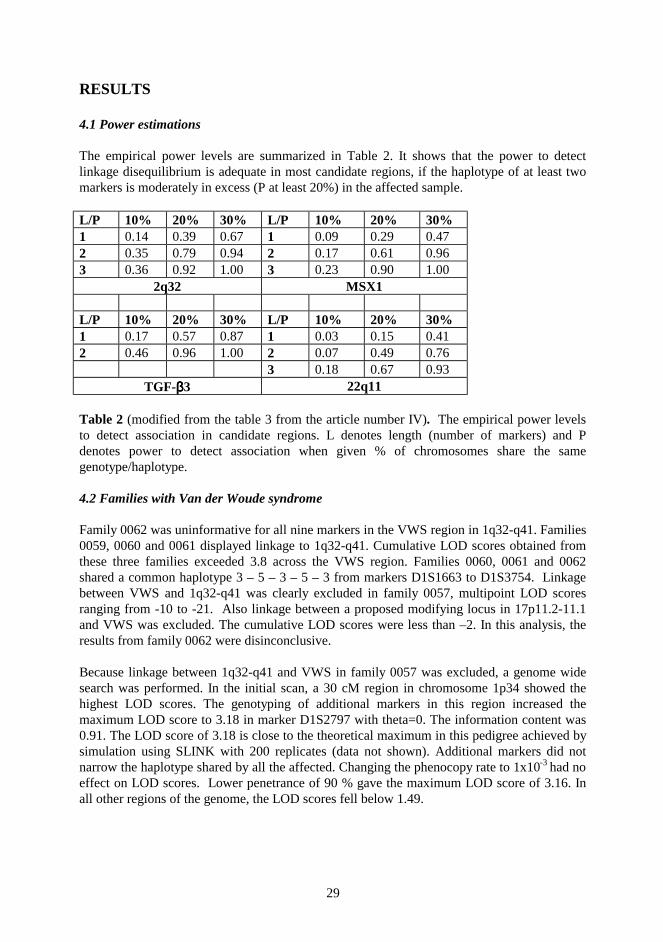
29
RESULTS
4.1 Power estimations
The empirical power levels are summarized in Table 2. It shows that the power to detectlinkage disequilibrium is adequate in most candidate regions, if the haplotype of at least twomarkers is moderately in excess (P at least 20%) in the affected sample.
L/P 10% 20% 30% L/P 10% 20% 30%1 0.14 0.39 0.67 1 0.09 0.29 0.472 0.35 0.79 0.94 2 0.17 0.61 0.963 0.36 0.92 1.00 3 0.23 0.90 1.00
2q32 MSX1
L/P 10% 20% 30% L/P 10% 20% 30%1 0.17 0.57 0.87 1 0.03 0.15 0.412 0.46 0.96 1.00 2 0.07 0.49 0.76
3 0.18 0.67 0.93TGF-ββββ3 22q11
Table 2 (modified from the table 3 from the article number IV). The empirical power levelsto detect association in candidate regions. L denotes length (number of markers) and Pdenotes power to detect association when given % of chromosomes share the samegenotype/haplotype.
4.2 Families with Van der Woude syndrome
Family 0062 was uninformative for all nine markers in the VWS region in 1q32-q41. Families0059, 0060 and 0061 displayed linkage to 1q32-q41. Cumulative LOD scores obtained fromthese three families exceeded 3.8 across the VWS region. Families 0060, 0061 and 0062shared a common haplotype 3 – 5 – 3 – 5 – 3 from markers D1S1663 to D1S3754. Linkagebetween VWS and 1q32-q41 was clearly excluded in family 0057, multipoint LOD scoresranging from -10 to -21. Also linkage between a proposed modifying locus in 17p11.2-11.1and VWS was excluded. The cumulative LOD scores were less than –2. In this analysis, theresults from family 0062 were disinconclusive.
Because linkage between 1q32-q41 and VWS in family 0057 was excluded, a genome widesearch was performed. In the initial scan, a 30 cM region in chromosome 1p34 showed thehighest LOD scores. The genotyping of additional markers in this region increased themaximum LOD score to 3.18 in marker D1S2797 with theta=0. The information content was0.91. The LOD score of 3.18 is close to the theoretical maximum in this pedigree achieved bysimulation using SLINK with 200 replicates (data not shown). Additional markers did notnarrow the haplotype shared by all the affected. Changing the phenocopy rate to 1x10⁻³ had noeffect on LOD scores. Lower penetrance of 90 % gave the maximum LOD score of 3.16. Inall other regions of the genome, the LOD scores fell below 1.49.
30
4.3 Families with non-syndromic cleft palate
4.3.1 Candidate regions
Linkage between non-syndromic cleft palate and candidate loci could not be detected. Inchromosome 2, the highest Z score (1.34, p=0.09, information content 0.61) was in locusD2S423. Markers in the candidate region 2q32 showed no evidence of linkage; the maximumZ score was 0.54 (p=0.29) and the information content was 0.78. In chromosome 22, thehighest Z value was 1.36 (p=0.09) with the information content of 0.77. In the TGFβ3 regionin chromosome 14, the Z score was 0.80 (p=0.20, information content 0.56). The MSX1region in chromosome 4 showed negative Z scores. The highest Z value in chromosome 4 was1.64 (p=0.06). Transmission/disequilibrium tests did not demonstrate any significant deviationfrom the expected values. No allelic association could be detected.
Marker D22S944 exhibited null alleles in 10 out of 24 families. Null alleles were carried byfourteen of the affected and 17 of the unaffected. The results were similar with a new set ofprimers. Individuals carrying null alleles seemed to be homozygous in that locus, butmendelian inheritance errors could be detected. No other mendelian errors occurred inchromosome 22. The marker D22S944 was checked in controls (28 Finnish SLE patients and63 of their healthy relatives) and again similar null alleles were found in 6 patients and 5 ofthe unaffected.
4.3.2 Genome-wide screen
When genome-wide screening nine multiplex families (chromosomes 1-22) (stage 1), nosignificant linkage could be detected. The highest NPL scores of all chromosomes in the ninelargest pedigrees are shown in Figures 5-6. The highest NPL score was seen in chromosome 1at the position of 50 cM from 1pter (Z=2.06, p=0.033, information content 0.58). NPL scoresreaching values over 1.5 were seen in chromosomes 2 (Z=1.97, p=0.038), 6 (Z=1.58,p=0.062), 11 (Z=1.60, p=0.061), 12 (Z=1.80, p=0.045) and 17 (Z=1.58, p= 0.062). Significantp-values (< 0.05) were seen in chromosomes 1, 2 and 12, at the positions of 50, 27 and 111cM, respectively.
Stage two consisted of an analysis of candidate region 1p34 and entire chromosomes 2 and 4with 24 multiplex families and with a denser marker map (~5 cM). When data from anadditional 15 pedigrees (pedigrees 10-24) were added, the NPL score in chromosome 1 at theposition of 50 cM fell to 1.31 (p=0.098). In the VWS-linked region in 1p34, the highest NPLscore was 1.52 (p=0.069, information content 0.86) at the position of 61 cM. When screeningthe entire chromosomes 2 and 4 in 24 multiplex pedigrees, the highest NPL scores were 2.29(p=0.016, information content 0.67) at the position of 27 cM, and 1.56 (p=0.064, inf. cont.0.67) at the position of 100 cM. In chromosome 2, NPL scores over 1.5 (p<0.05) were seen at~5-30 cM. In chromosome 4, the initial scan with nine families did not show any linkage butadditional families and a denser marker map raised a narrow and shallow peak of 1.56(p=0.064) at 100 cM. The NPL scores over the entire chromosomes 2 and 4 are shown inFigure 7. The NPL scores in the VWS-linked region in 1p34 are shown in Figure 7.
31
4.4 COL2A1, COL11A1 and COL11A2 sequence variations in patients with Robinsequence, non-syndromic micrognathia and CPO
Less than 20 % of variations detected in CSGE were in exons. Nine nucleotide substitutionscause one amino acid to chance to another, or creates a translation-termination codon. Six ofthese substitutions were found only in patients and in none of the 150 controls.
Two unrelated patients (35 and 39) with Robin sequence had a C to T transition in exon 17 inCOL2A1. The mother of patient 39 was homozygous for the nucleotide chance. She hasCPO. The RT-PCR of patient 35 did not show any splicing defect. Both alleles wereexpressed. The father of patient 35 also had the nucleotide change but he does not have anycleft.
Patient 26 with Robin sequence was found to carry an insertion of a T at the donor site ofintron 50 at nucleotide position +3 in COL11A1. This variation has previously been reportedin a patient with Marshall syndrome (Annunen et al. 1999). The parents of patient 26 did nothave the mutation. Patient 22 with Robin sequence had a G to A transition in IVS45+3 inCOL11A1, and the patient’s mother carried the same mutation. This variation has alsopreviously been reported in a patient with Marshall syndrome. Both alleles were expressed inthis patient with Marshall syndrome. Two unrelated patients (20 and 33) with RS had a T toA transition in IVS31-92 in COL11A1. Both the patients have inherited the mutations from aparent who is healthy and without any cleft.
Patient 62 with Robin sequence was found to have a C to T transition in exon 4 in COL11A2.This mutation changes Arg to translation termination. The patient’s father had the samenucleotide change and he was reported to have a high arched palate and small upturned nose.Patient 53 with non-syndromic micrognathia had a C to T change in exon 13 in COL11A2.
32
DISCUSSION
5.1 About the aim of the study
Cleft palate is one of the most common congenital malformations. Its incidence varies amongand between races but, in practise, it is seen worldwide. Treatment of a cleft patient demandsa highly specialised team of professionals. Surgical reconstruction of cleft palate requiresspecial technical skills. Because of secondary problems (ear infections, speech delay, growthdisturbances of facial bones, dental problems) related to oral clefts, patients must be regularlyfollowed up during their growing period. Thus, cleft palate causes a medical and social burdento the patient and financial costs to the society. Perhaps in the future, gene therapy can beapplied also to orofacial clefts, but it should be emphasized that there are many openquestions in gene therapy, and that gene transfer has not yet been used as evidence-basedtreatment in any disease.
A family with a child with an oral cleft, should always be given an opportunity to meet aclinical geneticist. This meeting should have two purposes: First of all, the family should beinformed about the mechanisms and the genetics of clefts. A very important goal of thisinformation is to vanish false assumptions and guilt. Secondly, an estimation of the recurrencerate should be given. To give this estimation, the physician should rule out cleft syndromes,which are inherited dominantly or recessively. To rule out these syndromes, the child and also,if necessary, other family members, should be carefully examined. One of the aims of ourstudy was to obtain new information about the mechanisms and the genetics of cleft palate tobe given to the families.
5.2 Methodological considerations
Cleft palate is considered to be a genetically complex disease. In addition to genetic factors,there are extrinsic factors that influence cleft formation. In non-syndromic CPO, there is noclear mendelian model for inheritance. Studies on cleft syndromes have taught us that defectsin functionally very different types of genes can cause cleft palate. Because of these facts, wehave been obliged to pay particular attention to phenotyping and choosing the right methodfor the linkage analysis. These problems are common to all gene-mapping projects concerningcomplex, multifactorial diseases (Lander and Schork 1994, Risch and Merikangas 1996)
The decision to choose patients with CPO and exclude patients with CL and CL/P was basedon previous observations on the epidemiology and embryology of oral clefts. A cleft of theprimary palate and the lip (one or both sides) can occur together with a cleft of the secondarypalate. However, if a proband has a cleft of the secondary palate, the recurrence risk of a cleftof the primary palate for relatives is no higher than the population risk, and vice versa (Fogh-Andersen 1942). This observation, combined with the observation on different timing inembryonic development (see above) leads us to assume that there might be partially differentetiological factors causing and disturbing either the closure of the primary palate only or thesecondary palate only. This assumption is also strengthened by the fact that there aresyndromes in which a cleft palate is a constant sign but a cleft lip is practically never seen.One should still keep in mind that in some patients these two forms of cleft occursimultaneously, and that there exist syndromes in which both forms can be seen separately ina particular pedigree.
33
Among CPO patients, it can not be assumed that all affected individuals with similar CPOphenotypes carry the same mutation, or that they carry mutations in a particular gene. Thereare several problems to be considered when working on genetically complex, multifactorialdiseases (Schork et al. 1997). In locus heterogeneity, defects in any gene can lead to a diseaseindependently of each other. Classical polygenic inheritance denotes a situation in which anumber of mutations in different genes must be transmitted to an individual before they resultin disease. In epistasis, the combined effect of two genes is “more” than or “different” fromthe sum of their separate effects (Frankel et al. 1996). The time-dependent expression of agene has its most pronounced deleterious effect at a particular developmental stage. Extrinsicfactors can modulate gene expression. Any combination of these variations in gene action canexist in cleft palate formation.
This fact inevitably leads to the question: Do we have enough power to find linkage or LD (ifone or both exist) in our study? Our simulations showed that in most candidate regions thepower to detect linkage disequilibrium is adequate, if the haplotype of at least two markers ismoderately in excess (P at least 20%) in the affected sample.
The other problem is that the penetrance and the model for inheritance of CPO are not known.Therefore, we chose to use non-parametric linkage analysis when screening genome-wide andcandidate regions. Sib-pair analysis does not require prior assumptions on parameters but,unfortunately, there are not enough affected sib-pairs with CPO in Finland. CPO has arelatively largeλ value and, thus, distant relatives offer greater power than sib pairs (Risch etal. 1990).
5.3 VWS
Previously, all the VWS families have shown linkage to 1q32-q41 (Murray et al. 1990,Schutte et al. 1996, Sertie et al. 1999, Wong et al. 1999, Houdayer et al. 1999). We found alarge Finnish family to be unlinked to 1q32-q41. It is remarkable that one affected individualonly carried a lip pit. The diagnosis was based on this sign. Usually lip pits are seen in∼ 80 %of patients. In this family, cleft palate was seen in all but one of the affected, and only one ofthe affected carried cleft lip and palate. This phenomenon can reflect the predomination of CPover CL/P in Finland (Lilius 1992). Other modifying loci or possibly extrinsic factors mightbe involved in determining the exact phenotype. It can also be speculated whether these twoobservations might imply a new subtype or variation of the Van der Woude syndrome.
In our study we found VWS in this large family to be linked to 1p34. Several interestingcandidate genes map to 1p34. CRTM (cartilage matrix protein, matrilin-1) has already beenexcluded as a major gene in non-syndromic CL/P families (Vintiner et al. 1993). Endothelin-A receptor–deficient mice manifest cranial neural crest defects (Clouthier et al. 1998).Endothelin is essential for normal postmigratory differentiation in neural crest (Maschhoff etBaldwin 2000). Endothelin 2 maps to the region implicated in family 0057. Multipleepiphyseal dysplasia is caused by mutations in COL9A2 (Muragaki et al 1996), and cleftpalate is seen in many skeletal dysplasias. The new finding on mutations in IRF6 causingVWS will teach us a lot about cleft formation in the near future.
In family 0062 the haplotypes of the father were IBS (3-5-3-5-3) in VWS region. Analysis ofextended haplotypes showed that the first affected child has inherited the maternal haplotypefrom her father and the other affected child has inherited the paternal haplotype from his
34
father. Both the parents of the father are unaffected. No clefts are known the have existed inprevious generations or in distant relatives. Interestingly, this haplotype is found also in twoother pedigrees with VWS (0060 and 0061) in affected members. It might be possible that thefather of family 0062 is a true homozygous for VWS causing mutation in 1q32-q41.
5.4 Non-syndromic cleft palate
On the basis of our study, it is obvious that 22q11, 2q32, MSX1 or TGF-β3 do not have majorroles in cleft palate formation in our study sample of the Finnish population. Although nolinkage nor association could be detected, these genes and genes in these regions might stillhave some influence on cleft formation.
The genome-wide scan did not find any significant linkage. However, several points should beemphasized. In the initial scan with nine pedigrees, the highest NPL scores were seen inchromosome 1p in the same region (48-52 cM, p<0.05), which was linked to VWS inpedigree 0057 (39-89 cM, p<0.0005). With fifteen additional families (10-24) and sixadditional markers in 1p34, the NPL scores fell below 1.52 (p=0.069). In chromosome 2 at~5-30 cM (2p24-p25) we found a suggestive linkage (Z>1.5 (p<0.05)), Zmax being 2.29(p=0.016). This is the first time it has been reported that 2p24-p25 shows any suggestivelinkage to CPO. An interesting region was also found in chromosome 12 at 111.0 cM (12q21)(Z=1.80, p<0.05). Marazita et al. had previously found significant associations for two loci inchromosome 12 but in different locations (78 and 166 cM).
5.5 Robin sequence
Robin sequence is a triad of cleft palate, micrognathia and glossoptosis (Robin P 1923).However, there is no convincing evidence that it is a true sequence. The high incidence oftwins among RS patients supports the theory that lack of space mechanically induces RS. Onthe other hand, a large proportion of RS patients have either Stickler or velocardiofacialsyndrome, and this fact shows that well-known mutations can lead to RS. Recent conclusionssuggest that RS is causally heterogeneous and also pathogenetically and phenotypicallyvariable (Cohen 1999).
In our study, we wanted to check whether mutations in genes involved in Stickler sdr causeRobin sequence, non-syndromic micrognathia or non-syndromic cleft palate. Two unrelatedpatient with non-syndromic RS had a similar mutation in exon 17 in COL2A1. The mother ofthe other patient was homozygous for the mutation. She has cleft palate only. The mutationdoes not change the encoded amino acid. No splicing defects could be detected and bothalleles were expressed. Thus, it is unlikely that this nucleotide change would be disease-causing, although it was not seen in controls. Three different nucleotide changes, which werenot seen in controls, were detected in COL11A1 in patients with non-syndromic RS. Two ofthese mutations have previously been described in patients with Marshall syndrome. Ourpatients did not show any clinical features suggesting Marshall syndrome, but differentphenotypes have been described in other diseases caused by collagen defects. Nucleotidechanges IVS45+3G>A and IVS50+3insT were now reported for the first time in patients withnon-syndromic RS. A nucleotide change IVS31+92T>A was detected in two patients withnon-syndromic RS. Both these patients had inherited the substitution from an unaffectedparent. All these three nucleotide changes in COL11A1 are likely to be disease-associated innon-syndromic RS. An American patient with non-syndromic RS carried, in exon 4 in

35
COL11A2, a mutation which changes a codon for arginine to a codon for translationtermination. It is likely that no functional protein is producted by the mutated allele. A patientwith non-syndromic micrognathia carried, in exon 13 in COL11A2, a mutation whichconverts arginine to trp. This change should also have some effect because trp is not found inthe non-triple helical region between the major and minor triple helical domains in fibrillarcollagens.
We found several interesting nucleotide changes in COL2A1, COL11A1 and COL11A2,mainly in patients with RS. They are not clearly disease-causing but disease-associated. Theetiopathology of Robin sequence needs further studies.
36
CONCLUSIONS
Three different sequence variations in COL11A1 were found in four out of 24 non-syndromicRS patients. Two of these variations have previously been reported in patients with Marshallsyndrome. Two different sequence variations in COL11A2 were detected, one in a RS patientand the other in a patient with micrognathia. An identical variation in COL2A1 was detectedin two unrelated patients with RS. None of these sequence variations were seen in controls.We conclude that these sequence variations can have impact on RS, CPO and micrognathia insome but not in majority of these patients.
We studied five Finnish VWS families and in four of them VWS was linked to previouslyreported VWS locus in 1q32-q41. Linkage to 1q32-q41 was clearly excluded in one largepedigree. When, analysing this pedigree, we found in a genome-wide scan a second locus forVWS in 1p34, which has not previously been reported.
Candidate regions 2q32 or 22q11 and candidate genes MSX1 or TGFβ3 do not play majorroles in cleft palate formation in Finnish families with non-syndromic cleft palate. Genome-wide scan in these large, multiplex families did not reveal any significant linkage althoughseveral interesting regions were found. The most interesting region is in 1p34 (48-52 cM),which is the same region that VWS in pedigree 0057 with was linked (39-89 cM). Weconclude that in non-syndromic cleft palate there is locus heterogeneity, but there is a strongevidence that in some families –yet an unknown- gene in 1p34 has impact on cleft formation.This region in 1p34 needs to be studied further on.
37
ACKNOWLEDGMENTS
First of all, I want to thank all the patients and their families who participated in this study.
I want to warmly thank my supervisor, Professor Juha Kere, who gave me the opportunity towork in the interesting field of genetics. Juha also provided me with excellent researchfacilities in the Department of Medical Genetics. My gratitude goes also to the former andpresent heads of the Department of Medical Genetics, Professors Albert de la Chapelle, PerttiAula and Leena Palotie.
I am very grateful to docents Mirja Somer and Markus Perola, the official reviewers of thisstudy, for their valuable advice and comments.
My sincerest gratitude goes to MD, PhD Jorma Rautio and Docent Jyri Hukki in the CleftCenter at Helsinki University Hospital. This study is based on the cleft register compiled byDr. Rautio. They also introduced me to medical aspects of orofacial clefts. I also thank Dr.Annukka Ritvanen from Stakes for her valuable help on epidemiology.
I want to thank my Swedish co-authors Professor Catharina Larsson and Doctors MyriamPeyrard-Janvid, Fung Ki Wong, Agneta Karsten, Ole Larson, Bin Tean Teh, Jan Huggare andSten Linder-Aronson.
My sincerest thanks go to Päivi Lahermo, Elisabeth Widen and Vesa Ollikainen from theFinnish Genome Center. Their admirable skills and professional attitude were extremelyimportant during this work.
I also thank Professor Leena Ala-Kokko and Doctor Miia Melkoniemi from Oulu University.
My warmest thanks go to laboratory technician Riitta Lehtinen, who always knew what to do.I also want to thank Siv Knaapila, who helped me many times with practical issues. ElinaLampainen, Minna Maunula and Sinikka Lindh often helped me and I want to thank them forall their assistance.
I acknowledge Jacqueline Välimäki for revising the language of my thesis.
It was a pleasure to work with many brilliant, young and warm-hearted colleagues, in thebeginning in the lab of “Aura-talo” and then later on the second floor in the Haartmaninstitute. I want to thank Doctors Kati Asumalahti, Siru Mäkelä, Katariina Hannula-Jouppi,Pia Höglund, Paula Kauppi, Tarja Laitinen and Hannes Lohi. Especially, I wish to thankDoctors Jaana Nopola-Hemmi and Sari Koskenmies, who shared with me the pain and the funof the “pipetting” and “mapping” complex diseases. Thank you for your friendship.
During these years I had a great opportunity to work in the Medical Department of TheFinnish Family Federation. I want to thank Professor Helena Kääriäinen and Doctors MaaritPeippo and Minna Pöyhönen for their ever-lasting encouragement, fruitful conversations and,above all, for introducing me to the fascinating field of clinical genetics.
38
I also warmly thank my colleagues, Professor Tuula Äärimaa, Docents Leena Haataja, LiisaMetsähonkala and Doctor Harri Arikka, in the Department of Pediatric Neurology in TurkuUniversity Hospital for their patient and warm attitude towards this long-lasting project.
My brother Mikael Koillinen helped me many times in problems with computers andcomputer programs. I am deeply grateful to him for advising me so patiently.
Finally, I want to thank my husband Valtteri.
This study was supported by the Academy of Finland, the Sigrid Juselius Foundation, theSwedish Medical Research Council, the Helsinki University Hospital research funds and theDuodecim Foundation.
Helsinki, November 2003
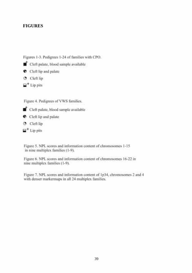
39
FIGURES
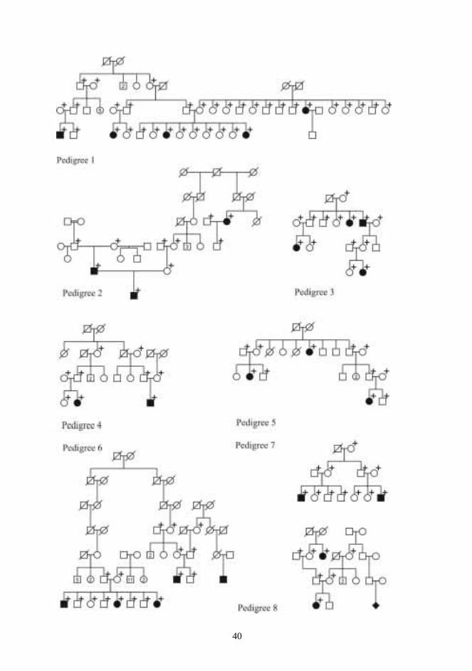
40
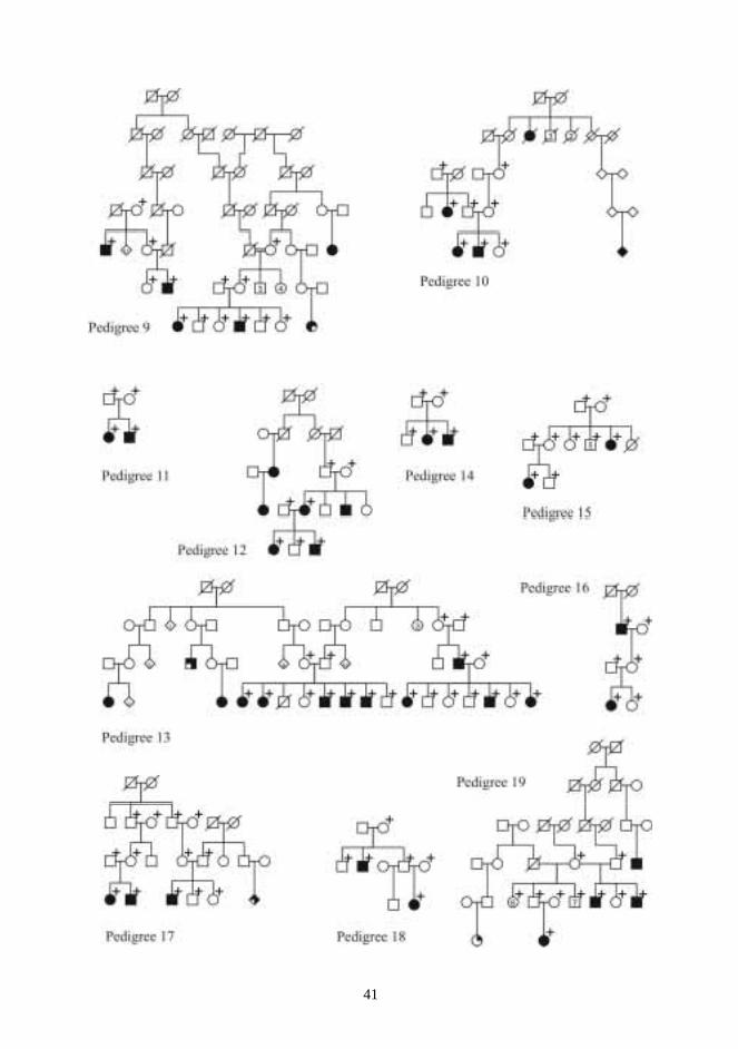
41
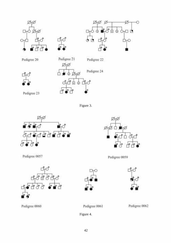
42
Figure 3.
Figure 4.
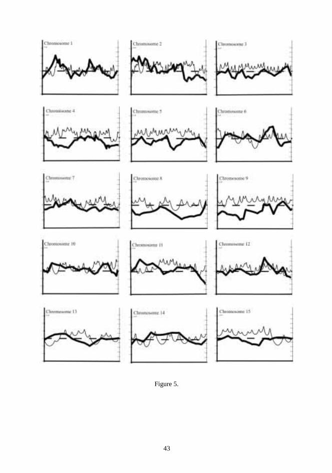
43
Figure 5.
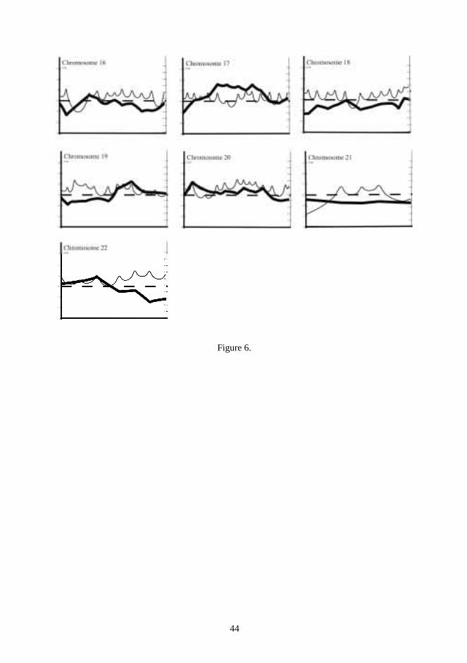
44
Figure 6.
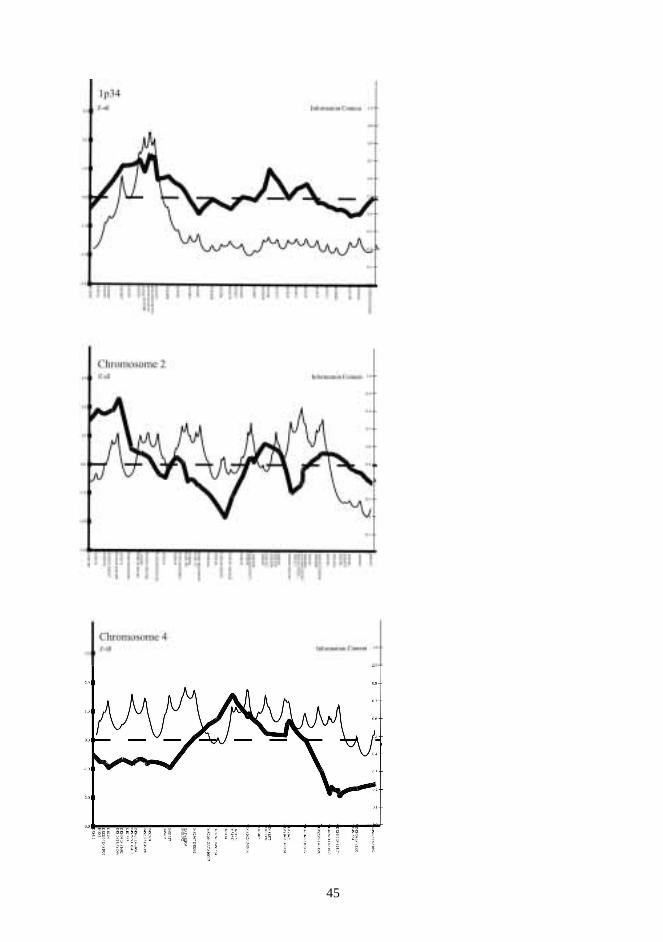
45
46
REFERENCES
Abbott BD, Birnbaum LS (1990) Retinoic acid-induced alterations in the expression of growth factors inembryonic mouse palatal shelves. Teratology 42(6):597-610
Abrishamchian AR, Khoury MJ, Calle EE (1994) The contribution of maternal epilepsy and its treatment to theetiology of oral clefts: a population based case-control study. Genet Epidem 11(4):343-351
Ala-Kokko L, Kvist A-P, Metsäranta M, Kivirikko KI, de Crombrugge B, Vuorio E (1995) Conservation of thesizes of 53 introns and over 100 intronic sequences for the binding of common transcription factors in the humanand mouse genes for type II procollagen (COL2A1). Biochem J 308:923-929
Ahmad NN, Ala-Kokko L, Knowlton RG (1991) Stop codon in the procollagen II gene (COL2A1) in a familywith the Stickler syndrome (arthro-ophthalmopathy). Proc Nat Acad Sci 88:6624-6627
Ahmad NN, McDonald-McGinn DM, Zackai EH, Knowlton RG, LaRossa D, DiMascio J, Prockop DJ (1993) Asecond mutation in the type II procollagen gene (COL2A1) causing Stickler syndrome (arthro-ophtalmopathy) isalso a premature termination codon. Am J Hum Genet 52:39-45
Altmuller J, Palmer LJ, Fischer H, Scherb H, Wjst M (2001) Review article: Genome wide scans of complexhuman diseases: true linkage is hard to find. Am J Hum Genet 69:936-950
Annunen S, Körkkö J, Czarny M et al Warman ML, Brunner HG, Kääriäinen H, Mulliken JB, Tranebjaerg L,Brooks DG, Cox GF, Cruysberg JR, Curtis MA, Davenport SLH, Friedrich CA, Kaitila I, Krawczynski MR,Latos-Bielenska A, Mukai S, Olsen BR, Shinno N, Somer M, Vikkula M, Zlotogora J, Prockop DJ, Ala-KokkoL (1999) Splicing mutations of 54 bp exons in the COL11A1 gene cause Marshall syndrome but other mutationscause overlapping Marshall/Stickler phenotypes. Am J Hum Genet 65:974-983.
Aula P, Autio S, Raivio K, Rapola J, Thoden CJ, Koskela SL, Yamashina I (1979) Salla disease. A newlysosomal storage disorder. Arch Neurol 36:88-94
Auranen M, Vanhala R, Varilo T, Ayers K, Kempas E, Ylisaukko-oja T, Sinsheimer J, Peltonen L, Järvelä I(2002) A genomwide screen for autism-spectrum disorders: evidence for a major susceptibility locus onchromosome 3q25-27. Am J Hum Genet 71:777-790
Balloux F (2001) EASYPOP (Version 1.7): A computer program for population genetics simulations. J Hered92:301-302
Barrow LL, van Bokhoven H, Daak-Hirsch S, Andersen T, van Beersum SEC, Gorlin R, Murray JC (2002)Analysis of the p63 gene in classical EEC syndrome, related syndromes, and non-syndromic orofacial clefts. JMed Genet 39:559-566
Beaty TH, Maestri NE, Hetmanski JB, Wyszynski DF, Vanderkolk CA, Simpson JC, McIntosh I, Smith EA,Zeiger JS, Raymond GV, Panny SR, Tifft CJ, Lewanda AF, Cristion CA, Wulfsberg EA (1997) Testing forinteraction between maternal smoking and TGFA genotype among oral cleft cases born in Maryland 1992-1996.Cleft Palate Craniofac J 34(5):447-54
Beaty TH, Wan H, Hetmanski JB, Fan YT, Zeiger JS, Liang KY, Chiu YF, Vanderkolk CA, Seifert KC,Wulfsberg EA, Raymond G, Panny SR, McIntosh I (2001) A case-control study of nonsyndromic oral clefts inMaryland. Ann Epidemiol 11(6):434-442
Beghi E, Annegers JF (2001) Collaborative Group for the Pregnancy Registries in Epilepsy Pregnancy registriesin epilepsy. Epilepsia 42(11):1422-1425
Benke PJ (1984) The isotretinoin teratogen syndrome. JAMA 251(24):3267-3269
Berg MJ, Fincham RW, Ebert BE, Schottelius DD (1988) Decrease of serum folates in healthy male volunteerstaking phenytoin. Epilepsia 29:67-73
47
Bienengraber V, Malek FA, Moritz KU, Fanghanel J, Gundlach KK, Weingartner J (2001) Is it possible toprevent cleft palate by prenatal administration of folic acid? An experimental study. Cleft Palate Craniofac J38(4):393-398
Boorman JG, Varma S, Ogilvie CM (2001) Velopharyngeal incompetence and chromosome 22q11 deletion.Lancet 10;357(9258):774
Botstein D, White RL, Skolnick M, Davis RW (1980) Construction of a genetic linkage map in man usingrestriction fragment polymorphisms. Am J Hum Genet 32:314-331
Braybrook C, Doudney K, Marcano ACB, Arnason A, Bjornsson A, Patton MA, Goodfellow PJ, Moore GE,Stanier P (2001) The T-box transcription factor gene TBX22 is mutated in X-linked cleft palate andankyloglossia. Nature Genet 29:179-183
Beiraghi S, Miller-Chisholm A, Kimberling WJ, Sun CE, Wang YF, Russell LJ, Khoshnevisan M, Storm AL,Long RE Jr, Witt PD, Mazaheri M, Diehl SR (1999) Confirmation of linkage of Van der Woude syndrome tochromosome 1q32: evidence of association with STR alleles suggests possible unique origin of the diseasemutation. J Craniofac Genet & Dev Biol. 19(3):128-34
Bocian M, Walker AP (1987) Lip pits and deletion 1q32-41. Am J Med Genet 26:437-443
Bonaiti C, Briard ML, Feingold J, Pavy B, Psaume J, Migne-Tufferaud G, Kaplan J (1982) An epidemiologicaland genetic study of facial clefting in France. I. Epidemiology and frequency in relatives. J Med Genet 19(1):8-15
Botto LD, May K, Fernhoff PM, Correa A, Coleman K, Rasmussen SA, Merritt RK, O'Leary LA, Wong LY,Elixson EM, Mahle WT, Campbell RM (2003) A population-based study of the 22q11.2 deletion: phenotype,incidence, and contribution to major birth defects in the population. Pediatrics 112:101-7
Brewer C, Holloway S, Zawalnyski P, Schinzel A, FitzPatrik D (1998) A chromosomal deletion map of humanmalformations. Am J Hum Genet 63:1163-1159
Brewer C, Holloway S, ZaWalnyski P, Schinzel A, FitzPatrick D (1999) A chromosomal duplication map ofmalformations: regions of suspected haplo- and triplolethality –and tolerance of segemental aneuplpody- inhumans. Am J Hum Genet 64:1702-1708
Brewer CM, Leek JP, Green AJ, Holloway S, Bonthron DT, Markham AF, FitzPatrick DR (1999) A locus forisolated cleft palate, located on human chromosome 2q32. Am J Hum Genet 65(2):387-396
Brown DM, Nichols BE, Weingeist TA, Sheffield VC, Kimura AE, Stone EM (1992) Procollagen II genemutation in Stickler syndrome. Arch Ophthal 110:1589-1593
Brown NL, Sandy JR (2003) Basic science in normal and abnormal palate development. Braz J Oral Sci 1(2):60-70
Brown NL, Yarram SJ, Mansell JP, Sandy JR (2002) Matrix metalloproteinases have a role in palatogenesis. JDent Res 81(12):826-30
Brown NL, Knott L, Halligan E, Yarram SJ, Mansell JP, Sandy JR (2003) Microarray analysis of murinepalatogenesis: Temporal expression of genes during normal palate development. Dev Growth Differ 45(2):153-65
Brunner HG, van Beersum SEC. Warman ML, Olsen BR, Ropers H-H, Mariman ECM (1994)A Stickler syndrome gene is linked to chromosome 6 near the COL11A2 gene. Hum Mol Genet 3: 1561-1564
Burdick AB, Bixler D, Puckett C (1985) Genetic analysis in families with Van der Woude syndrome. J CraniofacGenet Dev Biol 5:181-208
48
Burdick AB (1986) Genetic epidemiology and control of genetic expression in van der Woude syndrome. JCraniofac Genet Dev Biol [Suppl] 2:99-105
Burgoon JM, Selhub J, Nadeau M, Sadler TW (2002) Investigation of the effects of folate deficiency onembryonic development through the establishment of folate deficient mouse model. Teratology 65(5):219-227
Campbell K, Flavin N, Ivens A, Robert B, Buckingham M, Williamson R (1989) The human homeobox geneHOX7 maps to 4p16.1 and is deleted in Wolf-Hirschhorn syndrome patients. Am J Hum Genet 45 (suppl.): A179
Carinci F, Pezzetti F, Scapoli L, Martinelli M, Avantaggiato A, Carinci P, Padula E, Baciliero U, Gombos F,Laino G, Rullo R, Cenzi R, Carls F, Tognon M (2003) Recent developments in orofacial cleft genetics. JCraniofac Surg 14(2):130-143
Carlson C, Papolos D, Pandita RK, Faedda GL, Veit S, Goldberg R, Shprintzen R, Kucherlapati R, Morrow B(1997) Molecular analysis of velo-cardio-facial syndrome patients with psychiatric disorders. Am J Hum Genet60:851-859
Carter CO, Evans K, Coffey R, Roberts JAF, Buck A, Roberts MF (1982) A family study of isolated cleft palate.J Med Genet 19: 329-331
Castanet M, Park SM, Smith A, Bost M, Leger J, Lyonnet S, Pelet A, Czernichow P, Chatterjee K, Polak M(2002) A novel loss-of-function mutation in TTF-2 is associated with congenital hypothyroidism, thyroidagenesis and cleft palate. Hum Mol Genet 15;11(17):2051-2059
Celli J, Duijf P, Hamel BCJ, Bamshad M, Kramer B, Smits APT, Newbury-Ecob R, Hennekam R CM, VanBuggenhout G, van Haeringen A, WoodsCG, van Essen AJ, de Waal R, Vriend G, Haber DA, Yang A, McKeonF, Brunner HG, van Bokhoven H (1999) Heterozygous germline mutations in the p53 homolog p63 are the causeof EEC syndrome. Cell 99:143-153
Christensen K, Fogh-Andersen P (1993) Isolated cleft palate in Danish multiple births, 1970-1990. Cleft PalateCraniofac J 30(5):469-474
Christensen K, Fogh-Andersen P (1994) Etiological subgroups in non-syndromic isolated cleft palate. A genetic-epidemiological study of 52 Danish birth cohorts. Clin Genet 46:329-335
Christensen K, Mitchell LE (1996) Familial recurrence-pattern analysis of non-syndromic isolated cleft palate-aDanish registry study. Am J Hum Genet 58:182-190
Christensen K, Olsen J, Norgaard-Pedersen B, Basso O, Stovring H, Milhollin-Johnson L, Murray JC (1999)Oral clefts, transforming growth factor alpha gene variants, and maternal smoking: a population-based case-control study in Denmark, 1991-1994. Am J Epidem 149(3):248-55
Chung KS, Jacenko O, Boyle P. Olsen BR, Nishimura I (1997) Craniofacial abnormalities in mice carrying adominant interference mutation in type X collagen. Dev Dyn 208(4):544-552
Culiat CT, Stubbs LJ, Woychik RP, Russell LB, Johnson DK, Rinchik EM (1995) Deficiency of the beta 3subunit of the type A gamma-aminobutyric acid receptor causes cleft palate in mice. Nat Genet 11(3):344-346
Clayton-Smith J, Donnai D (1995) Fetal valproate syndrome. J Med Genet 32(2):724-727
Clementi M, Tenconi, Forabosco P (1997) Inheritance of cleft palate in Italy. Evidence for a major autosomalrecessive locus. Hum Genet 100:204-209
Clifton-Bligh RJ, Wentworth JM, Heinz P, Crisp MS, John R, Lazarus JH, Ludgate M, Chatterjee VK (1998)Mutation of the gene encoding human TTF-2 associated with thyroid agenesis, cleft palate and choanal atresia.Nat Genet 19(4):399-401
Clouthier DE, Williams SC, Yanagisawa H, Wieduwilt M, Richardson JA, Yanagisawa M. (1998) Cranial andcardiac neural crest defects in endothelin-A receptor-deficient mice. Development. 125:813-824
49
Cohen MM Jr (1999) Robin sequences and complexes: causal heterogeneity andpathogenetic/phenotypic variability. Am J Med Genet 4;84(4):311-5
Collins FS, Brooks LD, Chakravarti A (1998) A DNA polymorphism discovery resource for research on humangenomic variation. Genome Res 8(12):1229-1231
Condie BG, Bain G, Gottlieb DI, Capecchi MR (1997) Cleft palate in mice with a targeted mutation in thegamma-aminobutyric acid-producing enzyme glutamic acid decarboxylase 67. Proc Natl Acad Sci 94(21):11451-11455
Cooper DN, Smith BA, Cooke HJ, Nieman S, Schmidtke J (1985) An estimate of unique DNA sequenceheterozygosity in the human genome. Hum Genet 69:201-205
Cruts M, Backhovens H, Theuns J, Clark FR, la Paslier D, Weissenbach J, Goate AM, Martin J-J, VanBroeckhoven C (1995) Genetic and physical characterization of the early-onset Alzheimer’s disease AD3 locuson chromosome 14q24.3. Hum Mol Genet 4(8):1355-1364
Curtis EJ, Fraser F, Warburton D (1961) Congenital cleft lip and palate. Am J Dis Child 102:853-857
Czeizel AE, Timar L, Sarkozi A (1999) Dose-dependent effect of folic acid on the prevention of orofacial clefts.Pediatrics 104(6):66
Davies AF, Stephens RJ, Olavesen MG, Heather L, Dixon MJ, Magee A, Flinter F, Ragoussis J (1995) Evidenceof a locus for orofacial clefting on human chromosome 6p24 and STS content map of the region. Hum MolGenet 4:121-128
Derynck R, Lindquist PB, Lee A, Wen D, Tamm J, Graycar JL, Rhee L, Mason AJ, Miller DA, Coffey RJ (1988)A new type of transforming growth factor-beta, TGF-beta 3. EMBO J 1;7(12):3737-3743
De Felice M, Ovitt C, Biffali E, Rodriguez-Mallon A, Arra C, Anastassiadis K, Macchia PE, Mattei MG,Mariano A, Scholer H, Macchia V, Di Lauro R (1998) A mouse model for hereditary thyroid dysgenesis and cleftpalate. Nat Genet 19(4):395-398
De la Chapelle A, Herva R, Koivisto M, Aula P (1981) A deletion in chromosome 22 can cause DiGeorgesyndrome. Hum Genet 57:253-256
De la Chapelle A (1993) Disease gene mapping in isolated populations: the example of Finland. J Med Genet30:857-865
De la Chapelle A, Wright FA (1998) Linkage disequilibrium mapping in isolated populations: the example ofFinland revisited. Proc Natl Acad Sci 95:12416-12423
Demanais F, Bonati-Pellie C, Briard ML, Feingold J (1984) An epidemiological and genetic study of facialclefting in France. Segregation analysis. J Med Genet 21:436-440
Demczuk S, Levy A, Aubry M, Croquette MF, Philip N, Prieur M, Sauer U, Bouvagnet P, Rouleau GA, ThomasG (1995) Excess of deletions of maternal origin in the DiGeorge/velo-cardio-facial syndromes. A study of 22new patients and review of the literature. Hum Genet 96(1):9-13
Digilio MC, Angioni A, De Santis M, Lombardo A, Giannotti A, Dallapiccola B, Marino B (2003) Spectrum ofclinical variability in familial deletion 22q11.2: from full manifestation to extremely mild clinical anomalies. ClinGenet 63(4):308-13.
Donis-Keller H, Green P, Helms C, Cartinhour S, Weiffenbach B, Stephens K, Keith TP, Bowden DW, SmithDR, Lander ES, Botstein D, Akots G, Rediker KS, Gravuis T, Brown VA, Rising MB, Parker C, Powers JA,Watt DE, Kauffman ER, Bricker A, Phipps P, Muller-Kahle H, Fulton TR, Ng S, Schumm JW, Braman JC,Knowlton RG, Barker DF, Crooks SM, Lincoln SE, Daly MJ, Abrahamsson J (1987) A genetic linkage map ofthe human genome. Cell 51(2):319-337
50
Edwards MJ, Agho K, Attia J, Diaz P, Hayes T, Illingworth A, Roddick LG (2003) Case-control study of cleft lipor palate after maternal use of topical corticosteroids during pregnancy. Am J Med Genet 120A:459-463
Ericson A, Källen B, Westerholm P (1986) Cigarette smoking as an etiologic factor in cleft lip and palate. Am JObstet Gynecol 135:348-351
Ericson A, Källen B (2001) Nonsteroidal anti-inflammatory drugs in early pregnancy. Reprod Toxicol 15:371-375
Fawcett LB, Buck S, Beckman DA, Brent RL (1996) Is there a no-effect dose for corticosteroid-induced cleftpalate? The contribution of endogenous corticosterone to the incidence of cleft palate in mice. Pediatr Res39(5):856-861
Ferguson M (1988) Palate development. Development 103 Suppl.:41-60
FitzPatrick D, Farrall M (1993) An estimation of the number of susceptibility loci for isolated cleft palate. JCraniofac Genet Dev Biol 13:230-235
FitzPatrick D, Raine PAM, Boorman JG (1994) Facial clefts in the west of Scotland in the period 1980-1984:epidemiology and genetic diagnoses. J Med Genet 31:126-129
Fogh-Andersen P (1942) Inheritance of harelip and cleft palate. Busck, Copenhagen
Francomano CA, Liberfarb RM, Hirose T, Maumenee IH, Streeten EA, Meyers DA, Pyeritz RE (1987) TheStickler syndrome: evidence for close linkage to the structural gene for type II collagen. Genomics 1:293-296
Frankel WM, Schork NJ (1996) Who’s afraid of epistasis? Nat Genet 14:371-373
Friis ML, Holm NV, Sindrup EH, Fogh-Andersen P, Hauge M (1986) facial clefts in sibs and children ofepileptic patients. Neurology 36(3):346-350
Friis ML (1989) Facial clefts and congenital heart defects in children of parents with epilepsy: genetic andenvironmental etiologic factors. Acta Neur Scan 79(6):433-459
Frosst P, Blom HJ, Milos R, Goyette P, Sheppard CA, Matthews RG, Boers GJ, den Heijer M, Kluijtmans LA,van den Heuvel LP (1995) A candidate genetic risk factor for vascular disease: a common mutation inmethylenetetrahydrofolate reductase. Nat Genet 10(1):111-113
Gato A, Martinez ML, Tudela C, Alonso I, Moro JA, Formoso MA, Ferguson MW, Martinez-Alvarez C (2002)TGF-beta(3)-induced chondroitin sulphate proteoglycan mediates palatal shelf adhesion. Dev Biol15;250(2):393-405
Gendron-Maguire M, Mallo M, Zhang M, Gridley T (1993) Hoxa-2 mutant mice exhibit homeotic transformationof skeletal elements derived from cranial neural crest. Cell 75(7):1317-31
Glover TW (1995) CATCHing a break on 22. Nat Genet 10:257-258
Goldberg R, Marion R, Borderon M, Wiznia A, Shprintzen R (1985) Phenotypic overlap between velo-cardio-facial syndrome and the DiGeorge sequence. Am J Hum Genet Suppl 37:A54
Goldberg R, Motzkin B, Marion R, Scambler P, Shprintzen R (1993) Velo-cardio-facial syndrome: a review of120 patients. Am J Med Genet 45:313-319
Gorlin BJ, Cohen MM, Levin LS (1990) Syndromes of the head and neck. New York. Oxford University Press3rd edition, 1990
Gyapay G, Morisette J, Vignal A, Dib C, Fizames C, Millasseau P, Marc S, Bernardi G, Lathrop M,Weissenbach J (1994) The 1993-1994 Genethon human genetic linkage map. Nat Genet 7 (Special issue):246
51
Hadano S, Nichol K, Brinkman RR, Nasir J, Martindale D, Koop BF, Nicholson DW, Scherer SW, Ikeda J-E,Hayden MR (1999) A yeast artificial chromosome-based physical map of the juvenile amyotrophic lateralsclerosis (ALS2) critical region on human chromosome 2q33-q34. Genomics 55:106-112
Hagberg B, Sourander P, Svennerholm PL (1968) Late infantile progressive encephalopathy with disturbedpolyunsaturated fat metabolism. Acta Paediatr Scand 57:495-49
Hampe J, Wienker T, Schreiber S, Nurnberg P (1998) POPSIM: a general population simulator program.Bioinformatics 14:458-464
Hayes C, Werler MM, Willett WC, Mitchell AA (1996) Case-control study of periconceptional folic acidsupplementation and oral clefts. Am J Epidemiol 143(12):1229-34
Hecht JT, Annegars JF, Kurland LT (1989) Epilepsy and clefting disorders: lack of evidence of familialassociation. Am J Med Genet 33(2):244-247
Hecht JT, Annegars JF (1990) Familial aggregation of epilepsy and clefting disorders: a review of literarure.Epilepsia 32(5):574-577
Hecht JT, Mulliken JB, Blanton SH (2002) Evidence for a cleft palate only locus on chromosome 4 near MSX1.Am J Med Genet 15;110(4):406-7
Hewitt JE, Clark LN, Ivens A, Williamson R (1991) Structure and sequence of the human homeobox geneHOX7. Genomics 11:670-678
Hiranuma H, Jikko A, Maeda T, Abe M, Fuchihata H (2000) Effect of X irradiation on secondary palatedevelopment in mice. Radiat Res 154:34-38
Hirvasniemi A, Leisti J (1991) An inherited form of childhood epilepsy associated with mental retardation. Am JHum Genet 49 suppl:147
Holder-Espinasse M, Abadie V, Cormier-Daire V, Beyler C, Manach Y, Munnich A, Lyonnet S, Couly G, AmielJ (2001) Pierre Robin sequence: a series of 117 consecutive cases. J Pediatr 139(4):588-90
Homanics GE, DeLorey TM, Firestone LL, Quinlan JJ, Handforth A, Harrison NL, Krasowski MD, Rick CE,Korpi ER, Makela R, Brilliant MH, Hagiwara N, Ferguson C, Snyder, Olsen RW (1997) Mice devoid of gamma-aminobutyrate type A receptor beta3 subunit have epilepsy, cleft palate, and hypersensitive behavior. Proc NatlAcad Sci 94(8):4143-4148
Houdayer C, Soupre V, Rosenberg-Bourgin M, Martinez H, Tredano M, Feldmann D, Feingold D, Feingold J,Aymard P, Munnich A, Le Bouc Y, Vazquez MP, Bahuau M (1999) Linkage analysis of 5 novel Van der Woudesyndrome kindreds to 1q32-41 markers further supports locus homogeneity of the disease trait. Ann Genet 42:69-74
Houdayer C, Portnoi M-F, Vialard F, Soupre V, Crumiere C, Taillemite J-L, Couderc R, Vazquez M-P, BahuauM (2001) Pierre Robin sequence and interstitial deletion 2q32.3-q33.2. Am J Med Genet 102:219-226
Hovatta I, Varilo T, Suvisaari J, Terwilliger JD, Ollikainen V, Arajärvi R, Juvonen H, Kokko-sahin M-L,Väisänen L, Mannila H, Lönnqvist J, Peltonen L (1999) A genomewide screen for schizophrenia genes in anisolated Finnish subpopulation suggesting multiple susceptibility loci.. Am J Hum Genet 65:1114-1124
Hwang SJ, Beaty TH, Panny SR, Street NA, Joseph JM, Gordon S, McIntosh I, Francomano CA (1995)Association study of transforming growth factor alpha (TGF alpha) TaqI polymorphism and oral clefts:indication of gene-environment interaction in a population-based sample of infants with birth defects. Am JEpidemol 141(7):629-36
Hästbacka J, de la Chapelle A, Kaitila I, Sistonen P, Weaver A, Lander E (1992) Linkage disequilibriummapping in isolated founder populations: diastrophic dysplasia in Finland. Nat Genet 2:204-211
52
Hästbacka J, de la Chapelle A, Mahtani MM, Clines G, Reeve-Daly MP, Daly M, Hamilton BA, Kusumi K,Trivedi B, Weaver A, Coloma A, Lovett M, Buckler A, Kaitila I, Lander ES (1994) The diastrophic dysplasiagene encodes a novel sulfate transporter: positional cloning by fine-structure linkage disequilibrium mapping.Cell 78:1073-1087
Iida H, Kast A, Tsunenari Y, Asakura M (1988) Corticosterone induction of cleft palate in mice dosed withorciprenaline sulfate. Teratology 38(1):15-27
Isaac C, Marsh K, Paznekas W, Dixon J, Dixon M, Jabs E, Meier UT (2000) Characterization of the nucleolargene product, treacle, in Treacher Collins syndrome. Mol Biol Cell 11(9):3061–3071
Ikonen E, Baumann M, Grön K, Syvänen AC, Enomaa N, Halila R, Aula P, Peltonen L (1991)Aspartylglucosaminuria: cDNA encoding human aspartylglicosaminidase and the missence mutation causing thedisease. EMBO J 10:51-58
Itikala PR, Watkins ML, Mulinare J, Moore CA, Liu Y (2001) Maternal multivitamin use and orofacial clefts inoffspring. Teratology 63(2):79-86
Jaber L, Nahmani A, Halpern GJ, Shohat M (2002) Facial clefting in an Arab town in Israel. Clin Genet61(6):448-453
Jakobsen LP, Molsted K, Christensen K (2003) Occurrence of cleft lip and palate in the Faroe Islands andGreenland from 1950 to 1999. Cleft Palate Craniofac J 40(4):426-430
Jeffreys A, Wilson WV, Thein S (1985) Individual-specific”fingerprints” of human DNA. Nature 316:76-79
Jenkins M, Stady C (1980) Dominant inheritance of cleft of the soft palate. Hum Genet 48:179-181
Jezewski PA, Vieira AR, Nishimura C, Ludwig B, Johnson M, O'Brien SE, Daack-Hirsch S, Schultz RE, WeberA, Nepomucena B, Romitti PA, Christensen K, Orioli IM, Castilla EE, Machida J, Natsume N, Murray JC (2003)Complete sequencing shows a role for MSX1 in non-syndromic cleft lip and palate. J Med Genet 40(6):399-407
Jiang R, Lan Y, Chapman HD, Shawber C, Norton CR, Serreze DV, Weinmaster G, Gridley T. (1998) Defects inlimb, craniofacial, and thymic development in Jagged2 mutant mice. Genes & Dev 12(7):1046-1057
Johnson VP, Swayze VW II, Sato Y, Andreasen NC (1996) Fetal alcohol syndrome: craniofacial and centralnervous manifestations. Am J Med Genet 61:329-339
Jones K (ed): Smith’s recognizable patterns of human malformation (1997) W.B. Saunders Company,Philadelphia. 5th edition
Jones M (1988) Etiology of facial clefts. Prospective evaluation of 428 patients. Cleft palate J 25:16-20
Jorde LB (1995) Linkage disequilibrium as a gene-mapping tool. Am J Hum Genet 56:11-14
Jugessur A, Lie RT, Wilcox AJ, Murray JC, Taylor JA, Saugstad OD, Vindenes HA, Abyholm F (2003) Variantsof developmental genes (TGFA, TGFB3, and MSX1) and their associations with orofacial clefts: A case-parenttriad analysis. Genet Epidemiol 24(3):230-9
Jugessur A, Wilcox AJ, Lie RT, Murray JC, Taylor JA, Ulvik A, Drevon CA, Vindenes HA, Abyholm F (2003)Exploring the effects of methylenetetrahydrofolate reductase gene variants C677T and A1298C on the risk oforofacial clefts in 261 Norwegian case-parent triads. Am J Epidemiol 15;157(12):1083-91
Jumlongras D, Bei M, Stimson JM, Wang WF, DePalma SR, Seidman CE, Felbor U, Maas R, Seidman JG,Olsen BR (2001) A nonsense mutation in MSX1 causes Witkop syndrome. Am J Hum Genet 69(1):67-74
Jutikkala E, Pirinen K (1996) History of Finland. Helsinki.WSOY
53
Kaartinen V, Voncken JW, Shuler C, Warburton D, Bu D, Heisterkamp N, Groffen J (1995) Abnormal lungdevelopment and cleft palate in mice lacking TGF-beta 3 indicates defects of epithelial-mesenchymal interaction.Nat Genet 11(4):415-421
Khoury MJ, Gomez-Farias L, Mulinare J (1989) Does maternal cigarette smoking during pregnancy cause cleftlip and palate in offspring? Am J Dis Child 143:333-337
Kinnunen E, Wiksröm E, Porras J, Palo J (1983) The epidemiology of multiple sclerosis in Finland: increase ofprevalence and stability of foci in high-risk areas. Acta Neur Scan 67:255-26
Knottnerus AC, de Jong DJ, Haumann TJ, Mulder JW (2001) Higher incidence of twins in infants with PierreRobin sequence Cleft Palate Craniofac J 38(3):284
Kondo S, Schutte BC, Richardson RJ, Bjork BC, Knight AS, Watanabe Y, Howard E, Ferreira de Lima RLL,Daack-Hirsch S, Sander A, McDonald-McGinn DM, Zackai EH, Lammer EJ, Aylsworth AS, Ardinger HH,Lidral AC, Pober PR, Moreno L, Arcos-Burgos R, Valencia C, Houdayer C, Bahuau M, Moretti-Ferreira D,Richieri-Costa A, Dixon MJ, Murray JC (2002) Mutations in IRF6 cause Van der Woude and poplitealpterygium syndromes. Nat Genet 32(2):285-289
Koskinen S, Martelin T, Notkola I-L, Notkola V, Pitkänen K (1994) Suomen väestö. Hämeenlinna. Gaudeamus.
Kruglyak L, Daly MJ, Reeve-Daly MP, Lander ES (1996) Parametric and nonparametric linkage analysis: aunified multipoint approach. Am J Hum Gen 58(6):1347-63
Kruglyal L (1999) Prospects for whole-genome linkage disequilibrium mapping of common disease genes. NatGenet 22:139-144
Kruglyak L, Nickerson DA (2001) Variation is the spice of life. Nat Genet 27(3):234-6
Kuivaniemi H, Tromp G, Prockop DJ (1997) Mutations in fibrillar collagens (types I, II, III, and XI), fibril-associated collagen (type IX), and network-forming collagen (type X) cause a spectrum of diseases of bone,cartilage, and blood vessels. Hum Mutat 9:300-315
Kuokkanen S, Gshwend M, Rioux JD, Daly M, Terwilliger JD, Tienari PJ, Wikström J, Palo J, Stein LD, HudsonTJ, Lander E, Peltonen L (1997) Genomewide scan of multiple sclerosis in Finnish multiplex families. Am JHum Genet 61:1379-1387
Kusumi K, Trivedi B, Weaver A, Coloma A, Lovett M, Buckler A, Kaitila I, Lander ES (1994) The diastrophicdysplasia gene encodes a novel sulfate transporter: positional cloning by fine-structure linkage disequilibriummapping. Cell 78:1073-1087
Laatikainen T, Ranta R, Nordstrom R (1996) Craniofacial morphology in twins with cleft lip and palate. CleftPalate Craniofac J 33(2)96-103
Lahermo P, Sajantila A, Sistonen P, Lukka M, Aula P, Peltonen L, Savontaus M-L (1996) The geneticrelationship between the Finns and the Finnish Saami (Lapps): Analysis of nuclear DNA and mtDNA. Am J HumGenet 58:1309-1322
Lahermo P, Savontaus M-L, Sistonen P, Beres J, de Knijff P, Aula P, Sajantila A (1999)Y chromosomalpolymorphisms reveal founding lineages in the Finns and the Saami. Eur J Hum Genet 7(4):447-58
Lahiri DK, Nurnberger JI (1991) A rapid non-enzymatic method for the preparation of HMW DNA from bloodfor RFLP studies. Nucl Acids Res 19:5444
Lander E, Schork N (1994) Genetic dissection of complex traits. Science 265:2037-2048
Larson M, Hellquist R, Jakobsson OP (1998) Morphology of isolated cleft palate in children, including Robinsequence, treated with one or two-stage operations. Scand J Plast Reconstr Hand Surg 32(2):193-201
54
Laitinen T, Kauppi P, Ignatius J, Ruotsalainen T, Daly MJ, Kääriäinen H, Kruglyak L, Laitinen H, de la ChapelleA, Lander ES, Laitinen LA, Kere J (1997) Genetic control of serum IgE levels and asthma: linkage and linkagedisequilibrium studies in an isolated population. Hum Mol Genet 6(12):2069-76
Lammer EJ, Chen DT, Hoar RM, Agnish ND, Benke PJ, Braun JT, Curry CJ, Fernhoff PM, Grix AW Jr, Lott IT(1985) Retinoic acid embryopathy. N Engl J Med 313(14):837-41
Lander E, Kruglyak L (1995) Genetic dissection of complex traits. Guidelines for interpreting and reportinglinkage results. Nat Genet 11:241-247
Lathrop GM, Lalouel JM, Julier C, Ott J (1984) Strategies for multilocus linkage analysis in humans. Proc NatlAcad Sci USA 81:3443-3446
Lees MM, Winter RM, Malcolm S, Saal HM, Chitty L (1999) Popliteal pterygium syndrome: a clinical study ofthree families and report of linkage to the Van der Woude locus on 1q32. J Med Genet 36(12):888-892
Lehesjoki A-E, Koskiniemi M, Norio R, Tirrito S, Sistonen P, Lander E, de la Chapelle A (1993) Localization ofthe EPM1 gene for progressive myoclonus epilepsy on chromosome 21: linkage disequilibrium allows highresolution mapping. Hum Mol Genet 2:1229-1234
Liberfarb RM, Levy HP, Rose PS, Wilkin DJ, Davis J, Balog JZ, Griffith AJ, Szymko-Bennett YM, Johnston JJ,Francomano CA (2003) The Stickler syndrome: genotype/phenotype correlation in 10 families with Sticklersyndrome resulting from seven mutations in the type II collagen gene locus COL2A1. Genet Med 5(1):21-27
Lidral AC, Murray JC, Buetow KH, Basart AM, Schearer H, Shiang R, Naval A, Layda E, Magee K, Megee W(1997) Studies of the candidate genes TGFB2, MSX1, TGFA and TFB3 in the etiology of cleft lip and palate inthe Philippines. Cleft Palate Craniofac J 34(1)1-6
Lidral AC. Romitti PA. Basart AM. Doetschman T. Leysens NJ. Daack-Hirsch S. Semina EV. Johnson LR.Machida J, Burds A, Parnell TJ, Rubenstein JL, Murray JC (1998) Association of MSX1 and TGFB3 withnonsyndromic clefting in humans. Am J Hum Genet 63(2):557-68
Lieff S, Olshan AF, Werler M, Strauss RP, Smith J, Mitchell A (1999) Maternal cigarette smoking duringpregnancy and risk of oral clefts in newborns. Am J Epidemiol 150(7):683-94
Lilius P. (1992) Epidemiology of cleft lip and palate in Finland 1975-1985. PhD thesis, University of Helsinki
Lorente C, Cordier S, Goujard J, Ayme, Bianchi F, Calzolari E, De Walle HE, Knill-Jones R (2000) Tobaccoand alcohol use during pregnancy and risk of oral clefts. Occupational Exposure and Congenital MalformationWorking Group. Am J Pub Health. 90(3):415-9
Machida J, Yoshira K, Funkhauser C, Natsume N, Kawai T, Murray J (1999) Transforming growth factor-�
(TGFA): Genomic structure, boundary sequences, and mutation analysis in nonsyndromic cleft lip/palate andcleft palate only. Genomics 61:237-242
Mahtani MM, Widen E, Lehto M, Thomas J, McCarthy M, Brayer J, Bryant B, Chan G, Daly M, Forsblom C,Kanninen T, Kirby A, Kruglyak L, Munnelly K, Parkkonen M, Reeve-Daly MP, Weaver A, Brettin T, Duyk G,Lander ES, Groop LC (1996) Mapping of a gene for type 2 diabetes associated with an insulin secretion defectby a genome scan in Finnish families. Nat Genet 14(1):90-4
McWilliams BJ (1991) Submucous clefts of the palate: how likely are they to be symptomatic? Cleft PalateCraniofac J 28(3):247-249
Marazita ML, Jaskoll T, Melnick M (1988) Corticosteroid-induced cleft palate in short-ear mice. J CraniofacGenet & Dev Biol 8(1):47-51
Marazita ML, Field LL, Cooper ME, Tobias R, Maher BS, Peanchitlertkajorn S, Liu Y (2002) Genome scan forloci involved in cleft lip with or without cleft palate, in Chinese multiplex families. Am J Hum Genet 71:349-364
55
Martin JF, Bradley A, Olson EN (1995) The paired-like homeo box gene MHox is required for early events ofskeletogenesis in multiple lineages. Genes Dev 9:1237-1249
Martinelli M, Scapoli L, Pezzetti F, Carinci F, Francioso F, Baciliero U, Padula E, Carinci P, Tognon M (2001)Linkage analysis of three candidate regions of chromosome 1 in nonsyndromic familial orofacial cleft. Ann HumGenet 65: 465-471
Marques IL, Barbieri MA, Bettiol H (1998) Etiopathogenesis of isolated Robin sequence. Cleft Palate CraniofacJ 35(6):517-525
Maschhoff KL, Baldwin HS (2001) Molecular determinants of neural crest migration. Am J Med Genet97(4):280-288
Mayne R, Brewton RG, Mayne PM, Baker JR (1993) Isolation and characterization of the chains of typeV/typeXI collagen present in bovine vitreous. J Biol Chem 268: 9381-9386
McGuirt WT, Prasad SD, Griffith AJ, Kunst HPM, Green GE, Shpargel K, Runge C, Huybrechts C, MuellerRF, Lynch E, King M-C, Brunner HG, Cremers CW, Takanosu M, Li S-W, Arita M, Mayne R, Prockop DJ,Van Camp G, Smith RHJ (1999) Mutations in COL11A2 cause non-syndromic hearing loss (DFNA13). NatGenet 23:413-419
Melkoniemi M, Brunner HG, Manouvrier S, Hennekam R, Superti-Furga A, Kääriäinen H, Pauli RM, van EssenT, Warman ML, Bonaventure J, Miny P, Ala-Kokko L (2000) Autosomal recessive disorderotospondylomegaepiphyseal dysplasia is associated with loss-of-function mutations in the COL11A2 gene. Am JHum Genet 66:368-377
Miettinen PJ, Chin JR, Shum L, Slavkin HC, Shuler CF, Derynck R, Werb Z (1999) Epidermal growth factorreceptor function is necessary for normal craniofacial development and palate closure. Nat Genet 22(1):69-73
Milan M, Astolfi G, Volpato S, Garani GP, Clementi M, Tenconi R, Boni S, Calzolari E (1994) 766 cases of oralclefs in Italy. Eur J Epidemiol 10:317-324
Mills JL, Scott JM, Kirke PN, McPartlin JM, Conley MR, Weir DG, Molloy AM, Lee YJ (1996) Homocysteineand neural tube defects. J Nutr 126(3):756-760
Mills JL, Kirke PN, Molloy AM, Burke H, Conley MR, Lee YJ, Mayne PD, Weir DG, Scott JM (1999)Methylenetetrahydrofolate reductase thermolabile variant and oral clefts. Am J Med Genet 86:71-74
Mitchell LE, Murray JC, O'Brien S, Christensen K (2001) Evaluation of two putative susceptibility loci for oralclefts in the Danish population. Am J Epidemiol 15;153(10):1007-15
Mitchell LE, Beaty TH, Lidral AC, Munger RG, Murray JF, Saal HM, Wyszynski DF (2002) Guidelines for thedesign and analysis of studies on nonsyndromic cleft lip and cleft palate in humans: Summary report from aworkshop of the international consortium for oral clefts. Cleft Palate Craniofac J 39(1)93-100
Montenegro MA, Rojas M, Dominguez S, Rosales CJ (1998) Differences in extracellular matrix components andcell density during normal and dexamethasone-treated secondary palate development in two strains of mice withdifferent susceptibility to glucocorticoid induced-clefting. J Craniofac Genet Dev Biol 18(2):100-106
Morton N (1955) Sequential tests for the detection of linkage. Am J Hum Genet 7:277-318
Morrow B, Goldberg R, Carlson C, Das Gupta R, Sirotkin H, Collins J, Dunham I, O’Donnell H, Scambler P,Shprintzen R, Kucherlapati R (1995) Molecular definition of the 22q11 deletions in velo-cardio-facial syndrome.Am J Hum Genet 56:1391-1403
Mullis K, Faloona F, Scharf S, Saiki R, Horn G, Erlich H (1986) Specific enzymatic amplification of DNA invitro: The polymerase chain reaction. Cold Spring Harbor Symposia on Quantative Biology 51:263-273
56
Munger RG, Romitti PA, Daack-Hirsch S, Burns TL, Murray JC, Hanson J (1996) Maternal alcohol use and riskof orofacial cleft birth defects. Teratology 54(1):27-33
Muragaki Y, Mariman ECM, van Beersum SEC, Perala M, van Mourik JBA, Warman ML, Olsen BR, HamelBCJ (1996) A mutation in the gene encoding the alpha-2 chain of the fibril-associated collagen IX, COL9A2,causes multiple epiphyseal dysplasia (EDM2). Nat Genet. 12:103-105
Murray JC, Nishimura DY, Buetow KH, Ardinger HH, Spence MA, Sparkes RS, Falk RE, Falk P.M. GardnerRJM, Harkness EM, Glinski LP, Pauli RM, Nakamura Y, Green PP, Schinzel A (1990) Linkage of an autosomaldominant clefting syndrome (Van der Woude) to loci on chromosome 1q. Am J Hum Genet 46:486-491
Murray JC, Buetow KH, Weber JL, Ludwigsen S, Scherpbier-Heddema T, Manion F, Quillen J, Sheffield VC,Sunden S, Duyk G, Weissenbach J, Gyapay J, Dib C, Morissette J, Lathrop GM; Vignal A, White R, MatsunamiN, Gerken S, Melis R, Albertsen H; Plaetke R, Odelberg S, Ward D, Dausset J, Cohen D, Cann H (1994) Acomprehensive human linkage map with centimorgan density. Science 265:2049-2054
Murray JC (1995) Face facts: genes, environment and clefts. Am J Hum Genet 57:227-232
Murray JC (2001) Time for T. Nat Genet 29:107-109
Murray JC (2002) Gene/environment causes of cleft lip and/or palate. Clin Genet 61(4):248-56
Männikkö M, Kestilä M, Holmberg C, Norio R, Ryynänen M, Olsen A, Peltonen L (1995) Tryggvason K. Finemapping and haplotype analysis of the locus for cangenital nephrotic syndrome on chromosome 19q13.1. Am JHum Genet 57:1377-1383
Nordström REA, Laatikainen T, Juvonen T, Ranta R (1996) Cleft-twin sets in Finland 1948-1987. Cleft PalateCraniofac J 33(4):340-347
Norio R (1966) Heredity in the congenital nephrotic syndrome. Academic dissertation. The University ofHelsinki.
Norio R, Nevanlinna H, Perheentupa J (1973) Hereditary diseases in Finland: rare flora in rare soil. Ann Clin Res5:109-141
Norio R (2000) Suomineidon geenit.Helsinki. Otava
Nugent P, Greene RM (1998) MSX-1 gene expression and regulation in embryonic palatal tissue. In Vitro CellDev Biol Anim 34(10):831-835
Nugent P, Ma L, Greene RM (1998) Differential expression and biological activity of retinoic acid-inducedTGFbeta isoforms in embryonic palate mesenchymal cells. J Cell Physiol 177(1):36-46
Nugent P, Sucov H, Pisano M, Greene R (1999) The role of RXR-alpha in retinoic acid-induced cleft palate asassessed with the RXR-alpha knockout mouse. Int J Dev Biol 43(6):567-570
Ollikainen V (2002) Simulation techniques for disease gene localization in isolated populations. PhD thesis.University of Helsinki
Ott J (1974) Estimation of the recombination fraction in human pedigrees: efficient computation of the likelihoodfor human linkage studies. Am J Hum Genet 26:588-597
Ott J (1989) Computer-simulation methods in human linkage analysis. Proc Nat Ac Sci 86:4175-4178
Onofre MA, Brosco HB, Taga R (1997) Relationship between lower-lip fistulae and cleft lip and/or palate in Vander Woude syndrome. Cleft Palate Craniofac J 34(3):261-265
Orioli D, Henkemeyer M, Lemke G, Klein R, Pawson T (1996) Sek4 and Nuk receptors cooperate in guidance ofcommissural axons and in palate formation. EMBO J 15(22):6035-6049
57
Owens JR, Jones JW, Harris F (1985) Epidemiology of facial clefting. Arch Dis Child 60:521-524
Padanilam BJ, Stadler HS, Mills KA, McLeod LB, Solursh M, Lee B, Ramirez F, Buetow KH, Murray JC (1992)Characterization of the human HOX 7 cDNA and identification of polymorphic markers. Hum Mol Genet1(6):407-410
Palo J (1967) Prevalence of phenylketonuria and some other metabolic disorders among mentally retardedpatients in Finland. Acta Neurol Scand 43:573-579
Paylor R, McIlwain KL, McAninch R, Nellis A, Yuva-Paylor LA, Baldini A, Lindsay EA (2001) Mice deletedfor the DiGeorge/velocardiofacial syndrome region show abnormal sensorimotor gating and learning andmemory impairments. Hum Mol Genet 1;10(23):2645-50
Peltonen L, Jalanko A, Varilo T (1999) Molecular genetics of the Finnish disease heritage. Hum Mol Genet8(19):1913-1923
Perheentupa J (1972) Suomalainen tautiperintö kliinikon ja tutkijan kannalta. Duodecim 88:1
Perola M, Kainulainen K, Pajukanta P, Terwilliger JD, Hiekkalinna T, Ellonen P, Kaprio J, Koskenvuo M,Kontula K, Peltonen L (2000) Genome-wide scan of predisposing loci for increased diastolic blood pressure inFinnish siblings. J Hypertens 18(11):1579-1585
Peters H, Neubuser A, Kratochwil K, Balling R (1998) Pax9-deficient mice lack pharyngeal pouch derivativesand teeth and exhibit craniofacial and limb abnormalities. Genes Dev 12(17):2735-2747
Pezzetti F, Scapoli L, Martinelli M, Carinci F, Bodo M, Carinci P, Tognon M (1998) A locus in 2p13-p14(OFC2), in addition to that mapped in 6p23, is involved in nonsyndromic familial orofacial cleft malformation.Genomics 15;50(3):299-305
Prescott N, Lees JM, Malcolm S, Winter R (1998) Non-syndromic cleft lip and palate: a genome-wide sib-pairanalysis. Am J Hum Genet Suppl 63:A1764
Prescott NJ, Lees MM, Winter RM, Malcolm S (2000) Identification of susceptibility loci for non-syndromiccleft lip with or without cleft palate in a two stage genome scan of affected sib pairs. Hum Genet 106:345-350
Proetzel G, Pawlowski SA, Wiles MV, Yin M, Boivin GP, Howles PN, Ding J, Ferguson MW, Doetschman T(1995) Transforming growth factor-beta 3 is required for secondary palate fusion. Nat Genet 11(4):409-414
Qiu M, Bulfone A, Ghattas I, Meneses JJ, Christensen L, Sharpe PT, Presley R, Pedersen RA, Rubenstein JL(1997) Role of the Dlx homeobox genes in proximodistal patterning of the branchial arches: mutations of Dlx-1,Dlx-2, and Dlx-1 and -2 alter morphogenesis of proximal skeletal and soft tissue structures derived from the firstand second arches.Dev Biol 185:165-184
Ranta S, Zhang Y, Ross B, Lonka L, Takkunen E, Messer A, Sharp J, Wheeler R, Kusumi K, Mole S, Lui W,Bento Soares M, de Fatima Bonaldo M, Hirvasniemi A, de la Chapelle A, Gilliam TC, Lehesjoki AE (1999) Theneuronal ceroid lipofuscinoses in human EPMR and mnd mutant mice are associated with mutations in CLN8.Nat Genet 23:233-236
Reish O, Finkelstein Y, Mesterman R, Nachmani A, Wolach B, Fejgin M, Amiel A (2003) Is isolated palatalanomaly an indication to screen for 22q11 region deletion? Cleft Palate Craniofac J 40(2):176-179
Richards AJ, Yates JRW, Williams R , Payne SJ, Pope FM, Scott JD, Snead MP (1996) A family with Sticklersyndrome type 2 has a mutation in the COL11A1 gene resulting in the substitution of glycine 97 by valine inalpha-1(XI) collagen. Hum Mol Genet 5:339-1343
Rintala AE, Ranta R (1981) Lower lip sinuses. Epidemiology of microforms and transverse sulci. Br J Plast Surg34:26-30
58
Risch N, Merikangas R (1996) The future of genetic studies of complex human diseases. Science 273:1516-1517
Ritvaniemi P, Hyland J, Ignatius J, Kivirikko KI, Prockop DI, Alakokko L (1993) A fourth example suggests thatpremature termination codons in the COL2A1 gene are a common cause of the Stickler syndrome: analysis of theCOL2A1 gene by denaturing gradient gel electrophoresis. Genomics 17:218-221
Robert E, Kallen B, Harris J (1996) The epidemiology of orofacial clefts. 1. Some general epidemiologicalcharacteristics. J Craniofac Genet Dev Biol 16(4):234-241
Robin P (1923) A fall of the base of the tongue considered as new cause of nasopharyngeal respiratoryimpairment. Bull Acad Natl Med 89:37
Rodriguez-Pinilla E, Martinez-Frias ML (1998) Corticosteroids during pregnancy and oral clefts: a case-controlstudy. Teratology 58:2-5
Rollnick BR, Kaye CI (1986) Mendelian inheritance of isolated nonsyndromic cleft palate Am J Med Genet24:465-473
Romitti PA, Lidral AC, Munger RG, Daack-Hirsch S, Burns TL, Murray JC (1999) Candidate genes fornonsyndromic cleft lip and palate and maternal cigarette smoking and alcohol consumption: evaluation ofgenotype-environment interactions from a population-based case-control study of orofacial clefts. Teratology.59(1):39-50
Ruiter E, Bongers E, Smeets D, Kuijpers-Jagtman A, Hamel B (2003) No justification of routine screening for22q11 deletions in patients with overt cleft palate. Clin Genet 64(3):216-219
Sachidanandam R, Weissman D, Schmidt SC, Kakol JM, Stein LD, Marth G, Sherry S, Mullikin JC, MortimoreBJ, Willey DL, Hunt SE, Cole CG, Coggill PC, Rice CM, Ning Z, Rogers J, Bentley DR, Kwok PY, Mardis ER,Yeh RT, Schultz B, Cook L, Davenport R, Dante M, Fulton L, Hillier L, Waterston RH, McPherson JD, GilmanB, Schaffner S, Van Etten WJ, Reich D, Higgins J, Daly MJ, Blumenstiel B, Baldwin J, Stange-Thomann N,Zody MC, Linton L, Lander ES, Altshuler D; International SNP Map Working Group (2001) A map of humangenome sequence variation containing 1.42 million single nucleotide polymorphisms. Nature 15;409(6822):928-933.
Saxen I (1975) Etiological variables in oral clefts. PhD thesis. University of Helsinki
Sander A, Schmelzle R, Murray J (1994) Evidence for microdeletion in 1q32-41 involving the gene responsiblefor Van der Woude syndrome. Hum Mol Genet 3(4):575-578
Sander A, Murray JC, Scherpbier-Heddema T, Buetow K, Weissenbach J, Zingg M, Ludwig K, Schmelzle R(1995) Microsatellite-based fine mapping of the van der Woude syndrome locus to an interval of 4.1 cM betweenD1S245 and D1S414. Am J Hum Genet 56:310-318
Santavuori P, Rapola J, Sainio K, Raitta C (1982) A variant of Jansky-Bielschowsky disease. Neuropediatr13:135-141
Sariola H, Frilander M, Heino T, jernvall J, Partanen J, Sainio K, Salminen M, Thesleff I (2003)Kehitysbiologia. Duodecim. Jyväskylä. Gummerus
Savukoski M, Klockars T, Holmberg V, Santavuori P, Lander ES, Peltonen L (1998) CLN5, a novel geneencoding a putative transmembrane protein mutated in Finnish variant late infantile neuronal ceroidlipofuscinosis. Nat Genet 19:286-288
Satokata I, Maas R (1994) Msx1 deficient mice exhibit cleft palate and abnormalities of craniofacial and toothdevelopment. Nat Genet 6(4):348-356
Scapoli L, Pezzetti F, Carinci F, Martinelli M, Carinci P, Tognon M (1997) Evidence of linkage to 6p23 andgenetic heterogeneity in nonsyndromic cleft lip with or without cleft palate. Genomics 43(2):216-220
59
Scapoli C, Collins A, Martinelli M, Pezzetti F, Scapoli L, Tognon M (1999) Combined segregation and linkageanalysis of nonsyndromic orofacial cleft in two candidate regions. Ann Hum Genet 63:17-25
Schinzel A, Kläusler M (1986) The Van der Woude syndrome (dominantly inherited lip pits and clefts). J MedGenet 23:291-294
Schleutker J, Laine AP, Haataja L, Renlund M, Weissenbach J, Aula P, Peltonen L (1995) Linkagedisequilibrium utilized to established a refined genetic position of the Salla disease locus on 6q14-q15. Genomics27:286-292
Schutte BC, Sander A, Malik M, Murray JC (1996) Refinement of the Van der Woude gene location andconstruction of a 3.5-Mb YAC contig and STS maps spanning the critical region in 1q32-41. Genomics 36:507-514
Schutte BC, Basart AM, Watanabe Y, Laffin JJ, Coppage K, Bjork BC, Daak-Hirsch S, Patil S, Dixon MJ,Murray JC (1999) Microdeletions at chromosome bands 1q32-q41 as a cause of Van der Woude syndrome. Am JMed Genet 84(2)145-150
Schutte BC, Murray JC (1999) The many faces and factors of orofacial clefts. Hum Mol Genet 8(10)1853-9
Schutte BC, Bjork BC, Coppage KB, Malik MI, Gregory SG, Scott DJ, Brentzell LM, Watanabe Y, Dixon MJ,Murray JC (2000) A preliminary gene map for the Van der Woude syndrome critical region derived from 900 kbof genomic sequence at 1q32-q41. Gen Res 10:81-94
Sertie AL, Sousa AV, Stenman S, Pavanello RC, Passos-Bueno MR (1999) Linkage analysis in a large Brazilianfamily with van der Woude syndrome suggests the existence of a susceptibility locus for cleft palate at 17p11.2-11-1. Am J Hum Genet 65:433-440
Shapira Y, Lubit E, Kuftinec MM, Borell G (1999) The distribution of clefts of the primary and secondarypalates by sex, type, and location. Angle Orthod 69(6):523-8
Shaw GM, Lammer EJ, Wasserman CR, O'Malley CD, Tolarova MM (1995) Risks of orofacial clefts in childrenborn to women using multivitamins containing folic acid periconceptionally. Lancet 346:393-396
Shaw GM, Wasserman CR, Lammer EJ, O'Malley CD, Murray JC, Basart AM, Tolarova MM (1996) Orofacialclefts, parental cigarette smoking, and transforming growth factor-alpha gene variants. Am J Hum Genet58(3):551-61
Shaw GM, Wasserman CR, Murray JC, Lammer EJ (1998) Infant TGF-alpha genotype, orofacial clefts, andmaternal periconceptional multivitamin use. Cleft Palate Craniofac J 35(4):366-70
Shaw GM, Lammer EJ (1999) Maternal periconceptional alcohol consumption and risk for orofacial clefts. JPediatr134 (3):298-303
Sheffield LJ, Reiss JA, Strohm K, Gilding M (1987): A genetic follow-up study of 64 patients with the PierreRobin complex. Am J Med Gen 28:25-36
Sheffield VC, weber JL, Buetow CH, Murray JC, Even DA, Wiles K, Gastier JM, Pulido JC, Yandava C, SundenSL, Mattes G, Businga T, McClain A, Beck J, Scherpier T, Gilliam J, Zhong J, Duyk GM (1995) A collection oftri- and teranucleotide repeat markers used to generate high quality, high resolution human genome-wide linkagemaps. Hum Mol Genet 4(10):1837-1844
Shields ED, Bixler D, Fogh-Andersen P (1981) Cleft palate: a genetic and epidemiologic investigation. ClinGenet 20:13-24
Shiang R, Lidral AC, Ardinger HH, Buetow KH, Romitti PA, Munger RG, Murray JC (1993) Association oftransforming growth-factor alpha gene polymorphisms with non-syndromic cleft palate only (CPO). Am J HumGenet 53:836-843
60
Singh SM, Murphy B, O'Reilly R (2002) Monozygotic twins with chromosome 22q11 deletion and discordantphenotypes: updates with an epigenetic hypothesis. J Med Genet 39(11):71.
Shiono PH, Klebanoff MA, Berendes HW (1986) Congenital malformations and maternal smoking duringpregnancy. Teratology 34:65-71
Shprintzen RJ, Siegel-Sadewitz VL, Amato J, Goldberg RB (1985) Anomalies associated with cleft lip, cleftpalate or both. Am J Med Genet 20:585-595
Sirko-Osadsa DA, Murray MA, Scott JA, Lavery MA, Warman ML, Robin NH (1998) Stickler syndromewithout eye involvement is caused by mutations in COL11A2, the gene encoding the alpha-2(XI) chain of typeXI collagen. J Pediatr 132: 368-371
Somer M, Ignatius J, Vehmanen P, Keinänen M, Haapanen ML (1997) CATCH-22: kromosomin 22mikrodeleetio monimuotoisen oireyhtymän taustalla. Duodecim 113(12):111-115
Spielman R, McGinnis R, Ewens W (1993) Transmission test for linkage disequilibrium: the insulin gene regionand insulin-dependent diabetes mellitus (IDDM). Am J Hum Genet 52:506-516
Stevenson RE, Hall JG, Goodman RM (eds) (1993) Human malformations and related anomalies. NewYork.Oxford University Press
Stickle G. (ed) (1991) Congenital malformations worldwide. A report from the international clearinghouse for thebirth defects monitoring system. Amsterdam. Elsevier Science Publishers B.V.
Statistics of Finland.Http://www.stat.fi/
Stevens C, Carey J, Shigeoka A (1990) DiGeorge anomaly and velo-cardio-facial syndrome. Pediatrics 85:526-530
Stickler GB, Belau PG, Farrell FJ, Jones JD, Pugh DG, Steinberg AG, Ward LE (1965)Hereditary progressive arthro-ophthalmopathy. Mayo Clin Pro 40:433-455
Stickler GB, Hughes W, Houchin P (2001) Clinical features of hereditary progressive arthro-ophthalmopathy(Stickler syndrome): a survey. Genet Med 3:192-196
Stoll C, Alembik Y, Dott B, Roth MB (1991) Epidemiological and genetic study in 207 cases of oral clefts inAlsace, north-eastern France. J Med Genet 28:325-329
Stoll C, Alembik Y, Dott B, Roth MB (2000) Associated malformationes in cases with oral clefts. Cleft PalateCraniofac J 37:41-47
Suzuki K, Hu D, Bustos T, Zlotogora J, Richieri-Costa A, Helms JA, Spritz JA (2000) Mutations of PVRL1,encoding a cell-cell adhesion molecule/herpesvirus receptor, in cleft lip/palate ectodermal dysplasia. Nat Genet25:427-430
Sözen MA, Suzuki K, Tolarova MM, Bustos T, Iglesias JEF, Spritz JA (2001) Mutation of PVRL1 is associatedwith sporadic, non-syndromic cleft lip/palate in northern Venezuela. Nat Genet 29:141-142
ten Dijke P, Geurts van Kessel AH, Foulkes JG, Le Beau MM (1988) Transforming growth factor type beta 3maps to human chromosome 14, region q23-q24. Oncogene 3(6):721-724.
Treacher Collins Syndrome Collaborative Group (1996) Positional cloning of a gene involved in thepathogenesis of Treacher Collins syndrome. Nat Genet 12:130-136
Tudela C, Formoso MA, Martinez T, Perez R, Aparicio M, maestro C, Del Rio A, Martinez E, Ferguson M,Martinez-Alvarez C (2002) TGF-beta3 is required for the adhesion and intercalation of medial edge epithelialcells during palate fusion. Int J Dev Biol 46(3):333-336
61
Tyan ML (1987) Vitamin A-enhanced cleft palate susceptibility associated with H-2. J Immunogenet 14(4-5):239-45
Van den Boogaard MJ, Dorland M, Beemer FA, van Amstel HK (2000) MSX1 mutation is associated withorofacial clefting and tooth agenesis in humans. Nat Genet 24(4):342-3.
Van den Elzen AP, Semmekrot BA, Bongers EM, Huygen PL, Marres HA (2001) Diagnosis and treatment of thePierre Robin sequence: results of a retrospective clinical study and review of the literature. Eur J Pediatr160(1):47-53
Vanderas AP (1987) Incidence of cleft lip, cleft palate, and cleft lip and palate among races: A review. CleftPalate J 24(3) 216-225.
Van der Woude A (1954) Fistula labii inferioris congenita and its association with cleft lip and palate. Am J HumGenet 6:244-256
Varilo T, Savukoski M, Norio R, Santavuori P, Peltonen L, Järvelä I (1996) The age of human mutation:genealogical and linkage disequilibrium analysis of the CLN5 mutation in the Finnish population. Am J HumGenet 58:506-512.
Varilo T (1999) The age of the mutations in the Finnish disease heritage; a genealogical and linkagedisequilibrium study. PhD thesis. University of Helsinki.
Warman ML, Abbott M, Apte SS, Hefferon T, McIntosh I, Cohn DI, Hecht JT, Olsen BR, Francomano CA(1993) A type X collagen mutation causes Schmid metaphyseal chondrodysplasia. Nat Genet 5:79-82
Vastardis H, Karimbux N, Guthua SW, Seidman JG, Seidman CE (1996) A human MSX1 homeodomainmissense mutation causes selective tooth agenesis. Nat Genet 13(4):417-421
Wegner C, Nau H (1992) Alteration of embryonic folate metabolism by valproate acid during organogenesis.Neurology 42 (suppl 5):17-24
Weeks DE, Lange K (1988) The affected-pedigree-member method of linkage analysis. Am J Hum Genet50:859-868
Weeks DE, Ott J, Lathrop GM (1990) SLINK: a general simulation program for linkage analysis. Am J HumGenet Suppl 47:A204
Verheijen FW, Verbeek E, Aula N, Beerens CEMT, Havelaar AC, Joosse M, Peltonen L, Aula P, Galjaard H,van der Spek PJ, Mancini GMS (1999) A new gene, encoding an anion transporter, is mutated in sialic acidstorage diseases. Nat Genet 23:462-465
Werler MM, Lammer EJ, Rosenberg L, Mitchell AA (1990) Maternal cigarette smoking during pregnancy inrelation to oral clefts. Am J Epidemiol 132:926-932
Werler MM, Lammer EJ, Rosenberg L, Mitchell AA (1991) Maternal alcohol use in relation to selected birthdefects. Am J Epidemiol 134:691-698
Vesa J, Hellsten E, Verkruyse L, Camp L, Rapola J, Santavuori P, Hofman S, Peltonen L (1995) Mutations inpalmittoyl protein thioesterase gene causing infantile neuronal ceroid lipofuscinosis. Nature 376:584-587
Vieira AR, Orioli IM, Murray JC. Maternal age and oral clefts: a reappraisal. Oral Surg Oral Med Oral PatholOral Radiol Endod 94(5):530-5
Vieira AR, Orioli IM (2002) Birth order and oral clefts: a meta-analysis. Teratology 66(5):205-208
Vieira AR, Romitti PA, Orioli IM, Castilla EE (2003) Inheritance of cleft palate in South America: evidence fora major locus recessive. Orthod Craniofac Res 6(2):83-7
62
Vieira AR, Orioli IM, Castilla EE, Cooper ME, Marazita MI, Murray JC (2003) MSX1 and TGF�3 contribute to
clefting in South America. J Dent Res 82(4):289-292
Vikkula M, Mariman ECM, Lui VCH, Zhidkova NI, Tiller GE, Goldring MB, van Beersum SEC, de WaalMalefijt MC, van der Hoogen FHJ, Ropers HH, Mayne R, Cheah KSE, Olsen BR, Warman ML, Brunner HG(1995) Autosomal dominant and recessive osteochondrodysplasias associated with the COL11A2 locus. Cell80:431-437
Wilkie AOM, Slaney SF, Oldridge M, Poole MD, Ashworth GJ, Hockley AD, Hayward RD, DavidDJ, Pulleyn L J, Rutland P, Malcolm S, Winter RM, Reardon W (1995) Apert syndrome result from localizedmutations of FGFR2 and is allelic with Crouzon syndrome. Nat Genet 9:165-172
Wilson D, Cross I, Wren C (1994) Minimum prevalence of chromosome 22q11 deletions. Am J Hum Genet55:A169
Vintiner GM, Lo KK, Holder SE, Winter RM, Malcolm S (1993) Exclusion of candidate genes from a role incleft lip with or without cleft palate: linkage and association studies. J Med Genet 30:773-778
Virrankoski (2001) Suomen historia 1-2. Jyväskylä. Gummerus
Witkop CJ Jr (1965) Genetic disease of the oral cavity. In: Tiecke RW Oral Pathology (1965) New York.McGraw-Hill Pp. 810-814
Wong FK, Karsten A, Larson O, Huggare J, Hagberg C, Larsson C, Teh BT, Linder-Aronson S (1999) Clinicaland genetic studies of Van der Woude syndrome in Sweden. Acta Odontol Scan 2:72-77
Woolley N, Holopainen P, Ollikainen V, Mustalahti K, Maki M, Kere J, Partanen J (2002) A new locus forcoeliac disease mapped to chromosome 15 in a population isolate. Hum Genet 111(1):40-45.
Vuoristo MM, Pihlajamaa T, Vanderberg P, Prockop DJ, Ala-Kokko L (1995) The human COL11A2 genestructure indicates that the gene has not evolved with the genes for the major fibrillar collagens. J Biol Chem270: 22873-22881
Wyszynski DF, Beaty TH, Maestri NE (1996) Genetics of oral clefts revisited. Cleft palate Craniofac J 33(5)406-416
Wyszynski DF, Beaty TH (1996) Review of the role of potential teratogens in the origin of human nonsyndromicoral clefts. Teratology 53:309-317
Wyszynski DF, Duffy DL, Beaty TH (1997) Maternal cigarette smoking and oral clefts: A meta-analysis. CleftPalate Craniofac J 34(3):206-210
Zhao Y, Guo YJ, Tomac AC, Taylor NR, Grinberg A, Lee EJ, Huang S, Westphal H (1999) Isolated cleft palatein mice with a targeted mutation of the LIM homeobox gene lhx8. Proc Natl Acad Sci 96(26):15002-15006
Zollino M, Lecce R, Fischetto R, Murdolo M, Faravelli F, Selicorni A, Butte C, Memo L, Capovilla G, Neri G(2003) Mapping the Wolf-Hirschhorn syndrome phenotype outside the currently accepted WHS critical regionand defining a new critical region, WHSCR-2. Am J Hum Genet 72(3):590-597
Öhman M, Oksanen L, Kaprio J, Koskenvuo M, Mustajoki P, Rissanen P, Salmi J, Kontula K, Peltonen L (2000)Genome-wide scan of obesity in Finnish sibpairs reveals linkage to chromosome Xq24. J Clin Endocrinol Metab85(9):3183-3190