Embed Size (px)
Citation preview

Entrance Quiz: Chapters 4 + 5
1. What are the 4 major macromolecules?2. A short polymer and a monomer are linked, what is the
by-product and term for this process?3. How many molecules of water are needed to completely
hydrolyze a polymer that is ten monomers long?4. Why are human sex hormones considered lipids?5. Identify the macromolecule A B C D

Entrance Quiz: Chapters 4 + 5
1. What are the 4 major macromolecules? LIPIDS, NUCLEIC ACIDS, PROTEIN, CARBOHYDRATES
2. A short polymer and a monomer are linked, what is the by-product and term for this process? WATER AND DEHYDRATION
3. How many molecules of water are needed to completely hydrolyze a polymer that is ten monomers long? 9
4. Why are human sex hormones considered lipids? THEY ARE HYDROPHOBIC AND NONPOLAR
5. Identify the macromolecule A B C DPROTEIN NUCLEIC ACID LIPID CARBO

LIPIDSLIPIDSFUNCTION:FUNCTION: Lipids help to store energy, protects Lipids help to store energy, protects
organs, insulate the body, and form cell membranes.
EXAMPLES:EXAMPLES: Lipids - include fats, phospholipids, cholesterol and steroids.
FOOD SOURCEFOOD SOURCE: Butter, cheese, meats, milk, : Butter, cheese, meats, milk, nuts, oils.nuts, oils.
STRUCTURESTRUCTURE: Monomer is a fatty acid and : Monomer is a fatty acid and glycerol. Polymer is a triglycerideglycerol. Polymer is a triglyceride
PROTEINPROTEINFUNCTION:FUNCTION: Proteins are used for Proteins are used for muscle movement,
are part of the cell membrane and are enzymes.EXAMPLES:EXAMPLES: Amylase, Collagen Amylase, CollagenFOOD SOURCES:FOOD SOURCES: Dairy, eggs, fish, meat, nuts, Dairy, eggs, fish, meat, nuts,
beans.beans. STRUCTURE:STRUCTURE: Monomer is an amino acid; Polymer is Monomer is an amino acid; Polymer is
protein in a polypeptide chainprotein in a polypeptide chain
Its structure is: Its structure is:

• There are only 20 amino acids but a million proteins
• WHY?
1) Different lengths
2) Different combination

CarbohydratesCarbohydratesFUNCTION:FUNCTION: Energy (for Mitochondria) Energy (for Mitochondria)
EXAMPLES:EXAMPLES: Glucose, Starch, Cellulose, Chitin Glucose, Starch, Cellulose, Chitin
FOOD SOURCES:FOOD SOURCES: Sugar, breads, cereal, Sugar, breads, cereal, fruits, milk, pasta, vegetables, ricefruits, milk, pasta, vegetables, rice
STRUCTURE:STRUCTURE: Glucose (monosaccharide) is Glucose (monosaccharide) is the monomer. Polyssacharide is the the monomer. Polyssacharide is the polymerpolymer

NUCLEIC ACIDSNUCLEIC ACIDSFUNCTION:FUNCTION: Transfers genetic information from one Transfers genetic information from one
generation to the next.generation to the next.
EXAMPLESEXAMPLES: DNA and RNA: DNA and RNAFOOD SOURCESFOOD SOURCES: All foods from animals and plants have : All foods from animals and plants have
DNADNASTRUCTURESTRUCTURE: Monomer is a nucleotide (P, S, and B): Monomer is a nucleotide (P, S, and B)
Its structure is:Its structure is:

http://highered.mcgraw-hill.com/sites/0072943696/
student_view0/chapter2/animation__protein_denaturation.
html

Make a model to show the primary, secondary, tertiary, a
quarternary structure of a protein
Minimum 10 amino acids—pick from each group
You must have the structure of the amino acids


2nd structure
of a protein
H-bonds
R groups are NOT
involved in H-bonds

Tertiary
• Interactions with the aqueous solvent, known as the hydrophobic effect results in residues with non-polar side-chains typically being buried in the interior of a protein.
• Conversely, polar amino acid side-chains tend to on the surface of a protein where they are exposed to the aqueous milieu.

• http://bcs.whfreeman.com/thelifewire/content/chp03/0302002.html

Copy this:
“ If I am going to be absent on the day of a test, I will contact Ms. Morris.”
TITLE page: Chemistry of Life
Chemistry of Life
Week 7-8
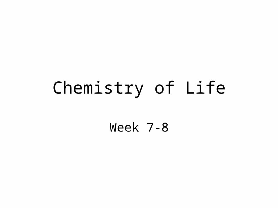
Overview• Living organisms and the
world they live in are subject to the basic laws of physics and chemistry.
• Biology is a multidisciplinary science, drawing on insights from other sciences.
• Life can be organized into a hierarchy of structural levels.
• At each successive level, additional emergent properties appear.

2.1 Matter consists of chemical elements in pure
form and in combinations called compounds.
• Organisms are composed of matter.– Matter is anything that takes up space and has
mass.– Matter is made up of elements.

2.1 Matter consists of chemical elements in pure
form and in combinations called compounds. • An element is a pure
substance that cannot be broken down into other substances by chemical reactions.
• There are 92 naturally occurring elements.
• Each element has a unique symbol, usually the first one or two letters of the name. Some of the symbols are derived from Latin or German names.

2.1 Matter consists of chemical elements in pure
form and in combinations called compounds. A compound is a pure substance consisting of two or more elements in a
fixed ratio.
• Table salt (sodium chloride or NaCl) is a compound with equal numbers of atoms of the elements chlorine and sodium.

Reflect on
• Blue green magnets
• White-red magnets

Essential Elements of Life• Essential elements
– Include carbon, hydrogen, oxygen, and nitrogen
– Make up 96% of living matter
• A few other elements– Make up the
remaining 4% of living matter

Trace elements
(a) Nitrogen deficiency (b) Iodine deficiency
• Are required by an organism in only minute quantities– But the absence of trace element can have
deadly effects
Figure 2.3

Radioactive Isotopes• Spontaneously give off particles and
energy– Alpha, beta, gamma radiation

Biological Uses for Radioactive IsotopesAPPLICATION Scientists use radioactive isotopes to label certain chemical substances, creating tracers that can be used to follow a metabolic process or locate the substance within an organism. In this example, radioactive tracers are being used to determine the effect of temperature on the rate at which cells make copies of their DNA.
DNA (old and new)
Ingredients includingRadioactive tracer (bright blue)
Human cells
Incubators
1 2 3
4 5 6
987
10°C 15°C 20°C
25°C 30°C 35°C
40°C 45°C 50°C
TECHNIQUE
2
1
The cells are placed in test tubes, their DNA is isolated, and unused ingredients are removed.
1 2 3 4 5 6 7 8 9
Ingredients for making DNA are added to human cells. One ingredient is labeled with 3H, a radioactive isotope of hydrogen. Nine dishes of cells are incubated at different temperatures. The cells make new DNA, incorporating the radioactive tracer with 3H.

Temperature (°C)
The frequency of flashes, which is recorded as counts per minute, is proportional to the amount of the radioactive tracer present, indicating the amount of new DNA. In this experiment, when the counts per minute are plotted against temperature, it is clear that temperature affects the rate of DNA synthesis—the most DNA was made at 35°C.
10 20 30 40 50
Optimumtemperaturefor DNAsynthesis
30
20
10
0Co
un
ts p
er
min
ute
(x 1
,00
0)
RESULTS
3
RESULTS
A solution called scintillation fluid is added to the test tubes and they are placed in a scintillation counter. As the 3H in the newly made DNA decays, it emits radiation that excites chemicals in the scintillation fluid, causing them to give off light. Flashes of light are recorded by the scintillation counter.
Figure 2.5

PET(positron-emission tomography)
Cancerous throat tissue

Figure 2.4 Tagging the Brain

Covalent BondsCovalent bonds can be
• Single—sharing one pair of electrons
• Double—sharing two pairs of electrons
• Triple—sharing three pairs of electrons C C
C H
N N

2.3 The formation and function of molecules depend on chemical bonding
between the atoms.
Electronegativity: the attractive force that an atomic nucleus exerts on electrons
Electronegativity depends on the number of positive charges (protons) and the distance between the nucleus and electrons.

Weak Chemical BondsHydrogen bonds: attraction
between the δ– end of one molecule and the δ+ hydrogen end of another molecule
Hydrogen bonds form between H and O and/or H and N.
Important with
water
DNA
Proteins

Van der Waals Interactions
• Van der Waals interactions– Occur when transiently positive and negative
regions of molecules attract each other

Structure and Function run from large scale body systems through molecules and atoms.
Structure and function are what Enzymes are all about
Morphine
Carbon
Hydrogen
Nitrogen
Sulfur
OxygenNaturalendorphin
(a) Structures of endorphin and morphine. The boxed portion of the endorphin molecule (left) binds to receptor molecules on target cells in the brain. The boxed portion of the morphine molecule is a close match.
(b) Binding to endorphin receptors. Endorphin receptors on the surface of a brain cell recognize and can bind to both endorphin and morphine.
Naturalendorphin
Endorphinreceptors
Morphine
Brain cellFigure 2.17

Why do medics pump wounded soldiers with morphine on the battlefield?

Concept 2.4: Chemical reactions make and break chemical bonds
• Chemical reactions– Convert reactants to products
Reactants Reaction Product
2 H2 O2 2 H2O
+
+

Life is the result of Chemical Reactions
• Photosynthesis– Is an example of a chemical reaction
Figure 2.18

Chemical Equilibrium
• Chemical equilibrium– Is reached when the forward and reverse
reaction rates are equal

Take out Water book
• Put notes and Hardy Weinberg lab in the center of the table
• Make sure there is a post it at the beginning of the lab
• If you want to go to a college talk and/or assembly, you MUST have an A or B and no Mi

Water: The Molecule That Supports All of Life
• Water is the biological medium here on Earth– All living organisms require
water more than any other substance
– Three-quarters of the Earth’s surface is submerged in water
– The abundance of water is the main reason the Earth is habitable

3.1: The polarity of water molecules results in hydrogen bonding
• The polarity of water molecules– Allows them to form hydrogen bonds with each other– Contributes to the various properties water exhibits
Hydrogenbonds
+
+
H
H+
+
–
–
– –

3.2: Four emergent properties of water contribute to Earth’s fitness for life
1. Cohesion
2. Moderation of Temperature
3. Insulation of bodies of water by floating ice
4. The solvent of life (universal solvent)

1. Cohesion• Cohesion – the hydrogen
bonds holding a substance together. (water – water)
• Adhesion – the hydrogen bonds holding one substance to another. (water – glass)
• Capillary Action – water transport in plants. Uses Cohesion and Adhesion– Transpiration
• Surface tension – measure of how difficult it is to stretch or break the surface of a liquid. – Water has a greater surface
tensions than most liquids

2. Moderation of Temperature
• Kinetic Energy – energy of motion
• Thermal Energy (heat) – total energy within a substance– Calorie – amount of
heat energy to heat 1g water by 1°C
– Kcal – 1000c• Temperature –
average kinetic energy per molecule (Celsius Scale)

2. Moderation of Temperature
• Specific heat – the amount of heat absorbed or loss for 1g of a substance to change its temperature by 1°C– Water has high
specific heat capacity compared to other substances
– 1 cal/g/°C

2. Moderation of Temperature
• Evaporation• Heat of vaporization –
the amount of heat 1g of a liquid must absorb to be converted to a gas
• Evaporative cooling – as a liquid evaporates the surface of the remaining liquid cools– This occurs because the
“hottest” molecules leave

3. Insulation of bodies of water by floating ice
Liquid water
Hydrogen bonds constantly break and re-form
Ice
Hydrogen bonds are stable
Hydrogen bond

3. Insulation of bodies of water by floating ice

4. Solvent of Life• Water is claimed to be the
universal solvent.– Solution – homogeneous
mixture of two or more substances in the same phase
– Solute – substance which is dissolved (in case of liquids, substance with the least amount
– Solvent – substance which is dissolving another
– Aqueous solution – solution involving water
– Hydration shell – pocket formed by water molecules in order to dissolve a substance

4. Solvent of life• Hydrophilic –
attracted to water– Can be dissolved– Unless molecule
is too large– Colloid –
stable suspension of fine molecules in a liquid. (blood, milk)
• Hydrophobic – repel water– Non-ionic, non-
polar, can’t form H-bonds

4. Solvent of Life
• Solute concentrations in aqueous solutions– Concentration =
g solute / ml solvent
– Molarity – moles solute / Liter solution

Acidic and Basic conditions affect living organisms
• Water can dissociate– Into hydronium ions and hydroxide ions
• H+ (hydrogen ion) is used to represent the hydronium ion
• Changes in the concentration of these ions– Can have a great affect on living organisms
• Only 1 in 554 mil pure water molecules will diss.
H
Hydroniumion (H3O+)
H
Hydroxideion (OH–)
H
H
H
H
H
H
+ –
+
Figure on p. 53 of water dissociating

Acids and Bases• Acids [H+]>[OH-]
• Bases [H+]<[OH-]• When acids dissolve in water, they
release hydrogen ions—H+ (protons).– H+ ions can attach to other molecules and
change their properties.
• Bases reduce H+ concentration byaccepting H+ ions and/or release OH- ions

Strong Acid
HCl is a strong acid—the dissolution is complete.
ClHHCl

Weak AcidOrganic acids have a carboxyl
group:
Weak acids: not all the acid molecules dissociate into ions.
HCOOHCOOH

Strong BaseNaOH is a strong base.
OHNaNaOH
The OH– absorbs H+ to form water.

Weak BasesWeak bases:
• Bicarbonate ion
• Ammonia
• Compounds with amino groups
323 COHHHCO
43 NHHNH
32 NHHNH
Acids, Bases, pHpH = negative log of the molar
concentration of H+ ions.
H+ concentration of pure water is 10–7 M, its pH = 7.
Lower pH numbers mean higher H+ concentration, or greater acidity.


Acids, Bases, buffers• Living organisms
maintain constant internal conditions, including pH.– Buffers help maintain
constant pH by accepting or donating H+ ions.
– They are kept in excess in systems
• A buffer is a weak acid and its corresponding base.
323 COHHHCO
•If you add 0.001 mole
of a stong acid to:
•1L of pure water
the pH will go from
7 2.0
•1L of blood the pH
will only decrease
from 7.4 7.3

Figure 2.17 Buffers Minimize Changes in pH

2.4 What Properties of Water Make It So Important in
Biology?Buffers illustrate the law of mass action: addition of reactant on one side of a reversible equation drives the system in the direction that uses up that compound.

2.4 What Properties of Water Make It So Important in
Biology?Life’s chemistry began in water.
Water and other chemicals may have come to Earth on comets.
Water was an essential condition for life to evolve.

FRQ
• The unique properties (characteristics) of water make life possible on Earth. Select three properties of water and:
a) for each property, identify and define the
property and explain it in terms of the physical/chemical nature of water.
b) for each property, describe one example of how the property affects the functioning of living organisms.

Build a carbohydrate
• Carbon (black)=4 bonds
• Hydrogen (white)=1 bond
• Oxygen (red)= 2bonds

Pick up 2 FRQ
• Look at the FRQs from 2 sample students
• Write advice to each student
• Rewrite your FRQs—why did you lose points?

Carbon
• Carbon atoms can form diverse molecules by bonding to four other atoms– Carbon has amazing ability to form molecules
because:• It has 4 valence electrons• It can form up to 4 covalent bonds• These can be single, double, or triple cov. Bonds• It can form large molecules.• These molecules can be chains, ring-shaped, or branched
– Isomers – are molecules that have the same molecular formula, but different in their arrangement of these atoms.
• This can result in different molecules with very different activities.

CarbohydratesCells use
glucose (monosaccharide) as an energy source.
Exists as a straight chain or ring form. Ring is more common—it is more stable.

Glucose=C6H12O6Carbon=black, Hydrogen=White, Oxygen=Red
• Carbohydrates: molecules in which carbon is flanked by hydrogen and hydroxyl groups.
H—C—OH
• Main Functions
– Energy source
– Carbon skeletons for many other molecules

Carbohydrates
CH2O

Quick on Carbon4.3 Characteristic chemical groups help
control how biological molecules function
FUNCTIONALGROUP
STRUCTURE
(may be written HO )
HYDROXYL CARBONYL CARBOXYL
OH
In a hydroxyl group (—OH), a hydrogen atom is bonded to an oxygen atom, which in turn is bonded to the carbon skeleton of the organic molecule. (Do not confuse this functional group with the hydroxide ion, OH–.)
When an oxygen atom is double-bonded to a carbon atom that is also bonded to a hydroxyl group, the entire assembly of atoms is called a carboxyl group (—COOH).
C
O O
C
OH
Figure 4.10
The carbonyl group ( CO) consists of a carbon atom joined to an oxygen atom by a double bond.

Some important functional groups of organic compounds
Acetic acid, which gives vinegar
its sour tatste
NAME OF
COMPOUNDS
Alcohols (their specific
names usually end in -ol)
Ketones if the carbonyl group is
within a carbon skeleton
Aldehydes if the carbonyl
group is at the end of the
carbon skeleton
Carboxylic acids, or organic
acids
EXAMPLE
Propanal, an aldehyde
Acetone, the simplest ketone
Ethanol, the alcohol
present in alcoholic
beverages
H
H
H
H H
C C OH
H
H
H
HH
H
H
C C H
C
C C
C C C
O
H OH
O
H
H
H H
H O
H
Figure 4.10

The amino group (—NH2) consists of a nitrogen atom bonded to two hydrogen atoms and to the carbon skeleton.
AMINO SULFHYDRYL PHOSPHATE
(may be written HS )
The sulfhydryl group consists of a sulfur atom bonded to an atom of hydrogen; resembles a hydroxyl group in shape.
In a phosphate group, a phosphorus atom is bonded to four oxygen atoms; one oxygen is bonded to the carbon skeleton; two oxygens carry negative charges; abbreviated P . The phosphate group (—OPO3
2–) is an ionized form of a phosphoric acid group (—OPO3H2; note the two hydrogens).
N
H
H
SH
O P
O
OH
OH
Figure 4.10
Quick on Carbon4.3 Characteristic chemical groups help
control how biological molecules function

Some important functional groups of organic compounds
Because it also has a carboxyl group, glycine is both an amine and a carboxylic acid; compounds with both groups are called amino acids.
Glycine EthanethiolGlycerol phosphate
O
C
HO
C
HH
N
H
H
H
C C SH
H
H H
H
H
OH
C C C O P O
OHHH
OH OH
Figure 4.10
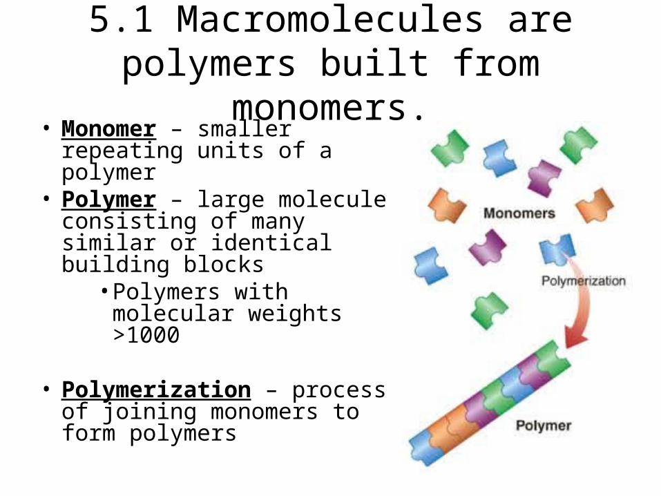
5.1 Macromolecules are polymers built from monomers.
• Monomer – smaller repeating units of a polymer
• Polymer – large molecule consisting of many similar or identical building blocks
• Polymers with molecular weights >1000
• Polymerization – process of joining monomers to form polymers

The synthesis and breakdown of polymers
• Dehydration synthesis (dehydration reaction) – synthesis reaction forming a byproduct of water
• Hydrolysis – degradation of a molecule using water to break down bonds– These processes
are often aided by enzymes

Dehydration Synthesis

Demonstrate
• Dehydration
• All the names for a polymer of glucose + glucose

The Diversity of Polymers
• Each cell has thousands of different kinds of macromolecules.– The inherent different between
human siblings reflect the variations in polymers:
• Especially DNA and proteins
• There are four major classes of biological macromolecules– Carbohydrates– Lipids– Proteins– Nucleic Acids

Carbohydrates• Monosaccharides:
simple sugars
• Disaccharides: two simple sugars linked by covalent bonds
• Oligosaccharides: three to 20 monosaccharides
• Polysaccharides: hundreds or thousands of monosaccharides—starch, glycogen, cellulose

Carbohydrates
• Monosaccharides have different numbers of carbons.– Trioses: three
carbons– structural isomers - glyceraldehyde
– Hexoses: six carbons—structural isomers
– Pentoses: five carbons

Carbohydrates• Monosaccharides bind together in
condensation reactions to form glycosidic linkages.
• Glycosidic linkages can be α or β.

Beta – glycosidic linkage

Alpha – glycosidic linkage

Carbohydrates• Oligosaccharides
may include other functional groups.
• Often covalently bonded to proteins and lipids on cell surfaces and act as recognition signals.
• ABO blood groups

Carbohydrates• Starch: storage of
glucose in plants – 1-4 glycosydic linkages between
alpha glucose
• Cellulose: very stable, good for structural components (cell walls of plants– 1-4 glycosydic linkages between
beta glucose
• Glycogen: storage of glucose in animals– 1-4 glycosydic linkages between
alpha glucose• with branching

Chitin
• Chitin, another important structural polysaccharide– Is found in the exoskeleton of arthropods– Can be used as surgical thread
(a) The structure of the chitin monomer.
O
CH2OH
OHH
H OH
H
NH
CCH3
O
H
H
(b) Chitin forms the exoskeleton of arthropods. This cicada is molting, shedding its old exoskeleton and emergingin adult form.
(c) Chitin is used to make a strong and flexible surgical
thread that decomposes after the wound or incision heals.
OH
Figure 5.10 A–C
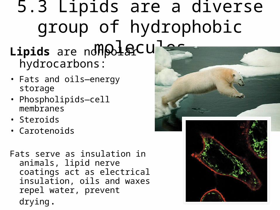
5.3 Lipids are a diverse group of hydrophobic molecules
Lipids are nonpolar hydrocarbons:
• Fats and oils—energy storage• Phospholipids—cell membranes• Steroids• Carotenoids
Fats serve as insulation in animals, lipid nerve coatings act as electrical insulation, oils and waxes repel water, prevent drying.

LipidsFats and oils are
triglycerides—simple lipids—made of three fatty acids and 1 glycerol.
Glycerol: 3 —OH groups—an alcohol
Fatty acid: nonpolar hydrocarbon with a polar carboxyl group—carboxyl bonds with hydroxyls of glycerol in an ester linkage.

Lipids• Saturated fatty acids:
no double bonds between carbons—it is saturated with hydrogen atoms.
• Unsaturated fatty acids: some double bonds in carbon chain.– monounsaturated: one
double bond
– polyunsaturated: more than one
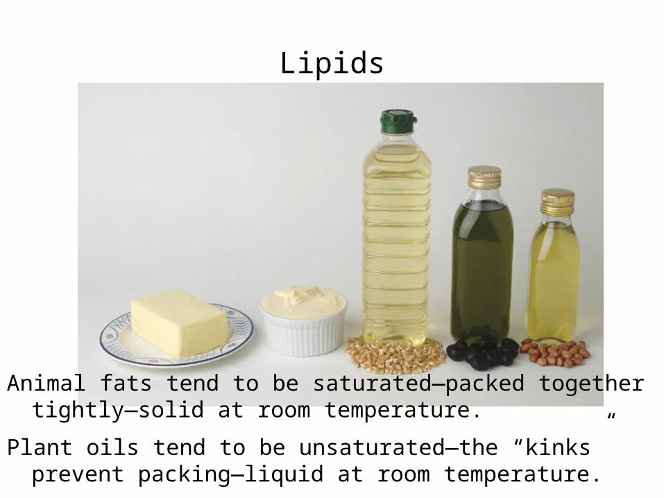
Lipids
Animal fats tend to be saturated—packed together tightly—solid at room temperature.
Plant oils tend to be unsaturated—the “kinks” prevent packing—liquid at room temperature.
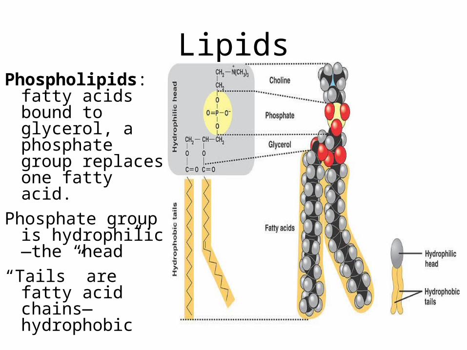
LipidsPhospholipids:
fatty acids bound to glycerol, a phosphate group replaces one fatty acid.
Phosphate group is hydrophilic—the “head”
“Tails” are fatty acid chains—hydrophobic

Lipid (phospholipid bilayer)

Lipid (Steroids)• Steroids
– Are lipids characterized by a carbon skeleton consisting of four fused rings
– Many hormones, including vertebrate sex hormones, are steroids produced from cholesterol
– Steroids play a role in regulating cell activities

Lipids (carotenoids)
Carotenoids: light-absorbing pigments

5.4 Proteins have many structures, resulting in a wide range of functions
Functions of proteins:
• Structural support• Protection• Transport• Catalysis• Defense• Regulation• Movement


Proteins• Proteins are made
from 20 different amino acids (monomeric units)
• Polypeptide chain: single, unbranched chain of amino acids– The chains are folded
into specific three dimensional shapes.
– Proteins can consist of more than one type of polypeptide chain.

Protein (polypeptide)
The composition of a protein: relative amounts of each amino acid present
The sequence of amino acids in the chain determines the protein structure and function.

Proteins• Amino acids have
carboxyl and amino groups—they function as both acid and base.
– The α carbon atom is asymmetrical.
– Amino acids exist in two isomeric forms:
• D-amino acids (dextro, “right”)
• L-amino acids (levo, “left”)—this form is found in organisms
Functional Group

Proteins (amino acids are grouped by characteristics)
O
O–
H
H3N+ C C
O
O–
H
CH3
H3N+ C
H
C
O
O–
CH3 CH3
CH3
C C
O
O–
H
H3N+
CH
CH3
CH2
C
H
H3N+
CH3
CH3
CH2
CH
C
H
H3N+ C
CH3
CH2
CH2
CH3N+
H
C
O
O–
CH2
CH3N+
H
C
O
O–
CH2
NH
H
C
O
O–
H3N+ C
CH2
H2C
H2N C
CH2
H
C
NonpolarGlycine (Gly) Alanine (Ala) Valine (Val) Leucine (Leu) Isoleucine (Ile)
Methionine (Met) Phenylalanine (Phe)
C
O
O–
Tryptophan (Trp) Proline (Pro)
H3C
Figure 5.17
S
O
O–

Proteins (amino acids are grouped by characteristics)
O–
OH
CH2
C C
H
H3N+
O
O–
H3N+
OH CH3
CH
C C
HO–
O
SH
CH2
C
H
H3N+ C
O
O–
H3N+ C C
CH2
OH
H H H
H3N+
NH2
CH2
OC
C C
O
O–
NH2 O
C
CH2
CH2
C CH3N+
O
O–
O
Polar
Electricallycharged
–O O
C
CH2
C CH3N+
H
O
O–
O– O
C
CH2
C CH3N+
H
O
O–
CH2
CH2
CH2
CH2
NH3+
CH2
C CH3N+
H
O
O–
NH2
C NH2+
CH2
CH2
CH2
C CH3N+
H
O
O–
CH2
NH+
NHCH2
C CH3N+
H
O
O–
Serine (Ser) Threonine (Thr)Cysteine
(Cys)Tyrosine
(Tyr)Asparagine
(Asn)Glutamine
(Gln)
Acidic Basic
Aspartic acid (Asp)
Glutamic acid (Glu)
Lysine (Lys) Arginine (Arg) Histidine (His)

Proteins• Amino acids
bond together covalently by peptide bonds to form the polypeptide chain.– Dehydration
synthesis

Proteins
A polypeptide chain is like a sentence:
• The “capital letter” is the amino group of the first amino acid—the N terminus.
• The “period” is the carboxyl group of the last amino acid—the C terminus.

ProteinsThe primary structure of a
protein is the sequence of amino acids.
The sequence determines secondary and tertiary structure—how the protein is folded.
The number of different proteins that can be made from 20 amino acids is enormous!
• Protein structure
–Primary
–Secondary
–Tertiary
–Quartinary

Proteins (primary structure)

Proteins (secondary structure)
Secondary structure:
• α helix—right-handed coil resulting from hydrogen bonding; common in fibrous structural proteins
• β pleated sheet—two or more polypeptide chains are aligned

Proteins (tertiary structure)Tertiary structure: Bending and folding results in a
macromolecule with specific three-dimensional shape.
The outer surfaces present functional groups that can interact with other molecules.

Proteins (tertiary structure)Tertiary structure
is determined by interactions of R-groups:
• Disulfide bonds• Aggregation of
hydrophobic side chains
• van der Waals forces
• Ionic bonds• Hydrogen
bonds

Proteins (Quartinary structure)• Quaternary
structure results from the interaction of subunits by:– hydrophobic
interactions
– van der Waals forces
– ionic bonds
– hydrogen bonds.
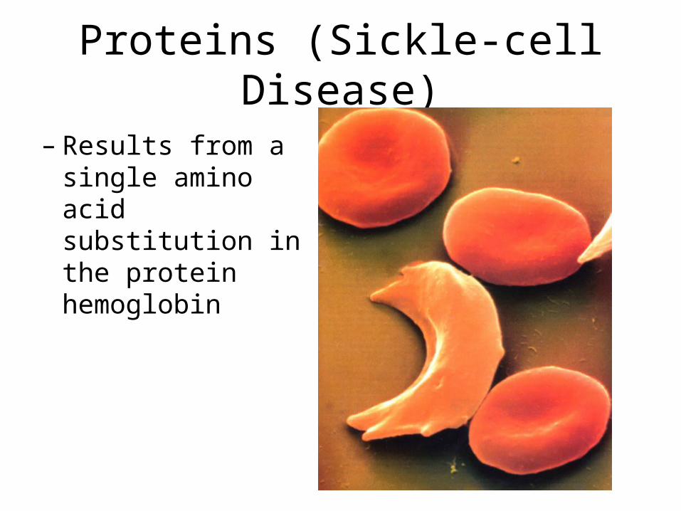
Proteins (Sickle-cell Disease)
– Results from a single amino acid substitution in the protein hemoglobin

Hemoglobin structure and sickle-cell disease
Fibers of abnormalhemoglobin deform cell into sickle shape.
Primary structure
Secondaryand tertiarystructures
Quaternary structure
Function
Red bloodcell shape
Hemoglobin A
Molecules donot associatewith oneanother, eachcarries oxygen.
Normal cells arefull of individualhemoglobinmolecules, eachcarrying oxygen
10 m 10 m
Primary structure
Secondaryand tertiarystructures
Quaternary structure
Function
Red bloodcell shape
Hemoglobin S
Molecules interact with one another tocrystallize into a fiber, capacity to carry oxygen is greatly reduced.
subunit subunit
1 2 3 4 5 6 7 3 4 5 6 721
Normal hemoglobin Sickle-cell hemoglobin. . .. . .
Figure 5.21
Exposed hydrophobic
region
Val ThrHis Leu ProGlulGlulGlu Val His Leu Thr ProValValGlu

Enzyme-Substrate Complex

Proteins (Denaturing)
• Conditions that affect secondary and tertiary structure:
• High temperature• pH changes• High concentrations of
polar molecules• Denaturation: loss of 3-
dimensional structure and thus function of the protein

Proteins (folding)• Proteins can sometimes fold incorrectly and bind to the wrong ligands.
• Chaperonins are proteins that help prevent this.
Hollowcylinder
Cap
Chaperonin(fully assembled)
Steps of ChaperoninAction: An unfolded poly- peptide enters the cylinder from one end.
The cap attaches, causing the cylinder to change shape insuch a way that it creates a hydrophilic environment for the folding of the polypeptide.
The cap comesoff, and the properlyfolded protein is released.
Correctlyfoldedprotein
Polypeptide
2
1
3
Figure 5.23

5.5 Nucleic acids store and transmit hereditary information
Nucleic acids: DNA—(deoxyribonucleic acid) and RNA—(ribonucleic acid)
Polymers (polynucleotides) — made of the monomeric units are nucleotides.
Nucleotides consist of a pentose sugar, a phosphate group, and a nitrogen-containing base.

5.5 Nucleic acids store and transmit hereditary information
DNA—deoxyribose
RNA—ribose
5.5 Nucleic acids store and transmit hereditary information
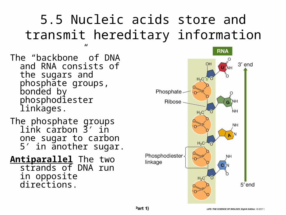
The “backbone” of DNA and RNA consists of the sugars and phosphate groups, bonded by phosphodiester linkages.
The phosphate groups link carbon 3′ in one sugar to carbon 5′ in another sugar.
Antiparallel The two strands of DNA run in opposite directions.

5.5 Nucleic acids store and transmit hereditary information
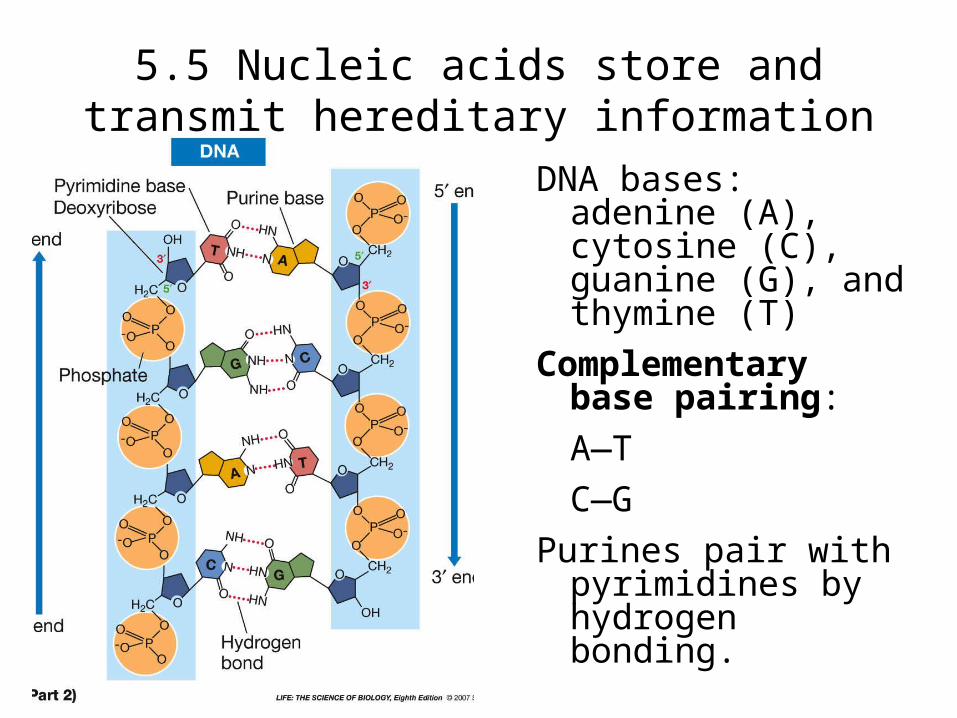
DNA bases: adenine (A), cytosine (C), guanine (G), and thymine (T)
Complementary base pairing:
A—T
C—G
Purines pair with pyrimidines by hydrogen bonding.

• A particular small polypeptide is nine amino acids long. Using three different enzymes to hydrolyze the polypeptide at various sites, we obtain the following five fragments (N denotes the amino end of the chain):
Ala-Leu-Asp-Tyr-Val-LeuTyr-Val-LeuN-Gly-Pro-LeuAsp-Tyr-Val-LeuN-Gly-Pro-Leu-Ala-LeuDetermine the primary structure of this polypeptide.– N-Gly-Pro-Leu-Ala-Leu-Asp-Tyr-Val-Leu– Asp-Tyr-Val-Leu-Gly-Pro-Leu-Ala-Leu– N-Gly-Pro-Leu-Ala-Leu-Ala-Leu-Asp-Tyr-Val-Leu– N-Gly-Pro-Leu-Asp-Tyr-Val-Leu-Tyr-Val-Leu

• (a) You are studying a cellular enzyme involved in breaking down fatty acids for energy. Looking at theR groups of the amino acids in the following figures, what amino acids would you predict to occur in the parts of the enzyme that interact with the fatty acids? *– non-polar– polar– electrically charged– polar and electrically charged– all of these

The 20 Amino Acids of Proteins

The 20 Amino Acids of Proteins (cont.)

• (b) You are studying a cellular enzyme involved in breaking down fatty acids for energy. Where would you predict to find the amino acids in the parts of the enzyme that interact with the fatty acids?– On the exterior surface of the enzyme– Sequestered in a pocket in the interior of the
enzyme– Randomly dispersed throughout the enzyme

• The R group or side chain of the amino acid serine is –CH2 –OH. The R group or side chain of the amino acid alanine is –CH3. Where would you expect to find these amino acids in globular protein in aqueous solution?– Serine would be in the interior, and alanine would be
on the exterior of the globular protein.– Alanine would be in the interior, and serine would be
on the exterior of the globular protein.– Both serine and alanine would be in the interior of the
globular protein.– Both serine and alanine would be on the exterior of
the globular protein.– Both serine and alanine would be in the interior and
on the exterior of the globular protein.

• (a) The sequence of amino acids of the enzyme lysozyme is known. Following is a list of amino acids and the number of each in the lysozyme molecule. Based on this list and the structures of the amino acids how many S-S bonds are possible in lysozyme?– 0– 2– 4– 6– 8

Amino Acids in the Lysozyme MoleculeType Number in
LysozymeType Number in
Lysozyme
Alanine 12 Leucine 8
Arginine 11 Lysine 6
Asparagine 13 Methionine 2
Aspartic acid 8 Phenylalanine
3
Cysteine 8 Proline 2
Glutamic acid
2 Serine 10
Glutamine 3 Threonine 7
Glycine 12 Tryptophan 6
Histidine 1 Tyrosine 3
Isoleucine 6 Valine 6

The 20 Amino Acids of Proteins

The 20 Amino Acids of Proteins (cont.)

• (b) The sequence of amino acids of the enzyme lysozyme is known. Following is a list of amino acids and the number of each in the lysozyme molecule. Based on this list and the structures of the amino acids is the net charge on lysozyme positive or negative?– positive– negative

Amino Acids in the Lysozyme MoleculeType Number in
LysozymeType Number in
Lysozyme
Alanine 12 Leucine 8
Arginine 11 Lysine 6
Asparagine 13 Methionine 2
Aspartic acid 8 Phenylalanine
3
Cysteine 8 Proline 2
Glutamic acid
2 Serine 10
Glutamine 3 Threonine 7
Glycine 12 Tryptophan 6
Histidine 1 Tyrosine 3
Isoleucine 6 Valine 6

The 20 Amino Acids of Proteins

The 20 Amino Acids of Proteins (cont.)

• Polymers of glucose units are used as temporary food storage in both plant and animal cells. Glucose units are connected to one another by 1, 4-linkages to make a linear polymer and by 1, 6-linkages to make branch points.

• (cont.) Polysaccharides of glucose unitsvary in size. The three most commonly encountered are:Type of
StarchCell Type Polymer
SizeAverage Number of 1,4-Bonds Between Branches
Amylopectin Plant 100,000,000 24 to 30
Amylos Plant 500,000 Linear
Glycogen Animal 3,000,000 8 to 12

• (cont.) When each polymer bond is made, a water molecule is released and becomes part of the cell water. How many water molecules were released during formation of each of the Glycogen?– 1,000,000
– 2,000,000
– 2,666,666
– 3,000,000
– 3,300,000