1
Elbow Dysplasia: Fragmented Coronoid Process !"#$%"& ()%& *+,+"-.%"/ 01"2+"/3 4.#5 6$7 89:;<=>;:8?? +@%-A7 @2)B"-+.C-D2@%-A5#)@ The elbow joint is comprised of three bones, the humerus in the upper forelimb, the radius and ulna in the lower forelimb (Fig 1). Elbow dysplasia is defined as any abnormality in the development of the elbow joint. Most theories involve abnormal or disparate growth between the radius and ulna or between the bones and surrounding muscles creating stress in certain regions of the joint. Fragmented coronoid process (FCP) is a condition that is a sequela to elbow dysplasia. The coronoid process is a semicircular cartilage surface along the ulna. The ulna and radius each support a portion of the weight and cartilage surface of the humerus. It is theorized that there is an abnormal force on the medial region of the coronoid process that results in damage to the cartilage and underlying bone. The result is weakened or complete fracture of the medial process (Fig 3). Diagnosis can be difficult because often plain radiographs of the area do not often delineate an obvious fracture. So one looks for secondary changes such as osteophyte (bone spur) formation or increased sclerosis (bone density) in the region of the coronoid (Fig 2). CT scan and MRI imaging give more detailed information and can be used in subtle cases. Other conditions that can occur as part of elbow dysplasia include ununited anconeal process (Fig 3) and OCD lesions Treatment: Current treatment involves the removal of the fragmented or fissured coronoid (subtotal coronoidectomy). Removal of the weakened bone and cartilage will resolve the lameness in the majority of dogs. However most dogs will still develop some degree of arthritic changes in the elbow in coming years. Newer and investigational procedures are being studied to evaluate altering the different weight bearing forces between the radius and ulna in a hope to spare future damage to the joint. Post-op Care: After subtotal coronoidectomy the dog should be limited in their activity for the first 4 weeks to permit healing of the joint. This means no running or jumping is permitted. Gradual increase in leash walks and the use of stretching exercises of the joint. Use of anti-inflammatory medication will also help speed recovery. Figure 1 Figure 2 Figure 3

Elbow Dysplasia: Fragmented Coronoid Process · Elbow Dysplasia: Fragmented Coronoid Process !"#$%"&’()%&’*+,+"-.%"/’01"2+"/3’4.#5’ 6$7’89:;;:8??’’’+@%-A7’@2)B"-+.C-D2@%-A5#)@

Embed Size (px)

Citation preview

Elbow Dysplasia: Fragmented Coronoid Process !"#$%"&'()%&'*+,+"-.%"/'01"2+"/3'4.#5'

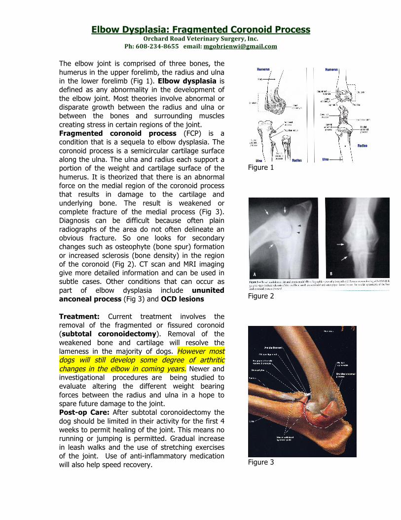
6$7'89:;<=>;:8??'''+@%-A7'@2)B"-+.C-D2@%-A5#)@ The elbow joint is comprised of three bones, the humerus in the upper forelimb, the radius and ulna in the lower forelimb (Fig 1). Elbow dysplasia is defined as any abnormality in the development of the elbow joint. Most theories involve abnormal or disparate growth between the radius and ulna or between the bones and surrounding muscles creating stress in certain regions of the joint. Fragmented coronoid process (FCP) is a condition that is a sequela to elbow dysplasia. The coronoid process is a semicircular cartilage surface along the ulna. The ulna and radius each support a portion of the weight and cartilage surface of the humerus. It is theorized that there is an abnormal force on the medial region of the coronoid process that results in damage to the cartilage and underlying bone. The result is weakened or complete fracture of the medial process (Fig 3). Diagnosis can be difficult because often plain radiographs of the area do not often delineate an obvious fracture. So one looks for secondary changes such as osteophyte (bone spur) formation or increased sclerosis (bone density) in the region of the coronoid (Fig 2). CT scan and MRI imaging give more detailed information and can be used in subtle cases. Other conditions that can occur as part of elbow dysplasia include ununited anconeal process (Fig 3) and OCD lesions Treatment: Current treatment involves the removal of the fragmented or fissured coronoid (subtotal coronoidectomy). Removal of the weakened bone and cartilage will resolve the lameness in the majority of dogs. However most dogs will still develop some degree of arthritic changes in the elbow in coming years. Newer and investigational procedures are being studied to evaluate altering the different weight bearing forces between the radius and ulna in a hope to spare future damage to the joint. Post-op Care: After subtotal coronoidectomy the dog should be limited in their activity for the first 4 weeks to permit healing of the joint. This means no running or jumping is permitted. Gradual increase in leash walks and the use of stretching exercises of the joint. Use of anti-inflammatory medication will also help speed recovery.

Figure 1

Figure 2

Figure 3