Embed Size (px)
Citation preview

HERNiNDEZ-ALFARO, ESCIJDER, AND MARCO
6. Ray DA: Lingt~al thyroid. Arch Surg 37:316, 1938 7. Wama Kulasuriya KAAS. Hcnth KB: Investigating a lingual
thyroid. lnt J Oral Maxillofac Surg 21:227, 1992 8. Declerck S. Casselman JW. Depondt M. et al: Lingual thyroid
imaging. J Beige Radio1 76:24 1. 1993 9. Guneri A. Ceryan K. lgci E. et al: Lingual thyroid: The diagnostic
value of magnetic resonance imaging, J Luyngol Otol 105:493. 1991 10. Vainktaris E. Semergidis T. Christopoulou P. et al: Lingual
thyroid: A new surgical approach-A case report. J Crdniomax- illofac Surg 22:307. 1994
227
11. James DW. Anthony PS. Claude D. et al: Evaluation and management of lingual thyroid. Ann Otol Rhino1 Lu-yngol lO5:312 1996
12. Kamat MR. Kulkarni JN. Desai PB, et al: Lingual thyroid: A review of 12 cases. Br J Surg 66:537. 1979
13. Paludetti G. Galli J. Almadori G, et al: Ectopic thyroid gland. Acta Otorhinolaryngol ltal 11:117. 1991
1-r. Bishan S. Atiyeh AA, Fadi FH. et al: Lingual thyroid: Tongue splitting incision for trdnsoral excision. J Luygol Otol 109:520. 1995
J Oral Maxlllofac Surg 58 227-232.2000
Joint Formation Between an Osteochondroma of the Coronoid Process and the Zygomatic Arch (Jacob Disease): Report of Case and Review of Literature
Federico Herndndez-Alfaro, MD, DDS, FEBOMFS, * &car Escuder, MD, f and Vicente Marco, MD*
Enlargement of the coronoid process of the mandible was first described by Langenbeck in 1853,’ and joint formation between the coronoid process and the zygoma was first described by Jacob in 1899.’ Subse- quently, enlargement of the coronoid process has been sporadically reported in the literature.‘,+‘3
This condition can be unilateral or bilateral. The latter is more frequent in young men, resembles the normal coronoid in shape, and is self-limited in growth.’ ’ The unilateral form usually grows progres- sively to form a mushroom-shaped enlargement of the process. Besides, in Jacob disease, a new joint forms between the coronoid process and the zygoma. The most consistent clinical feature of this condition is reduction of mouth opening. Treatment consists of coronoid resection through an intraoral or extraoral approach. Histologically, most of the lesions show a bony growth capped by cartilage. Numerous factors
‘Professor, Department of Oral and Maxillofacial Surgery, Univer-
sitat lntemacional de Catalunya; Chief, Unit of Maxillofacial Deformi-
ties, Hospital General de Catalunya and Teknon Medical Center,
Barcelona, Spain.
tFormer Chief Resident, Department of Oral and Maxillofacial Surgery, Bellvitge University Hospital, Barcelona, Spain.
#Chief, Department of Pathology, Hospital Generdl de Catalunya
and Teknon Medical Center, Barcelona, Spain.
Address correspondence and reprint requests to Dr Alfaro;
Cirurgia Maxillofacial, Centre Medic Teknon. Marques de Vil-
ldlonga. 12. 08017 Barcelona. Spain; e-mail: [email protected]
b 2ooO Amerlcon Assouat~on of Oral and Maxlllofoctcl Surgeons
0278.2391/00/58020017$3 00/O
have been suggested in the pathogenesis of coronoid process enlargement,3,~,“,a’n,‘.1 but nothing has been suggested regarding the pathogenesis of the new joint.
Because of the history, which includes an insidious clinical onset, this condition has often been over- looked and treated initially as a temporomandibular joint (TMn disorder. We report a case of Jacob disease that illustrates the importance of a proper differential diagnosis when faced with a patient having restricted mouth opening.
Review of the literature
Only 8 reported cases of Jacob disease were found (Table 1). There was one in the English literature,15 5 in the French literature (including the original descrip tion),‘~‘G’Y 1 in the Belgian literature,“’ and 1 in the Czeck literature.*0 Of the 8 previous cases, there were 7 in males and 1 in a female, with a mean age of 28 years and a range of 13 to 62 years. Three of the cases were bilateral. All of them except the first were treated surgically by unilateral or bilateral coronoidec- tomy. One caseI was approached extraorally (no more details given); the others were treated by an intraoral approach. Only 1 case was treated under regional anesthesia. I6
Report of Case
A 22-year-old man was referred to our department with a history of limitation of mouth opening that began 2 years before and was initially diagnosed by a dentist as a left TMJ disorder. The patient underwent 6 months of bite appliance
228 JACOB DISEASE: CASE REPORT AND REVIEW OF LITERATURE
Case Author Year (yr) Treatment Comments
Jacob2 1899 62 Hallam15 1947 18 Ginestet et al’s 1956 19 Chemin et alI6 1958 20 Van de Vijver’* 1962 18 Dechaume et al” 1964 13 Goudot et aI” 1989 45 Rames and OrbanZo 1990 36 Her&ndez-Alfaro et al 1996 22
therapy, and subsequently 5 arthroscopic procedures were performed without any improvement in mouth opening. FlnalIy the patient was referred with a diagnosis of left TMJ ankylosis for open TMJ surgery.
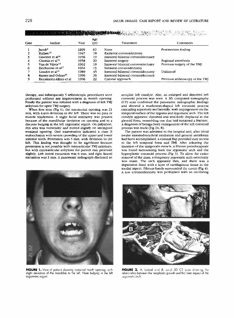
When fust seen (Fig 1). the interincisal opening was 21 mm, with 4-mm deviation to the left There was no pain or muscle tenderness. A slight facial assymetry was present because of the mandibular deviation on opening and to a discrete bulging in the left zygomatic region. On palpation, this area was nontender and moved slightly on attempted maximal opening. Oral examination indicated a class 11 malocclusion with severe crowding of the upper and lower anterior teeth. Protrusion was 5 mm, with deviation to the left. This finding was thought to be significant because protrusion is not possible with intra-articular TMJ ankylosis, but with extra-articular ankylosis the patient may protrude slightly. Left lateral excursion was 6 mm, and right lateral excursion was 3 mm. A panoramic radiograph disclosed an
None Extraoral coronoidectomy Intraoral bilateral coronoidectomy Intraoral surgery Intraoral bilateral coronoidectomy Intraoral coronoidectomy Intraoral bilateral coronoidectomy Intraoral bilateral coronoidectomy Coronal approach
Postmortem finding
Regional anesthesia Previous surgery of the TMJ
Unilateral
Previous arthroscopy of the TMJ
atrophic left condyle. Also, an enlarged and distorted left coronoid process was seen. A 3D computed tomography (CT) scan confirmed the panoramic radiographic findings and showed a mushroom-shaped left coronoid process extending superiorly and laterally, with impingement on the temporal surface of the zygoma and zygomatic arch. The left condyle appeared distorted and anteriorly displaced in the glenoid fossa, resembling one that had sustained a fracture. A diagnosis of benign bony enlargement of the left coronoid process was made <Fig 2A, B).
The patient was admitted to the hospital and, after blind awake nasoendotracheal intubation and general anesthesia had been accomplished, a coronal flap provided easy access to the left temporal fossa and TMJ. After releasing the insertion of the temporalis muscle, a fibrous pseudocapsule was found surrounding both the zygomatic arch and the hyperplastic coronoid process (Fig 3). To allow for easier removal of the mass, a temporary zygomatic arch ostectomy was made. The arch appeared thin, and there was a depression lined with a layer of cartilaginous tissue in the medial aspect. Fibrous bands surrounded the cavity (Fig 4). A low coronoidectomy was performed wirh an oscillating
FIGURE 1. View of patient showin 3,
restricted mouth opening, with slight deviation of the mandible to t e left. Note bulging in the leh zygomotic region.
FIGURE 2. A, lateral and 6, oxio 30 CT scan showing the relationship between the neoplastic growth and the inner aspect of the zygomatic arch.

HERNANDEZ-ALFARO, ESCIIDER. AND MARCO 229
FIGURE after the c ~ng distorl fibromuscl mass lorrc
Int nOI
of lis
root )erollve
opi srooch the arch o sue coverer
sb nd
‘9
‘If?,“+
IOW-
the the
saw. The process was then removed after releasing some fibrous insertions. This immediately allowed a SZ-mm inter- incisal opening. The zygomatic arch was repositioned with 2 miniplates after removal of the librocartilaginous tissue and smoothing of the remaining irregular bony surface.
The TMJ was then investigated through an incision made in the capsule. The disc appeared distorted and perforated. Thus, a discectomy was done followed by an interpositional
pedicled flap of temporalis muscle and fascia (Fig 5). A drain was inserted, and the Hap was closed. Maximal interincisal opening was maintained with a rubber wedge left in place for 2-i hours. Recovery after surgeq WJS uneventhll, and the patient was discharged 43 hours later. Thereafter, jaw stretching exercises maintained a stable interincisal opening of 47 mm 6 months postoperatively (Fig 6).
The coronoid specimen resembled a mandibular condyle

230 JACOB DISEASE: CASE REPORT AND REVIEW OF LITERATLJRE
FIGURE 5. View of the left TMJ. Note the distorted fibrous disc (a) and temporalis muscle and fascia flop for reconstruction (b).
with fibromuscular insertions (Fig 7). Microscopically, the sections showed fibrous, cartilaginous, and bony elements irregularly arranged. A diagnosis of osteochondroma was made (Figs 8, 9). The cartilage lining the cavity of the zygomatic arch was disorganized and uncalcihed. Synovial tissue was attached to both the hyperplastic coronoid process and the zygoma.
Discussion
Symptomatic enlargement of the coronoid process is a rare condition. Since the first reported case by
FIGURE 6. Normal opening 6 months after surgery
FIGURE 7. View of resected mushroom-shaped coronoid specimen.
Langenbeck,’ much confusion has existed regarding the nature and pathogenesis of this condition. Although there are not enough epidemiologic data regarding the prevalence of this process, asymptomatic cases are probably more frequent than previously thought.” Honig et al*’ examined the panoramic radiographs of a randomly selected sample of 2,000 patients and found a prevalence of 0.5%. A much lower prevalence of the Jacob disease should be expected.
Some have advocated trauma as a possible causative event in the development of the hyperplasia. The influence of functional alterations in the shape and structure of the coronoid process has been proposed by others.7*9 Isberg et al6 pointed out that hyperactiv- ity of the temporalis muscle, which is often present together with internal derangements of the TMJ, is likely to promote coronoid hyperplasia through a reactive process in response to pull of the tendon. Nothing has been suggested regarding the pathogen- esis of the new joint, and it is still a subject of discussion whether the Jacob disease is a particular variety of coronoid process hyperplasia or a com- pletely different clinical entity.
This case was initially diagnosed as TMJ dysfunction and managed as such. However, several panoramic radiographs obtained at the onset of symptoms al- ready showed coronoid enlargement. Therefore, in this case, TMJ dysfunction was most probably second- ary to the surgical manipulation involved in the multiple arthroscopic procedures and to lack of func- tion for several years.

HERNANDEZALFARO, ESCUDER, AND MARCO 231
FIGURE 8. low-power photomi- crograph of the specimen [hema- toxylln and eosn, orlglnol magnifl- cation x 16)
Diagnosis of this entity can be made easily from a panoramic radiograph and careful clinical examina- tion. Although a Waters’ radiograph is very useful in showing the coronoid hyperplasia, and its relation
FIGURE 9. High-power microphotographs (hemotoxylin and eosin, original magnification ~401 showing detail of Fig 8. Note the hyollne cartilage and the underlying bone with irregular trabeculoe.
with the zygoma,” we found, as other authors did previously,19,z3 that 3D CT imaging is essential to complete the diagnosis and especially to plan the surgery. In this case, such imaging helped in deciding the surgical approach and confirmed the disturbed condition of the homolateral condyle.
Different approaches have been advocated to treat this condition. Most of the previously reported cases of coronoid hyperplasia and Jacob disease had been treated through an intraoral approach, although limita- tions of this approach are well recognized.8 Extraoral approaches also have been described. Ostrofsky and Lownie” treated 5 of 9 patients through a submandibu- lar approach, advocating that it is safer when the full extent of the problem is unknown. Both the intraoral and the submandibular approach are insufficient in cases in which the coronoid is large enough to be trapped over the arch, as was the situation in this patient. Other reports have proposed a surgical ap preach directly over the arch.9 This approach, beside leaving an aesthetically unacceptable scar, risks injury to the upper branches of the facial nerve and should be avoided. The coronal flap, recommended in previ- ous reports,“’ offers an excellent approach to the region, while avoiding visible scars and allowing for complete visualization and treatment of this condi-
tion. Also, considering the amount of debridement that has to be done with the fibrous and muscular insertions on the coronoid, we think that this ap preach should be used in the following situations: 1) When the size and position of the lesion prevent removal by an intraoral approach. This can easily be determined from the CT scan; 2) In cases with concomitant involvement of the TMJ; 3) In bilateral cases. In this patient, the coronal approach allowed removal of the lesion, thorough debridement of the

232 JACOB DISEASE: CASE REPORT AND REVIEW OF LITERATURE
fibrous adhesions, and TMJ reconstruction with a temporalis flap. Temporary removal of the arch has been shown to facilitate removal of the hyperplasic coronoid process.” In this case, it also allowed for complete removal of the adhesions and smoothing of the inner aspect of the zygomatic arch.
Since Shackelford and Brown’ first reported 2 cases of enlargement of the coronoid process, there has been much confusion with regard to the basic nature of this entity. Differences in the proportion of cartilagi- nous and bony elements in the specimen have justi- fied several histologic diagnoses, namely, osteochon- droma, osteoma, cartilage-capped exostosis, and hyperplasia. 24 Osteochondromas are benign neo- plasms developing most frequently between the ages of 10 and 30 years,25 as in most of the patients with the Jacob disease. They probably arise from the perios- teum, which forms areas of metaplastic cartilage.” The lesion in this case consisted of a mushroom- shaped process with fibrous, cartilaginous, and bony tissue, and it had the well-described cartilaginous cap.
The first report of this condition by Jacob’ de- scribed involvement of the malar bone and bulging into the temporal fossa, thus reducing the space and allowing premature contact with the hyperplasic coronoid process. Few reports have mentioned im- pingement of the process on the inner aspect of the zygomatic arch.“l In this case, impingement on the arch was accompanied by the presence of a concavity covered by cartilage. Both bony sides of the lesion were surrounded by a pseudocapsule consisting of fibrous and synovial tissue.
References 1. Iangenbeck B: Angeborene Kleinert der llnterkiefer. Langen-
beck’sArch 1:451, 1861 2. Jacob 0: Une cause rare de constriction permanente des
machoires. Bull et Mem de la Societi Anatomique de Paris 1:917, 1899
3. Bra&ford JF: An unusual osteochondroma from the coronoid process of the mandible. BrJ Radio1 25:555. 1952
4. Bronstein SL. Osborne JJ: Mandibular limitation due to bilateral coronoid enlargement: Management by surgery and physical therapy. J Craniomandib Pnct 3:58. 1984
5. Carpentier JP. Sadania JB, Cajuzaa A: Cause rare d’intubation difBcile: Ia maladie de langenbeck. Ann Fr Anesth Reanim 10:297, 1991
6.
7.
8.
9.
10.
11.
12.
13.
14.
15.
16.
17.
18.
19.
20.
21.
22.
23.
24.
25.
Isberg A. lsacsson G. Nah KS: Mandibular coronoid process locking: A prospective study of frequency and association with internal derangement of the temporomandibular joint. Oral Surg Oral Med Oral Pdthol63:275, 1987 McLaughlin PM, Hopper C. Bowley ND: Hyperplasia of the mandibular coronoid process: An analysis of 31 cases and a review of the literature. J Ordl Maxillofac Surg 53:250. 1995 Rowe NL: Bilaterdl developmental hyperplasia of the mandibu- lar coronoid process: A report of two cases. Br J Ordl Surg 1:90. 1963 Shackelford RT. Brown WH: Restricted jaw motion due to osteochondroma of the coronoid process. J Bone Joint Surg Am 1:107, 1949 Shim RB. Lister RL: Limited mandibular movements due to enlargement of the coronoid processes. J Oral Surg 16:183. 1958
Totsuka Y, Fukuda H. Lizurd T. et al: Osteochondroma of the coronoid process of the mandible. J Crdniomaxillofac Surg 18:27. 1990
Tucker MR. Bonner Guilford MD, Howard CW: Coronoid process hyperplasia causing restricted opening and facial asym- metry. Oral Surg 58: 130. 198-t Van Zile WN. Johnson WB: Bilateral coronoid process exosto- ses simulating partial ankylosis of the temporomandibular joint: Report of a case. J Oral Surg 15:72. 195-’ Van de Vijver LM: Een zetdzamr van beperking van de mondo- petting: de zietke van 0. Jacob. Acta Stomatol Belg 59: 187. 1962 Hallam JW: Exostosis of the coronoid process of the mandible and true joint formation with zygomatic arch. Br J Surg 34:432. 1947 Chemin J, Berchcr J, Ginestet G, et al: La maladie de 0. Jacob. Cah Odonto-stomatologie 8: 17, 1958 Dechaume M. Grellet M. Benneau M, et al: Constriction permanmtc des michoires d’origine cxtrd-articulaire coronoi’do malaire: Maladie de Jacob. Rev Stomatol65:5 13. 1964 Ginestet G. Dupuis A. Merville L. et al: Constriction de machoires d’originc coronoido-malaire. Bulletin officiel de la societe de Stomatologie et de Chirurgie Maxillo-Faciale de France. Seance du 18 decembre 1956 Goudot P, Guilbert F, Buthiau D. et al: Apport de I’imagerie modeme dans I’exploration des dysmorphoses coronoi’do- malaires. Rev Stomatol Chir Maxillofac 90:42-f. 1989 Rames P, Urban F: Hyperplazie koroidnich vybezku mandibuly: Kasuistika. Prakt zubni Iek 38: nr 9:277. 1990 Honig JF. Met-ten HA, Halling F, et al: Rotgenologische studie zur hautigkeit der asymptomatischen processuscoronoideus vergroserung. Schweiz Monatschr Zahnheilkund 103:281, 1993 Ostrofsky MK, Lownie JF: Zygomatico-coronoid ankylosis.
J Oral Surg 35:752. 1977 Takahashi A. Has-Zong W, Munkami S. et al: Diagnosis of coronoid process hyperplasia by three-dimensional computed tomographic imaging. Dentomaxillofac Radio1 22: 149, 1993 Farrar WB. McCarty WL: A Clinical Outline ofTemporomandibu- lar Joint Diagnosis and Treatment (ed 7). Mongomery, AL, Normandie Publications. 1982, pp 7-8 Sarnat BG, Engel MB: A serial study of mandibular growth after removal of the condyle in the Macara rhesus monkey. Plast Reconstr Surg 7:364. 1951
![Non-Traumatic Fracture of an Osteochondroma Mimicking ... · an osteochondroma, with most published accounts associated with trauma [3, 9, 10]. Fractures through an osteochondroma](https://img.pdfslide.us/doc/110x75/5dd14475d6be591ccb65063f/non-traumatic-fracture-of-an-osteochondroma-mimicking-an-osteochondroma-with.jpg)