Embed Size (px)
Citation preview
www.eda-egypt.org • Codex : 117/1801
I . S . S . N 0 0 7 0 - 9 4 8 4
Oral Surgery
EGYPTIANDENTAL JOURNAL
Vol. 64, 1047:1054, April, 2018
* Lecturer of Oral and Maxillofacial Surgery, Faculty of Oral and Dental Medicine, Cairo University.
INTRODUCTION
The Temporomandibular joint (TMJ) is the movable joint that connects the skull with the mandible, it represents one of the most complex joints in the human body that plays an important role in mastication and occlusion (1). Ankylosis is
a Greek word means “stiff joint” Ankylosis of the TMJ is a disabling condition which entails fusion of the mandibular condyle with the base of the skull, this leads to problems in mastication, digestion, speech, cosmetics, additionally it may cause disturbances of facial growth and acute compromise of the respiratory airway(2).
THE CORONOID PROCESS GRAFT: A NEW REVOLUTION IN MANAGEMENT OF TEMPOROMANDIBULAR JOINT ANKYLOSIS
Hesham Fattouh*
ABSTRACT
Objectives: The aim of the present study is to compare the clinical outcomes of the widely used autogenous costochondral grafts with autogenous coronoid process grafts for condylar reconstruction in treatment of unilateral temporomandibular joint (TMJ) ankylosis in adults.
Patients and Methods: Nine patients of unilateral TMJ ankylosis in age group older than 18 years were selected from the outpatient clinic of the Department of Oral and Maxillofacial Surgery, Faculty of Oral and Dental Medicine, Cairo University, they were 6 males and 3 females, the condyle was reconstructed after gap arthroplasty in 4 patients with costochondral grafts and with coronoid process graft in 5 patients. Clinical and radiographic examination was done postoperatively to compare between the 2 groups.
Results: The clinical outcomes in both groups were satisfactory and comparable with no significant differences between the 2 groups in the measurements before and after the operation regarding mouth opening, lateral excursion and mandibular deviation. Complications related to the donor site were much higher in the costochondral group.
Conclusion: Autogenous coronoid process is a promising alternative graft to the traditional costochondral graft for condylar reconstruction in adult patients with TMJ ankylosis.
KEYWORDS: TMJ - ankylosis - Reconstruction - coronoid process grafts - costochondral grafts.
(1048) Hesham FattouhE.D.J. Vol. 64, No. 2
TMJ ankylosis usually results from trauma (13–96%), infection (10–40%) or systemic disease (10%) such as rheumatoid arthritis and ankylosing spondylitis (3), the hypothesis proposed for trauma cases is that intra-articular haematoma, scarring and the formation of excessive bone leads to hypomobility(4).
Ankylosisis is classified to true ankylosis (intracapsular) or pseudoankylosis (extracapsular), true ankylosis results from any condition that leads to fibrous or bony union between the articular surfaces of the TMJ, false ankylosis results from pathological conditions not directly related to the joint but causing mechanical obstruction of the normal jaw movement, such as coronoid process enlargement, zygomatic arch fracture or scarring resulting from irradiation, surgery or infection(5).
The ultimate objectives for management of TMJ ankylosis is to establish normal movement and function of the jaw, restore appearance, achieve normal growth and occlusion and prevent relapse (6). Although a variety of techniques for the treatment of TMJ ankylosis have been described in the literature, there is no published agreement regarding the best treatment (3). Procedures for treatment of TMJ ankylosis that have been described in the literature include gap arthroplasty, interpositional gap arthroplasty, and/or joint reconstruction (7).
Condylar reconstruction after release of ankylosis is a great challenge for the clinician to rebuild a structurally and functionally acceptable neo-condyle. Several autogenous and alloplastic grafts have been developed for condylar reconstruction(8). The utilization of the costochondral graft (CCG) as a neocondyle has been popular because of accessibility and ease of the technique (9). However, the growth characteristics of CCG are dissimilar to those of the condyle because its continued and harmonious growth is an exception rather than a rule and is totally unpredictable, with chances of recurrence and mandibular overgrowth in the
affected side(10), also donor site complications such as pneumothorax, pleural tear and effusion, pneumonia, empyema and occasional fracture have been reported (9).
The coronoid process is an excellent donor graft site that has been widely used in the craniomaxillofacial field for a long time as in reconstruction of orbital floor deformities (11). In patients with TMJ ankylosis, the disadvantages of donor site morbidity and exploration of two surgical sites occurred from obtaining autogenous grafts from distant sites can be overcomed when the used grafts are derived from the immediate vicinity such as the resected coronoid process (12).
The aim of the present study is to investigate the efficacy of the coronoid process as a free graft for reconstruction of the condyle in management of TMJ ankylosis.
PATIENTS AND METHODS
A prospective study was performed on 9 patients with age ranged from 19 to 26 years with unilateral TMJ ankylosis selected and categorized from the outpatient clinic of the Department of Oral and Maxillofacial Surgery, Faculty of Oral and Dental Medicine, Cairo University, patients were 6 males and 3 females and all were presented with long-standing, restricted mouth opening with malocclusion and with various degree of facial asymmetry according to the duration of the ankylosis. Patients were excluded if they had a previous surgery for ankylosis.
Patients were distributed to one of the two groups of the present study according to the intended graft material used in condylar reconstruction, the patient himself had chosen the graft type after explaining the advantages and disadvantages of each graft to him preoperatively; 4 condyles were reconstructed with costochondral graft and 5 with coronoid process grafts. All the operations were done by the same surgeon and all the clinical examinations were carried out by the same investigator.
THE CORONOID PROCESS GRAFT (1049)
Preoperative assessment included a thorough history to determine the cause of ankylosis, clinical parameters included mouth opening, lateral excursion and protrusive movements were measured preoperative and at regular follow-up intervals at 3-6 and 12 months. Radiographic analysis included panoramic and cone beam computed tomography (CBCT) scan, CBCT scanning was used to clarify the status of the TMJ and to detect the outlining of the osseous mass.
Surgical technique
All patients were treated under general anesthesia with fiberoptic nasoendotracheal intubation. A preauricular incision with a temporal extension was made to expose the ankylosed condyle. We followed the protocol proposed by Kaban et al (13); after releasing the ankylotic mass by gap arthroplasty (1.5–2 cm), intraoral coronoidectomy was performed in ipsilateral side. Mouth opening was then tested and when restriction still exists(less than 30 mm), the contralateral coronoid process was exposed via separate intraoral incision and resected in the same manner, Pterygomassetericsling release was performed at the side of ankylosis only. After obtaining a satisfactory mouth opening, the condyle was reconstructed by one of the following 2 free graft types:
Group I (Costochondral graft):
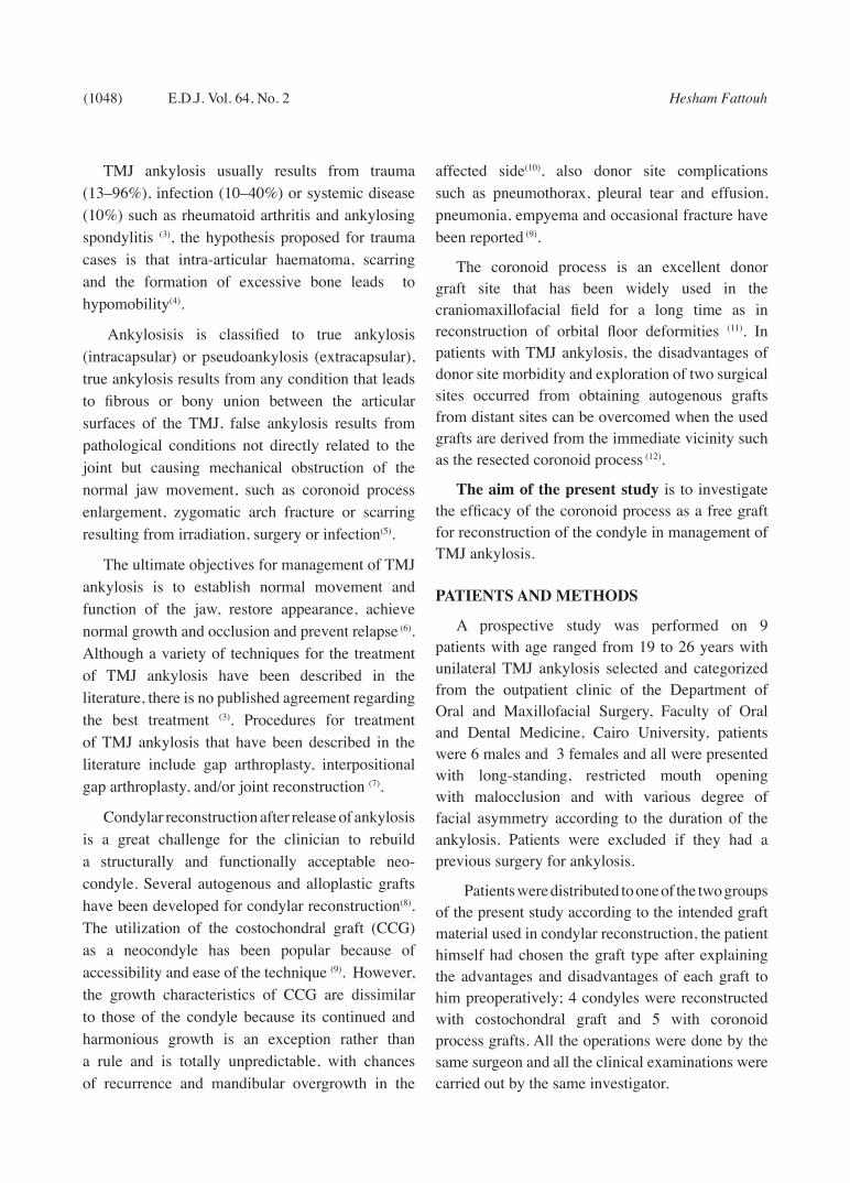
Carefully planned submammary incision for the fifth or the sixth rib was done to harvest the graft from the right side together with 6-7 mm of costal cartilage and about 6–7 cm bone. Care was taken not to damage the perichondrium at the costochondral junction which will be useful in the recipient site, the donor site was checked for any pleural tear. The costal cartilage and the bone were then trimmed and rounded to resemble the condylar morphology. The teeth were put in the desired occlusion with the help of temporary intermaxillary fixation. The rib was applied on the lateral surface of the ramus and fixation was carried out with at least three lag screws(Fig. 1A-1B).
Group II (Coronoid process graft)
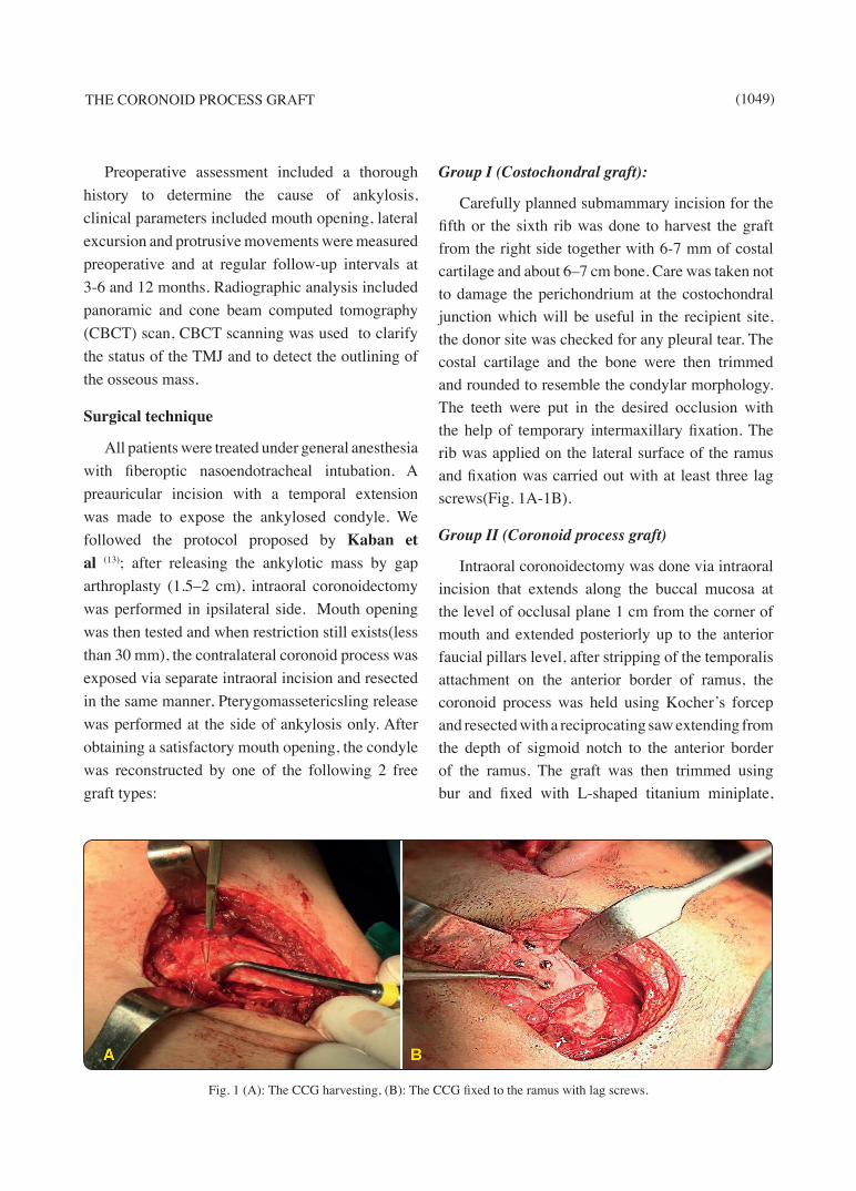
Intraoral coronoidectomy was done via intraoral incision that extends along the buccal mucosa at the level of occlusal plane 1 cm from the corner of mouth and extended posteriorly up to the anterior faucial pillars level, after stripping of the temporalis attachment on the anterior border of ramus, the coronoid process was held using Kocher’s forcep and resected with a reciprocating saw extending from the depth of sigmoid notch to the anterior border of the ramus. The graft was then trimmed using bur and fixed with L-shaped titanium miniplate,
Fig. 1 (A): The CCG harvesting, (B): The CCG fixed to the ramus with lag screws.
(1050) Hesham FattouhE.D.J. Vol. 64, No. 2
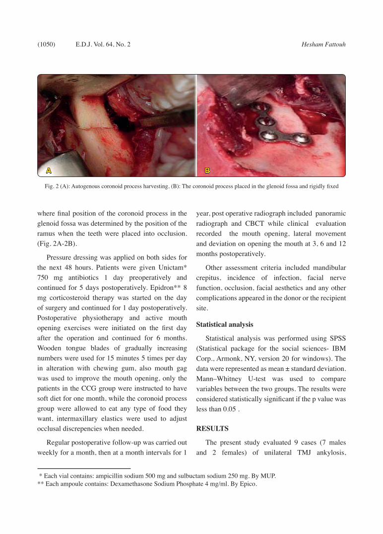
where final position of the coronoid process in the glenoid fossa was determined by the position of the ramus when the teeth were placed into occlusion.(Fig. 2A-2B).
Pressure dressing was applied on both sides for the next 48 hours. Patients were given Unictam* 750 mg antibiotics 1 day preoperatively and continued for 5 days postoperatively. Epidron** 8 mg corticosteroid therapy was started on the day of surgery and continued for 1 day postoperatively. Postoperative physiotherapy and active mouth opening exercises were initiated on the first day after the operation and continued for 6 months. Wooden tongue blades of gradually increasing numbers were used for 15 minutes 5 times per day in alteration with chewing gum, also mouth gag was used to improve the mouth opening, only the patients in the CCG group were instructed to have soft diet for one month, while the coronoid process group were allowed to eat any type of food they want, intermaxillary elastics were used to adjust occlusal discrepencies when needed.
Regular postoperative follow-up was carried out weekly for a month, then at a month intervals for 1
year, post operative radiograph included panoramic radiograph and CBCT while clinical evaluation recorded the mouth opening, lateral movement and deviation on opening the mouth at 3, 6 and 12 months postoperatively.
Other assessment criteria included mandibular crepitus, incidence of infection, facial nerve function, occlusion, facial aesthetics and any other complications appeared in the donor or the recipient site.
Statistical analysis
Statistical analysis was performed using SPSS (Statistical package for the social sciences- IBM Corp., Armonk, NY, version 20 for windows). The data were represented as mean ± standard deviation. Mann–Whitney U-test was used to compare variables between the two groups. The results were considered statistically significant if the p value was less than 0.05 .
RESULTS
The present study evaluated 9 cases (7 males and 2 females) of unilateral TMJ ankylosis,
Fig. 2 (A): Autogenous coronoid process harvesting, (B): The coronoid process placed in the glenoid fossa and rigidly fixed
* Each vial contains: ampicillin sodium 500 mg and sulbuctam sodium 250 mg. By MUP.** Each ampoule contains: Dexamethasone Sodium Phosphate 4 mg/ml. By Epico.
THE CORONOID PROCESS GRAFT (1051)
8 patients had right side ankylosis and only 1 patient had left side ankylosis, patients were divided to 2 groups with a very similar preoperative data, after the gap arthroplasty procedure, the first group was reconstructed by CCG (4 patients), while the second one was reconstructed with coronoid process graft (5 patients). The mean age was 21.5±2.38 in group I and 22±2.54 in group II with no significant differences between the two groups. Trauma was the main etiological factor in 7 patients (78%), while middle ear infection was the etiological factor in only 1 patient of each group (22%).
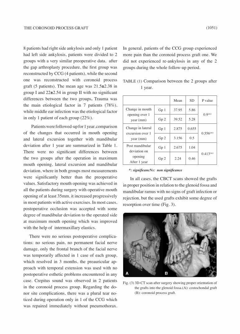
Patients were followed-up for 1 year, comparison of the changes that occurred in mouth opening and lateral excursion together with mandibular deviation after 1 year are summarized in Table 1. There were no significant differences between the two groups after the operation in maximum mouth opening, lateral excursion and mandibular deviation, where in both groups most measurements were significantly better than the preoperative values. Satisfactory mouth opening was achieved in all the patients during surgery with operative mouth opening of at least 35mm, it increased progressively in most patients with active exercises. In most cases, postoperative occlusion was accepted with some degree of mandibular deviation to the operated side at maximum mouth opening which was improved with the help of intermaxillary elastics.
There were no serious postoperative complica-tions: no serious pain, no permanent facial nerve damage, only the frontal branch of the facial nerve was temporarily affected in 1 case of each group, which resolved in 3 months, the preauricular ap-proach with temporal extension was used with no postoperative esthetic problems encountered in any case. Crepitus sound was observed in 2 patients in the coronoid process group. Regarding the do-nor site complications, there was a plural tear no-ticed during operation only in 1 of the CCG which was repaired immediately without pneumothorax.
In general, patients of the CCG group experienced more pain than the coronoid process graft one. We did not experienced re-ankylosis in any of the 2 groups during the whole follow-up period.
TABLE (1) Comparison between the 2 groups after 1 year.
Mean SD P value
Change in mouth opening over 1
year (mm)
Gp 1 37.95 5.860.9(ns)
Gp 2 39.52 5.28
Change in lateral excursion over 1
year (mm)
Gp 1 2.875 0.6550.556(ns)
Gp 2 3.156 0.5
Post mandibular deviation on
openingAfter 1 year
Gp 1 2.675 1.04
0.413(ns)
Gp 2 2.24 0.46
*: significancNs: non significance
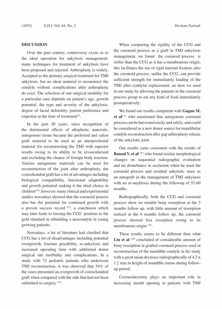
In all cases, the CBCT scans showed the grafts in proper position in relation to the glenoid fossa and mandibular ramus with no signs of graft infection or rejection, but the used grafts exhibit some degree of resorption over time (Fig. 3).
Fig. (3) 3D-CT scan after surgery showing proper orientation of the grafts into the glenoid fossa.(A): costochondal graft (B): coronoid process graft.
(1052) Hesham FattouhE.D.J. Vol. 64, No. 2
DISCUSSION
Over the past century, controversy exists as to the ideal operation for ankylosis management; many techniques for treatment of ankylosis have been proposed and rejected. Arthroplasty is widely. Accepted as the primary surgical treatment for TMJ ankylosis, but no ideal material to reconstruct the condyle without complications after arthroplasty do exist. The selection of one surgical modality for a particular case depends on patient’s age, growth potential, the type and severity of the ankylosis, degree of facial deformity, patient preference and expertise at the time of treatment(3).
In the past 20 years, since recognition of the detrimental effects of alloplastic materials, autogenous tissue became the preferred and safest graft material to be used as an interpositional material for reconstructing the TMJ with superior results owing to its ability to be revascularized and excluding the chance of foreign body reaction. Various autogenous materials can be used for reconstruction of the joint after arthroplasty, the costochondral graft has a lot of advantages including biological compatibility, functional adaptability and growth potential making it the ideal choice in children(14), however many clinical and experimental studies nowadays showed that the coronoid process also has the potential for continued growth with a proven success record (15), a conclusion which may later leads to loosing the CCG position as the gold standard in rebuilding a neocondyle in young growing patients.
Nowadays, a lot of literature had clarified that CCG has a lot of disadvantages including potential overgrowth, fracture possibility, re-ankylosis and increased operating time with additional donor surgical site morbidity and complications, In a study with 72 pediatric patients who underwent TMJ reconstruction, it was observed that 54% of the cases presented an overgrowth of costochondral graft when compared with the side that had not been submitted to surgery (16).
When comparing the rigidity of the CCG and the coronoid process as a graft in TMJ ankylosis management, we found the coronoid process is stiffer than the CCG as it has a membranous origin, this facilitates the use of rigid internal fixation, also the coronoid process, unlike the CCG, can provide sufficient strength for immediately loading of the TMJ after condylar replacement, an item we used in our study by allowing the patients in the coronoid process group to eat any kind of food immediately postoperatively.
We found our results competent with Gagan M. et al(17) who mentioned that autogenous coronoid process can be harvested easily and safely, and could be considered as a new donor source for mandibular condyle reconstruction after gap arthroplasty release of the ankylotic joint.
Our results came consistent with the results of Bansal V. et al( 18 ) who found similar morphological changes on sequential radiographic evaluation and no disturbance in occlusion when he used the coronoid process and residual ankylotic mass as an autograft in the management of TMJ ankylosis with no re-anyklosis during the followup of 52-60 months.
Radiographically, both the CCG and coronoid process show no notable bony resorption at the 3 months follow up, with little amount of resorption noticed at the 6 months follow up, the coronoid process showed less resorption owing to its membranous origin (19).
These results seems to be different than what Liu et al (20) concluded of considerable amount of bony resorption in grafted coronoid process used in reconstruction of the mandible condyle in his study with a great mean decrease radiographically of 4.2 ± 1.2 mm in height of mandible ramus during follow-up period.
Coronoidectomy plays an important role in increasing mouth opening in patients with TMJ
THE CORONOID PROCESS GRAFT (1053)
ankylosis as its presence leads to mechanical restriction of mouth opening and possible re-ankylosis, the resected coronoid process is usually elongated due to the hyperactivity of the temporalis muscle(21), an issue which favorites its placement after gap arthroplasty to restore the height of the mandible ramus if used as a new donor site, also this will avoid exploration of two surgical sites with the probability for donor site morbidity as access to the coronoid process of the mandible is readily obtained and proximal to the recipient site.
Considering the better exposure that it gives to both the ankylotic mass and the coronoid process, we prefer the modified preauricular approach with temporal extension in all cases, however, we experienced varying degree of temporary weakness of the facial nerve with that approach, this have been reported by various authors ranging from 1.5% to 50%(22).
In our study we found patients acceptance of the coronoid process graft procedure high, probably due to the lack of cutaneous scarring and reported pain which we found in the CCG group, on the contrary, coronoid graft group reported minimal discomfort as unusual traction feelings on the temporalis muscle.
On the other hand, regarding the limitation of the coronid graft, we think it is technically difficult to use the coronoid process for condylar reconstruction if it is involved in the ankylotic mass in severe ankylosis condition or if large graft segment is needed. Also, some degrees of crepitus were mentioned by 2 patients in the coronoid process group, which may be attributable to the fact that the surface of the coronoid process is stiffer, more pointed and is not covered by cartilage like the CGG, this is competent with Weina Z. et al (23 ) who found the same crepitus in his reconstruction of the TMJ after ankylosis using the coronoid process.
CONCLUSION
No ideal material could be used for TMJ reconstruction after gap arthroplasty in all cases; each case should be assessed separately. The coronoid process as a graft material to reconstruct the condyle after gap arthroplasty in TMJ ankylosis produced good and comparable results to the costochondral grafts, specially in the patients with TMJ ankylosis, where the coronoid process is not involved in the ankylotic process..
RECOMMENDATION
It is impossible to draw conclusions that may be applicable universally from a study of such small sample and duration, so further studies on a larger population number and for a greater follow-up will be required.
REFERENCES
1. Potter JK and Dierks EJ: Vascularized options for recon-struction of the mandibular condyle. SeminPlastSurg 22: 156160, 2008.
2. Babu L, Jain MK, Ramesh C, Vinayaka N. Is aggressive gap arthroplasty essential in the management of temporo-mandibular joint ankylosis? A prospective clinical study of 15 cases. Br J Oral MaxillofacSurg51: 473–8, 2013.
3. Salha R, Sidebottom AJ. Management of the temporoman-dibular joint in rheumatoid disorders. Br J Oral Maxillo-facSurg51: 191–8, 2013.
4. Erol B, Tanrikulu R, Go¨rgu¨n B. A clinical study on anky-losis of the temporomandibular joint. J Craniomaxillofac-Surg34:100–6, 2006.
5. Elgazzar RF, Abdelhady AI, Saad KA, Elshaal MA, Hus-sain MM, Abdelal SE, et al. Treatment modalities of TMJ ankylosis: Experience in Delta Nile, Egypt. Int J Oral MaxillofacSurg 39:333–42, 2010.
6. Rowe NL: Ankylosis of the temporomandibular joint. Parts 1, 2, and 3. J R CollSurgEdinb 27:67, 1982.
7. Khadka A, Hu J. Autogenous grafts for condylar recon-struction in treatment of TMJ ankylosis: current concepts and considerations for the future. Int J Oral Maxillofac-Surg41:94–102, 2012.
(1054) Hesham FattouhE.D.J. Vol. 64, No. 2
8. Wolford LM, Cottrell DA, Henry C: Sternoclavicular grafts for temporomandibular joint reconstruction. J Oral MaxillofacSurg 52:119, 1994.
9. Wen-Ching-Ko E, Huang CS, Chen YR: Temporomandib-ular joint reconstruction in children using costochondral grafts. J Oral MaxillofacSurg 57:789, 1999.
10. El-Sayed KM. Temporomandibular joint reconstruction with costochondral graft using modified approach. Int J Oral MaxillofacSurg 37:897–902, 2008.
11. Mintz SM, Ettinger A, Schmakel T, Gleason M. Contra-lateral coronoid process bone grafts for orbital floor recon-struction: an anatomic and clinical study. J Oral and Maxil-lofacSurg 56(10):1140–1145, 1998
12. Hu W, Thadani S, Mukul SK, et al. Autogeneous coronoid process asfree graft for reconstruction of mandibular con-dyle in patients with tem-poromandibularankylosis. Oral MaxillofacSurg18:313–23, 2014.
13. Kaban LB, PerrottDH ,Fisher K. A protocol for manage-ment of temporo-mandibularjointankylosis. J Oral Maxil-lofacSurg48:1145–52, 1990.
14. Ma J, Jiang H, Liang L. Interpositionalarthroplasty versus reconstruction arthroplasty for temporomandibular joint ankylosis: a systematic review and meta-analysis. J Cra-niomaxillofacSurg43:1202–1207, 2015
15. Zhu SS, HuJ, Li J, Luo E, Liang X, Feng G. Free grafting of autogenous coronoid process for condylar reconstruction in patients with temporo- mandibularjointankylosis. Oral Surg Oral Med Oral Pathol Oral RadiolEndod106:662–7, 2008.
16. Movahed R, Mercuri L. Management of temporomandibu-lar joint ankylosis. OralMaxillofacSurgClin N Am 27:27–35, 2015.
17. Gagan Mehta, Shadab Mohammad, Hari Ram. Re-Assess-ment of Coronoid as a Graft for Condylar Reconstruction in TMJ Ankylosis Patients: A Prospective Study and Lit-erature Review. J. Maxillofac. Oral Surg.; 16(2):175-180. 2017.
18. Bansal V, Mowar A, Dubey P. Coronoid process and re-sidual ankylotic mass as anautograft in the management of ankylosis of the temporomandibular joint in young ado-lescent patients:a retrospective clinical investigation. Br J Oral Maxillofac Surg. Apr;54(3):280-5. 2016.
19. ZhuSS, HuJ, LiN, ZhouHX, LuoE .Autogenous coronoid processas a new donor source for reconstruction of man-dibular condyle: an exper- imental study on goats. Oral Surg Oral Med Oral Pathol Oral RadiolEndod101:572–80, 2006.
20. Liu Y, Li J, Hu J, Zhu S, Luo E, Hsu Y. Autogenous coro-noid process pedicled on temporal muscle grafts for recon-struction of the mandible condylar in patients with Tem-poromandibular joint ankylosis. Oral Surg Oral Med Oral Pathol Oral RadiolEndod109:203–210, 2010.
21. Isberg AM, McNamara JA Jr, Carlson DS, Isacsson G Cor-onoid process elongation in rhesus monkeys (Macacamu-latta) after experimentally induced mandibular hypomobil-ity: a cephalometric and histologic study. Oral Surg Oral Med Oral Pathol Oral RadiolEndod. 70:704–710, 1990.
22. Shaker AA. The use of costochondral grafts in the man-agement of temporomandibular joint ankylosis. Egyptian Journal of Plastic and Reconstructive Surgery 27:73–83.14, 2003.
23. Weina Zhang, BinGub, JingHucBr J Oral Maxillofac Surg. Dec;52(10):928-33, 2014.