Embed Size (px)
Citation preview
This article appeared in a journal published by Elsevier. The attachedcopy is furnished to the author for internal non-commercial researchand education use, including for instruction at the authors institution
and sharing with colleagues.
Other uses, including reproduction and distribution, or selling orlicensing copies, or posting to personal, institutional or third party
websites are prohibited.
In most cases authors are permitted to post their version of thearticle (e.g. in Word or Tex form) to their personal website orinstitutional repository. Authors requiring further information
regarding Elsevier’s archiving and manuscript policies areencouraged to visit:
http://www.elsevier.com/authorsrights
Author's personal copy
MICE LACKING THE TRANSCRIPTIONAL COACTIVATOR PGC-1aEXHIBIT ALTERATIONS IN INHIBITORY SYNAPTIC TRANSMISSIONIN THE MOTOR CORTEX
S. E. DOUGHERTY, a� A. F. BARTLEY, b� E. K. LUCAS, a
J. J. HABLITZ, b L. E. DOBRUNZ b* AND R. M. COWELL a*
aUniversity of Alabama Birmingham, Department of Psychiatry
& Behavioral Neurobiology, United States
bUniversity of Alabama Birmingham, Department of Neurobiology,
Evelyn F. McKnight Brain Institute, Civitan International Research
Center, United States
Abstract—Peroxisome proliferator-activated receptor ccoactivator 1a (PGC-1a) is a transcriptional coactivator
known to regulate gene programs in a cell-specific manner
in energy-demanding tissues, and its dysfunction has been
implicated in numerous neurological and psychiatric disor-
ders. Previous work from the Cowell laboratory indicates
that PGC-1a is concentrated in inhibitory interneurons and
is required for the expression of the calcium buffer parvalbu-
min (PV) in the cortex; however, the impact of PGC-1a defi-
ciency on inhibitory neurotransmission in the motor cortex
is not known. Here, we show that mice lacking PGC-1a exhi-
bit increased amplitudes and decreased frequency of spon-
taneous inhibitory postsynaptic currents in layer V
pyramidal neurons. Upon repetitive train stimulation at the
gamma frequency, decreased GABA release is observed.
Furthermore, PV-positive interneurons in PGC-1a �/� mice
display reductions in intrinsic excitability and excitatory
input without changes in gross interneuron morphology.
Taken together, these data show that PGC-1a is required
for normal inhibitory neurotransmission and cortical PV-
positive interneuron function. Given the pronounced motor
dysfunction in PGC-1a �/� mice and the essential role of
PV-positive interneurons in maintenance of cortical excit-
atory:inhibitory balance, it is possible that deficiencies in
PGC-1a expression could contribute to cortical hyperexcit-
ability and motor abnormalities in multiple neurological
disorders. � 2014 IBRO. Published by Elsevier Ltd. All rights
reserved.
Key words: peroxisome proliferator-activated receptor gam-
ma coactivator 1a, parvalbumin, inhibitory neurotransmis-
sion, motor cortex, interneuron.
INTRODUCTION
Peroxisome proliferated-activated receptor c coactivator
1a (PGC-1a) is a transcriptional coactivator which, by
interacting with different transcription factors, initiates
cell and tissue-specific gene programs. Since the
discovery of PGC-1a in 1998 (Puigserver et al., 1998),
many studies have suggested that a reduction in its levels
and/or activity plays a role in neurological disorders
including Parkinson Disease (Zheng et al., 2010), Alzhei-
mer Disease (Qin et al., 2009; Sheng et al., 2012),
Huntington Disease (Cui et al., 2006; Taherzadeh-Fard
et al., 2009; Chaturvedi et al., 2010), schizophrenia
(Christoforou et al., 2007; Jiang et al., 2013b), anxiety dis-
orders (Hettema et al., 2011) and multiple sclerosis (Witte
et al., 2013). Studies with whole body and neuron-specific
PGC-1a �/� mice indicate that PGC-1a is required for
the expression of a subset of metabolic and neuronal tran-
scripts (Lin et al., 2004; Lucas et al., 2010, 2012; Ma
et al., 2010), but the physiological consequences of these
transcriptional changes are not clear. Elucidating the
impact of PGC-1a deficiency on neuronal function will
give us insight into its contribution to neuronal dysfunction
in various disorders.
The PGC-1a protein is highly concentrated in
GABAergic cell populations throughout the brain (Cowell
et al., 2007; Jiang et al., 2013b), and PGC-1a �/� mice
exhibit deficiencies in the expression of the calcium buffer
protein parvalbumin (PV) in forebrain regions including
cortex, hippocampus, and striatum (Lucas et al., 2010).
In these regions, PV is expressed by a subset of GABAer-
gic interneurons that exhibit fast-spiking (FS) and non-
adapting properties (Kawaguchi, 1993; Kawaguchi and
Kondo, 2002; Tepper and Bolam, 2004) and entrain local
pyramidal neurons to generate gamma oscillations
http://dx.doi.org/10.1016/j.neuroscience.2014.04.0230306-4522/� 2014 IBRO. Published by Elsevier Ltd. All rights reserved.
*Corresponding authors. Address: Department of Neurobiology,University of Alabama at Birmingham, 1825 University Boulevard,SHEL 902, Birmingham, AL 35294, United States. Tel: +1-205-934-7923 (L.E. Dobrunz). Address: Department of Psychiatry & Behav-ioral Neurobiology, University of Alabama at Birmingham, 1720 7thAvenue South, SC 729, Birmingham, AL 35294, United States. Tel:+1-205-975-7466 (R.M. Cowell).
E-mail addresses: [email protected] (L. E. Dobrunz), [email protected] (R. M. Cowell).
� These authors contributed equally to the data collection andpreparation of this manuscript.Abbreviations: ACSF, artificial cerebral spinal fluid; AHP,afterhyperpolarization; ANOVA, analysis of variance; AP, actionpotential; CCK, cholecystokinin; CNQX, 6-cyano-7-nitroquinoxaline-2,3-dione; DL-APV, DL-2-amino-5-phosphonoovaleric acid; EGFP,enhanced green fluorescent protein; EGTA, ethylene glycoltetraacetic acid; EPSC, excitatory postsynaptic current; FS, fast-spiking; GAD, glutamic acid decarboxylase; HEPES, hydroxyethylpiperazineethanesulfonic acid; IPSC, inhibitory postsynaptic current;NMDA, N-methyl-D-aspartate; P, postnatal day; PBS, phosphatebuffered saline; PGC-1a, peroxisome proliferator-activated receptor ccoactivator 1a; PV, parvalbumin; TRITC, tetramethylrhodamine; TTX,tetrodotoxin.
Neuroscience 271 (2014) 137–148
137

Author's personal copy
(Wang and Buzsaki, 1996; Bartos et al., 2002;
Vreugdenhil et al., 2003; Sohal et al., 2009). Interestingly,
mice lacking PGC-1a show pronounced motor abnormal-
ities and decreased PV protein expression in the motor
cortex by 4 weeks of age (Lucas et al., 2012), suggesting
that the motor cortex may be particularly dependent on
PGC-1a for proper function. Previous investigations of
inhibitory neurotransmission in the hippocampus of PGC-1a�/� mice (Lucas et al., 2010) suggest that inhibition is
enhanced in this region, similar to what is observed in
PV �/� mice (Vreugdenhil et al., 2003). However, it is
possible that inhibition in the cortex is affected differen-
tially by a lack of PGC-1a; it is therefore important to
evaluate the impact of PGC-1a deficiency in the cortex,
with relevance for disorders in which cortical PGC-1a defi-
cits have been reported, including Parkinson Disease (Zheng
et al., 2010) and Alzheimer Disease (Qin et al., 2009).
In light of the deficiency in PV expression in the cortex
of PGC-1a �/� mice and the profound motor dysfunction
in these animals, we sought to determine the
physiological impact of PGC-1a deletion on inhibitory
neurotransmission in the motor cortex. We hypothesized
that mice lacking PGC-1a would exhibit altered
inhibitory transmission onto cortical pyramidal neurons
and that PV+ interneurons would be especially
affected. In order to investigate the potential role of
PGC-1a in cortical inhibitory neurotransmission, we
utilized motor cortex acute slices from a PGC-1a �/�mouse model (Lin et al., 2004). Our data show that, in
contrast to results from the PGC-1a �/� hippocampus,
a loss of PGC-1a leads to alterations in basal GABA
release in the cortex, concurrent with reduced GABA
release upon gamma frequency stimulation. Furthermore,
in PGC-1a �/� mice expressing enhanced green fluores-
cent protein (EGFP) specifically in PV+ cells, we found
that FS interneurons have a reduced firing rate in
response to current injections, suggesting that PGC-1afunctions in a cell-autonomous manner to regulate inter-
neuron excitability. Additionally, PV+ cells showed
reduced synaptic excitatory activity, suggesting that
PV+ cells could be less active in the motor cortex of
PGC-1a �/� mice. Taken together these data suggest
that reductions in PGC-1a expression are associated with
deficiencies in inhibitory neurotransmission and synaptic
function in the cortex. These results have implications
for understanding the impact of PGC-1a alterations on
cortical network signaling in disease, as synchronization
of firing by PV+ interneurons is critical for normal cortical
output and higher cognitive processing (Sohal et al.,
2009).
EXPERIMENTAL PROCEDURES
Animals
All experimental protocols were approved by the
Institutional Animal Care and Use Committee of the
University of Alabama at Birmingham. PGC-1a �/�mice (generous gift of Jiandie Lin, University of
Michigan, (Lin et al., 2004)) were maintained on a
C57BL/6J genetic background and housed two to five in
a cage at 26 ± 2 �C room temperature with food and
water ad libitum. All experiments were conducted with
4-week-old male and female PGC-1a +/+ and �/� litter-
mates generated by breeding PGC-1a +/� mice.
For pyramidal neuron recordings, PGC-1a +/+ and
�/� mice were used. For targeted interneuron
recordings, mice from the PGC-1a �/� line were
crossed with mice expressing EGFP under the control
of the glutamic acid decarboxylase (GAD67) promoter
(G42 line; JAX#7677) to generate EGFP-positive PGC-
1a +/+ and EGFP-positive PGC-1a �/� littermates.
This mouse line was chosen for two reasons: (1) EGFP
is only expressed in the PV+ subset of inhibitory
interneurons, primarily in cortex (Chattopadhyaya et al.,
2004; Bartley et al., 2008) and (2) EGFP expression is
not dependent on the activity of the PV promoter (which
would be expected to be reduced in the absence of
PGC-1a). All experiments were conducted in accordance
with the Guide for the Care and Use of Laboratory Ani-
mals adopted by the U.S. National Institutes of Health.
For pyramidal neuron recordings
Whole-cell recordings. Mice aged postnatal day (P) 27
to P33 were anesthetized with isoflourane and then
decapitated. Brains were placed in ice-cold artificial
cerebral spinal fluid (ACSF) containing the following (in
mM): 125 NaCl, 3.5 KCl, 0.5 CaCl2, 3.5 MgCl2, 26
NaHCO3 and 10 D-glucose. The ACSF was bubbled
with 95% O2/5% CO2. Coronal brain slices (300-lmthick) containing motor cortex were cut using a
Vibratome (Ted Pella, Inc., Riverside, CA, USA). The
slices were kept for 30 min at 37 ± 1 �C and then
stored at room temperature (22 ± 1 �C). Slices were
perfused continuously with oxygenated recording ACSF
containing the following (in mM): 125 NaCl, 3.5 KCl, 2.0
CaCl2, 2.0 MgCl2, 26 NaHCO3 and 10 D-glucose at
room temperature. Whole-cell patch clamp recordings
were acquired from visually identified pyramidal neurons
in layer five of the motor cortex. Position in cortex was
verified through inclusion of 0.4% biocytin in the internal
solution followed by streptavidin staining (Invitrogen,
Carlsbad, California, US, s32355). Recordings were
conducted on a Zeiss AxioExaminer microscope (Carl
Zeiss, Thornwood, NY, USA). Cells were voltage
clamped at �70 mV, using internal solution containing
the following (in mM): 129 CsCl, 2 MgATP, 10 EGTA,
10 HEPES, 0.2 GTP and 2 QX-314, pH 7.2. Pipette tip
resistance was 2–5 M. Voltage clamp recordings were
obtained using a PC505A amplifier (Warner
Instruments, Hamden, CT, USA) controlled by Clampex
8.0 software via a Digidata 1322A interface (Molecular
Devices, Union City, CA, USA), filtered at 5 kHz and
digitized at 10 kHz. Input resistance and series
resistance were monitored by applying a 10-mV voltage
step. Spontaneous IPSCs (sIPSCs) and evoked IPSCs
were pharmacologically isolated with CNQX (6-cyano-
7-nitroquinoxaline-2,3-dione) (10 lM) and DL-APV
(DL-2-amino-5-phosphonoovaleric acid) (50 lM).
Miniature IPSCs (mIPSCs) were recorded, on a
separate cohort of animals, in the presence of CNQX,
D-APV, and TTX (tetrodotoxin) (1 lM).
138 S. E. Dougherty et al. / Neuroscience 271 (2014) 137–148

Author's personal copy
Stimulation. Synaptic responses were evoked with a
bipolar stimulating electrode consisting of a twisted pair
of 25-lm Formvar-insulated nichrome wires. The
electrode was positioned in layer V of the motor cortex.
For each cell the stimulation threshold for evoking
IPSCs was determined, and the stimulus intensity was
set at twice the threshold intensity. A series of paired
stimulations at 20-, 30-, and 100-ms intervals were
applied to elucidate paired-pulse ratios. For gamma train
recordings, 34 stimulations were applied in 500 ms to
evoke a response in patched pyramidal cells (66 Hz).
Recordings were taken at 32 ± 1 �C.
Data analyses. Analyses of sIPSCs and mIPSCs were
performed using the EVAN event analysis software
(generously provided by Istvan Mody, UCLA, CA, USA)
and Clampfit 8.0 software which focused on amplitude,
inter-event interval, rise time, and decay time of events.
T50, which is 50% of the peak decay time, was used as
a measure of decay time. All sIPSCs/mIPSCs that fit the
template and passed visual inspection were included in
the analysis. Analyses of all other electrophysiological
experiments were performed using Clampfit 8.0,
GraphPad Prism, and Microsoft Excel. To calculate the
paired-pulse ratio, the amplitude of the second inhibitory
postsynaptic current (IPSC) was measured after
subtracting the first IPSC, and divided by the amplitude
of the first IPSC relative to the baseline set immediately
before the first stimulus. Train stimulation was analyzed
by measuring the charge transfer (area under the curve)
for 1 s after stimulation. The amplitude of the first pulse
was also measured, as was the ratio of the last to first
response (IPSC34/IPSC1). Response amplitudes were
measured by taking the peak value minus the value
immediately before stimulation. For Fig. 3E, we
calculated IPSCn/IPSC1 for each pulse in the train and
determined the standard deviations within the train for
each cell. A two-tailed student’s t-test assuming unequal
variance was utilized to assess statistical significance.
Values were considered statistically significant when the
p-value was less than 0.05.
For interneuron recordings
Slice preparation. Experiments were conducted in
300-lm acute brain slices prepared from of P27 to P33
mice. The animals used were obtained from crossing
the PGC-1a heterozygous (Lucas et al., 2010) line with
the G42 mouse line. In the G42 mouse line, GFP was only
expressed in a subset of neocortical PV+ inhibitory neu-
rons, the FS basket cells (Chattopadhyaya et al., 2004;
Bartley et al., 2008). The mice were anesthetized with iso-
flurane and decapitated, and their brains were removed
rapidly. Coronal slices of the brain were cut using a vibrat-
ing microtome (VT1000S; Leica, Bannockburn, IL, USA).
Slicing and dissection of the cortex was done in ice-cold
(1–3 �C) dissecting solution containing the following (in
mM): 87 NaCl, 3 KCl, 0.5 CaCl2, 7.0 MgCl2, 1.25
NaH2PO4, 26 NaHCO3, 75 sucrose and 20 glucose,
bubbled with 95% O2–5% CO2, pH 7.35–7.45. Slices
were stored in a holding chamber containing a slightly
modified ACSF (see below) for approximately 30 min at
30 to 32 �C and then transferred to room temperature.
Modified ACSF contained (in mM) 1 CaCl2 and 2 MgCl2.
Slices were bubbled with 95% O2–5% CO2 for P1 h
before recording.
Intrinsic firing assessment. During the experiment,
slices were held in a submersion recording chamber
perfused (3–4 mLs/min) with ACSF composed of (in
mM): 126 NaCl, 3 KCl, 2 CaCl2, 1 MgCl2, 1.25
NaH2PO4, 26 NaHCO3, and 20 glucose. The solution
was bubbled with 95% O2/5% CO2, and the pH was
between 7.35 and 7.45. Picrotoxin (100 lM) was added
to the external solution to block inhibitory synaptic
responses mediated by GABAA receptors; 50 lMD-APV was added to prevent N-methyl-D-aspartate
(NMDA) receptor-mediated currents. All intrinsic firing
experiments were performed at 28 to 30 �C.Interneurons expressing EGFP were identified visually
using infrared differential inference contrast optics and
epifluorescent optics on a Nikon (New York) E600FN
upright microscope. Targeted layer IV interneurons were
patched in the voltage-clamp configuration and recorded
in current-clamp configuration while maintaining a
holding potential of �60 ± 1 mV using an Axopatch
200B amplifier (Molecular Devices). Patch electrodes
(4–6 MX) were filled with internal solution composed of
the following (in mM): 150 K-gluconate, 0.1 EGTA, 3
NaCl, 6 KCl, 10 HEPES, 10 Na-ATP, and 0.3 GTP. pH
was adjusted to 7.3 with KOH. The resting potential was
measured immediately after break-in, and input
resistance was measured in voltage-clamp with a 400-
ms, �8-mV step from a �60-mV holding potential.
Firing frequency versus injected current plots (F–I plots)
were made by measuring the initial firing frequency of a
spike train evoked by a series of incremental 600-ms
current steps at intervals of 50 or 100 pA. The spike
threshold potential was defined as the membrane
potential, in a 5-ms window preceding spike peak, at
which the third derivative was maximum (an inflection
point). The action potential (AP) amplitude was
calculated from the spike threshold to the peak of the
AP. The AP half-width is the duration of the AP by the
500-pA current step. Afterhyperpolarization (AHP) was
calculated from the spike threshold to the peak of the
AHP. The access resistance and holding current
(<200 pA) were monitored continuously. Recordings
were rejected if either access resistance or holding
current increased >20% during the experiment.
Electrophysiological interneuron classification. To
distinguish FS interneurons from non-FS (NFS)
interneurons, depolarizing current steps were used to
analyze the firing response of each interneuron. Single-
spike properties were determined on spikes elicited by
near threshold current injection. Spike-frequency
adaptation was quantified by the ratio between the last
and first interspike intervals in spike trains evoked by
600-ms depolarizing steps. Cells were classified as FS if
they had the following (Rotaru et al., 2011): (1) by the
500-pA current step the firing frequency reached at least
S. E. Dougherty et al. / Neuroscience 271 (2014) 137–148 139

Author's personal copy
150 Hz; (2) narrow spikes (duration at half peak amplitude
60.6 ms); (3) large afterhyperpolarizing potentials
(amplitude P14 mV); and (4) absence of significant
spike-frequency adaptation (adaptation ratio61.5). These
criteria may exclude some FS neurons. Spontaneous
AMPA-mediated excitatory postsynaptic current (EPSCs)
(sEPSCs) were pharmacologically isolated with 100 lMpicrotoxin and 50 lM D-APV.
Biocytin filling of interneurons. Brain slices were
prepared as described above. Interneurons expressing
eGFP were identified visually using epifluorescent optics
on a Nikon E600FN upright microscope (Melville, New
York, USA). Patch electrodes (4–6 MX) were filled with
internal solution containing 0.4% biocytin. Cells were
patched for 15–25 min to allow for adequate filling of the
processes. After filling, slices were immediately placed
in 4% paraformaldehyde (PFA) for 24–72 h and stored
in phosphate buffered saline (PBS) until processing.
Slices were washed in PBS, incubated in 10% methanol
and 3.5% H2O2 in PBS for 10 min, washed in PBS, and
incubated in tetramethylrhodamine (TRITC) conjugated
steptavidin (Jackson Immunoresearch, West Grove, PA,
USA) in 0.3% phosphate buffered saline with Triton
X-100 (PBST) for 2 h. After washes with PBS, slices
were mounted onto charged microscope slides and
coverslipped with Prolong Antifade Gold (Invitrogen).
Neurolucida software (MBF Bioscience, Williston, VT,
USA) was used to trace biocytin-labeled interneurons.
As axons were not reliably labeled, only soma and
dendrite characteristics were measured. Variables of
interest included soma size, dendrite length and volume,
and number of dendrites, branch points, and branches.
Data analyses. All statistics were performed using
Origin software (Origin Lab Corporation, 2002) and
statistical significance was p< 0.05. Data are presented
as means ± SE and sample number (n) refers to cell
number for electrophysiological experiments. Statistical
comparisons for electrophysiological data were made
using Student’s t-test or a one-way analysis of variance
(ANOVA) followed by Tukey’s posthoc analysis. In
figures and table, ⁄indicates a statistically significant
difference.
RESULTS
Basal GABA release is reduced in the motor cortex ofPGC-1a �/� mice
To assess basal inhibitory neurotransmission, we initially
evaluated both spontaneous and miniature IPSCs in the
motor cortex from PGC-1a �/� mice around P30. This
age was chosen based on evidence that these mice
exhibit structural changes in the brain at older ages
(Lucas et al., 2012) and that PGC-1a expression in the
cortex peaks around P14–21 (Cowell et al., 2007). The
motor cortex is of particular interest because decreases
in PV protein expression in PGC-1a �/� mice are very
robust in this region (Lucas et al., 2010), and these mice
have pronounced motor deficits (Lucas et al., 2012).
Whole-cell voltage clamp recordings were performed on
layer V pyramidal neurons in the motor cortex in the pres-
ence of APV and CNQX. The observed sIPSCs exhibited
increased amplitudes (Fig. 1B, B0, p= 0.0363,
t(19) = 2.252) and decreased frequency (Fig. 1C, C0,
p= 0.0265, t(14) = 1.993) in PGC-1a �/� mice as com-
pared to PGC-1a +/+ littermates. There were no signif-
icant differences in event kinetics as measured by rise
time (2.12 ± 0.51 ms in PGC-1a +/+ mice and
2.38 ± 0.24 ms in PGC-1a �/� mice; p= 0.627) or time
Fig. 1. Global ablation of PGC-1a results in alterations in basal
GABA release. (A) Representative traces of sIPSCs from
PGC-1a +/+ and PGC-1a �/� animals (B) Cumulative probability
plot of sIPSC amplitudes for PGC-1a +/+ and PGC-1a �/� groups.
Curves are shifted to the right in the PGC-1a�/� group, indicating an
increase in amplitude. Insert (B0) shows a histogram summarizing
effects on mean amplitude. (C) Similar to B but showing cumulative
probability plots of inter-event intervals. (C0) sIPSC frequency
(extrapolated from inter-event interval) was reduced in the
PGC-1a �/� animals as evidenced by a lengthening of the inter-
event interval. The observed changes in sIPSCs are indicative of
alterations in basal GABA release from the cortical interneurons.
Student’s t-test ⁄p< 0.05 n= 3 animals/group, approx. 10 cells per
condition.
140 S. E. Dougherty et al. / Neuroscience 271 (2014) 137–148

Author's personal copy
to decay to 50% of peak value (T50 2.69 ± 0.35 ms in
PGC-1a +/+ mice and 3.54 ± 0.23 ms in PGC-1a�/� mice; p= 0.07).
AP-mediated activity and spontaneous vesicle fusion
events (miniature IPSCs, mIPSCs) can be differentially
regulated (Ramirez and Kavalali, 2011). Therefore, we
pharmacologically isolated mIPSCs with bath application
of TTX to eliminate AP-mediated activity. The observed
mIPSCs exhibited decreased amplitudes (Fig. 2B, B0,
p= 0.0231, t(14) = 2.551) and decreased frequencies
(Fig. 2C, C0, p= 0.0426, t(14) = 1.853) in PGC-1a �/�mice as compared to those measured in +/+ littermates.
There were no significant differences in event kinetics as
measured by rise time (1.82 ± 0.23 ms in PGC-1a +/+
mice and 2.18 ± 0.16 ms in PGC-1a �/� mice;
p= 0.19) or time to decay to 50% of peak value (T50
2.86 ± 0.36 ms in PGC-1a +/+ mice and
3.14 ± 0.30 ms in PGC-1a �/� mice; p= 0.8). These
alterations suggest that PGC-1a differentially affects
spontaneous vesicle release and AP-mediated activity;
mIPSCs are smaller and less frequent whereas AP-regu-
lated events are larger in magnitude but still less frequent.
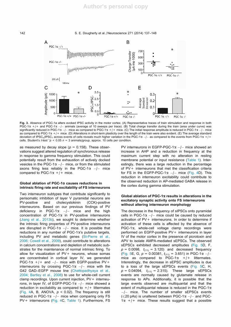
Global ablation of PGC-1a is associated with reducedevoked cortical GABA release with gamma frequencystimulation
In order to further investigate the presynaptic contribution
to the altered GABA neurotransmission in these mice, we
recorded evoked IPSCs at a stimulus intensity set to be
twice the intensity threshold for producing detectible
IPSCs, and measured short-term plasticity in response
to paired stimuli and short stimulus trains. In response
to paired-pulse stimulation, which has been shown to
indirectly measure presynaptic release (Dobrunz et al.,
1997; Dobrunz and Stevens, 1997), we saw no change
in PGC-1a �/� mice in the paired-pulse ratio of inhibitory
responses onto pyramidal neurons in the motor cortex
(data not shown). To evaluate how a lack of PGC-1aaffects evoked GABA release during stimulation trains,
we applied repetitive trains of stimuli in the gamma
frequency range, which mimics the frequency at which
PV+ interneurons entrain the cortical network (Traub
et al., 1996; Wang and Buzsaki, 1996). Example
traces are shown in Fig. 3A; while responses from
PGC-1a +/+ mice consistently showed short-term
depression during the train, responses from
PGC-1a �/� mice were more variable. Some cells in
PGC-1a �/� mice showed even larger short-term
depression whereas others showed a mixture of
facilitation and depression. We observed a reduction in
overall charge transfer in the PGC-1a �/� mice as
compared to PGC-1a +/+ mice (Fig. 3B, p= 0.0044,
t(18) = 3.257). This was caused in part by a reduction in
the initial response size (Fig. 3C, p= 0.025,
t(7) = 2.838). When we compared the amplitude of the
final response to the amplitude of the first response to
evaluate short-term plasticity over the stimulation period,
we found that the ratio of the last response to the first
response was consistently <1 in PGC-1a +/+ mice,
while the ratio was larger and more variable in
PGC-1a �/� mice (Fig. 3D, p= 0.0082, t(18) = 2.967).
The decrease in short-term depression (increase in
IPSC34/IPSC1) is consistent with a decrease in the initial
release probability, which could also contribute to the
observed reduction in the initial response size. Further,
the stimulation to stimulation variability across events,
measured as the standard deviation of IPSCn/IPSC1
across all stimuli in the train, is significantly larger in cells
from the PGC-1a �/� mice (Fig. 3E, p< 0.0001,
t(64) = 6.996). There is no change in event decay kinetics
Fig. 2. Global ablation of PGC-1a results in alterations in spontane-
ous inhibitory vesicle fusion events. (A) Representative traces of
mIPSCs from PGC-1a +/+ and PGC-1a �/� animals. (B, C)
Cumulative probability plots of amplitude and interval, respectively.
(B0) Quantification revealed that the mIPSC amplitude was reduced in
the PGC-1a �/� animals as compared to PGC-1a +/+ mice. (C0)
mIPSC frequency (extrapolated from inter-event interval) was
reduced in the PGC-1a �/� animals as evidenced by a lengthening
of the inter-event interval. Student’s t-test ⁄p< 0.05 n= 3 animals/
group, approx. 10 cells per condition.
S. E. Dougherty et al. / Neuroscience 271 (2014) 137–148 141
Author's personal copy
as measured by decay slope (p= 0.158). These obser-
vations suggest altered regulation of synchronous release
in response to gamma frequency stimulation. This could
potentially result from the exhaustion of actively docked
vesicles in the PGC-1a �/� mice, or from the stimulated
axons firing less reliably in the PGC-1a �/� mice
compared to PGC-1a +/+ mice.
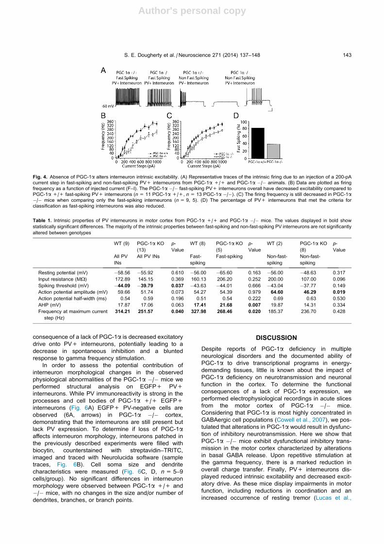
Global ablation of PGC-1a causes reductions inintrinsic firing rate and excitability of FS interneurons
Two interneuron subtypes that contribute significantly to
perisomatic inhibition of layer V pyramidal neurons are
PV-positive and cholecystokinin (CCK)-positive
interneurons. Based on our previous findings of PV
deficiency in PGC-1a �/� mice and the high
concentration of PGC-1a in PV-positive interneurons
(Jiang et al., 2013b), we sought to determine whether
the intrinsic firing properties of PV-positive interneurons
are disrupted in PGC-1a �/� mice. It is possible that
reductions in any number of PGC-1a’s putative targets,
including PV and metabolic genes (St-Pierre et al.,
2006; Cowell et al., 2009), could contribute to alterations
in calcium concentrations and depletion of metabolic sub-
strates for the maintenance of normal intrinsic firing. To
allow for visualization of PV+ neurons, whose somas
are concentrated in cortical layer IV, we generated
PGC-1a +/+ and �/� mice with EGFP-positive PV+
interneurons by crossing PGC-1a �/� mice with the
G42 GAD–EGFP mouse line (Chattopadhyaya et al.,
2004; Bartley et al., 2008) to use for whole-cell current
clamp recordings. Upon current injection, PV+ interneu-
rons, in layer IV, of EGFP-PGC-1a �/� mice showed a
reduction in excitability as compared to +/+ littermates
(Fig. 4A, B, ANOVA, p< 0.02). The frequency is still
reduced in PGC-1a �/� mice when comparing only FS
PV+ interneurons (Fig. 4C, Table 1). Furthermore, FS
PV interneurons in EGFP-PGC-1a �/� mice showed an
increase in AHP and a reduction in frequency at the
maximum current step with no alteration in resting
membrane potential or input resistance (Table 1). Inter-
estingly, there was a large reduction in the percentage
of PV+ interneurons that met the classification criteria
for FS in the EGFP-PGC-1a �/� mice (Fig. 4D). This
reduction in interneuron excitability could contribute to
the observed reduction in AP-mediated GABA release in
the cortex during gamma stimulation.
Global ablation of PGC-1a results in alterations in theexcitatory synaptic activity onto FS interneuronswithout altering interneuron morphology
The decrease in the frequency of sIPSCs onto pyramidal
cells in PGC-1a �/� mice could be caused by reduced
activation of PV+ interneurons. In order to determine if
activation of these cells is affected by the absence of
PGC-1a, whole-cell voltage clamp recordings were
performed on EGFP-positive PV+ interneurons in layer
IV of the motor cortex in the presence of picrotoxin and
APV to isolate AMPA-mediated sEPSCs. The observed
sEPSCs exhibited decreased amplitudes (Fig. 5D, F,
p= 0.0098, t(11) = 3.120) and decreased frequency
(Fig. 5E, G, p= 0.00381, t(11) = 3.651) in PGC-1a �/�mice as compared to PGC-1a +/+ littermates.
Interestingly, the decrease in sEPSC amplitudes is due
to a loss of the large sEPSCs events (Fig. 5C, H,
p= 0.04094, t(11) = 2.315). These large sEPSCs
events are normally caused by glutamate release in
response to APs. Additionally, it is possible that the
large events observed are multiquantal and that the
extent of multiquantal release is reduced in the PGC-1a�/� mice. The number of smaller sEPSCs events
(620 pAs) is unaltered between PGC-1a �/� and PGC-
1a +/+ mice. These results suggest that a possible
Fig. 3. Absence of PGC-1a alters evoked IPSC activity in the motor cortex. (A) Representative traces of train stimulation and response in both
PGC-1a +/+ and PGC-1a �/� animals (average of 10 sweeps per trace). (B) Total charge transfer during the train (area under curve) was
significantly reduced in PGC-1a �/� mice as compared to PGC-1a +/+ mice. (C) The initial response amplitude is reduced in PGC-1a �/� mice
as compared to PGC-1a +/+ mice. (D) Alterations in short-term plasticity over the length of the train were also evident. (E) The average standard
deviation of IPSCn/IPSC1 across events of cells reveals much higher variation in the PGC-1a �/� as compared to the events from PGC-1a +/+
cells. Student’s t-test ⁄p< 0.05 n= 3 animals/group, approx. 10 cells per condition.
142 S. E. Dougherty et al. / Neuroscience 271 (2014) 137–148
Author's personal copy
consequence of a lack of PGC-1a is decreased excitatory
drive onto PV+ interneurons, potentially leading to a
decrease in spontaneous inhibition and a blunted
response to gamma frequency stimulation.
In order to assess the potential contribution of
interneuron morphological changes in the observed
physiological abnormalities of the PGC-1a �/� mice we
performed structural analysis on EGFP+ PV+
interneurons. While PV immunoreactivity is strong in the
processes and cell bodies of PGC-1a +/+ EGFP+
interneurons (Fig. 6A) EGFP+ PV-negative cells are
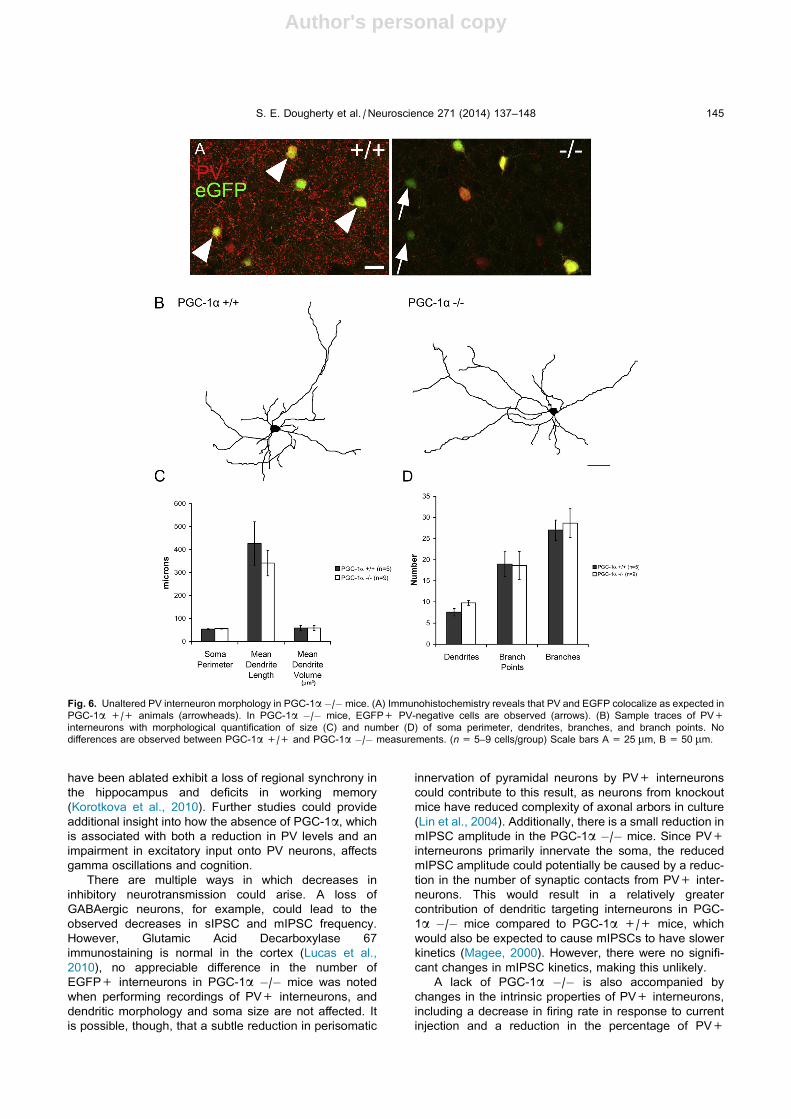
observed (6A, arrows) in PGC-1a �/� cortex,
demonstrating that the interneurons are still present but
lack PV expression. To determine if loss of PGC-1aaffects interneuron morphology, interneurons patched in
the previously described experiments were filled with
biocytin, counterstained with streptavidin–TRITC,
imaged and traced with Neurolucida software (sample
traces, Fig. 6B). Cell soma size and dendrite
characteristics were measured (Fig. 6C, D, n= 5–9
cells/group). No significant differences in interneuron
morphology were observed between PGC-1a +/+ and
�/� mice, with no changes in the size and/or number of
dendrites, branches, or branch points.
DISCUSSION
Despite reports of PGC-1a deficiency in multiple
neurological disorders and the documented ability of
PGC-1a to drive transcriptional programs in energy-
demanding tissues, little is known about the impact of
PGC-1a deficiency on neurotransmission and neuronal
function in the cortex. To determine the functional
consequences of a lack of PGC-1a expression, we
performed electrophysiological recordings in acute slices
from the motor cortex of PGC-1a �/� mice.
Considering that PGC-1a is most highly concentrated in
GABAergic cell populations (Cowell et al., 2007), we pos-
tulated that alterations in PGC-1a would result in dysfunc-
tion of inhibitory neurotransmission. Here we show that
PGC-1a �/� mice exhibit dysfunctional inhibitory trans-
mission in the motor cortex characterized by alterations
in basal GABA release. Upon repetitive stimulation at
the gamma frequency, there is a marked reduction in
overall charge transfer. Finally, PV+ interneurons dis-
played reduced intrinsic excitability and decreased excit-
atory drive. As these mice display impairments in motor
function, including reductions in coordination and an
increased occurrence of resting tremor (Lucas et al.,
Fig. 4. Absence of PGC-1a alters interneuron intrinsic excitability. (A) Representative traces of the intrinsic firing due to an injection of a 200-pA
current step in fast-spiking and non-fast-spiking PV+ interneurons from PGC-1a +/+ and PGC-1a �/� animals. (B) Data are plotted as firing
frequency as a function of injected current (F–I). The PGC-1a �/� fast-spiking PV+ interneurons overall have decreased excitability compared to
PGC-1a +/+ fast-spiking PV+ interneurons (n= 11 PGC-1a +/+, n= 13 PGC-1a �/�). (C) The firing frequency is still decreased in PGC-1a�/� mice when comparing only the fast-spiking interneurons (n= 9, 5). (D) The percentage of PV+ interneurons that met the criteria for
classification as fast-spiking interneurons was also reduced.
Table 1. Intrinsic properties of PV interneurons in motor cortex from PGC-1a +/+ and PGC-1a �/� mice. The values displayed in bold show
statistically significant differences. The majority of the intrinsic properties between fast-spiking and non-fast-spiking PV interneurons are not significantly
altered between genotypes
WT (9) PGC-1a KO
(13)
p-
Value
WT (8) PGC-1a KO
(5)
p-
Value
WT (2) PGC-1a KO
(8)
p-
Value
All PV
INs
All PV INs Fast-
spiking
Fast-spiking Non-fast-
spiking
Non-fast-
spiking
Resting potential (mV) �58.56 �55.92 0.610 �56.00 �65.60 0.163 �56.00 �48.63 0.317
Input resistance (MX) 172.89 145.15 0.369 160.13 206.20 0.252 200.00 107.00 0.096
Spiking threshold (mV) �44.09 �39.79 0.037 �43.63 �44.01 0.666 �43.04 �37.77 0.149
Action potential amplitude (mV) 59.66 51.74 0.073 54.27 54.39 0.979 64.60 46.29 0.019
Action potential half-width (ms) 0.54 0.59 0.196 0.51 0.54 0.222 0.69 0.63 0.530
AHP (mV) 17.87 17.06 0.063 17.41 21.68 0.007 19.87 14.31 0.334
Frequency at maximum current
step (Hz)
314.21 251.57 0.040 327.98 268.46 0.020 185.37 236.70 0.428
S. E. Dougherty et al. / Neuroscience 271 (2014) 137–148 143

Author's personal copy
2012), it is intriguing to postulate that the physiological
alterations in cortical function contribute to hyperactivity
via overexcitation of downstream neuronal targets.
In the absence of PGC-1a, PV expression is reduced
by approximately 80% in the cortex (Lucas et al., 2010);
as such, it is possible that a loss of PV could be contrib-
uting to the synaptic deficits in these mice. While the elec-
trophysiological responses to gamma frequency
stimulation are similar in the hippocampus of PGC-1a�/� and PV �/� mice (Schwaller et al., 1999; Caillard
et al., 2000; Vreugdenhil et al., 2003; Lucas et al.,
2010), motor impairment is much more severe in PGC-
1a �/� mice than PV �/� mice (Schwaller et al., 1999;
Farre-Castany et al., 2007; Lucas et al., 2012). Therefore
it is possible that alterations in other PGC-1a-dependenttranscripts in PV+ interneurons, such as metabolic regu-
lators (Lin et al., 2004; Finck and Kelly, 2006; St-Pierre
et al., 2006) or synaptic proteins (St-Pierre et al., 2006;
Cowell et al., 2009) could be causing changes in inhibitory
neurotransmission. Considering the reduced excitatory
drive onto PV+ interneurons in PGC-1a �/� mice, it is
possible that while PGC-1a is not highly concentrated in
pyramidal neurons, its loss could have an effect on pyra-
midal neuron function. In fact, previous work suggests
that deletion of PGC-1a in excitatory neurons can cause
structural abnormalities (vacuolizations) in the cortex
(Ma et al., 2010), although the functional consequences
of PGC-1a deletion from pyramidal neurons have not
been evaluated. Additionally, we previously suggested
that the observed higher facilitation in the hippocampus
was due to a deficiency of PV at presynaptic terminals
and reduced PV-mediated calcium buffering. In contrast,
we observed a reduction in IPSC amplitude and charge
transfer in the cortex; we believe that changes in
the expression of transcripts, in addition to PV, could be
occurring in the cortex, contributing to the observed
changes in cortical physiology.
PV interneurons are described as primarily FS and
non-adapting (Kawaguchi, 1993; Kawaguchi and Kondo,
2002; Tepper and Bolam, 2004). These intrinsic proper-
ties, together with their perisomatic targeting of pyramidal
neurons (Freund and Katona, 2007), make PV+ inter-
neurons ideal modulators of network oscillations in cortex,
particularly gamma oscillations (Freund, 2003; Bartos
et al., 2007; Sohal, 2012). PV�/� mice have an
enhancement of kainate-induced gamma oscillations in
hippocampal CA3 (Vreugdenhil et al., 2003). However,
mice in which NMDA receptors onto PV interneurons
Fig. 5. Global ablation of PGC-1a results in decreased spontaneous excitatory activity onto parvalbumin interneurons in the motor cortex. (A)
Representative traces of sEPSCs onto PV+ interneurons in PGC-1a +/+ and PGC-1a �/� animals. (B) Representative traces of sEPSCs in
PGC-1a +/+ and PGC-1a �/� animals on an expanded time scale. (C) Histogram plot of sEPSCs onto PV+ interneurons from PGC-1a +/+
(black) and PGC-1a �/� (gray) animals. The larger amplitude sEPSCs are diminished in the PGC-1a �/� animals (D, E) Cumulative probability
plots of amplitude and inter-event interval, respectively. (F) Quantification revealed that the sEPSC amplitude was reduced in the PGC-1a �/�animals compared to PGC-1a +/+. (G) sEPSC frequency was reduced in the PGC-1a �/� animals. (H) The maximum amplitude of sEPSCs is
reduced on the PV+ interneurons in the PGC-1a �/� animals. Student’s t-test ⁄p< 0.05 (PGC-1a +/+ n= 6; PGC-1a �/� n= 8).
144 S. E. Dougherty et al. / Neuroscience 271 (2014) 137–148
Author's personal copy
have been ablated exhibit a loss of regional synchrony in
the hippocampus and deficits in working memory
(Korotkova et al., 2010). Further studies could provide
additional insight into how the absence of PGC-1a, whichis associated with both a reduction in PV levels and an
impairment in excitatory input onto PV neurons, affects
gamma oscillations and cognition.
There are multiple ways in which decreases in
inhibitory neurotransmission could arise. A loss of
GABAergic neurons, for example, could lead to the
observed decreases in sIPSC and mIPSC frequency.
However, Glutamic Acid Decarboxylase 67
immunostaining is normal in the cortex (Lucas et al.,
2010), no appreciable difference in the number of
EGFP+ interneurons in PGC-1a �/� mice was noted
when performing recordings of PV+ interneurons, and
dendritic morphology and soma size are not affected. It
is possible, though, that a subtle reduction in perisomatic
innervation of pyramidal neurons by PV+ interneurons
could contribute to this result, as neurons from knockout
mice have reduced complexity of axonal arbors in culture
(Lin et al., 2004). Additionally, there is a small reduction in
mIPSC amplitude in the PGC-1a �/� mice. Since PV+
interneurons primarily innervate the soma, the reduced
mIPSC amplitude could potentially be caused by a reduc-
tion in the number of synaptic contacts from PV+ inter-
neurons. This would result in a relatively greater
contribution of dendritic targeting interneurons in PGC-
1a �/� mice compared to PGC-1a +/+ mice, which
would also be expected to cause mIPSCs to have slower
kinetics (Magee, 2000). However, there were no signifi-
cant changes in mIPSC kinetics, making this unlikely.
A lack of PGC-1a �/� is also accompanied by
changes in the intrinsic properties of PV+ interneurons,
including a decrease in firing rate in response to current
injection and a reduction in the percentage of PV+
Fig. 6. Unaltered PV interneuron morphology in PGC-1a �/�mice. (A) Immunohistochemistry reveals that PV and EGFP colocalize as expected in
PGC-1a +/+ animals (arrowheads). In PGC-1a �/� mice, EGFP+ PV-negative cells are observed (arrows). (B) Sample traces of PV+
interneurons with morphological quantification of size (C) and number (D) of soma perimeter, dendrites, branches, and branch points. No
differences are observed between PGC-1a +/+ and PGC-1a �/� measurements. (n= 5–9 cells/group) Scale bars A = 25 lm, B = 50 lm.
S. E. Dougherty et al. / Neuroscience 271 (2014) 137–148 145

Author's personal copy
interneurons that met the criteria to be classified as FS.
These changes in PV+ cell firing rate could be caused
by changes in metabolic genes regulated by PGC-1a(Finck and Kelly, 2006); there is abundant evidence for
a high metabolic requirement for cortical PV+ interneu-
rons (reviewed in Jiang et al., 2013a). A loss of PV itself
could contribute to the decreased firing rate, as loss of
the calcium buffer in PV knockout mice has been shown
to alter intrinsic excitability of GABAergic neurons in the
reticular thalamic nucleus (Alberi et al., 2013). However
it is also possible that other factors contribute to this
reduction. In addition, there was a reduction in the fre-
quency and amplitude of sEPSCs onto PV+ interneurons
in the PGC-1a �/� mice. Altogether, these changes
could cause a decrease in spontaneous AP activity in
PV+ cells, which could contribute to the decrease in
sIPSC frequency observed in layer V pyramidal cells.
The observed increase in sIPSC amplitude seems con-
trary to this line of reasoning; however, it is possible that
the increase in sIPSC amplitude could be an indirect con-
sequence of the reduced mIPSC frequency. The loss of
the smallest events would bias the sIPSC measurements
in favor of larger events.
The change in intrinsic excitability could also
contribute to the decrease in IPSCs onto pyramidal cells
at gamma frequency, if the presynaptic axons from
PV+ interneurons are not able to fire as reliably at
66 Hz in the PGC-1a �/� mice. The observed reduction
in initial response size could contribute to the reduced
charge transfer observed. Though when given in train
stimulation if the first event is small we observed that
the size of the subsequent responses can still increase
over time. Alternatively, the decrease in GABA release
during gamma frequency stimulation could be caused by
the depletion of vesicles in the PGC-1a �/� mice.
Because short-term depression during trains of stimuli is
governed in part by the size of the readily releasable
vesicle pool (Dobrunz, 2002), this could be caused by a
reduction in the number of readily releasable vesicles
(Pozzo-Miller et al., 1999), which are thought to equal
the number of docked vesicles (Schikorski and Stevens,
2001). A change in the number of readily releasable ves-
icles might be expected to also alter the paired-pulse ratio
(Walters et al., 2014), which was seen at dentate gyrus
synapses from PGC-1a �/� mice (Lucas et al., 2010)
but not the cortical synapses studied here. However,
other examples have shown no difference in the paired-
pulse ratio between two types of synapses but differences
in short-term plasticity during longer physiological trains
(Speed and Dobrunz, 2009).
Our studies of evoked inhibitory transmission utilized
perisomatic (inter-layer V) stimulation in efforts to
primarily activate axons from GABAergic cell
populations that target the soma of pyramidal neurons.
As previously discussed, PGC-1a is concentrated
primarily in GABAergic neurons and tightly regulates
PV; therefore, it is tempting to postulate that the main
subtype of interneuron contributing to the observed
physiological changes is the PV+ interneuron. In fact,
we found that the excitatory input onto and the firing
rate of PV+ interneurons are reduced in the absence of
PGC-1a. However, it is possible that other interneuron
populations also contribute to alterations in inhibitory
neurotransmission. For example, CCK+ interneurons
also target the perisomatic regions of pyramidal neurons
and are thought to work in concert with PV+ cells to
regulate oscillations (reviewed in (Freund and Katona,
2007)). It is possible that PGC-1a is playing a regulatory
role on transcription within the CCK+ interneuron sub-
type. Cell-specific PGC-1a ablation studies could shed
light on the role of these individual populations in the
observed synaptic dysfunction and allow for differentiation
of interneuron susceptibility in response to loss of PGC-1a.Here we show that genetic ablation of the coactivator
PGC-1a results in overt alterations in GABAergic
transmission in the motor cortex that are different from
what was previously seen in the hippocampus. The
reduction in GABAergic transmission during high-
frequency stimulation could result in hyperexcitability
and contribute to the observed motor dysfunction in
these animals. Although further studies are required to
identify additional PGC-1a-regulated transcripts that
could influence interneuron function and definitively link
deficits in inhibitory neurotransmission to the motor
abnormalities, these studies highlight the critical role for
PGC-1a in the maintenance of normal cortical inhibition.
Acknowledgments—This work was funded by National
Institutes of Health (NIH) Grant 5K01MH077955 (R.M.C.),
1R01NS070009-04 (R.M.C.), 1R01MH098534-02 (L.E.D.),
5P30NS047466-09 (J.J.H.), NIH Blueprint Grant NS57098, and
the Civitan McNulty Scientist (R.M.C.) and Emerging Scholar
(E.K.L.) Awards. We would like to acknowledge the work of
Grace Nix in performing immunuohistochemistry for PV/EGFP
images. We would like to thank the members of the Hablitz labo-
ratory for additional instruction and aid in electrophysiological
training.
REFERENCES
Alberi L, Lintas A, Kretz R, Schwaller B, Villa AE (2013) The calcium-
binding protein parvalbumin modulates the firing 1 properties of
the reticular thalamic nucleus bursting neurons. J Neurophysiol
109(11):2827–2841.
Bartley AF, Huang ZJ, Huber KM, Gibson JR (2008) Differential
activity-dependent, homeostatic plasticity of two neocortical
inhibitory circuits. J Neurophysiol 100(4):1983–1994.
Bartos M, Vida I, Frotscher M, Meyer A, Monyer H, Geiger JR, Jonas
P (2002) Fast synaptic inhibition promotes synchronized gamma
oscillations in hippocampal interneuron networks. Proc Natl Acad
Sci U S A 99(20):13222–13227.
Bartos M, Vida I, Jonas P (2007) Synaptic mechanisms of
synchronized gamma oscillations in inhibitory interneuron
networks. Nat Rev Neurosci 8(1):45–56.
Caillard O, Moreno H, Schwaller B, Llano I, Celio MR, Marty A (2000)
Role of the calcium-binding protein parvalbumin in short-term
synaptic plasticity. Proc Natl Acad Sci U S A 97(24):
13372–13377.
Chattopadhyaya B, Di Cristo G, Higashiyama H, Knott GW, Kuhlman
SJ, Welker E, Huang ZJ (2004) Experience and activity-
dependent maturation of perisomatic GABAergic innervation in
primary visual cortex during a postnatal critical period. J Neurosci
24(43):9598–9611.
Chaturvedi RK, Calingasan NY, Yang L, Hennessey T, Johri A, Beal
MF (2010) Impairment of PGC-1alpha expression,
146 S. E. Dougherty et al. / Neuroscience 271 (2014) 137–148
Author's personal copy
neuropathology and hepatic steatosis in a transgenic mouse
model of Huntington’s disease following chronic energy
deprivation. Hum Mol Genet 19(16):3190–3205.
Christoforou A, Le Hellard S, Thomson PA, Morris SW, Tenesa A,
Pickard BS, Wray NR, Muir WJ, Blackwood DH, Porteous DJ,
Evans KL (2007) Association analysis of the chromosome 4p15-
p16 candidate region for bipolar disorder and schizophrenia. Mol
Psychiatry 12(11):1011–1025.
Cowell RM, Blake KR, Russell JW (2007) Localization of the
transcriptional coactivator PGC-1alpha to GABAergic neurons
during maturation of the rat brain. J Comp Neurol 502(1):1–18.
Cowell RM, Talati P, Blake KR, Meador-Woodruff JH, Russell JW
(2009) Identification of novel targets for PGC-1alpha and histone
deacetylase inhibitors in neuroblastoma cells. Biochem Biophys
Res Commun 379(2):578–582.
Cui L, Jeong H, Borovecki F, Parkhurst CN, Tanese N, Krainc D
(2006) Transcriptional repression of PGC-1alpha by mutant
huntingtin leads to mitochondrial dysfunction and
neurodegeneration. Cell 127(1):59–69.
Dobrunz LE (2002) Release probability is regulated by the size of the
readily releasable vesicle pool at excitatory synapses in
hippocampus. Int J Dev Neurosci 20(3–5):225–236.
Dobrunz LE, Stevens CF (1997) Heterogeneity of release probability,
facilitation, and depletion at central synapses. Neuron
18(6):995–1008.
Dobrunz LE, Huang EP, Stevens CF (1997) Very short-term plasticity
in hippocampal synapses. Proc Natl Acad Sci U S A
94(26):14843–14847.
Farre-Castany MA, Schwaller B, Gregory P, Barski J, Mariethoz C,
Eriksson JL, Tetko IV, Wolfer D, Celio MR, Schmutz I, Albrecht U,
Villa AE (2007) Differences in locomotor behavior revealed in
mice deficient for the calcium-binding proteins parvalbumin,
calbindin D-28k or both. Behav Brain Res 178(2):250–261.
Finck BN, Kelly DP (2006) PGC-1 coactivators: inducible regulators
of energy metabolism in health and disease. J Clin Invest
116(3):615–622.
Freund TF (2003) Interneuron diversity series: rhythm and mood in
perisomatic inhibition. Trends Neurosci 26(9):489–495.
Freund TF, Katona I (2007) Perisomatic inhibition. Neuron
56(1):33–42.
Hettema JM, Webb BT, Guo AY, Zhao Z, Maher BS, Chen X, An SS,
Sun C, Aggen SH, Kendler KS, Kuo PH, Otowa T, Flint J, van den
Oord EJ (2011) Prioritization and association analysis of murine-
derived candidate genes in anxiety-spectrum disorders. Biol
Psychiatry 70(9):888–896.
Jiang Z, Cowell RM, Nakazawa K (2013a) Convergence of genetic
and environmental factors on parvalbumin-positive interneurons
in schizophrenia. Front Behav Neurosci 7:116.
JiangZ,RompalaGR,ZhangS,CowellRM,NakazawaK (2013b)Social
isolation exacerbates schizophrenia-like phenotypes via oxidative
stress in cortical interneurons. Biol Psychiatry 73(10):1024–1034.
Kawaguchi Y (1993) Physiological, morphological, and histochemical
characterization of three classes of interneurons in rat
neostriatum. J Neurosci 13(11):4908–4923.
Kawaguchi Y, Kondo S (2002) Parvalbumin, somatostatin and
cholecystokinin as chemical markers for specific GABAergic
interneuron types in the rat frontal cortex. J Neurocytol 31(3–5):
277–287.
Korotkova T, Fuchs EC, Ponomarenko A, von Engelhardt J, Monyer
H (2010) NMDA receptor ablation on parvalbumin-positive
interneurons impairs hippocampal synchrony, spatial
representations, and working memory. Neuron 68(3):557–569.
Lin J, Wu PH, Tarr PT, Lindenberg KS, St-Pierre J, Zhang CY,
Mootha VK, Jager S, Vianna CR, Reznick RM, Cui L, Manieri M,
Donovan MX, Wu Z, Cooper MP, Fan MC, Rohas LM, Zavacki
AM, Cinti S, Shulman GI, Lowell BB, Krainc D, Spiegelman BM
(2004) Defects in adaptive energy metabolism with
CNS-linked hyperactivity in PGC-1alpha null mice. Cell 119(1):
121–135.
Lucas EK, Markwardt SJ, Gupta S, Meador-Woodruff JH, Lin JD,
Overstreet-Wadiche L, Cowell RM (2010) Parvalbumin deficiency
and GABAergic dysfunction in mice lacking PGC-1alpha. J
Neurosci 30(21):7227–7235.
Lucas EK, Dougherty SE, McMeekin LJ, Trinh AT, Reid CS, Cowell
RM (2012) Developmental alterations in motor coordination and
medium spiny neuron markers in mice lacking pgc-1alpha. PLoS
One 7(8):e42878.
Ma D, Li S, Lucas EK, Cowell RM, Lin JD (2010) Neuronal
inactivation of peroxisome proliferator-activated receptor gamma
coactivator 1alpha (PGC-1alpha) protects mice from diet-induced
obesity and leads to degenerative lesions. J Biol Chem
285(50):39087–39095.
Magee JC (2000) Dendritic integration of excitatory synaptic input.
Nat Rev Neurosci 1(3):181–190.
Pozzo-Miller LD, Gottschalk W, Zhang L, McDermott K, Du J,
Gopalakrishnan R, Oho C, Sheng ZH, Lu B (1999) Impairments in
high-frequency transmission, synaptic vesicle docking, and
synaptic protein distribution in the hippocampus of BDNF
knockout mice. J Neurosci 19(12):4972–4983.
Puigserver P, Wu Z, Park CW, Graves R, Wright M, Spiegelman BM
(1998) A cold-inducible coactivator of nuclear receptors linked to
adaptive thermogenesis. Cell 92(6):829–839.
Qin W, Haroutunian V, Katsel P, Cardozo CP, Ho L, Buxbaum JD,
Pasinetti GM (2009) PGC-1alpha expression decreases in the
Alzheimer disease brain as a function of dementia. Arch Neurol
66(3):352–361.
Ramirez DM, Kavalali ET (2011) Differential regulation of
spontaneous and evoked neurotransmitter release at central
synapses. Curr Opin Neurobiol 21(2):275–282.
Rotaru DC, Yoshino H, Lewis DA, Ermentrout GB, Gonzalez-Burgos
G (2011) Glutamate receptor subtypes mediating synaptic
activation of prefrontal cortex neurons: relevance for
schizophrenia. J Neurosci 31(1):142–156.
Schikorski T, Stevens CF (2001) Morphological correlates of
functionally defined synaptic vesicle populations. Nat Neurosci
4(4):391–395.
Schwaller B, Dick J, Dhoot G, Carroll S, Vrbova G, Nicotera P, Pette
D, Wyss A, Bluethmann H, Hunziker W, Celio MR (1999)
Prolonged contraction-relaxation cycle of fast-twitch muscles in
parvalbumin knockout mice. Am J Physiol 276(2 Pt
1):C395–C403.
Sheng B, Wang X, Su B, Lee HG, Casadesus G, Perry G, Zhu X
(2012) Impaired mitochondrial biogenesis contributes to
mitochondrial dysfunction in Alzheimer’s disease. J Neurochem
120(3):419–429.
Sohal VS (2012) Insights into cortical oscillations arising from
optogenetic studies. Biol Psychiatry 71(12):1039–1045.
Sohal VS, Zhang F, Yizhar O, Deisseroth K (2009) Parvalbumin
neurons and gamma rhythms enhance cortical circuit
performance. Nature 459(7247):698–702.
Speed HE, Dobrunz LE (2009) Developmental changes in short-term
facilitation are opposite at temporoammonic synapses compared
to Schaffer collateral synapses onto CA1 pyramidal cells.
Hippocampus 19(2):187–204.
St-Pierre J, Drori S, Uldry M, Silvaggi JM, Rhee J, Jager S,
Handschin C, Zheng K, Lin J, Yang W, Simon DK, Bachoo R,
Spiegelman BM (2006) Suppression of reactive oxygen species
and neurodegeneration by the PGC-1 transcriptional coactivators.
Cell 127(2):397–408.
Taherzadeh-Fard E, Saft C, Andrich J, Wieczorek S, Arning L (2009)
PGC-1alpha as modifier of onset age in Huntington disease. Mol
Neurodegener 4:10.
Tepper JM, Bolam JP (2004) Functional diversity and specificity of
neostriatal interneurons. Curr Opin Neurobiol 14(6):685–692.
Traub RD, Whittington MA, Colling SB, Buzsaki G, Jefferys JG (1996)
Analysis of gamma rhythms in the rat hippocampus in vitro and
in vivo. J Physiol 493(Pt 2):471–484.
Vreugdenhil M, Jefferys JG, Celio MR, Schwaller B (2003)
Parvalbumin-deficiency facilitates repetitive IPSCs and gamma
oscillations in the hippocampus. J Neurophysiol 89(3):1414–1422.
Walters BJ, Hallengren JJ, Theile CS, Ploegh HL, Wilson SM,
Dobrunz LE (2014) A catalytic independent function of the
S. E. Dougherty et al. / Neuroscience 271 (2014) 137–148 147

Author's personal copy
deubiquitinating enzyme USP14 regulates hippocampal synaptic
short-term plasticity and vesicle number. J Physiol 592(Pt 4):
571–586.
Wang XJ, Buzsaki G (1996) Gamma oscillation by synaptic inhibition
in a hippocampal interneuronal network model. J Neurosci
16(20):6402–6413.
Witte ME, Nijland PG, Drexhage JA, Gerritsen W, Geerts D, van Het
Hof B, Reijerkerk A, de Vries HE, van der Valk P, van Horssen J
(2013) Reduced expression of PGC-1alpha partly underlies
mitochondrial changes and correlates with neuronal loss in
multiple sclerosis cortex. Acta Neuropathol 125(2):231–243.
Zheng B, Liao Z, Locascio JJ, Lesniak KA, Roderick SS, Watt ML,
Eklund AC, Zhang-James Y, Kim PD, Hauser MA, Grunblatt E,
Moran LB, Mandel SA, Riederer P, Miller RM, Federoff HJ,
Wullner U, Papapetropoulos S, Youdim MB, Cantuti-Castelvetri I,
Young AB, Vance JM, Davis RL, Hedreen JC, Adler CH, Beach
TG, Graeber MB, Middleton FA, Rochet JC, Scherzer CR (2010)
PGC-1alpha, a potential therapeutic target for early intervention in
Parkinson’s disease. Sci Transl Med 2(52):52ra73.
(Accepted 15 April 2014)(Available online 24 April 2014)
148 S. E. Dougherty et al. / Neuroscience 271 (2014) 137–148