Embed Size (px)
Citation preview
Loss of the Histidine Kinase DhkD Results in MobileMounds during Development of DictyosteliumdiscoideumCharles K. Singleton*, Yanhua Xiong
Department of Biological Sciences, Vanderbilt University, Nashville, Tennessee, United States of America
Abstract
Background: Histidine kinases are receptors for sensing cellular and environmental signals, and in response to theappropriate cue they initiate phosphorelays that regulate the activity of response regulators. The Dictyosteliumdiscoideum genome encodes 15 histidine kinases that function to regulate several processes during the multicellulardevelopmental program, including the slug to culmination transition, osmoregulation, and spore differentiation. Whilethere are many histidine kinases, there is only a single response regulator, RegA. Not surprisingly given theubiquitous involvement of cAMP in numerous processes of development in Dictyostelium, RegA is a cAMPphosphodiesterase that is activated upon receiving phosphates through a phosphorelay. Hence, all of the histidinekinases characterized to date regulate developmental processes through modulating cAMP production. Here weinvestigate the function of the histidine kinase DhkD.Principal Findings: The dhkD gene was disrupted, and the resulting cells when developed gave a novel phenotype.Upon aggregation, which occurred without streaming, the mounds were motile, a phenotype termed the pollywogstage. The pollywog phenotype was dependent on a functional RegA. After a period of random migration, thepollywogs attempted to form fingers but mostly generated aberrant structures with no tips. While prestalk andprespore cell differentiation occurred with normal timing, proper patterning did not occur. In contrast, wild typemounds are not motile, and the cAMP chemotactic movement of cells within the mound facilitates proper prestalk andprespore patterning, tip formation, and the vertical elongation of the mound into a finger.Conclusions: We postulate that DhkD functions to ensure the proper cAMP distribution within mounds that in turnresults in patterning, tip formation and the transition of mounds to fingers. In the absence of DhkD, aberrant cellmovements in response to an altered cAMP distribution result in mound migration, a lack of proper patterning, and aninability to generate normal finger morphology.
Citation: Singleton CK, Xiong Y (2013) Loss of the Histidine Kinase DhkD Results in Mobile Mounds during Development of Dictyostelium discoideum.PLoS ONE 8(9): e75618. doi:10.1371/journal.pone.0075618
Editor: Adrian John Harwood, Cardiff University, United Kingdom
Received June 24, 2013; Accepted August 15, 2013; Published September 25, 2013
Copyright: © 2013 Singleton et al. This is an open-access article distributed under the terms of the Creative Commons Attribution License, which permitsunrestricted use, distribution, and reproduction in any medium, provided the original author and source are credited.
Funding: The work was supported by the Vanderbilt College of Arts and Sciences. The funders had no role in study design, data collection and analysis,decision to publish, or preparation of the manuscript.
Competing interests: The authors have declared that no competing interests exist.
* E-mail: [email protected]
Introduction
Histidine kinases are receptors in the major mechanism ofsignal transduction in bacteria, the so-called two-componentsignaling systems that mediate numerous physiologicalresponses to various environmental signals and conditions [1].Most often histidine kinases are integral membrane proteinswhose extracellular domain serves to recognize and bind asignaling ligand that in turn activates the intracellular kinasedomain. Autophosphorylation of a histidine residue begins aphosphorelay in which the phosphate is passed to an aspartatein a downstream component, or several his-asp passes occuramong multiple component proteins. The terminal aspartate
acceptor is termed a response regulator whosephosphorylation activates the regulator domain, which in manyinstances in bacteria is a transcription factor. Variouspermutations of this basic signaling system exist, includinghistidine kinases acting as phosphatases to reverse the flow ofphosphates within the relay.
Phosphorelay signaling systems have been found in severaleukaryotes, including plants, yeast and fungi, and Amoebozoa[1]. Dictyostelium discoideum stands out among theseeukaryotes because of the relatively large number of histidinekinases in its genome [2]. Several of the 15 histidine kinases ofDictyostelium have been characterized, and they function in anumber of different processes during the multicellular
PLOS ONE | www.plosone.org 1 September 2013 | Volume 8 | Issue 9 | e75618
developmental program, including spore encapsulation, sporedormancy, osmoregulation, prespore to spore differentiation,and the slug to culmination transition [3-11].
While the functions of and the signals/ligands that activatethe histidine kinases of Dictyostelium are varied, all of thekinases appear to regulate phosphorelays that terminate in asingle response regulator, RegA [12-14]. The regulatorydomain of RegA is a cAMP phosphodiesterase, andphosphorylation of RegA by a phosphorelay activates thisactivity. As has been amply documented, cAMP is pervasive inmediating and regulating numerous processes during thedevelopment of Dictyostelium cells into a multicellular organism[15]. For instance, chemotaxis to cAMP mediates aggregationof starving individual cells resulting in the initial multicellularstructure, that being a mound. Within the mounds, differentialcAMP chemotaxis, along with differential cell adhesion [16], ofthe newly formed prestalk and prespore cell types (arising inpart due to cAMP stimulation of protein kinase A) results incomplex movements of the cells that generate the formation ofa tip composed of various prestalk cell types [17]. The tipdirects a vertical elongation of the mound into a finger and slug.cAMP chemotaxis also drives cell movements within the slugsthat lead to slugs being motile [17].
Herein we characterize the function of the histidine kinaseDhkD by disrupting the dhkD gene and observing the resultingphenotype. DhkD is a relatively large protein that possessestwo histidine kinase and two receiver domains on theintracellular side of a single pass transmembrane domain andextracellular PAS and PAC domains that likely are involved inligand binding (http://www.uniprot.org/uniprot/Q54SP4).Development of the dhkD null cells reveals a novel phenotypein that motility is conferred to the usually non-motile mounds.For a 3 to 4 hour period, the mounds, termed pollywogs due totheir similar appearance to tadpoles, migrate randomly.Prestalk and prespore cell types are generated with normaltiming and are initially scattered throughout the pollywogs asthey are in wild type non-motile mounds. However, the typicalpatterning that subsequently results as wild type mounds formtips and the tipped mounds transition to fingers does not occurin the dhkD developing entities. The net result is very few fruitsare formed by the mutant cells. The pollywog phenotype isrescued by disrupting the regA gene in the dhkD null cells,suggesting that dhkD functions by modulating the cAMPphosphodiesterase activity of RegA, like other characterizedhistidine kinases of Dictyostelium.
We postulate that in response to an as yet unknown signal,DhkD activates RegA to regulate the production of cAMP andensure the necessary cAMP distribution within a mound. Theresulting chemotactic cell movements in response to the cAMPdistribution normally mediate proper patterning of the prestalkand prespore cells, tip formation, and the vertical elongation ofthe mound into a finger. In the absence of DhkD, we suggestthat the cAMP environment within a mound is altered such thatthe chemotactic cell movements result in mound migrationinstead of finger formation.
Materials and Methods
Disruption of dhkDA disruption construct, pdhkD-4, was made as follows. The
blasticidin resistance gene cassette from pBSR519 [18] wasinserted into a BamHI site between a 658 bp 5’ region of thedhkD gene (covering most of exons one and two) and an 812bp 3’ region (corresponding to codons 310-580) that had beencloned into the pGEM T-easy vector (Promega). Thus, the bsrcassette replaced the first half of the first catalytic domain. The5’ fragment was generated by PCR using primers dhkd-5 anddhkd-6, while the 3’ fragment was made with dhkd-3 anddhkd-4 (table 1). Digestion with EcoRI was carried out torelease the dhkD/bsr fusion prior to transformation intoDictyostelium Ax4 cells via electroporation. To check fordisruption, genomic DNA was isolated from blasticidin resistantclones and used as a template in a PCR reaction with ablasticidin specific primer and a dhkD primer downstream ofthe cloned 3’ region. Several disrupted isolates independentlyderived from multiple transformations gave the same aberrantphenotypes and showed no detectable dhkD mRNA. One suchstrain was named BS170 and was used for the experiments inthis paper. Similar disruptions were also made in a strain nullfor regA (DBS0236257, Dictyostelium Stock Center) resultingin a strain, BS171, that is doubly disrupted in regA and dhkD.The dhkD disruption plasmid used in this instance waspdhkD-12 for which the bsr cassette of pdhkD-4 was replacedwith a hygromycin cassette [19].
Fusing the dhkD promoter to lacZPCR using primers dhkd-17 and dhkd-23 (table 1) gave a
1174 bp fragment corresponding to the 5’ upstream sequencesand the first seven codons of the dhkD gene. After sequencing,the fragment was used to replace the ecmAO promoter inpecmAO-i-α-gal (BglII and XbaI digestion) and thus becamefused to a rapid turnover version of β-galactosidase. Theresulting plasmid was named pdhkD-11 and was transformedinto Ax4. LacZ constructs for the pre-stalk- and pre-spore-
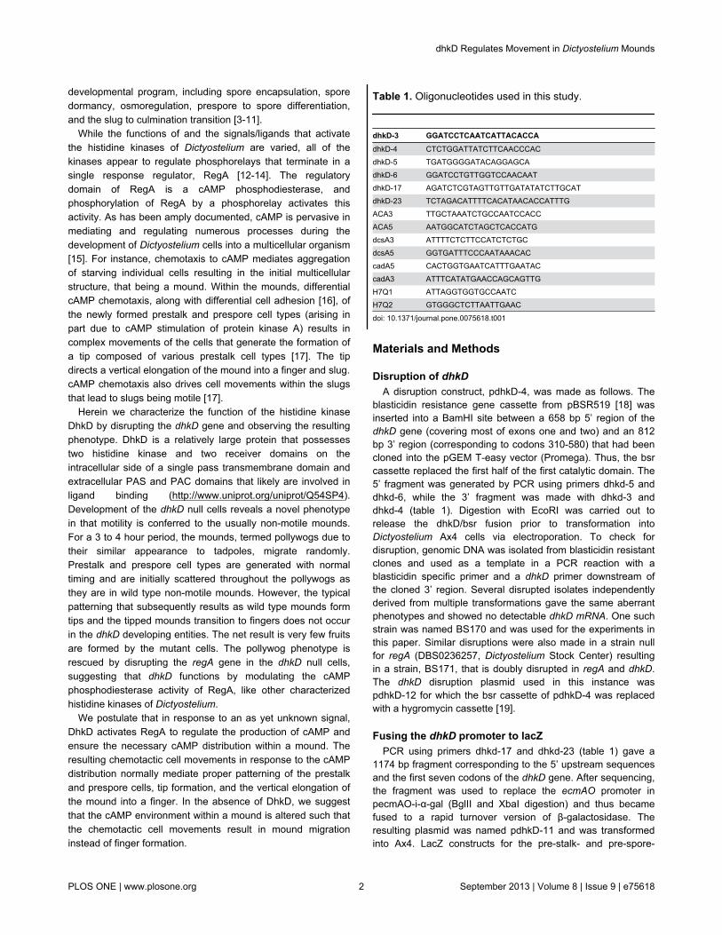
Table 1. Oligonucleotides used in this study.
dhkD-3 GGATCCTCAATCATTACACCAdhkD-4 CTCTGGATTATCTTCAACCCACdhkD-5 TGATGGGGATACAGGAGCAdhkD-6 GGATCCTGTTGGTCCAACAATdhkD-17 AGATCTCGTAGTTGTTGATATATCTTGCATdhkD-23 TCTAGACATTTTCACATAACACCATTTGACA3 TTGCTAAATCTGCCAATCCACCACA5 AATGGCATCTAGCTCACCATGdcsA3 ATTTTCTCTTCCATCTCTGCdcsA5 GGTGATTTCCCAATAAACACcadA5 CACTGGTGAATCATTTGAATACcadA3 ATTTCATATGAACCAGCAGTTGH7Q1 ATTAGGTGGTGCCAATCH7Q2 GTGGGCTCTTAATTGAAC
doi: 10.1371/journal.pone.0075618.t001
dhkD Regulates Movement in Dictyostelium Mounds
PLOS ONE | www.plosone.org 2 September 2013 | Volume 8 | Issue 9 | e75618
specific promoters ecmAO, ecmA, ecmB, and pspA weregenerously provided by K. Jermyn and J. Williams and weretransformed into Ax4 and into BS170. Staining for β-galactosidase activity was carried out as described [20].
Cell growth and developmentStrains were maintained and grown in HL-5 medium [21] at
21°C. For development, cells were grown in the presence ofKlebsiella pneumoniae on plates, harvested, and excessbacteria removed by differential centrifugation prior to platingcells on nitrocellulose filters for standard development [22].Plating cells at lower than normal densities or to examine slugswas done on 2% agar. For slugs, the plates were kept in a lightproof box or in a box with a small pinhole to allow low intensity,directional light.
RT-PCRRNA was isolated using Trizol (InVitrogen). RT-PCR was
carried out as described [23] using various primer pairs aslisted in the results section and shown in table 1. For eachprimer pair, RNA concentrations, annealing temperatures, andcycle numbers were optimized to maximize sensitivity tovariations in RNA levels between samples: acaA, 15 cycleswith an annealing temperature (30 seconds) from 59° to 50°followed by 10 cycles at 55°; cadA, 20 cycles with an annealingtemperature (30 seconds) from 55° to 45° followed by 13cycles at 43°; dcsA, 15 cycles with an annealing temperature(30 seconds) from 55° to 50° followed by 10 cycles at 52°;dhkD, 25 cycles with an annealing temperature (45 seconds) of55°. In each case, differences in mRNA levels of two to ten-foldwere readily detected. Controls with no reverse transcriptasewere included to demonstrate no genomic DNA contaminationexisted. Oligonucleotides specific for the H7 gene were usedas an internal control as a constitutively transcribed geneduring growth and development [24].
Microscopy and image processingDeveloping cells and β-galactosidase results were
photographed with a Leica MZ16 stereomicroscope equippedwith a Q-Imaging Retiga 1300 camera and Q-Imaging software.Images were imported into Microsoft PowerPoint, labeled, andsaved as a PDF file. The figures were cropped and sized usingAdobe Photoshop CS6. Time-lapse photography was carriedout using a Leica DM6000B microscope with a 5X objectiveand SimplePCI software. 3 to 5 percent of the developing cellswere expressing GFP (transformed with pTX-GFP [25]) toenhance the ability to observe cell movement. The time-lapsesequence was begun once the developing cells showed signsof obvious mound formation and was continued past the fingerstage. Pictures under fluorescent light were taken at 3-minuteintervals. The movies in the supporting materials show a 2.5-hour portion of the time-lapse sequence, beginning at themound stage. The movies were compiled using 3 frames (9minutes real time) per second.
Results
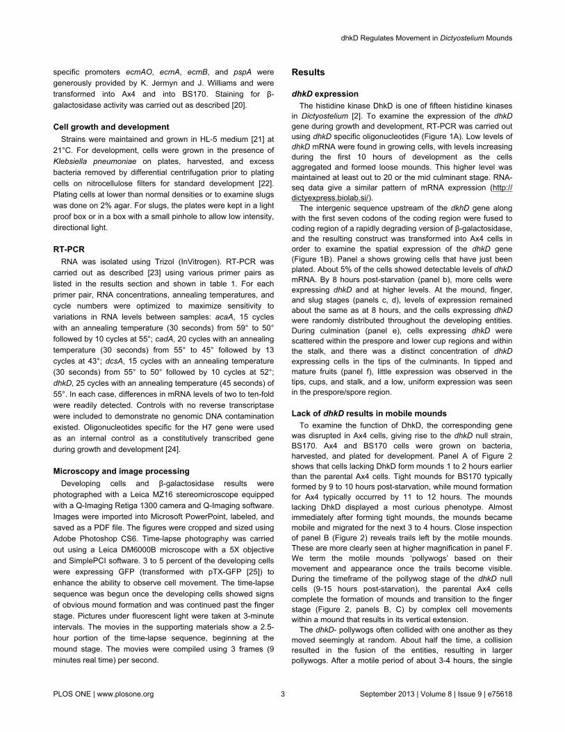
dhkD expressionThe histidine kinase DhkD is one of fifteen histidine kinases
in Dictyostelium [2]. To examine the expression of the dhkDgene during growth and development, RT-PCR was carried outusing dhkD specific oligonucleotides (Figure 1A). Low levels ofdhkD mRNA were found in growing cells, with levels increasingduring the first 10 hours of development as the cellsaggregated and formed loose mounds. This higher level wasmaintained at least out to 20 or the mid culminant stage. RNA-seq data give a similar pattern of mRNA expression (http://dictyexpress.biolab.si/).
The intergenic sequence upstream of the dkhD gene alongwith the first seven codons of the coding region were fused tocoding region of a rapidly degrading version of β-galactosidase,and the resulting construct was transformed into Ax4 cells inorder to examine the spatial expression of the dhkD gene(Figure 1B). Panel a shows growing cells that have just beenplated. About 5% of the cells showed detectable levels of dhkDmRNA. By 8 hours post-starvation (panel b), more cells wereexpressing dhkD and at higher levels. At the mound, finger,and slug stages (panels c, d), levels of expression remainedabout the same as at 8 hours, and the cells expressing dhkDwere randomly distributed throughout the developing entities.During culmination (panel e), cells expressing dhkD werescattered within the prespore and lower cup regions and withinthe stalk, and there was a distinct concentration of dhkDexpressing cells in the tips of the culminants. In tipped andmature fruits (panel f), little expression was observed in thetips, cups, and stalk, and a low, uniform expression was seenin the prespore/spore region.
Lack of dhkD results in mobile moundsTo examine the function of DhkD, the corresponding gene
was disrupted in Ax4 cells, giving rise to the dhkD null strain,BS170. Ax4 and BS170 cells were grown on bacteria,harvested, and plated for development. Panel A of Figure 2shows that cells lacking DhkD form mounds 1 to 2 hours earlierthan the parental Ax4 cells. Tight mounds for BS170 typicallyformed by 9 to 10 hours post-starvation, while mound formationfor Ax4 typically occurred by 11 to 12 hours. The moundslacking DhkD displayed a most curious phenotype. Almostimmediately after forming tight mounds, the mounds becamemobile and migrated for the next 3 to 4 hours. Close inspectionof panel B (Figure 2) reveals trails left by the motile mounds.These are more clearly seen at higher magnification in panel F.We term the motile mounds ‘pollywogs’ based on theirmovement and appearance once the trails become visible.During the timeframe of the pollywog stage of the dhkD nullcells (9-15 hours post-starvation), the parental Ax4 cellscomplete the formation of mounds and transition to the fingerstage (Figure 2, panels B, C) by complex cell movementswithin a mound that results in its vertical extension.
The dhkD- pollywogs often collided with one another as theymoved seemingly at random. About half the time, a collisionresulted in the fusion of the entities, resulting in largerpollywogs. After a motile period of about 3-4 hours, the single
dhkD Regulates Movement in Dictyostelium Mounds
PLOS ONE | www.plosone.org 3 September 2013 | Volume 8 | Issue 9 | e75618
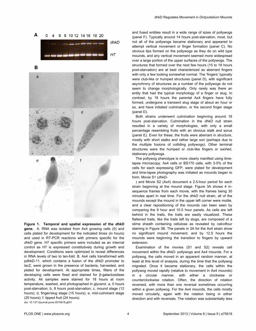
Figure 1. Temporal and spatial expression of the dhkDgene. A. RNA was isolated from Ax4 growing cells (0) andcells plated for development for the indicated times (in hours)and used in RT-PCR reactions with primers specific for thedhkD gene. H7 specific primers were included as an internalcontrol as H7 is expressed constitutively during growth anddevelopment. Conditions were optimized to reveal differencesin RNA levels of two to ten-fold. B. Ax4 cells transformed withpdhkD-11, which contains a fusion of the dhkD promoter tolacZ, were grown in the presence of bacteria, harvested, andplated for development. At appropriate times, filters of thedeveloping cells were fixed and stained for β-galactosidaseactivity. All samples were stained for 18 hours at roomtemperature, washed, and photographed in glycerol. a, 0 hourspost-starvation; b, 8 hours post-starvation; c, mound stage (12hours); d, finger/slug stage (15 hours); e, mid-culminant stage(20 hours); f, tipped fruit (24 hours).doi: 10.1371/journal.pone.0075618.g001
and fused entities result in a wide range of sizes of pollywogs(panel F). Typically around 14 hours post-starvation, most, butnot all of the pollywogs became stationary and appeared toattempt vertical movement or finger formation (panel C). Noobvious tips formed on the pollywogs as they do on wild typemounds, and any vertical movement seemed more widespreadover a large portion of the upper surfaces of the pollywogs. Thestructures that formed over the next few hours (15 to 18 hourspost-starvation) are at best characterized as aberrant fingerswith only a few looking somewhat normal. The ‘fingers’ typicallywere club-like or humped structures (panel D), with significantasynchrony of structures as a number of the pollywogs do notseem to change morphologically. Only rarely was there anentity that had the typical morphology of a finger or slug. Incontrast, by 18 hours the parental Ax4 fingers have fullyformed, undergone a transient slug stage of about an hour orso, and have initiated culmination, or the second finger stage(panel D).
Both strains underwent culmination beginning around 18hours post-starvation. Culmination in the dhkD null strainresulted in a variety of morphologies, with only a smallpercentage resembling fruits with an obvious stalk and sorus(panel E). Even for these, the fruits were aberrant in structure,mostly with short stalks and rather large sori (perhaps due tothe multiple fusions of colliding pollywogs). Other terminalstructures were the humped or club-like fingers or swirled,stationary pollywogs.
The pollywog phenotype is more clearly manifest using time-lapse microscopy. Ax4 cells or BS170 cells, with 3-5% of thecells for each expressing GFP, were plated for developmentand time-lapse photography was initiated as mounds began toform. Movie S1 (dhkD-
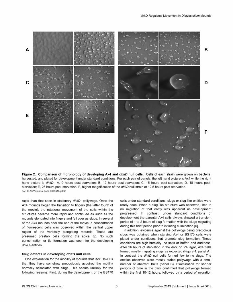
) and Movie S2 (Ax4) document a 2.5-hour period for eachstrain beginning at the mound stage. Figure 3A shows 4 in-sequence frames from each movie, with the frames being 30minutes apart in real time. For the dhkD null strain, all of themounds except the mound in the upper left corner were motile,and a clear repositioning of the mounds can been seen bycomparing the 9 hour and 10.5 hour panels. As cells are leftbehind in the trails, the trails are easily visualized. Theseflattened trails, like the trails left by slugs, are composed of aslime sheath containing cellulose as revealed by calcoflourstaining in Figure 3B. The panels in 3A for the Ax4 strain showno significant mound movement, and by 12.5 hours themounds were beginning the transition to fingers by upwardextension.
Examination of the movies (S1 and S2) reveals cellmovement within the dhkD- pollywogs and Ax4 mounds. For apollywog, the cells moved in an apparent random manner, atleast at this level of analysis, during the time that the pollywogmigrated. Once it became stationary, the cells within thepollywog moved rapidly (relative to movement in Ax4 mounds)in a circular manner, with either a clockwise orcounterclockwise rotation. Often, the direction of rotationreversed, with more than one reversal sometimes occurringwithin a given pollywog. For the Ax4 mounds, the cells mostlymoved circularly, again with the rotation being in eitherdirection and with reversals. The rotation was substantially less
dhkD Regulates Movement in Dictyostelium Mounds
PLOS ONE | www.plosone.org 4 September 2013 | Volume 8 | Issue 9 | e75618
rapid than that seen in stationary dhkD- pollywogs. Once theAx4 mounds began the transition to fingers (the latter fourth ofthe movie), the rotational movement of the cells within thestructures became more rapid and continued as such as themounds elongated into fingers and fell over as slugs. In severalof the Ax4 mounds near the end of the movie, a concentrationof fluorescent cells was observed within the central upperregion of the vertically elongating mounds. These arepresumed prestalk cells forming the apical tip. No suchconcentration or tip formation was seen for the developingdhkD- entities.
Slug defects in developing dhkD null cellsOne explanation for the mobility of mounds that lack DhkD is
that they have somehow precociously acquired the motilitynormally associated with slugs. This seems unlikely for thefollowing reasons. First, during the development of the BS170
cells under standard conditions, slugs or slug-like entities wererarely seen. When a slug-like structure was observed, little tono migration of that entity was apparent as developmentprogressed. In contrast, under standard conditions ofdevelopment the parental Ax4 cells always showed a transientperiod of 1 to 2 hours of slug formation with the slugs migratingduring this brief period prior to initiating culmination [6].
In addition, evidence against the pollywogs being precociousslugs was obtained when starving Ax4 or BS170 cells wereplated under conditions that promote slug formation. Theseconditions are high humidity, no salts or buffer, and darkness.After 26 hours of starvation in the dark on 2% agar, Ax4 cellsformed mostly migrating slugs as expected (Figure 4, panel A).In contrast the dhkD null cells formed few to no slugs. Theentities observed were mostly curled pollywogs with a smallnumber of aberrant fruits (panel B). Examination for shorterperiods of time in the dark confirmed that pollywogs formedwithin the first 10-12 hours, followed by a period of migration
Figure 2. Comparison of morphology of developing Ax4 and dhkD null cells. Cells of each strain were grown on bacteria,harvested, and plated for development under standard conditions. For each pair of panels, the left hand picture is Ax4 while the righthand picture is dhkD-. A, 9 hours post-starvation; B, 12 hours post-starvation; C, 15 hours post-starvation; D, 18 hours post-starvation; E, 26 hours post-starvation; F, higher magnification of the dhkD null strain at 12.5 hours post-starvation.doi: 10.1371/journal.pone.0075618.g002
dhkD Regulates Movement in Dictyostelium Mounds
PLOS ONE | www.plosone.org 5 September 2013 | Volume 8 | Issue 9 | e75618
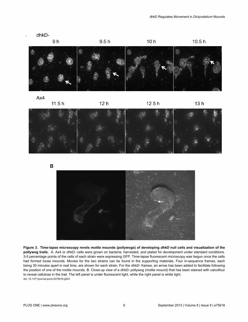
Figure 3. Time-lapse microscopy revels motile mounds (pollywogs) of developing dhkD null cells and visualization of thepollywog trails. A. Ax4 or dhkD- cells were grown on bacteria, harvested, and plated for development under standard conditions.3-5 percentage points of the cells of each strain were expressing GFP. Time-lapse fluorescent microscopy was begun once the cellshad formed loose mounds. Movies for the two strains can be found in the supporting materials. Four in-sequence frames, eachbeing 30 minutes apart in real time, are shown for each strain. For the dhkD- frames, an arrow has been added to facilitate followingthe position of one of the motile mounds. B. Close-up view of a dhkD- pollywog (motile mound) that has been stained with calcoflourto reveal cellulose in the trail. The left panel is under fluorescent light, while the right panel is white light.doi: 10.1371/journal.pone.0075618.g003
dhkD Regulates Movement in Dictyostelium Mounds
PLOS ONE | www.plosone.org 6 September 2013 | Volume 8 | Issue 9 | e75618
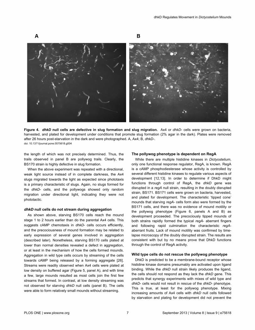
the length of which was not precisely determined. Thus, thetrails observed in panel B are pollywog trails. Clearly, theBS170 strain is highly defective in slug formation.
When the above experiment was repeated with a directional,weak light source instead of in complete darkness, the Ax4slugs migrated towards the light as expected since phototaxisis a primary characteristic of slugs. Again, no slugs formed forthe dhkD- cells, and the pollywogs showed only randommigration under directional light, indicating they were notphototactic.
dhkD null cells do not stream during aggregationAs shown above, starving BS170 cells reach the mound
stage 1 to 2 hours earlier than do the parental Ax4 cells. Thissuggests cAMP chemotaxis in dhkD- cells occurs efficiently,and the precociousness of mound formation may be related toearly expression of several genes involved in aggregation(described later). Nonetheless, starving BS170 cells plated atlower than normal densities revealed a defect in aggregation,or at least in the mechanism of how the cells formed mounds.Aggregation in wild type cells occurs by streaming of the cellstowards cAMP being released by a forming aggregate [26].Streams were readily observed when Ax4 cells were plated atlow density on buffered agar (Figure 5, panel A), and with timea few, large mounds resulted as most cells join the first fewstreams that formed. In contrast, at low density streaming wasnot observed for starving dhkD null cells (panel B). The cellswere able to form relatively small mounds without streaming.
The pollywog phenotype is dependent on RegAWhile there are multiple histidine kinases in Dictyostelium,
only one functional response regulator, RegA, is known. RegAis a cAMP phosphodiesterase whose activity is controlled byseveral different histidine kinases to regulate various aspects ofdevelopment [12,13]. In order to determine if DhkD mightfunctions through control of RegA, the dhkD gene wasdisrupted in a regA null strain, resulting in the doubly disruptedstrain, BS171. BS171 cells were grown on bacteria, harvested,and plated for development. The characteristic ‘tipped cone’mounds that starving regA- cells form also were formed by theBS171 cells, and there was no evidence of mound motility orthe pollywog phenotype (Figure 6, panels A and B) asdevelopment proceeded. The precociously tipped mounds ofboth strains rapidly formed the typical regA- aberrant fingersand following rapid culmination the characteristic regA-aberrant fruits. Lack of mound motility was confirmed by time-lapse microscopy of the doubly disrupted strain. The results areconsistent with but by no means prove that DhkD functionsthrough the control of RegA activity.
Wild type cells do not rescue the pollywog phenotypeDhkD is predicted to be a membrane-bound receptor whose
histidine kinase domains presumably are activated upon ligandbinding. While the dhkD null strain likely produces the ligand,the cells should not respond as they lack the dhkD gene. Thispredicts that synergy experiments with mixes of wild type anddhkD- cells would not result in rescue of the dhkD- phenotype.This is true, at least for the pollywog phenotype. Mixingincreasing amounts of Ax4 cells with dhkD null cells followedby starvation and plating for development did not prevent the
Figure 4. dhkD null cells are defective in slug formation and slug migration. Ax4 or dhkD- cells were grown on bacteria,harvested, and plated for development under conditions that promote slug formation (2% agar in the dark). Plates were removedafter 26 hours post-starvation in the dark and were photographed. A, Ax4; B, dhkD-.doi: 10.1371/journal.pone.0075618.g004
dhkD Regulates Movement in Dictyostelium Mounds
PLOS ONE | www.plosone.org 7 September 2013 | Volume 8 | Issue 9 | e75618
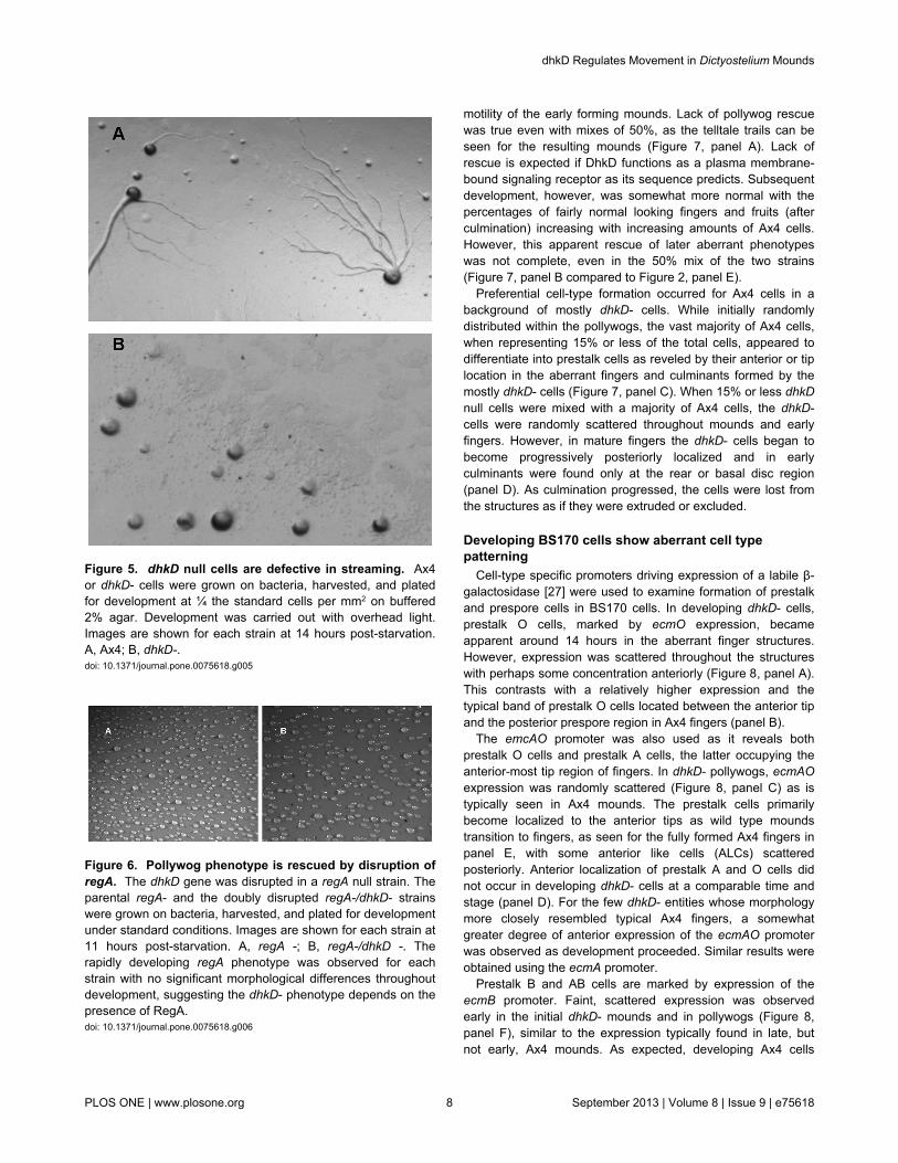
Figure 5. dhkD null cells are defective in streaming. Ax4or dhkD- cells were grown on bacteria, harvested, and platedfor development at ¼ the standard cells per mm2 on buffered2% agar. Development was carried out with overhead light.Images are shown for each strain at 14 hours post-starvation.A, Ax4; B, dhkD-.doi: 10.1371/journal.pone.0075618.g005
Figure 6. Pollywog phenotype is rescued by disruption ofregA. The dhkD gene was disrupted in a regA null strain. Theparental regA- and the doubly disrupted regA-/dhkD- strainswere grown on bacteria, harvested, and plated for developmentunder standard conditions. Images are shown for each strain at11 hours post-starvation. A, regA -; B, regA-/dhkD -. Therapidly developing regA phenotype was observed for eachstrain with no significant morphological differences throughoutdevelopment, suggesting the dhkD- phenotype depends on thepresence of RegA.doi: 10.1371/journal.pone.0075618.g006
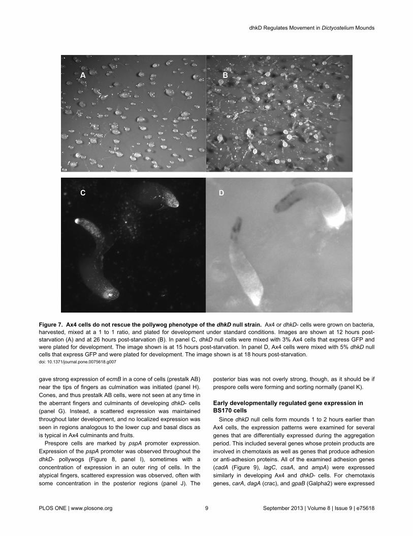
motility of the early forming mounds. Lack of pollywog rescuewas true even with mixes of 50%, as the telltale trails can beseen for the resulting mounds (Figure 7, panel A). Lack ofrescue is expected if DhkD functions as a plasma membrane-bound signaling receptor as its sequence predicts. Subsequentdevelopment, however, was somewhat more normal with thepercentages of fairly normal looking fingers and fruits (afterculmination) increasing with increasing amounts of Ax4 cells.However, this apparent rescue of later aberrant phenotypeswas not complete, even in the 50% mix of the two strains(Figure 7, panel B compared to Figure 2, panel E).
Preferential cell-type formation occurred for Ax4 cells in abackground of mostly dhkD- cells. While initially randomlydistributed within the pollywogs, the vast majority of Ax4 cells,when representing 15% or less of the total cells, appeared todifferentiate into prestalk cells as reveled by their anterior or tiplocation in the aberrant fingers and culminants formed by themostly dhkD- cells (Figure 7, panel C). When 15% or less dhkDnull cells were mixed with a majority of Ax4 cells, the dhkD-cells were randomly scattered throughout mounds and earlyfingers. However, in mature fingers the dhkD- cells began tobecome progressively posteriorly localized and in earlyculminants were found only at the rear or basal disc region(panel D). As culmination progressed, the cells were lost fromthe structures as if they were extruded or excluded.
Developing BS170 cells show aberrant cell typepatterning
Cell-type specific promoters driving expression of a labile β-galactosidase [27] were used to examine formation of prestalkand prespore cells in BS170 cells. In developing dhkD- cells,prestalk O cells, marked by ecmO expression, becameapparent around 14 hours in the aberrant finger structures.However, expression was scattered throughout the structureswith perhaps some concentration anteriorly (Figure 8, panel A).This contrasts with a relatively higher expression and thetypical band of prestalk O cells located between the anterior tipand the posterior prespore region in Ax4 fingers (panel B).
The emcAO promoter was also used as it reveals bothprestalk O cells and prestalk A cells, the latter occupying theanterior-most tip region of fingers. In dhkD- pollywogs, ecmAOexpression was randomly scattered (Figure 8, panel C) as istypically seen in Ax4 mounds. The prestalk cells primarilybecome localized to the anterior tips as wild type moundstransition to fingers, as seen for the fully formed Ax4 fingers inpanel E, with some anterior like cells (ALCs) scatteredposteriorly. Anterior localization of prestalk A and O cells didnot occur in developing dhkD- cells at a comparable time andstage (panel D). For the few dhkD- entities whose morphologymore closely resembled typical Ax4 fingers, a somewhatgreater degree of anterior expression of the ecmAO promoterwas observed as development proceeded. Similar results wereobtained using the ecmA promoter.
Prestalk B and AB cells are marked by expression of theecmB promoter. Faint, scattered expression was observedearly in the initial dhkD- mounds and in pollywogs (Figure 8,panel F), similar to the expression typically found in late, butnot early, Ax4 mounds. As expected, developing Ax4 cells
dhkD Regulates Movement in Dictyostelium Mounds
PLOS ONE | www.plosone.org 8 September 2013 | Volume 8 | Issue 9 | e75618
gave strong expression of ecmB in a cone of cells (prestalk AB)near the tips of fingers as culmination was initiated (panel H).Cones, and thus prestalk AB cells, were not seen at any time inthe aberrant fingers and culminants of developing dhkD- cells(panel G). Instead, a scattered expression was maintainedthroughout later development, and no localized expression wasseen in regions analogous to the lower cup and basal discs asis typical in Ax4 culminants and fruits.
Prespore cells are marked by pspA promoter expression.Expression of the pspA promoter was observed throughout thedhkD- pollywogs (Figure 8, panel I), sometimes with aconcentration of expression in an outer ring of cells. In theatypical fingers, scattered expression was observed, often withsome concentration in the posterior regions (panel J). The
posterior bias was not overly strong, though, as it should be ifprespore cells were forming and sorting normally (panel K).
Early developmentally regulated gene expression inBS170 cells
Since dhkD null cells form mounds 1 to 2 hours earlier thanAx4 cells, the expression patterns were examined for severalgenes that are differentially expressed during the aggregationperiod. This included several genes whose protein products areinvolved in chemotaxis as well as genes that produce adhesionor anti-adhesion proteins. All of the examined adhesion genes(cadA (Figure 9), lagC, csaA, and ampA) were expressedsimilarly in developing Ax4 and dhkD- cells. For chemotaxisgenes, carA, dagA (crac), and gpaB (Galpha2) were expressed
Figure 7. Ax4 cells do not rescue the pollywog phenotype of the dhkD null strain. Ax4 or dhkD- cells were grown on bacteria,harvested, mixed at a 1 to 1 ratio, and plated for development under standard conditions. Images are shown at 12 hours post-starvation (A) and at 26 hours post-starvation (B). In panel C, dhkD null cells were mixed with 3% Ax4 cells that express GFP andwere plated for development. The image shown is at 15 hours post-starvation. In panel D, Ax4 cells were mixed with 5% dhkD nullcells that express GFP and were plated for development. The image shown is at 18 hours post-starvation.doi: 10.1371/journal.pone.0075618.g007
dhkD Regulates Movement in Dictyostelium Mounds
PLOS ONE | www.plosone.org 9 September 2013 | Volume 8 | Issue 9 | e75618
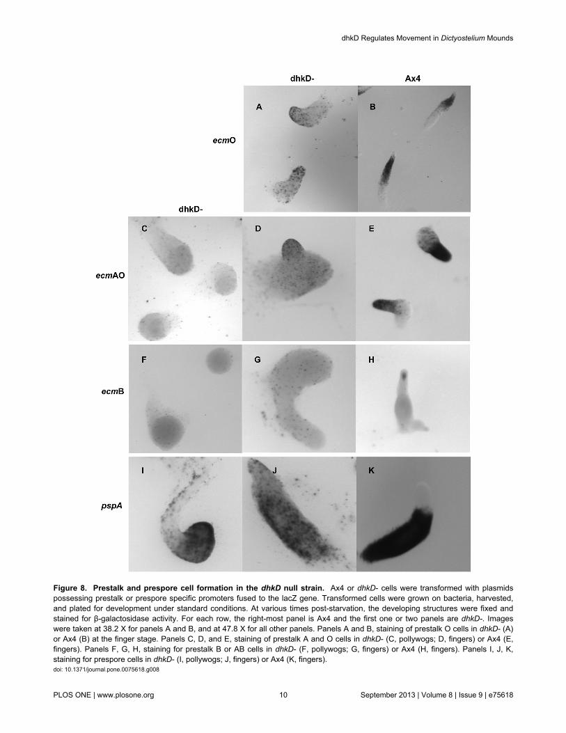
Figure 8. Prestalk and prespore cell formation in the dhkD null strain. Ax4 or dhkD- cells were transformed with plasmidspossessing prestalk or prespore specific promoters fused to the lacZ gene. Transformed cells were grown on bacteria, harvested,and plated for development under standard conditions. At various times post-starvation, the developing structures were fixed andstained for β-galactosidase activity. For each row, the right-most panel is Ax4 and the first one or two panels are dhkD-. Imageswere taken at 38.2 X for panels A and B, and at 47.8 X for all other panels. Panels A and B, staining of prestalk O cells in dhkD- (A)or Ax4 (B) at the finger stage. Panels C, D, and E, staining of prestalk A and O cells in dhkD- (C, pollywogs; D, fingers) or Ax4 (E,fingers). Panels F, G, H, staining for prestalk B or AB cells in dhkD- (F, pollywogs; G, fingers) or Ax4 (H, fingers). Panels I, J, K,staining for prespore cells in dhkD- (I, pollywogs; J, fingers) or Ax4 (K, fingers).doi: 10.1371/journal.pone.0075618.g008
dhkD Regulates Movement in Dictyostelium Mounds
PLOS ONE | www.plosone.org 10 September 2013 | Volume 8 | Issue 9 | e75618
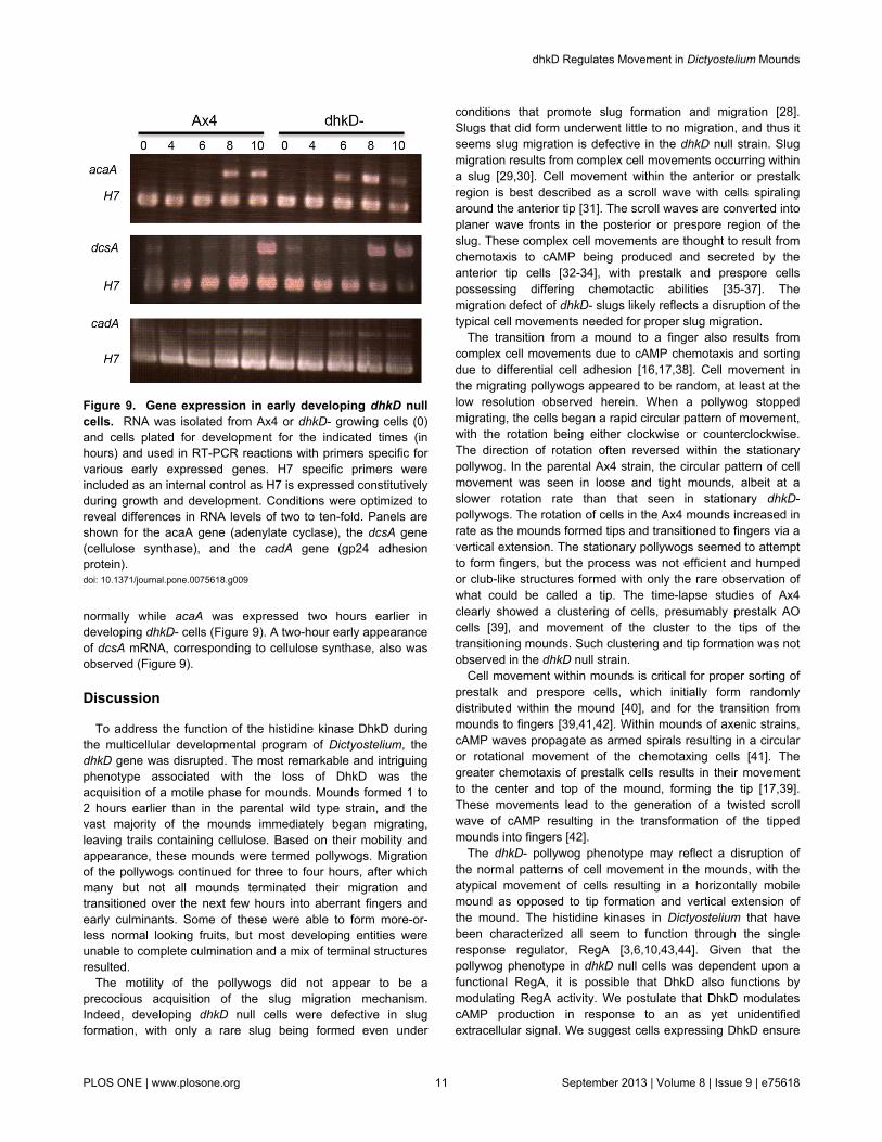
normally while acaA was expressed two hours earlier indeveloping dhkD- cells (Figure 9). A two-hour early appearanceof dcsA mRNA, corresponding to cellulose synthase, also wasobserved (Figure 9).
Discussion
To address the function of the histidine kinase DhkD duringthe multicellular developmental program of Dictyostelium, thedhkD gene was disrupted. The most remarkable and intriguingphenotype associated with the loss of DhkD was theacquisition of a motile phase for mounds. Mounds formed 1 to2 hours earlier than in the parental wild type strain, and thevast majority of the mounds immediately began migrating,leaving trails containing cellulose. Based on their mobility andappearance, these mounds were termed pollywogs. Migrationof the pollywogs continued for three to four hours, after whichmany but not all mounds terminated their migration andtransitioned over the next few hours into aberrant fingers andearly culminants. Some of these were able to form more-or-less normal looking fruits, but most developing entities wereunable to complete culmination and a mix of terminal structuresresulted.
The motility of the pollywogs did not appear to be aprecocious acquisition of the slug migration mechanism.Indeed, developing dhkD null cells were defective in slugformation, with only a rare slug being formed even under
Figure 9. Gene expression in early developing dhkD nullcells. RNA was isolated from Ax4 or dhkD- growing cells (0)and cells plated for development for the indicated times (inhours) and used in RT-PCR reactions with primers specific forvarious early expressed genes. H7 specific primers wereincluded as an internal control as H7 is expressed constitutivelyduring growth and development. Conditions were optimized toreveal differences in RNA levels of two to ten-fold. Panels areshown for the acaA gene (adenylate cyclase), the dcsA gene(cellulose synthase), and the cadA gene (gp24 adhesionprotein).doi: 10.1371/journal.pone.0075618.g009
conditions that promote slug formation and migration [28].Slugs that did form underwent little to no migration, and thus itseems slug migration is defective in the dhkD null strain. Slugmigration results from complex cell movements occurring withina slug [29,30]. Cell movement within the anterior or prestalkregion is best described as a scroll wave with cells spiralingaround the anterior tip [31]. The scroll waves are converted intoplaner wave fronts in the posterior or prespore region of theslug. These complex cell movements are thought to result fromchemotaxis to cAMP being produced and secreted by theanterior tip cells [32-34], with prestalk and prespore cellspossessing differing chemotactic abilities [35-37]. Themigration defect of dhkD- slugs likely reflects a disruption of thetypical cell movements needed for proper slug migration.
The transition from a mound to a finger also results fromcomplex cell movements due to cAMP chemotaxis and sortingdue to differential cell adhesion [16,17,38]. Cell movement inthe migrating pollywogs appeared to be random, at least at thelow resolution observed herein. When a pollywog stoppedmigrating, the cells began a rapid circular pattern of movement,with the rotation being either clockwise or counterclockwise.The direction of rotation often reversed within the stationarypollywog. In the parental Ax4 strain, the circular pattern of cellmovement was seen in loose and tight mounds, albeit at aslower rotation rate than that seen in stationary dhkD-pollywogs. The rotation of cells in the Ax4 mounds increased inrate as the mounds formed tips and transitioned to fingers via avertical extension. The stationary pollywogs seemed to attemptto form fingers, but the process was not efficient and humpedor club-like structures formed with only the rare observation ofwhat could be called a tip. The time-lapse studies of Ax4clearly showed a clustering of cells, presumably prestalk AOcells [39], and movement of the cluster to the tips of thetransitioning mounds. Such clustering and tip formation was notobserved in the dhkD null strain.
Cell movement within mounds is critical for proper sorting ofprestalk and prespore cells, which initially form randomlydistributed within the mound [40], and for the transition frommounds to fingers [39,41,42]. Within mounds of axenic strains,cAMP waves propagate as armed spirals resulting in a circularor rotational movement of the chemotaxing cells [41]. Thegreater chemotaxis of prestalk cells results in their movementto the center and top of the mound, forming the tip [17,39].These movements lead to the generation of a twisted scrollwave of cAMP resulting in the transformation of the tippedmounds into fingers [42].
The dhkD- pollywog phenotype may reflect a disruption ofthe normal patterns of cell movement in the mounds, with theatypical movement of cells resulting in a horizontally mobilemound as opposed to tip formation and vertical extension ofthe mound. The histidine kinases in Dictyostelium that havebeen characterized all seem to function through the singleresponse regulator, RegA [3,6,10,43,44]. Given that thepollywog phenotype in dhkD null cells was dependent upon afunctional RegA, it is possible that DhkD also functions bymodulating RegA activity. We postulate that DhkD modulatescAMP production in response to an as yet unidentifiedextracellular signal. We suggest cells expressing DhkD ensure
dhkD Regulates Movement in Dictyostelium Mounds
PLOS ONE | www.plosone.org 11 September 2013 | Volume 8 | Issue 9 | e75618
proper cAMP waves that in turn mediate proper cell movementwithin mounds, and this movement typically leads to tip andfinger formation. Without DhkD, distortions in cAMP productionlead to altered cell movement within the pollywogs that in turndrive pollywog migration instead of tip and finger formation. It isinteresting that in Ax4 mounds DhkD expressing cells did notappear to be spatially localized or patterned in a manner thatmight be expected for cells ensuring proper cAMP waves.Instead, DhkD positive cells were scattered throughout themound, seemingly at random. A higher resolution and morecomplete study of cell movement and of cAMP waveproduction in pollywogs, and a more detailed examination ofthe movement of DhkD positive cells in wild type mounds andearly fingers, are warranted.
The lack of proper patterning of prestalk and prespore celltypes observed in developing dhkD null cells supports ourproposed model of DhkD function. Timing of the appearance ofeach cell type in the developing dhkD- entities was notsubstantially different from that seen in the parental Ax4 cells.In contrast, though, differences in spatial patterning wereobserved between developing wild type and mutant cells.
Using the ecmO promoter as a marker, no band of prestalkO cells was observed in dhkD- while in Ax4 cells the typicalpstO band of cells between the anterior tips and the posteriorprespore region were readily apparent. Instead, ecmOexpressing cells were scattered throughout the dhkD-pollywogs and in the humped finger-like structures. TheecmAO promoter gave a similar scattered expression patternand thus did not reveal localized prestalk A cells that aretypically found at the anterior tips of Ax4 late mounds andfingers. Lack of proper patterning also was seen for prestalk Bcells, revealed by expression of the ecmB promoter, with onlyscattered cells observed in pollywogs and finger-like structures.The prestalk AB cone of cells in the tips of wild type fingers thatmark the initiation of culmination were never observed indhkD-.
Together, the results with the prestalk specific promoterssuggest no anterior tip or tip-like structure forms in developingdhkD null cells. As mentioned, a lack of tip formation wasconfirmed in the time-lapse studies. While prestalk cells doarise, they do not sort or localize properly. Patterning is aprocess dependent on differential cAMP chemotaxis and cellmovements within the transitioning mounds [17,39]. Essentiallythe same lack of proper patterning was observed for presporecells, although for these cells a weak posterior bias was seen.For both prestalk and prespore cells, there appeared to befewer of each than observed in wild type developing structures,or the promoters were more weakly expressed in dhkD- cells.For all cell types examined except prestalk B cells, developingdhkD- structures whose morphology was somewhat moresimilar to the wild type morphology showed a more wild typepatterning of prestalk cells, and some of these entities wereable to form fruits, albeit usually being short and having largesori.
Examination of the expression at the level of mRNA revealedthat adhesion related genes were transcribed normally in thedhkD null strain, namely those of cadA, lagC, csaA, and ampA.While this suggests the patterning defects do not arise from
defects in cell adhesion, the production of the adhesionproteins themselves were not examined. Nonetheless, thedevelopmental phenotype and behavior of the dhkD null strainwas very different from that seen in mutant strains lacking oroverexpressing cell adhesion proteins involved during moundand finger formation [16,45-47].
Most chemotaxis genes that were examined were expressednormally, including carA, dagA (crac), and gpaB (Galpha2).The exception was acaA, which encodes adenylate cyclase.Expression of acaA began about 2 hours earlier in developingdhkD null cells as compared to the parental Ax4 cells. Earlierthan normal expression may be related to the 1 to 2 hourearlier mound formation seen in dhkD-.
Normally, developing Dictyostelium cells stream in responseto relays of cAMP pulses during the initial stages ofaggregation. Near the end of aggregation, there is a change inbehavior such that streaming is inefficient and instead the cellssimply aggregate to complete the formation of tight mounds[48]. Interestingly, the early streaming period depends onRegA, and streams are not found in regA null strains [48]. It isbelieved that the map kinase ERK2 inhibits RegA activityduring early aggregation [49], yet to date no histidine kinasehas been identified that activates RegA during this time period.Given that DhkD appears to function through modulating RegAactivity and that aggregating dhkD null cells do not stream,even at early times, it is possible that DhkD may modulateRegA activity during early aggregation and this modulation maybe necessary for streaming of the cells.
Supporting Information
Movie S1. Developing dhkD null cells. dhkD- cells weregrown on bacteria, harvested, and plated for developmentunder standard conditions. Time-lapse was carried out using aLeica DM6000B microscope with a 5X objective. 3 to 5 percentof the developing cells were expressing GFP to enhance theability to observe cell movement. The time-lapse sequence wasbegun once the developing cells showed signs of obviousmound formation and was continued past the “finger” stage.Pictures under fluorescent light were taken at 3-minuteintervals. The movie is a 2.5-hour portion of the time-lapsesequence, beginning at the mound stage. The movie wascompiled using 3 frames (9 minutes real time) per second.(AVI)
Movie S2. Developing Ax4 cells. Ax4 cells were grown onbacteria, harvested, and plated for development understandard conditions. Time-lapse was carried out using a LeicaDM6000B microscope with a 5X objective. 3 to 5 percent of thedeveloping cells were expressing GFP to enhance the ability toobserve cell movement. The time-lapse sequence was begunonce the developing cells showed signs of obvious moundformation and was continued past the finger stage. Picturesunder fluorescent light were taken at 3-minute intervals. Themovie is a 2.5-hour portion of the time-lapse sequence,beginning at the mound stage. The movie was compiled using3 frames (9 minutes real time) per second.
dhkD Regulates Movement in Dictyostelium Mounds
PLOS ONE | www.plosone.org 12 September 2013 | Volume 8 | Issue 9 | e75618
(AVI)
Acknowledgements
We thank Keith Jermyn, Jeff Williams, and the DictyosteliumStock Center for providing plasmids.
Author Contributions
Conceived and designed the experiments: CKS YX. Performedthe experiments: CKS YX. Analyzed the data: CKS YX.Contributed reagents/materials/analysis tools: YX. Wrote themanuscript: CKS.
References
1. Capra EJ, Laub MT (2012) Evolution of two-component signaltransduction systems. Annu Rev Microbiol 66: 325-347. doi:10.1146/annurev-micro-092611-150039. PubMed: 22746333.
2. Anjard C, Loomis W (2006). ictyostelium Histidine Kinases: 1-18.3. Anjard C, Loomis WF (2005) Peptide signaling during terminal
differentiation of Dictyostelium. Proc Natl Acad Sci U S A 102:7607-7611. doi:10.1073/pnas.0501820102. PubMed: 15897458.
4. Kirsten JH, Xiong YH, Dunbar AJ, Rai M, Singleton CK (2005)Ammonium transporter C of Dictyostelium discoideum is required forcorrect prestalk gene expression and for regulating the choice betweenslug migration and culmination. Dev Biol 287: 146-156. doi:10.1016/j.ydbio.2005.08.043. PubMed: 16188250.
5. Singleton CK, Kirsten JH, Dinsmore CJ (2006) Function of ammoniumtransporter A in the initiation of culmination of development inDictyostelium discoideum. Eukaryot Cell 5: 991-996. doi:10.1128/EC.00058-06. PubMed: 16835443.
6. Singleton CK, Zinda MJ, Mykytka B, Yang P (1998) The histidinekinase dhkC regulates the choice between migrating slugs and terminaldifferentiation in Dictyostelium discoideum. Dev Biol 203: 345-357. doi:10.1006/dbio.1998.9049. PubMed: 9808785.
7. Thomason PA, Sawai S, Stock JB, Cox EC (2006) The histidine kinasehomologue DhkK/Sombrero controls morphogenesis in Dictyostelium.Dev Biol 292: 358-370. doi:10.1016/j.ydbio.2006.01.010. PubMed:16473345.
8. Wang N, Shaulsky G, Escalante R, Loomis WF (1996) A two-component histidine kinase gene that functions in Dictyosteliumdevelopment. EMBO J 15: 3890-3898. PubMed: 8670894.
9. Zinda MJ, Singleton CK (1998) The hybrid histidine kinase dhkBregulates spore germination in Dictyostelium discoideum. Dev Biol 196:171-183. doi:10.1006/dbio.1998.8854. PubMed: 9576830.
10. Schuster SC, Noegel AA, Oehme F, Gerisch G, Simon MI (1996) Thehybrid histidine kinase DokA is part of the osmotic response system ofDictyostelium. EMBO J 15: 3880-3889. PubMed: 8670893.
11. Anjard C, Loomis WF (2008) Cytokinins induce sporulation inDictyostelium. Development 135: 819-827. doi:10.1242/dev.018051.PubMed: 18216168.
12. Shaulsky G, Fuller D, Loomis WF (1998) A cAMP-phosphodiesterasecontrols PKA-dependent differentiation. Development 125: 691-699.PubMed: 9435289.
13. Thomason PA, Traynor D, Cavet G, Chang W-T, Harwood AJ et al.(1998) An intersection of the cAMP/PKA and two-component signaltransduction systems in Dictyostelium. EMBO J 17: 2838-2845. doi:10.1093/emboj/17.10.2838. PubMed: 9582277.
14. Thomason PA, Traynor D, Stock JB, Kay RR (1999) The RdeA-RegAsystem, a eukaryotic phospho-relay controlling cAMP breakdown. J BiolChem 274: 27379-27384. doi:10.1074/jbc.274.39.27379. PubMed:10488068.
15. Schaap P (2011) Evolution of developmental cyclic adenosinemonophosphate signaling in the Dictyostelia from an amoebozoanstress response. Development, growth &. Differentiation 53:452-462.
16. Siu C-H, Sriskanthadevan S, Wang J, Hou L, Chen G et al. (2011)Regulation of spatiotemporal expression of cell-cell adhesion moleculesduring development of Dictyostelium discoideum. Development, growth&. Differentiation 53: 518-527.
17. Weijer CJ (2004) Dictyostellium morphogenesis. Curr Opin Genet Dev14: 392-398. doi:10.1016/j.gde.2004.06.006. PubMed: 15261655.
18. Puta F, Zeng C (1998) Blasticidin resistance cassette in symmeyricalpolylinkers for insertional inactivation of genes in Dictyostelium. FoliaBiol 44: 185-188.
19. Egelhoff TT, Brown SS, Manstein DJ, Spudich JA (1989) Hygromycinresistance as a selectable marker in Dictyostelium discoideum. Mol CellBiol 9: 1965-1968. PubMed: 2546056.
20. Dingermann T, Reindl N, Werner H, Hildebrandt M, Nellen W et al.(1989) Optimization and in situ detection of Escherichia coli beta-
galactosidase gene expression in Dictyostelium discoideum. Gene 85:353-362. doi:10.1016/0378-1119(89)90428-9. PubMed: 2516830.
21. Cocucci SM, Sussman M (1970) RNA in cytoplasmic and nuclearfractions of cellular slime mold amebaes. J Cell Biol 45: 399-407. doi:10.1083/jcb.45.2.399. PubMed: 5535143.
22. Singleton CK, Delude RL, McPherson CE (1987) Characterization ofgenes which are deactivated upon the onset of development inDictyostelium discoideum. Dev Biol 119: 433-441. doi:10.1016/0012-1606(87)90047-9. PubMed: 3803712.
23. Pekovich SR, Martin PR, Singleton CK (1998) Thiamine deficiencydecreases steady-state mRNA levels for transketolase and pyruvatedehydrogenase but not for a-ketoglutarate dehydrogenase in threehuman cell types. J Nutr 128: 683-687. PubMed: 9521628.
24. Zhang Q (1995) Studies of H7 gene function and regulation of itsexpression by a bidirectional promoter in Dictyostelium discoideum. Ph.D. thesis, Vanderbilt University.
25. Levi S, Polyakov M, Egelhoff TT (2000) Green fluorescent protein andepitope tag fusion vectors for Dictyostelium discoideum. Plasmid 44:231-238. doi:10.1006/plas.2000.1487. PubMed: 11078649.
26. Kriebel PW, Barr VA, Parent CA (2003) Adenylyl cyclase localizationregulates streaming during chemotaxis. Cell 112: 549-560. doi:10.1016/S0092-8674(03)00081-3. PubMed: 12600317.
27. Jermyn KA, Duffy KT, Williams JG (1989) A new anatomy of theprestalk zone in Dictyostelium. Nature 340: 144-146. doi:10.1038/340144a0. PubMed: 2739736.
28. Newell PC, Ross FM (1982) Genetic analysis of the slug stage ofDictyostelium discoideum. J Gen Microbiol 128: 1639-1652.
29. Vasiev B, Weijer CJ (2003) Modelling of Dictyostelium discoideum slugmigration. J Theor Biol 223: 347-359. doi:10.1016/S0022-5193(03)00103-6. PubMed: 12850454.
30. Umeda T, Inouye K (2004) Cell sorting by differential cell motility: amodel for pattern formation in Dictyostelium. J Theor Biol 226: 215-224.doi:10.1016/j.jtbi.2003.08.016. PubMed: 14643191.
31. Siegert F, Weijer CJ (1992) 3-dimensional scroll waves organizeDictyostelium slugs. Procedings Natl Acad Sciences, USA 89: 6433.doi:10.1073/pnas.89.14.6433.
32. Dormann D, Weijer C, Siegert F (1997) Twisted scroll waves organizeDictyostelium mucoroides slugs. J Cell Sci 110: 1831-1837. PubMed:9296384.
33. Dormann D, Weijer CJ (2001) Propagating chemoattractant wavescoordinate periodic cell movement in Dictyostelium slugs. Development128: 4535-4543. PubMed: 11714678.
34. Bretschneider T, Siegert F, Weijer CJ (1995) Three-dimensional scrollwaves of cAMP could direct cell movement and gene expression inDictyostelium slugs. Proc Natl Acad Sci U S A 92: 4387-4391. doi:10.1073/pnas.92.10.4387. PubMed: 7753816.
35. Matsukuma S, Durston AJ (1979) Chemotactic cell sorting inDictyostelium discoideum. J Embryol Exp Morphol 50: 243-251.PubMed: 222874.
36. Mee JD, Tortolo DM, Coukell MB (1986) Chemotaxis-associatedproperties of separated prestalk and prespore cells of Dictyosteliumdiscoideum. Biochem Cell Biol 64: 722-732. doi:10.1139/o86-099.
37. Sternfeld J, David CN (1981) Cell sorting during pattern formation inDictyostelium. Differentiation 20: 10-21. doi:10.1111/j.1432-0436.1981.tb01150.x.
38. Siu C-H (2004) Regulation of cell-cell adhesion during Dictyosteliumdevelopment. Semin Cell Dev Biol 15: 633-641. doi:10.1016/S1084-9521(04)00090-4. PubMed: 15561583.
39. Clow PA, Chen T, Chisholm RL, McNally JG (2000) Three-dimensionalin vivo analysis of Dictyostelium mounds reveals directional sorting ofprestalk cells and defines a role for the myosin II regulatory light chainin prestalk cell sorting and tip protrusion. Development 127: 2715-2728.PubMed: 10821769.
40. Thompson CRL, Reichelt S, Kay RR (2004) A demonstration of patternformation without positional information in Dictyostelium. Dev Growth
dhkD Regulates Movement in Dictyostelium Mounds
PLOS ONE | www.plosone.org 13 September 2013 | Volume 8 | Issue 9 | e75618
Differ 46: 363-369. doi:10.1111/j.1440-169x.2004.00753.x. PubMed:15367204.
41. Siegert F, Weijer CJ (1995) Spiral and concentric waves Dictyorganizemulticellular Dictyostelium mounds. Curr Biol 5: 937–943. doi:10.1016/S0960-9822(95)00184-9. PubMed: 7583152.
42. Vasiev B, Weijer CJ (1999) Modeling chemotactic cell sorting duringDictyostelium discoideum mound formation. Biophys J 76: 595-605.doi:10.1016/S0006-3495(99)77228-0. PubMed: 9929466.
43. Giusti C, Luciani M-F, Ravens S, Gillet A, Golstein P (2010) Autophagiccell death in Dictyostelium requires the receptor histidine kinase DhkM.Mol Biol Cell 21: 1825-1835. doi:10.1091/mbc.E09-11-0976. PubMed:20375146.
44. Tekinay T, Ennis HL, Wu MY, Nelson M, Kessin RH et al. (2003)Genetic interactions of the E3 ubiquitin ligase component FbxA withcyclic AMP metabolism and a histidine kinase signaling pathway duringDictyostelium discoideum development. Eukaryot Cell 2: 618-626. doi:10.1128/EC.2.3.618-626.2003. PubMed: 12796307.
45. Roisin-Bouffay C, Jang W, Caprette DR, Gomer RH (2000) A precisegroup size in Dictyostelium is generated by a cell-counting factor
modulating cell-cell adhesion. Mol Cell 6: 953-959. doi:10.1016/S1097-2765(05)00082-1. PubMed: 11090633.
46. Varney TR, Ho H, Petty C, Blumberg DD (2002) A novel disintegrindomain protein affects early cell type specification and patternformation in Dictyostelium. Development 129: 2381-2389. PubMed:11973270.
47. Wang J, Hou LS, Awrey D, Loomis WF, Firtel RA et al. (2000) Themembrane glycoprotein gp150 is encoded by the lagC gene andmediates cell-cell adhesion by heterophilic binding during Dictyosteliumdevelopment. Dev Biol 227: 734-745. doi:10.1006/dbio.2000.9881.PubMed: 11071787.
48. Das S, Rericha EC, Bagorda A, Parent CA (2011) Direct biochemicalmeasurements of signal relay during Dictyostelium development. J BiolChem 286: 38649-38658. doi:10.1074/jbc.M111.284182. PubMed:21911494.
49. Maeda M, Lu SJ, Shaulsky G, Miyazaki Y, Kuwayama H et al. (2004)Periodic signaling controlled by an oscillatory circuit that includesprotein kinases ERK2 and PKA. Science 304: 875-878. doi:10.1126/science.1094647. PubMed: 15131307.
dhkD Regulates Movement in Dictyostelium Mounds
PLOS ONE | www.plosone.org 14 September 2013 | Volume 8 | Issue 9 | e75618