Embed Size (px)
Citation preview

Molecular and Cellular Probes (1992) 6, 477-487
Detection of the thermostable direct hemolysin gene (tdh)and the thermostable direct hemolysin-related hemolysin
gene (trh) of Vibrio parahaemolyticus by polymerase chainreaction
Jun Tada,' Tetsuo Ohashi, Naoyuki Nishimura,' Yoshinari Shirasaki, 1Hiroko Ozaki,1 Shigeru Fukushima,'* Jun Takano, Mitsuaki Nishibuchi 2
and Yoshifumi Takeda2
' Central Research Laboratory, Shimadzu Corporation, 1 Nishinokyo- Kuwabaracho,Nakagyo-ku, Kyoto 604, Japan and 2 Department of Microbiology, Faculty of Medicine,
Kyoto University, Konoe-cho, Yoshida, Sakyo-ku, Kyoto 606, Japan
(Received 8 June 1992, Accepted 23 June 1992)
Polymerase chain reaction (PCR) protocols were established for specific detection of the tdh and trhgenes, the virulence marker genes of Vibrio parahaemolyticus encoding two related hemolysins. Thetdh and trh genes are known to have sequence divergence of up to 3 .3% and 16%, respectively .Attempts were made to find suitable primer pairs and annealing temperatures to detect each genewithout fail . DNAs extracted from 36 representative strains of V. parahaemolyticus were used in theinitial screening with various combinations of primer pairs and annealing temperatures . Thecombinations of primer pairs and annealing temperatures selected were then tested with DNAsextracted from 227 more strains of V. parahaemolyticus and from 133 bacterial strains belonging to40 species other than V. parahaemolyticus . PCR protocols (primer pairs and annealing temperatures)were established that gave identical results to those obtained with the tdh- and trh-specificpolynucleotide probes. These protocols established for the tdh and trh genes could detect 400 fg (100cells) of cellular DNA carrying the respective gene . Spike experiments demonstrated that thesensitivities of the established PCRs were reduced by a factor of 10 4-105 by an inhibitor(s) present in anormal faecal sample, indicating the need for either DNA extraction or enrichment of the faecalsample in alkaline peptone water for 4 h before the PCR of faecal samples .
KEYWORDS : Vibrio parahaemolyticus, thermostable direct hemolysin gene, PCR .
INTRODUCTION
Vibrio parahaemolyticus, a natural inhabitant ofestuarine and marine environments, often causesseafood-borne gastroenteritis .' , ' Although themechanism of its pathogenic effect is still not clearlyunderstood, epidemiological studies have suggesteda very strong association of thermostable directhemolysin (TDH) with disease .` Production of TDH isresponsible for a defined characteristic called the
* Author to whom correspondence should be addressed .
0890-8508/92/060477 + 11 $08 .00/0 477
Kanagawa phenomenon,' which is manifested asbeta-type hemolysis on a special blood agar calledWagatsuma agar.' Since the Kanagawa phenomenonreaction is difficult to judge, immunological methodshave been developed to identify TDH-producingstrains specifically,$ and of these, bead-enzyme-linked immunosorbent assay' has been shown to behighly sensitive for detecting TDH . A DNA probe
© 1992 Academic Press Limited

478
J. Tada et alL
method to detect the gene (tdh) encoding TDH is also
MATERIALS AND METHODSsensitive and has been used successfully to demon-strate that not only all Kanagawa phenomenon-positive strains but also some Kanagawa phenom-enon-negative strains, mostly from clinical sources,have the tdh gene.' Subsequently, the tdh genescloned from Kanagawa phenomenon-negative strainswere found to encode biologically active TDHs,10 12
indicating that all strains having the tdh gene arepotentially virulent . The nucleotide sequences of thetdh genes of V. parahaemolyticus so farreported", " ,"` show sequence variations of up to3-3% .
Recently, a Kanagawa phenomenon-negative clini-cal strain not possessing the tdh gene was found toproduce a TDH-related hemolysin (TRI-1) ." Like thetdh gene, the gene (trh) encoding TRH has a 567 bycoding region, but is only 68% homologous to thetdh gene . 17 A molecular epidemiologic study withtdh- and trh-specific DNA probes demonstrated astrong association of the two genes with clinicalstrains, suggesting that TRH as well as TDH is animportant virulence factor ." Furthermore, significantsequence variation was very recently found in the trhgenes of various strains . The trh probe-positive strainsmanifesting various intensities of hybridization signalscould be subdivided into two sub-groups representedby trh 1 (formerly trh) and trh 2 genes ." The trh 1 andtrh 2 genes show 84% homology and the trh 2 gene-bearing strains are also considered potentially viru-lent ." .The polymerase chain reaction (PCR) method is
usually more rapid and sensitive than a DNA oroligonucleotide probe-based hybridization methodfor detecting a target gene in bacteria . But in usingthe PCR method for detecting the tdh and trh genesat a high detection rate, possible mismatch(es) of thePCR primers due to the existence of natural variationsin their sequences'" 1,13-15,19 must be avoided. Theprimary aim of this study was to establish PCRprotocols giving identical, or nearly identical resultsto those obtained by the DNA (=polynucleotide)probe method. For this purpose, we first screenedvarious combinations of primer pairs and annealingtemperatures using DNAs extracted from representa-tive strains. The selected PCR protocols werescreened further with DNAs extracted from a muchlarger number of organisms. Some enteropathogenicbacteria present in faecal samples could be detectedby the PCR method with 20 or without 21 prior DNAextraction from the faecal samples . To assess thepractical applicability of the established PCR proto-cols to faecal samples, we carried out a spike experi-ment to evaluate the need for DNA extraction fromfaecal samples .
Bacterial strains
The bacterial strains used in this study are listed inTable 1 . The strains belonging to the genus Vibriowere described previously." Other strains wereobtained from the American Type Culture Collection,Japan Collection of Microorganisms and the Institutefor Fermentation, Osaka. The presence or absence ofthe tdh and trh genes in Vibrio strains was deter-mined in previous studies by DNA colony blot hybri-dization tests with DNA probes specific to the tdhand trh genes."' Strains that gave weak or very weakhybridization signals with a trh gene probe" areincluded in the trh-positive group in Table 1, butwhen necessary (see below), the trh genes in variousstrains were classified into trh 1 and trh 2 genes byDNA colony blot and Southern blot hybridizationanalyses as reported elsewhere."
DNA extraction from bacterial cells
Pure cultures of the test organisms grown on appro-priate agar media were harvested and used for DNAextraction. Cellular DNA was extracted by standardprocedures using phenol and chloroform ."
PCR primers
Oligonucleotide (19- or 20-mer) primers weredesigned based on the reported nucleotide sequenceof the tdh 2 gene cloned from strain WP1 11 and thetrh (=trh 1) gene cloned from strain AQ4037 . 17 Thedesignations and nucleotide sequences of theprimers are listed in Table 2. The primers weresynthesized by the (3-cyanoethyl phosphoramiditemethod with an automated DNA synthesizer (Cyc-lone plusTM DNA synthesizer, Milligen, Burlington,MA, USA) and purified using a high-performanceliquid chromatography apparatus (LC-6A, ShimadzuCo., Kyoto, Japan) equipped with a reversed phasecolumn (Cosmosil C18, Nacalai Tesque, Kyoto, Japan) .
PCR amplification
PCR amplification was performed in a 30 pl mixturecomposed of 10 mm Tris-HCI (pH 9 .0), 50 mm KCI,1-5 mm MgCl2, 0-01% gelatin, 0 .6µM each of theprimers, 02 mm each of the four deoxynucleosidetriphosphates (Ultrapure dNTP Set, Pharmacia LKBBiotechnology, Uppsala, Sweden), 0 .05% Tween 20,

Table 1 . Detections of the tdh and trh genes by PCR assay in DNAs extracted from Vibrio parahaemolyticus andother organisms
Organism
PCR for tdh and trh genes of Vibrio parahaemolyticus
479
Presence or absence* of
Results§ of PCR assay forNo. of strains
tdh gene'
trh gene$
tdh gene¶
trh gene I
Vibrio parahaemolyticus
25
+
+
+
+110
+
-
+
-56
-
+
-
+72
-Vibrio cholerae, 01
10
-
-
-Vibrio cholerae, non-OI
1
+
+
-73
-
-Vibrio mimicus
2
+
-
+2
-
-Vibrio furnissii
4
-
-
-Vibrio fluvialis
5
-
-
-Vibrio metschnikovii
1
-Bacillus cereus
1
NT
NT
-Bacillus subtilis
1
NT
NT
-Staphylococcus aureus
1
NT
NTStaphylococcus epidermidis
1
NT
NT
-Salmonella typhimurium
1
NT
NT
-
-Salmonella enteritidis
1
NT
NT
-
-Clostridium perfringens
1
NT
NT
-Campylobacter jejuni
1
NT
NT
-Campylobacter coli
1
NT
NT
-Escherichia coli
1
NT
NT
-
-Yersinia enterocolitica
1
NT
NT
-Shigella dysenteriae
1
NT
NT
-Shigella flexneri
1
NT
NT
-Shigella sonnei
1
NT
NTBacteroides fragilis
1
NT
NTBacteroides vulgatus
1
NT
NT
-
-Enterococcus faecalis
1
NT
NT
-
-Klebsiella pneumoniae
1
NT
NT
-
-Proteus mirabilis
1
NT
NT
-Proteus vulgaris
1
NT
NT
-Citrobacter freundii
1
NT
NT
-Streptococcus pyogenes
1
NT
NT
-
-Streptococcus pneumoniae
1
NT
NT
-Haemophilus influenzae
1
NT
NT
-Neisseria gonorrhoeae
1
NT
NT
-Neisseria meningitidis
1
NT
NT
-
-Listeria monocytogenes
1
NT
NT
-
-Lactobacillus acidophilus
1
NT
NTBifidobacterium adolescentis
1
NT
NTFusobacterium nucleatum
1
NT
NT
-Propionibacterium acnes
1
NT
NT
-
-Veillonella atypica
1
NT
NTPseudomonas aeruginosa
1
NT
NT
-
-Corynebacterium diphtheriae
1
NT
NT
-
-Peptostreptococcus anaerobius
1
NT
NT
-
-
+, present . -, absent . NT, not tested .j Determined by the DNA colony blot hybridization test with a tdh gene probe in previous studies ."'
Determined by the DNA colony blot hybridization test with a trh gene probe in a previous study ." Strains exhibiting weakor very weak hybridization signalst 8 were included in the gene-positive (+) group in order to include both the trh 1 and trh 2sub-group."
§ +, only specific amplicons of expected sizes (see Table 3) were detected by gel electrophoresis . -, no amplicons weredetected by gel electrophoresis .
¶ Primer pairs D1-D2, D5-D2 and D5-D3 were used at an annealing temperature of 55 °C. The results obtained with thethree primer pairs were identical .
II Primer pair R2-R6 was used at an annealing temperature of 55 °C.

* Sequence in the 5' to 3' (left to right) direction . The primer sequences of the tdh and trh genesare identical to those of the tdh 2 gene" and the trh (=trh1) gene", respectively . The numerals atboth ends correspond to the nucleotide positions of the coding regions of the respective genes,numbered from the bases (=position 1) of the N termini of the mature proteins .
j' The Td value, temperature at which 50% of the primer is dissociated was calculated by themethod of Suggs et al .26
0 .05% Nonidet P-40, 0 . 75 U of Taq polymerase (Per-kin-Elmer Cetus Corp ., Norwalk, CT, USA), and tem-plate DNA (1 ng, unless otherwise specified). The PCRwas carried out for 35 cycles in a DNA thermal cycler(Perkin-Elmer Cetus Corp .) ; one cycle consisted ofdenaturation at 94 °C for 1 min, primer annealing at aspecified temperature for 1 min, and extension at72 °C for 1 min .
Detection of amplified DNAs
PCR-amplified DNAs (amplicons) were usuallydetected by agarose gel electrophoresis . A 5 µl por-tion the PCR-amplified mixture was separated in 3%agarose (Agarose-ME, Nacalai Tesque) gel by electro-phoresis at 3 to 4 V cm"' for 1 h in TAE buffer [40 mmTris-acetate (pH 8. 0), 20 mm EDTAI containing0 . 5 gg ml-' ethidium bromide . After electrophoresis,
the gel was examined with a u .v. (at 302 nm) transil-
luminator (UVP Inc., San Gabriel, CA, USA) and
photographed. The target-specific amplicons were
identified by their size, which was determined bytheir mobility relative to those of molecular weightmarkers (Hin cli-digested phage OX174 DNA) .
To confirm the specificity of the amplicons and toassess the sensitivity of the PCR assay, we examinedselected samples by Southern blot hybridization . ForSouthern blotting, 5 µl of 500-fold diluted or undi-luted (sensitivity test) PCR-amplified mixture was sub-jected to agarose gel electrophoresis as describedabove. The gel was treated with 0-5 M NaOH contain-ing 1 . 5 M NaCl at room temperature for 30 min,
followed by neutralization with 0-5 M Tris-HCI (pH 8-0)containing 1 . 5 M NaCl and 1 mm EDTA at roomtemperature for 30 min . The amplicons were trans-
ferred to a nylon membrane (Hybond TM , AmershamInternational, Buckinghamshire, UK) in 10 X SSC over-
night [1 x SSC is 0-15 rn NaCl, 15 mm trisodiumcitrate(pH 7 . 0)1 . After washing with 2 X SSC, the membranewas dried with 3 MM paper (Whatman Biosystems,Maidstone, Kent, UK). Probe DNAs specific to the tdhand the trh genes were prepared from recombinantplasmids pCVD518 9 and pKTN101,' 8 respectively, asdescribed previously . The probes were radiolabelledwith 32P in a multiprime DNA labelling system (Amer-sham International). The specific activities of thelabelled probes were 3 X 10⁸ to 6 X 10⁸ cpm µg' .Hybridization was performed in 5 X SSPE [1 X SSPE is0 . 15 m NaCl, 18 mm sodium phosphate, 1 mm EDTA,(pH 8.0)], 1 % SDS, 5 X Denhardt's mixture (1 X Den-hardt's mixture is 0-02% Ficoll, 0-02% polyvinylpyrro-lidone, 0-02% bovine serum albumin) supplementedwith 0-1 mg ml"' poly A (Boehringer Mannheim Bio-
chemicals, Indianapolis, IN, USA) and a radiolabelledprobe (7 . 5 X 10⁶-1 . 5 X 10' cpm) at 65 °C overnight .After hybridization, the membrane was washed with1 X SSPE and 0. 1 % SDS twice at 65°C for 20 min, air-dried and exposed to X-ray film overnight (Fuji RX, FujiFilm Co., Kanagawa, Japan) .
Examination of spiked faeces by PCR
A 0. 1 g portion of normal human faeces was sus-pended in 10 ml of phosphate-buffered saline
480 J . Tada et al .
Table 2. PCR primers used in this study
Target Calculatedgene Designation Nucleotide base sequence* Td (°C)1
tdh D1 82-CCATCTGTCCCTTTTCCTGC-101 62D2 454-CCAAATACATTTTACTTGG -436 56D3 506-CCACTACCACTCTCATATGC-487 60D4 534-GTTGGATATACACATTACC -516 52D5 256-GGTACTAAATGGCTGACATC-275 58D6 275-GATGTCAGCCATTTAGTACC-256 58
trh R1 82-CCATCCATACCTTTTCCTTC-101 58R2 256-GGCTCAAAATGGTTAAGCG -275 56R3 256-GCCTCAAAATGGTTAAGCGC-276 60R4 461-GTTTCATCCAAATACGTTAC-442 54R5 465-TGGCGTTTCATCCAAATACG-446 58R6 505-CATTTCCGCTCTCATATGC -486 56

(pH 7 . 0) . After vigorous mechanical shaking(3000 rpm) for 60 s, insoluble particulate matter wasremoved by low-speed centrifugation and aliquots ofthe suspension were mixed with an equal volume(100 µl) of serially diluted bacterial culture (overnightculture of WP1 or AQ4037 in LB broth with 1NaCI23) . The numbers of viable cells in the cultureswere determined by a plate count method. Thespiked sample was subjected to PCR without (directPCR) or with a prior treatment ; i .e . extraction of DNAfrom the spiked faeces or enrichment culture of thespiked faeces . For direct PCR of the spiked faeces, thesample was heated at 95 °C for 5 min and a 5 tlportion was subjected to PCR. For extraction of DNAfrom the spiked faeces, the sample (200 µl) wasincubated first with 400 pl of lysozyme solution(1 mg ml - ' at 37°C for 10 min and then with 20 .tl ofproteinase K solution (20 mg ml - ') and 20 .tl of 20%sodium dodecylsulfate solution at room temperaturefor 1 h. DNA was extracted from the lysate by a seriesof phenol-chloroform treatments [400 .d of 10 mmTris-1 mm EDTA (pH 80-saturated phenol, 400 µl ofchloroform-isoamyl alcohol (24 :1 v/v), and 400 lal ofchloroform] and precipitated with 40 µl of 3 M sodiumacetate solution and 800 µl of cold ethanol . Then itwas rinsed with 70% ethanol, dried, and dissolved in200 gl of 10 mm Tris-1 m m EDTA solution (pH 8 . 0) . A5 µl portion of the DNA solution was subjected toPCR. For the enrichment method, the spiked sample(200 µl) was inoculated into 1 ml of alkaline peptonewater 24 and incubated with shaking at 100 rpm at37°C for 4 h. The culture was heated at 95°C for 5 minand a 5 pl portion was subjected to PCR . PCR wasperformed as described above except that the totalvolume of the reaction mixture was increased to50 µl . A 5 µl portion of the PCR-amplified mixture wasexamined by gel electrophoresis as described aboveto detect the specific amplicons .
RESULTS
Screening of PCR primers and annealingtemperatures
The nucleotide sequences of five representative tdh(tdh 1-tdh 5) genes"," and the trh (=trh 1) gene" ofV. parahaemolyticus were compared. The tdh genegroup and the trh (trh 1) gene differed by as much as31-32%, sequence heterogeneity being observedthroughout the coding regions . Therefore, it wasimpossible to design a PCR primer pair for simultan-eous detections of the tdh and trh genes. However,five regions containing sequences that were wellconserved among the five tdh genes but were signi-
PCR for tdh and trh genes of Vibrio parahaemolyticus 481
ficantly different from those of the trh 1 gene weredetected . We thus decided to design primers target-ing these regions for detecting the tdh and trh genesseparately .
The tdh 2 gene is one of the two tdh gene copiescarried by Kanagawa phenomenon-positive strainsand is the gene mainly responsible for production ofextracellular TDH . ",'3,25 Therefore, we selected) thisgene as a representative of the tdh genes. Six PCRprimers for the tdh gene targeted to the tdh 2 gene-specific sequences within the five conserved regionswere prepared (D1-D6 in Table 2) . Primers D11, D2and D4 did not have any mismatch with the fourother tdh genes. Primer D3 had 1 by and 2 bymismatches with two and one other tdh gene, re-spectively . Primer D5 had a 1 by mismatch with thefour other tdh genes. Seven pairs of the tdh primers(Table 3) were used for amplification of the DNAsextracted from 36 selected strains of V. parahaernoly-ticus . Of the 36 strains, 17 strains had both the tdhand trh genes, 17 the tdh gene only, and two neithergene. The PCR products were assayed by gel electro-phoresis. All seven primer pairs gave specific arnpli-cons of the expected sizes (Table 3) at annealingtemperatures of 50°C and below . But small amountsof additional non-specific amplicons (differing in sizefrom the specific amplicons) were also observed inmany cases. When the annealing temperature wasraised to 55 °C, three primer sets, D1-D2, D5-D2 andD5-D3, gave the specific amplicons only and theseamplicons were detected only with the 34 tdh-carrying strains . But results with the four other primerpairs were unsuccessful at 55 °C, no specific ampli-cons being obtained with some tdh-bearing strainsand non-specific amplicons with some other strains.The D1-D3 pair gave no specific amplicons with onetdh-positive strain and a non-specific amplicon withone strain . The D1-D4 pair gave non-specific ampli-cons with three tdh-positive strains. The D1-D6 pairgave non-specific amplicons with two strains. TheD5-D4 pair gave non-specific amplicons with onestrain . Therefore, the three successful primer pairsand an annealing temperature of 55 °C were chosenfor more extensive tests with DNAs extracted from227 more strains of V. parahaemolyticus . The resultsobtained with the three primer pairs were identicaland that the results completely agreed with thoseobtained with the tdh gene probe. The resultsobtained with these three probes for all 263 testedstrains of V. parahaemolyticus are summarized inTable 1 .
Initially, four PCR primers were designed for detec-tion of the trh (=trh 1) gene (R1-R5 in Table 2) .Experiments with DNAs extracted from the 36selected strains were carried out with four primer

482
J. Tada et al.
39402
182
Table 3. Pairs of PCR primers used in this study
* Designations of primers are the same as in Table 2.The numerals correspond to the nucleotide positions of the coding regions
of the respective genes, numbered from the bases (= position 1) of the N terminiof the mature proteins . See legend to Table 2 for detailed explanation .
pairs, R1-R4, R1-R5, R3-R4 and R3-R5 (Table 3) . Ofthe four primer pairs, R3-R5 gave the most successfulresults at an annealing temperature of 55 °C, but notbelow, in detecting the trh gene (found to be trh 1later) of the 36 test strains . At this point in our study,the sequence of the trh 2 gene and the distributionsof the trh 1 and trh 2 genes in the test strains of V .parahaemolyticus were determined in a separatestudy." The primers R1, R2, R3, R4 and R5 were foundto have 1 bp, 2 bp, 2 bp, 4 by and 3 by mismatches,respectively, with the trh 2 gene. Therefore, the PCRwith the primer pair R3-R5 was considered specificfor the trh 1 gene, but not for the trh 2 gene .Accordingly, an additional primer R6 derived fromthe trh 1 sequence but having no mismatch with thetrh 2 gene was designed (Table 2) . The primer pairsR3-R5 and R2-R6, the latter being designed to detectboth the trh 1 and the trh 2 gene, were then testedwith DNAs extracted from 263 strains, including theabove 36 strains, of V. parahaemolyticus . The R3-R5
Table 4 . Results of PCR assay for detection of the trh1 and trh2 genes in DNAextracted from 263 strains of Vibrio parahaemolyticus .
Results of PCR assay with primerPresence or absence* of
pair / annealing temperatureNo. ofstrains
trh1 gene
trh2 gene
++
primer pair gave varying results, particularly with trh2-positive strains, at an annealing temperature of55°C (results not shown) . The R2-R6 primer pairdetected both the trh 1 and the trh 2 genes and non-specific amplicons were not observed at this anneal-ing temperature (Tables 1 and 4) . The R3-R5 primerpair detected trh 1-positive strains with very highspecificity when the annealing temperature wasincreased to 60 °C (Table 4) .
Specificity of PCR
The above screening test suggested that the primerpairs D1-D2, D5-D2 and D5-D3 for the tdh gene andthe primer pair R2-R6 for the trh (both trh 1 and trh 2)gene, both used at an annealing temperature of 55°C,would be successful . The specificities of these PCRprotocols were further assessed by amplifying theDNA templates extracted from organisms other than
R2-R6 / 55 °C
R3-R5 / 60°C
*+, present; -, absent . Determined by DNA colony blot and Southern blot hybridizationanalyses with the trh 1- and trh 2-specific DNA probes in a separate study ."
Target gene Designation* Amplified region'Length (bp) ofamplicon
tdh D1-D2 82-454 373D1-D3 82-506 425D1-D4 82-534 453D1-D6 82-275 194D5-D2 256-454 199D5-D3 256-506 251D5-D4 256-534 279
trh R1-R4 82-461 380R1-R5 82-465 384R3-R4 256-461 206R3-R5 256-465 210R2-R6 256-505 250

V. parahaemolyticus . These included 98 strainsbelonging to 5 species of the genus Vibrio and 35strains belonging to 35 species spanning over 25other genera (Table 1) . The three pairs of the primersfor the tdh gene gave identical results . The PCRresults obtained for the Vibrio species agreed withthose obtained with the tdh and trh gene probes;only rare strains of Vibrio cholerae non-O1 andVibrio mimicus possessing the tdh gene211 werepositive by the PCR for the tdh gene. All the organ-isms other than Vibrio gave negative results (noamplicons at all) with any of the primer pairs .
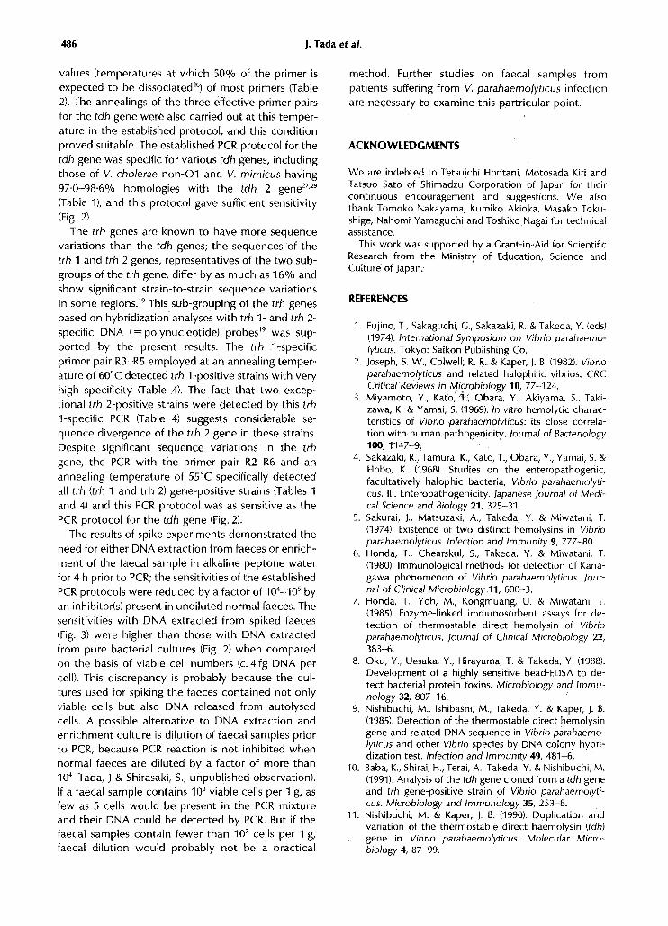
The specificities of the amplicons were confirmedby Southern blot hybridization analyses with the tdhand trh (=trh 1) gene probes (Fig . 1). The DNAsextracted from representative strains of V. parahae-molyticus were used as templates : tdh-positive andtrh 1-negative strains (lanes 2-5), tdh-negative andtrh 1-positive strains (lanes 6-9), tdh- and trh 1-positive strains (lanes 11-14), and tdh- and trh 1-negative strains (lanes 15 and 16) . Hybridization wascarried out under high-stringency conditions to dis-tinguish tdh- and trh 1-specific hybridizations . The trh2 gene sequences produce weak or no hybridizationsignals with the trh 1-specific probe under this con-dition in Southern blot analysis." Therefore, to avoidconfusion, trh 2 gene-positive strains were not in-
(a)
(b)I 2 3 4 5 6 7 8 9
10 II 12 13 14 15 16
1 2 3 4 5 6 7 8 9
10
(c)
( d )1 2 3 4 5 6 7 8 9
101 2 3 4 5 6 7 8 9
10 II 12 13 14 15 16
PCR for tdh and trh genes of Vibrio parahaemolyticus
483
cluded . The DNA fragments amplified from the tdh(Figs la and 1c, lanes 2-5 and 11-14) and trh (Figs lband 1d, lanes 6-9 and 11-14) genes hybridized onlywith the respective probes . No other PCR productswere detected either by gel electrophoresis or bySouthern blot hybridization .
Sensitivity of PCR
The sensitivities of the PCR methods established forthe tdh and trh genes were evaluated with the DNAsextracted from strains WP1 and AQ4037, respect-ively . WP1, a representative Kanagawa phenomenon-positive strain, carries two tdh gene copies" but hasno trh gene." AQ4037 carries the trh 1 gene, whichwas used for nucleotide sequencing, 17 but does nothave the tdh gene." The D5-D3 and R2-R6 primerpairs for the tdh and trh gene, respectively, were bothemployed at an annealing temperature of 55 °C. ThePCR-amplified mixtures were serially diluted and ex-amined by gel electrophoresis and Southern blotanalysis. The limits of detection of the tdh and trhgenes were both 400 fg DNA (corresponding to c . 100cells) by gel electrophoresis (Figs 2a and 2b) and 40 fgDNA (c. 10 cells) by Southern blot hybridizationanalysis (Figs 2c and 2d) .
I I 12 13 14 15 16
1I 12 13 14 15 16
Fig. 1 . Analysis of PCR products of DNAs extracted from 14 representative strains of V. parahaemolyticus .DNA samples were extracted from the following strains: lanes 2-5, tdh-positive and trh-negative strains ; lanes6-9, tdh-negative and trh-positive strains; lanes 11-14, tdh-positive and trh-positive strains ; lanes 15 and 16,tdh-negative and trh-negative strains . (a) and (c), PCR amplification of the tdh gene with primer pair D5-D3annealed at 55 °C. (b) and (d), PCR amplification of the trh gene with primer R2-R6 annealed at 55°C . (a) and(b), Analysis by agarose gel electrophoresis . A 5 pl portion of the PCR-amplified mixture was separated in 3%agarose gel containing ethidium bromide and the amplicons were located under u .v . light . (c) and (d),Analysis by Southern blot hybridization with specific DNA probes . A 5lal portion of 500-fold dilutedPCR-amplified mixture was separated by 3% agarose gel electrophoresis, transferred to a nylon membrane,and hybridized with a 32 P-labelled DNA probe specific to the tdh gene (c) or the trh gene (d). The expectedpositions of the specific amplicons relative to those of the molecular weight markers (Hin cll-digested OX174DNA, lanes 1 and 10) are indicated by arrowheads .

484
2 3 4 5 6
(c)I
2
3
4
5
6
7
8
Examination of spiked faeces by PCR
7 8
The need for pretreatment of faecal samples was
investigated to apply the established PCR protocol fordetection of tdh- and/or trh-positive organisms in
spiked faecal samples . Suspensions of a normal faecal
sample were mixed with known numbers of viable
cells of WP1 (tdh + , trh - ) or AQ4037 (tdh -, trh + ) . The
spiked samples were subjected to PCR with or with-
out treatment (by DNA extraction or enrichment inalkaline peptone water) prior to PCR . The primer pairs
D5-D3 and R2-R6 were both used at an annealing
temperature of 55 °C for detection of the tdh gene of
J . Tada et al .
2 3 4 5 6 7
(d )I
2
3
4
5
6
7
8
Fig. 2. Analysis of PCR products to assess the sensitivities of PCR for the tdh and trh genes. (a) and (c), PCRamplification of the tdh gene with primer pair D5-D3 annealed at 55 °C. DNA extracted from V .parahaemolyticus strains WP1 carrying the tdh (tdh 1 and tdh 2) genes was amplified . (b) and (d), PCRamplification of the trh gene with primer pair R2-R5 annealed at 55 °C. DNA extracted from AQ4037 carryingthe trh 1 gene [(b) and (d)1 was amplified . The estimated amounts of cellular DNA present in each of theinitial PCR mixtures were zero (DNA not added, lane 2), 4 fg (lane 3), 40 fg (lane 4), 400 fg (lane 5), 4 pg (lane6), 40 pg (lane 7), and 400 pg (lane 8) . Samples (5 µl) of the PCR-amplified mixture were analyzed by gelelectrophoresis(see legend to Fig . 1) [(a) and (b)] and samples (5 µp of the PCR products (undiluted) wereanalysed by Southern blot hybridization (see legend to Fig . 1) [(c) and (d)1 . The blots were hybridized with theDNA probes specific to the tdh gene (c) and the trh gene (d) . Molecular weight markers (Hin cll-digestedOX174 DNA) were loaded in lane 1 . The expected positions of the specific amplicons relative to those of themolecular weight marker are indicated by arrowheads .
WP1 and the trh gene of AQ4037, respectively. The
limits of detection (minimum numbers of spiked cells
needed to obtain a positive result) after the different
treatments were determined (Fig . 3) . For suspensionof a normal faecal sample, spiking with at least the
order of 10 5 cells was necessary to detect the tdh and
trh genes by the PCR (Fig . 3a, lane 8 and Fig . 3b, lane
8). When the DNA extraction method was employed,
the detection limits for the tdh And trh genes were
reduced by factors of 105 and 10", respectively, (Fig .3c, lane 3 and Fig. 3d, lane 4). The enrichment
method also reduced the detection limits drastically,
resulting in higher sensitivities than the DNA extrac-

3 4
PCR for tdh and trh genes of Vibrio parahaemolyticus
5 6 7 8 9
.4-
tion method; a single viable cell in a faecal samplecould be detected by this method (Fig . 3e, lane 3 andFig. 3f, lane 3). No amplicon was observed withunspiked faeces (Figs 3a-3f, lane 2), confirming thespecificity of the established PCR protocol .
DISCUSSION
The purpose of this study was to establish PCRprotocols to detect tdh- and/or trh gene-positivestrains of V. parahaemolyticus . The main problem inachieving this goal was variations in the nucleotidesequences of the genes . Considerable differencesbetween the sequences of the tdh and trh genes (31-
3 4 5 6 8 9
Fig . 3 . Detections of the tdh and trh genes in spiked faeces by PCR. (a), (c) and (e), PCR for detection of thetdh gene in faeces spiked with strain WP1 . The primer pair D5-D3 was employed at an annealingtemperature of 55 °C. The estimated numbers of viable cells of WP1 in the initial PCR mixtures were 0 (notspiked, lane 2), 1 . 5 (lane 3), 1 . 5 x 10 (lane 4), 1 . 5 x 10 2 (lane 5), 1 .5 X 10 3 (lane 6), 1 . 5 X 10^ (lane 7), 1 . 5 x 10 5(lane 8) and 1 . 5 x 10⁵ (lane 9) . (b), (d) and (f), PCR for detection of the trh gene in faeces spiked with strainAQ4037. The primer pair R2-R6 was employed at an annealing temperature of 55°C . The estimated numbersof viable cells of AQ4037 in the initial PCR mixtures were 0 (not spiked, lane 2), 1 . 2 (lane 3), 1 . 2 x 10 (lane 4),12 x 102 (lane 5), 12 x 10 3 (lane 6), 12 x 10° (lane 7), 12 x 10 5 (lane 8), 12 x 106 (lane 9) . PCR amplification wasperformed on the spiked samples directly ((a) and (b)], on the DNA extracted from the spiked samples [(c) and(d)], and on enriched cultures of the spiked samples [(e) and (f)] . The expected positions of the specificamplicons relative to those of the molecular weight markers (Hin cll-digested 0X174 DNA, lanes 1) areindicated by arrows .
485
32% sequence divergence"-' 9) prevented establish-ment of a primer pair for simultaneous detections ofthe tdh and trh genes, but minor sequence variationswithin each gene group (tdh and trh) were overcomeby extensive screening of primer pairs and annealingtemperatures . As in gene probe methods, separatePCR protocols for the tdh and trh genes were estab-lished . The nucleotide sequences of the tdh genes ofV. parahaemolyticus so far reported",",' 3-15 showrelatively small sequence diversity (less than 3 . 3%) .Nevertheless, four of the seven primer pairs targetedat the conserved regions were unsuccessful in aninitial screening test at an annealing temperature of55°C. This temperature appeared considerably strin-gent because it was very close to the calculated Td
486
J. Tada et al.
values (temperatures at which 50% of the primer isexpected to be dissociated 26) of most primers (Table2). The annealings of the three effective primer pairsfor the tdh gene were also carried out at this temper-ature in the established protocol, and this conditionproved suitable. The established PCR protocol for thetdh gene was specific for various tdh genes, includingthose of V. cholerae non-O1 and V. mimicus having97.0-98 .6% homologies with the tdh 2 gene 27,29
(Table 1), and this protocol gave sufficient sensitivity(Fig. 2) .The trh genes are known to have more sequence
variations than the tdh genes; the sequences of thetrh 1 and trh 2 genes, representatives of the two sub-groups of the trh gene, differ by as much as 16% andshow significant strain-to-strain sequence variationsin some regions ." This sub-grouping of the trh genesbased on hybridization analyses with trh 1- and trh 2-specific DNA (=polynucleotide) probes 19 was sup-ported by the present results. The trh 1-specificprimer pair R3-R5 employed at an annealing temper-ature of 60 °C detected trh 1-positive strains with veryhigh specificity (Table 4). The fact that two excep-tional trh 2-positive strains were detected by this trh1-specific PCR (Table 4) suggests considerable se-quence divergence of the trh 2 gene in these strains .Despite significant sequence variations in the trhgene, the PCR with the primer pair R2-R6 and anannealing temperature of 55°C specifically detectedall trh (trh 1 and trh 2) gene-positive strains (Tables 1and 4) and this PCR protocol was as sensitive as thePCR protocol for the tdh gene (Fig. 2).
The results of spike experiments demonstrated theneed for either DNA extraction from faeces or enrich-ment of the faecal sample in alkaline peptone waterfor 4 h prior to PCR; the sensitivities of the establishedPCR protocols were reduced by a factor of 10 4-105 byan inhibitor(s) present in undiluted normal faeces . Thesensitivities with DNA extracted from spiked faeces(Fig. 3) were higher than those with DNA extractedfrom pure bacterial cultures (Fig . 2) when comparedon the basis of viable cell numbers (c . 4 fg DNA percell) . This discrepancy is probably because the cul-tures used for spiking the faeces contained not onlyviable cells but also DNA released from autolysedcells. A possible alternative to DNA extraction andenrichment culture is dilution of faecal samples priorto PCR, because PCR reaction is not inhibited whennormal faeces are diluted by a factor of more than104 (Tada, J & Shirasaki, S ., unpublished observation) .If a faecal sample contains 10 8 viable cells per 1 g, asfew as 5 cells would be present in the PCR mixtureand their DNA could be detected by PCR . But if thefaecal samples contain fewer than 10' cells per 1 g,faecal dilution would probably not be a practical
method. Further studies on faecal samples frompatients suffering from V. parahaemolyticus infectionare necessary to examine this partricular point.
ACKNOWLEDGMENTS
We are indebted to Tetsuichi Hontani, Motosada Kiri andTatsuo Sato of Shimadzu Corporation of Japan for theircontinuous encouragement and suggestions. We alsothank Tomoko Nakayama, Kumiko Akioka, Masako Toku-shige,shige, Nahomi Yamaguchi and Toshiko Nagai for technicalassistance .
This work was supported by a Grant-in-Aid for ScientificResearch from the Ministry of Education, Science andCulture of Japan.
REFERENCES
1 . Fujino, T., Sakaguchi, G ., Sakazaki, R . & Takeda, Y . (eds)(1974) . International Symposium on Vibrio parahaemo-lyticus . Tokyo: Saikon Publishing Co .
2 . Joseph, S. W., Colwell, R. R . & Kaper, J . B . (1982). Vibrioparahaemolyticus and related halophilic vibrios . CRCCritical Reviews in Microbiology 10, 77-124 .
3 . Miyamoto, Y., Kato, T :, Obara, Y ., Akiyama, S ., Taki-zawa, K. & Yamai, S . (1969) . In vitro hemolytic charac-teristics of Vibrio parahaemolyticus: its close correla-tion with human pathogenicity . Journal of Bacteriology100,1147-9.
4. Sakazaki, R ., Tamura, K ., Kato, T ., Obara, Y ., Yamai, S . &Hobo, K . (1968). Studies on the enteropathogenic,facultatively halophic bacteria, Vibrio parahaemolyti-cus . III . Enteropathogenicity . Japanese Journal of Medi-cal Science and Biology 21, 325-31 .
5 . Sakurai, J ., Matsuzaki, A ., Takeda, Y. & Miwatani, T .(1974). Existence of two distinct hemolysins in Vibrioparahaemolyticus . Infection and Immunity 9, 777-80.
6 . Honda, T ., Chearskul, S., Takeda, Y . & Miwatani, T .(1980) . Immunological methods for detection of Kana-gawa phenomenon of Vibrio parahaemolyticus . Jour-nal of Clinical Microbiology 11, 600-3 .
7 . Honda, T., Yoh, M., Kongmuang, U . & Miwatani, T .(1985) . Enzyme-linked immunosorbent assays for de-tection of thermostable direct hemolysin of Vibrioparahaemolyticus . Journal of Clinical Microbiology 22,383-6 .
8 . Oku, Y., Uesaka, Y., Hirayama, T . & Takeda, Y. (1988) .Development of a highly sensitive bead-ELISA to de-tect bacterial protein toxins . Microbiology and Immu-nology 32, 807-16 .
9 . Nishibuchi, M ., Ishibashi, M ., Takeda, Y . & Kaper, J . B.(1985) . Detection of the thermostable direct hemolysingene and related DNA sequence in Vibrio parahaemo-lyticus and other Vibrio species by DNA colony hybri-dization test. Infection and Immunity 49, 481-6 .
10 . Baba, K ., Shirai, H ., Terai, A., Takeda, Y . & Nishibuchi, M.(1991) . Analysis of the tdh gene cloned from a tdh geneand trh gene-positive strain of Vibrio parahaemolyti-cus . Microbiology and Immunology 35, 253-8 .
11 . Nishibuchi, M . & Kaper, J . B. (1990) . Duplication andvariation of the thermostable direct haemolysin (tdh)gene in Vibrio parahaemolyticus . Molecular Micro-biology 4, 87-99 .

12. Yoh, M., Honda, T ., Miwatani, T . & Nishibuchi, M . (1991).Characterization of thermostable direct hemolysinsencoded by four representative tdh genes of Vibrioparahaemolyticus . Microbial Pathogenesis 10, 165-72 .
13 . lida, T . & Yamamoto, K . (1990) . Cloning and expressionof two genes encoding highly homologous hemolysinsfrom a Kanagawa phenomenon-positive Vibrio para-haemolyticus T4750 strain . Gene 93, 9-15 .
14. Nishibuchi, M. & Kaper, J . B . (1985) . Nucleotide se-quence of the thermostable direct hemolysin gene ofVibrio parahaemolyticus . Journal of Bacteriology 162,558-64 .
15 . Taniguchi, H., Hirano, H ., Kubomura, S ., Higashi, K . &Mizuguchi, Y. (1986) . Comparison of the nucleotidesequences of the genes for the thermostable directhemolysin and the thermostable hemolysin fromVibrio parahaemolyticus . Microbial Pathogenesis 1,425-32 .
16. Honda, T., Ni, Y . & Miwatani, T . (1988). Purification andcharacterization of a hemolysin produced by a clinicalisolate of Kanagawa phenomenon-negative Vibrioparahaemolyticus and related to the thermostabledirect hemolysin . Infection and immunity 56, 961-5 .
17. Nishibuchi, M ., Taniguchi, T ., Misawa, T ., Khaeomanee-iam, V., Honda, T. & Miwatani, T . (1989) . Cloning andnucleotide sequence of the gene (trh) encoding thehemolysin related to the thermostable direct hemoly-sin of Vibrio parahaemolyticus . Infection and Immunity57, 2691-7.
18. Shirai, H ., Ito, H ., Hirayama, T, Nakamoto, Y ., Naka-bayashi, N ., Kumagai K ., Takeda, Y. & Nishibuchi, M .(1990) . Molecular epidemiologic evidence for associ-ation of thermostable direct hemolysin (TDH) and TDH-related hemolysin of Vibrio parahaemolyticus withgastroenteritis . Infection and Immunity 58, 3568-73 .
19 . Kishishita, M., Matsuoka, N ., Kumagai, K., Yamazaki, S.,Takeda, Y . & Nishibuchi, M . (1992). Sequence variationin thermostable direct hemolysin-related hemolysin(trh) gene of Vibrio parahaemolyticus . Applied andEnvironmental Microbiology 58, 2449-57.
20. Olive, M. D. (1989). Detection of enterotoxigenicEscherichia coli after polymerase chain reaction ampli-fication with a thermostable DNA polymerase . Journalof Clinical Microbiology 27, 261-5.
PCR for tdh and trh genes of Vibrio parahaemolyticus
487
21 . Shirai, H ., Nishibuchi, M ., Ramamurthy, T ., Blhatta-charya, S . K ., Pal, S . C . & Takeda . Y . (1991) . Polymerasechain reaction for detection of the cholera enterotoxinoperon of Vibrio cholerae. Journal of Clinical Micro-biology 29, 2517-21 .
22. Maniatis, T., Fritsch, E. F. & Sambrook, J . (1982). Molecu-lar Cloning. A Laboratory Manual. pp. 436-478 ColdSpring Harbor, NY : Cold Spring Harbor Laboratory .
23 . Miller, J . H . (1972) . Experiments in Molecular Genetics.p. 433 . Cold Spring Harbor, NY: Cold Spring HarborLaboratory .
24. Leininger, H . V . (1976) . Equipment, media, reagents,routine tests and strains . In : Compendium of Methodsfor Microbiological Examination of Foods. (Speck, M. L.ed.) pp . 10-94 . Washington, DC : American PublicHealth Association .
25. Nishibuchi, M., Kumagai, K . & Kaper, J . B . (1991) .Contribution of the tdh 1 gene of Kanagawa phenom-enon-positive Vibrio parahaemolyticus to productionof extracellular thermostable direct hemolysin . Mi-crobial Pathogenesis 11, 453-60 .
26. Suggs, S . V ., Hirose, T ., Miyake, T., Kawashima, E . H .,Johnson, M . J ., Itakura, K . & Wallace, R . B . (1981) . Use ofsynthetic oligodeoxyribonucleotides for the isolation ofspecific cloned DNA sequences . In : DevelopmentalBiology Using Purified Genes. (Brown, D . D . & Fox, C . F .,eds) pp . 683-93 . New York: Academic Press .
27. Baba, K ., Shirai, H ., Terai, A ., Kumagai, K ., Takeda, Y . &Nishibuchi, M . (1991) . Similarity of the tdh gene-bearingplasmids of Vibrio cholera non-O1 and Vibrio parahae-molyticus . Microbial Pathogenesis 10, 61-70 .
28. Nishibuchi, M ., Khaeomanee-iam, V., Honda, T ., Kaper,J. B. & Miwatani, T. (1990) . Comparative analysis of thehemolysin genes of Vibrio cholerae non-O1, V. mimi-cus and V. hollisae that are similar to the tdh gene ofV. parahaemolyticus . FEMS Microbiology Letters 67,251-6 .
29. Terai, A ., Shirai, H ., Yoshida, 0 ., Takeda, Y. & Nishibu-chi, M . (1990) . Nucleotide sequence of the thermos-table direct hemolysin gene (tdh gene) of Vibrio mimi-cus and its evolutionary relationship with the tdh genesof Vibrio parahaemolyticus . FEMS Microbiology Letters71, 319-24.


![The Thermostable Direct Hemolysin from Grimontia hollisaeCauses Acute Hepatotoxicity ... · 2015-05-20 · [1,2]. Grimontia hollisae (previously named V. hollisae) has been frequently](https://img.pdfslide.us/doc/110x75/5e27808c4970c608ed7c29f7/the-thermostable-direct-hemolysin-from-grimontia-hollisaecauses-acute-hepatotoxicity.jpg)