Embed Size (px)
Citation preview

Vol. 58, No. 11INFECTION AND IMMUNITY, Nov. 1990, p. 3601-36120019-9567/90/113601-12$02.00/0Copyright © 1990, American Society for Microbiology
Intracellular Hemolysin-Producing Listeria monocytogenes StrainsInhibit Macrophage-Mediated Antigen Processing
CHRISTOPHER W. CLUFF,t MARY GARCIA, AND H. KIRK ZIEGLER*Department of Microbiology and Immunology, Emory University
School of Medicine, Atlanta, Georgia 30322
Received 14 February 1990/Accepted 2 August 1990
We found that virulent hemolysin-producing (Hly+) Listeria monocytogenes strains inhibit antigen processingand presentation when added to macrophages in vitro. Avirulent Hly- bacteria caused little or no inhibition.Live Hly+ bacteria inhibited presentation of both heat-killed L. monocytogenes and ovalbumin. Severalobservations indicate that hemolysin produced by intracellular bacteria was responsible for the inhibition.First, inhibition was observed even when extracellular bacteria were removed after a brief 10-min bacterialuptake period. Second, inhibition was not prevented by the addition of cholesterol, a substance whichinactivates soluble hemolysin. Third, only very high concentrations of soluble hemolysin were inhibitory.Under conditions which inhibit antigen presentation (105 per well), macrophages retained normal levels of Ia,maintained normal morphology, and were not permeable when assayed by chromium release. The uptake andcatabolism of 35S-labeled live bacteria by macrophages were similar for both Hly+ and Hly- bacteria. Only asmall decrease in uptake and catabolism of surface-iodinated heat-killed L. monocytogenes by macrophagespretreated with inhibitory numbers of live Hly+ bacteria was observed. Additionally, macrophages pretreatedwith live Hly+ bacteria and fixed 1.5 h later were able to effectively present an ovalbumin peptide (amino acids323 to 339) to the T-cell hybridoma DO11.10. Hemolysin-producing bacteria inhibited the presentation ofantigens that need processing better than they did of antigens that do not require a processing event. Thus, wehave demonstrated inhibition of an intracellular antigen processing pathway by hemolysin-producing L.monocytogenes, which may contribute to the virulence of this pathogen.
Listeria monocytogenes is a facultative intracellular gram-positive bacterium that is responsible for causing severeinfections, usually in newborns and immunocompromisedindividuals (17, 40). The sulfhydryl-dependent hemolysinsecreted by L. monocytogenes has been shown to be animportant virulence factor. All clinical isolates of L. mono-cytogenes have been shown to be hemolytic (19, 20, 38), andnonhemolytic mutants obtained by transposon mutagenesishave been shown to be avirulent in mice (15, 23). Althoughhemolysin production does not appear to affect uptake ofbacteria by macrophages, production of this toxin has beenshown to enhance the survival of L. monocytogenes inmouse peritoneal macrophages (29, 37).The importance of T-cell-mediated immunity in recovery
from listeriosis has been established (13, 24, 26, 34, 45, 53).Both CD4+ and CD8+ T cells appear to be involved in asuccessful anti-L. monocytogenes immune response (11, 25,31). The induction of bactericidal macrophages by gammainterferon (7, 27) and the production of tumor necrosisfactor-a (33) have been recognized as crucial events inbacterial clearance.
In attempts to gain insight into the events that take placeduring infection with virulent strains of L. monocytogenes,we have examined the effects of live bacteria on macrophagefunctions in vitro. Using a modified assay system (8, 35, 44,46-48, 50-52), we have shown that macrophages treated withhemolytic strains of L. monocytogenes present antigenspoorly to class II major histocompatibility complex (MHC)-restricted T cells (9). In this study we have investigated the
* Corresponding author.t Present address: Rocky Mountain Laboratories, Hamilton, MT
59840.
mechanism by which hemolysin inhibits macrophage-medi-ated antigen presentation. Intracellular interference with anantigen-processing pathway is demonstrated, and a possiblemechanism(s) of inhibition is discussed in relation to currentmodels of antigen presentation.
MATERIALS AND METHODSMedia. Peritoneal lavage was performed with Hanks bal-
anced salt solution containing 0.06% bovine serum albumin,10 mM HEPES (N-2-hydroxyethylpiperazine-N'-2-ethane-sulfonic acid) buffer, 2 mM L-glutamine, and heparin (10U/ml). This medium, minus the heparin, was used for cellwashing procedures. For cell culture prior to fixation ofmacrophages, the following medium was used: RPMI 1640containing 5% fetal calf serum (FCS), 10 mM HEPES, 2 mML-glutamine, 0.075% sodium bicarbonate, and 0.5 mM so-dium pyruvate. T cells were added to the macrophages inthis culture medium supplemented with 50 ,ug of gentamicinper ml. Dulbecco modified Eagle medium (DMEM) contain-ing 10% FCS, penicillin (50 U/ml), streptomycin (50 pug/ml),and 2 mM L-glutamine was used in assays for interleukin-2(IL-2) production with HT-2 cells. HT-2 cells were main-tained in DMEM supplemented with 2 mM L-glutamine, 10%FCS, and 10% Rat-T Monoclone (Collaborative ResearchInc., Bedford, Mass.) as a source of IL-2.
L. monocytogenes preparation. L. monocytogenes wasprepared as described previously (9, 48). Hemolysin-produc-ing (Hly+) L. monocytogenes were used for immunization.Bacteria used in vitro were grown in 50 ml of brain heartinfusion broth (BHI) for 15 h at 37°C with an inoculum of 109bacteria and then washed twice with cold phosphate-buff-ered saline (PBS). The concentration of bacteria was deter-mined with a spectrophotometer and confirmed by colonycounts on blood-agar (tryptic soy agar with 5% sheep blood).
3601
on June 4, 2018 by guesthttp://iai.asm
.org/D
ownloaded from

3602 CLUFF ET AL.
Examination of colonies grown on blood-agar plates con-firmed the presence or absence of hemolysin production forall L. monocytogenes strains used. Prior to addition tomacrophages, bacteria were diluted to proper concentrationsin ice-cold culture medium. The Hly- transposon TnJ545mutant and the Hly+ revertant, designated CNL85/162 andCNL85/163, respectively, were constructed and character-ized as described previously (15). Medium used for thegrowth of the transposon-containing mutant was supple-mented with tetracycline (10 j,g/ml). The strains used in theexperiment shown in Fig. 6 were obtained from the Ameri-can Type Culture Collection (L. monocytogenes strainsATCC 43250, 43251, 43248, and 43249). These strains havebeen described before (36).T cells and macrophages. Female BALB/c or C3HeB/FeJ
mice 8 to 12 weeks old were purchased from JacksonLaboratories, Bar Harbor, Maine. T cells were purified fromthe peritoneal exudates of C3HeB/FeJ mice infected intra-peritoneally with 1 x 10⁴ to 5 x 10⁴ live L. monocytogenes.One week after infection, the mice received an intraperito-neal injection of 10% proteose peptone (1.5 ml) and werekilled 3 days later. T-cell enrichment was accomplished byremoving adherent cells with culture dishes and nylon wool.Nonadherent cells were then treated with anti-Iak serum andcomplement. Such T cells were "functionally pure" becausethey would not respond well to antigen unless macrophageswere added (48). The T cells active in this system have beencharacterized as class II MHC restricted, Lyl+, CD8-, la-,Thyl+ cells (44).The I-Ad-restricted, antiovalbumin T-cell hybridoma (des-
ignated DO11.10) has been described previously (41).L. monocytogenes-immune mice were also used as the
macrophage source for most experiments. Peritoneal exu-date cells (PEC) were harvested as described above andincubated for 2 h at 37°C in tissue culture vessels to allowmacrophage adherence. The nonadherent cells were re-moved by washing. Macrophages used for the experimentsshown in Fig. 4, SB, 6, and 7 were harvested from theperitoneal cavity of BALB/c mice injected intraperitoneally3 days before sacrifice with 100 ,ug of concanavalin A in 1 mlof PBS.
Assay for antigen processing and presentation. The abilityof the macrophages to process and present antigen to T cellswas measured by the enhanced production of IL-2 after 24 hin culture. Assays were performed in 96-well plates (Costar).In each well, 105 T cells were added to macrophages derivedfrom 2 x 10⁵ PEC. Live bacteria or heat-killed L. monocy-togenes (HKLM) were added as described in the text andfigure legends. Macrophages were fixed as described previ-ously (9, 48). T cells were added, and after 18 h, superna-tants were collected. Supernatants (80 ,ul of 1:2 and 1:20dilutions) were added to IL-2-dependent HT-2 cells (20 ,ul at1.5 x 105 cells per ml in 96-well plates). After 24 h, tritiatedthymidine (20 ,ul; 0.025 mCi/ml) was added, and thymidineincorporation was determined 16 h later. Results are ex-pressed as net cpm (cpm from cultures with T cells minuscpm from parallel cultures without T cells).For the experiment shown in Fig. 5, soluble listerial
proteins (SLP) (50 jig/ml) were added along with T cells tomacrophages that had been treated with live bacteria andfixed. The preparation of SLP has been described before(48). Ovalbumin was obtained from Sigma Chemical Co. (St.Louis, Mo.), and the ovalbumin peptide was constructed bysolid-phase peptide synthesis and purified by reverse-phasehigh-pressure liquid chromatography in the MicrochemistryDepartment at Emory University. The processing require-
ments of these antigens has been described previously (41,48).Radioimmunoassay for detection of Ta. The radioimmuno-
assay technique has been described previously in detail (49).Briefly, fixed macrophages were incubated with anti-IAk(10-2.16) or control immunoglobulin G2b myeloma protein.The next incubation was with rabbit anti-mouse immuno-globulin, and the final incubation was with 125I-Staphylococ-cus aureus protein A. The plate was cut with a hot wire, andradioactivity bound in each well was determined by a gammacounter. Ia is expressed as net cpm (specific minus nonspe-cific binding).
Assay for antigen uptake and catabolism by macrophages.(i) Intrinsically labeled live bacteria. Hly+ and Hly- strains ofL. monocytogenes were grown overnight in BHI, washedthree times with cold PBS, and suspended in 4 ml of coldPBS. A 1-ml amount of each suspension was added to 9 mlof RPMI 1640 (minus methionine) along with 0.1 ml (approx.1 mCi) of 35S-labeled methionine (ICN Radiochemicals,Irvine, Calif.). The bacteria were incubated while rotatingfor 6 h, washed three times with cold PBS, and suspended in5 ml of cold PBS. This labeling procedure did not alterhemolysin production. Bacterial concentrations were deter-mined with a spectrophotometer, and dilutions were made incold culture medium. Dilutions of live bacteria (100 ,ul perwell) were added to the macrophages. The plates werecentrifuged (2,000 rpm, 5 min) and returned to the incubatorfor 10 min. The unbound bacteria were removed by washing,the plates were incubated at 37°C for various periods of time,and the supematants and lysates were collected. Trichloro-acetic acid (TCA) precipitation was performed on eachsample. The amount of radioactivity associated with thesoluble and precipitable fractions was determined as de-scribed previously (8). In some experiments the number ofviable bacteria associated with macrophages was monitoredby colony counts (37).
(ii) Extrinsically labeled HKLM. HKLM were surfacelabeled with 1251 by the chloramine T method (18, 52).Macrophages were treated with live Hly+ and Hly- L.monocytogenes and washed, and radiolabeled HKLM (105cpm per well) were added 30 min later. The plates werecentrifuged (2,000 rpm, 5 min) to speed adherence, and, aftera 10-min incubation at 37°C, the plates were washed toremove unbound radiolabeled HKLM. The remainder of theassay was carried out according to the catabolism protocoldescribed for the 35S-labeled live bacteria.Chromium release studies. PEC were plated at 2 x 10⁵ per
well (200 ,ul) in 96-well plates, with 2 ,uCi of 51Cr added toeach well. The plates were incubated at 37°C for 2 h, and thewells were washed. Dilutions of live bacteria were added tothe appropriate wells. The plates were centrifuged (2,000rpm, 5 min) and returned to the incubator. After 10 min theunbound bacteria were removed by washing, and the plateswere incubated for a total of 90 min. At this time thesupernatants and lysates were collected, and the amount ofradioactivity associated with each was determined.
Preparation and quantitation of soluble hemolysin. L.monocytogenes were grown for 18 to 24 h at 37°C withconstant aeration in BHI supplemented with 1% glucose.The bacteria were removed, and phenylmethylsulfonyl flu-oride and EDTA were added to final concentrations of 100mM and 500 mM, respectively. Proteins were precipitatedby addition of solid ammonium sulfate (53 g/100 ml) andstirring for 8 to 12 h at 4°C (20). The precipitate wassuspended in PBS (1/100 original volume) and dialyzed
INFECT. IMMUN.
on June 4, 2018 by guesthttp://iai.asm
.org/D
ownloaded from
INTRACELLULAR LISTERIAE INHIBIT ANTIGEN PROCESSING
30--
25+
_0=ZOl8 20.F:0>-X)2 15t CL
_J%O
I--
5.
01
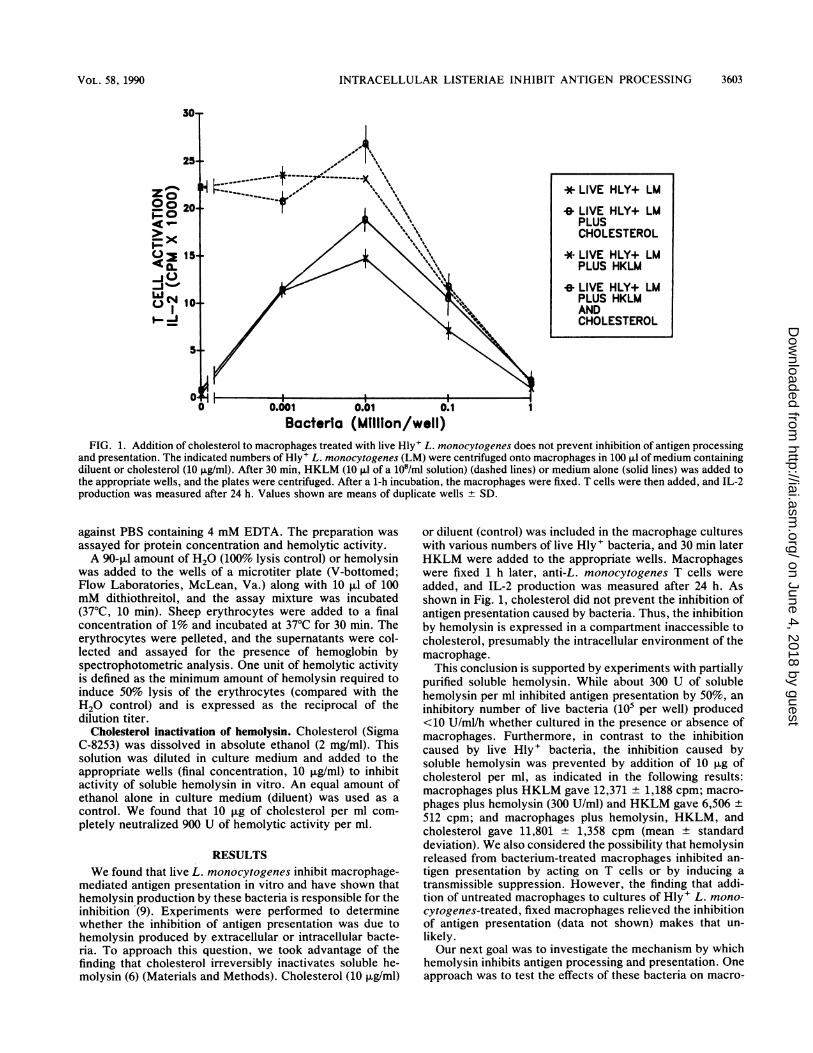
Bacteria (Million/well)FIG. 1. Addition of cholesterol to macrophages treated with live Hly+ L. monocytogenes does not prevent inhibition of antigen processing
and presentation. The indicated numbers of Hly+ L. monocytogenes (LM) were centrifuged onto macrophages in 100 ,ul of medium containingdiluent or cholesterol (10 j±g/ml). After 30 min, HKLM (10 ,ul of a 108/ml solution) (dashed lines) or medium alone (solid lines) was added tothe appropriate wells, and the plates were centrifuged. After a 1-h incubation, the macrophages were fixed. T cells were then added, and IL-2production was measured after 24 h. Values shown are means of duplicate wells + SD.
against PBS containing 4 mM EDTA. The preparation wasassayed for protein concentration and hemolytic activity.A 90-,ul amount of H20 (100% lysis control) or hemolysin
was added to the wells of a microtiter plate (V-bottomed;Flow Laboratories, McLean, Va.) along with 10 ,ul of 100mM dithiothreitol, and the assay mixture was incubated(37°C, 10 min). Sheep erythrocytes were added to a finalconcentration of 1% and incubated at 37°C for 30 min. Theerythrocytes were pelleted, and the supernatants were col-lected and assayed for the presence of hemoglobin byspectrophotometric analysis. One unit of hemolytic activityis defined as the minimum amount of hemolysin required toinduce 50% lysis of the erythrocytes (compared with theH20 control) and is expressed as the reciprocal of thedilution titer.
Cholesterol inactivation of hemolysin. Cholesterol (SigmaC-8253) was dissolved in absolute ethanol (2 mg/ml). Thissolution was diluted in culture medium and added to theappropriate wells (final concentration, 10 ,ug/ml) to inhibitactivity of soluble hemolysin in vitro. An equal amount ofethanol alone in culture medium (diluent) was used as a
control. We found that 10 ,ug of cholesterol per ml com-pletely neutralized 900 U of hemolytic activity per ml.
RESULTSWe found that live L. monocytogenes inhibit macrophage-
mediated antigen presentation in vitro and have shown thathemolysin production by these bacteria is responsible for theinhibition (9). Experiments were performed to determinewhether the inhibition of antigen presentation was due tohemolysin produced by extracellular or intracellular bacte-ria. To approach this question, we took advantage of thefinding that cholesterol irreversibly inactivates soluble he-molysin (6) (Materials and Methods). Cholesterol (10 ,ug/ml)
or diluent (control) was included in the macrophage cultureswith various numbers of live Hly+ bacteria, and 30 min laterHKLM were added to the appropriate wells. Macrophageswere fixed 1 h later, anti-L. monocytogenes T cells were
added, and IL-2 production was measured after 24 h. Asshown in Fig. 1, cholesterol did not prevent the inhibition ofantigen presentation caused by bacteria. Thus, the inhibitionby hemolysin is expressed in a compartment inaccessible tocholesterol, presumably the intracellular environment of themacrophage.
This conclusion is supported by experiments with partiallypurified soluble hemolysin. While about 300 U of solublehemolysin per ml inhibited antigen presentation by 50%, an
inhibitory number of live bacteria (105 per well) produced<10 U/ml/h whether cultured in the presence or absence ofmacrophages. Furthermore, in contrast to the inhibitioncaused by live Hly+ bacteria, the inhibition caused bysoluble hemolysin was prevented by addition of 10 ,ug ofcholesterol per ml, as indicated in the following results:macrophages plus HKLM gave 12,371 + 1,188 cpm; macro-
phages plus hemolysin (300 U/ml) and HKLM gave 6,506 +
512 cpm; and macrophages plus hemolysin, HKLM, andcholesterol gave 11,801 + 1,358 cpm (mean ± standarddeviation). We also considered the possibility that hemolysinreleased from bacterium-treated macrophages inhibited an-
tigen presentation by acting on T cells or by inducing a
transmissible suppression. However, the finding that addi-tion of untreated macrophages to cultures of Hly+ L. mono-
cytogenes-treated, fixed macrophages relieved the inhibitionof antigen presentation (data not shown) makes that un-
likely.Our next goal was to investigate the mechanism by which
hemolysin inhibits antigen processing and presentation. Oneapproach was to test the effects of these bacteria on macro-
* LIVE HLY+ LM
* LIVE HLY+ LMPLUSCHOLESTEROL
* LIVE HLY+ LMPLUS HKLM
* LIVE HLY+ LMPLUS HKLMANDCHOLESTEROL
VOL. 58, 1990 3603
on June 4, 2018 by guesthttp://iai.asm
.org/D
ownloaded from

3604 CLUFF ET AL.
A14T
2-
C
'OT90+
80-
70*
60-Io
PERCENT soRELEASE
MACROPHAGESTREATED FOR1.5 HRS WITH:
0 HLY+ LM
EI HLY- LU
40+
3o4-I
20-
10-
01
0 0.001 0.01 0.1
Bacteria (Million/well)
BT-7
6-~
5j
0
0
O 4-
2 3-
0221)
l-
0 0.001 0.01 0.1Bacteria (Million/well)
phage catabolic activity, membrane permeability, Ia expres-sion, and antigen presentation under identical conditions.Because inhibition was caused by intracellular bacteria anduptake was similar for Hly+ and Hly- bacteria (Fig. 2Dlegend and below), this and subsequent experiments wereperformed by removing unbound live bacteria after a brief10-min uptake period. Antigen presentation was inhibited bytreatment with Hly+ but not Hly- bacteria (Fig. 2A). Fiftypercent inhibition of antigen presentation was noted when105 bacteria were added per well. This dose corresponds toapproximately 1 bacterium per macrophage.The number of viable Hly+ and Hly- bacteria present
during this 1- to 2-h period of antigen processing andpresentation was monitored. The number of viable bacteriaassociated with macrophages immediately after the 10-minuptake period was not significantly different for Hly+ andHly- bacteria. After 1.5 h in culture, a small but significant
0 0.001 0.01 0.1Bacteria (Million/well)
FIG. 2. Effects of live Hly' L. monocytogenes on macrophagefunctions important for effective antigen presentation. The indicatednumbers of Hly+ or Hly- L. monocytogenes (LM) were centrifugedonto macrophages in 100 ,ul of culture medium. The plates wereincubated for 10 min to allow uptake, and the unbound bacteria wereremoved by washing. (A) Antigen presentation. HKLM (10 RI;108/ml in culture medium) were added to the appropriate wells after30 min, and the plates were centrifuged. The macrophages werefixed 1 h later, T cells were added, and IL-2 production wasmeasured after 24 h. Symbols: x, live Hly+ bacteria; 0, live Hly-bacteria; , no HKLM; -- -, plus HKLM. (B) Ia expression.The macrophages were fixed after 90 min and assayed for Iaexpression by radioimmunoassay as described in Materials andMethods. Symbols: x, Hly+; 0, Hly-. (C) Chromium release.During the adherence period (prior to addition of live bacteria),macrophages were incubated with 51Cr (2 ,uCi per well). The amountof chromium released into the supernatant and the quantity retainedby the macrophages were determined 90 min after the live bacteriawere centrifuged onto the monolayers. Percent release = (cpmreleased into the supernatant/total cpm taken up by macrophages) x100. (D) Catabolism. Live Hly+ and Hly- L. monocytogenes wereintrinsically labeled with [35S]methionine. Live bacteria (106 per wellin 100 ,ul) were centrifuged onto the macrophages. Catabolism wasmeasured by determining the amount of radioactivity associatedwith the TCA-soluble (sol) and -precipitable (ppt) fractions of theculture supernatants (supn) and macrophage lysates (lys) aftervarious periods of time. Uptakes of these two strains were found tobe similar: 39.8% of added cpm for Hly+ and 38.5% for Hly-bacteria. For all experiments, values shown are means of duplicatewells + SD.
preferential survival and/or replication of Hly+ bacteria wasnoted; 22% + 8% more viable Hly+ than Hly- bacteria wereobserved. These relatively small differences cannot accountfor the major (10- to 100-fold) differences in antigen presen-tation when Hly+ and Hly- bacteria are compared.
Ta expression (Fig. 2B) was not significantly decreased bytreatment with Hly+ bacteria. Interestingly, a small yetsignificant increase in Ta expression was noted with Hly- butnot Hly+ bacteria. Treatment of macrophages with inhibi-tory doses of Hly+ bacteria (105 per well) did not altermacrophage permeability (Fig. 2C). It is clear from boththese studies, as well as from microscopic observations ofmacrophage morphology (9, 37), that gross changes in mac-rophage membrane structure and viability are not observed
I 0
zo00< --> &8
jCLX-j )
_j o
INFECT. IMMUN.
on June 4, 2018 by guesthttp://iai.asm
.org/D
ownloaded from

INTRACELLULAR LISTERIAE INHIBIT ANTIGEN PROCESSING
Daoi
a60
(nLa0z
20L0)40'-i
I--0
U.
0 20.
* HLY+LM
*& HLY-LM
TIME (HRS)
TIME: 24 HRS
HLY+ LM HLY- LMBACTERIAL STRAIN
* SUPN:TCASOL
0I SUPN:TCAPPT
0 LYS:TCASOL
0 LYS:TCAPPT
FIG. 2-Continued
at a 1:1 bacterium-macrophage ratio. Higher concentrationsof L. monocytogenes (106 per well), however, did cause 51Crrelease from labeled macrophages, but it should be notedthat the increased 51Cr release (about 30% above back-ground) caused by Hly+ bacteria was observed only atbacterial numbers 10-fold higher than those that could pro-
duce inhibitory effects on antigen presentation.To measure antigen uptake and catabolism, 35S-labeled
live L. monocytogenes and surface-iodinated HKLM were
used as described in Materials and Methods. Live 35S-labeled Hly+ and Hly- L. monocytogenes were centrifugedonto macrophages, and unbound bacteria were removed.TCA-soluble and -precipitable fractions of the culture super-
natants and lysates were collected, and the radioactivityassociated with each fraction was determined. This proce-dure allowed us to determine the uptake of live bacteria, as
well as the amount of protein catabolized and released as
TCA-soluble material. Our data indicate that after 2 h, therewas no difference in either uptake (39.8% for Hly+ and38.5% for Hly- bacteria) or catabolic activity (Fig. 2D)between macrophages treated with 35S-radiolabeled Hly+ or
Hly- L. monocytogenes. At much later times (24 h), mac-
rophages cultured with Hly+ bacteria retained about twotimes more TCA-precipitable material than those culturedwith Hly- bacteria.Uptake and catabolism also were evaluated with surface-
iodinated HKLM. The labeled HKLM were added to mac-
rophages pretreated for 30 min with various numbers of liveHly+ or Hly- L. monocytogenes. The unbound radiolabeledbacteria were removed, and the fate of the radiolabel was
monitored as described above. Only small inhibitory effectswere observed. Pretreatment of macrophages with Hly+bacteria modestly inhibited uptake of HKLM, by 20% (Fig.3A). Although both Hly- (Fig. 3B) and Hly+ (Fig. 3C)bacteria slightly inhibited the catabolism of ingested 1251_labeled HKLM when added at 106 per well, only the Hly+
strain caused a small inhibition of this activity when added at105 per well.Another approach to studying the mechanism of inhibition
by hemolysin involved the use of antigens that differ inrequirements for antigen processing. We used the T-cellhybridoma DO11.10, which recognizes an ovalbumin pep-tide (amino acids 323 to 339) in association with I-Ad. Incontrast to the native protein, which requires processing, thepeptide can be effectively presented to DO11.10 by fixedmacrophages (Fig. 4). The other antigenic system used was
HKLM and SLP; unlike HKLM, SLP can be presented byfixed macrophages as described previously (48).Macrophages were exposed to various concentrations of
live Hly+ L. monocytogenes for 1.5 h, washed, and fixed. Inone case, HKLM were present 1 h prior to fixation. Inanother protocol, T cells were added along with SLP afterfixation. Macrophages pretreated with Hly+ L. monocyto-genes were inhibited from presenting HKLM, but once
fixed, they were able to effectively present the antigens inthe SLP preparation to the specific T cells (Fig. 5).
In another series of experiments, we determined whetherpresentation of the ovalbumin peptide would be inhibitedwhen it was added prior to fixation. Macrophages were
treated with live Hly+ or Hly- strains of L. monocytogenes,followed by addition of ovalbumin or ovalbumin peptide.The macrophages were fixed after 2 h, DO11.10 cells were
added, and IL-2 production was measured after 24 h. TheHly- strains caused only modest inhibition of ovalbuminpresentation. The addition of .106 Hly+ bacteria per wellalmost completely inhibited the presentation of ovalbumin(Fig. 6A) yet only partially reduced the presentation of theovalbumin peptide (Fig. 6B). The results obtained in manyindependent experiments with 105 bacteria per well are
presented in Table 1. Hly+ bacteria caused approximately50% inhibition of ovalbumin presentation without a signifi-cant reduction in the response with the peptide.
2CL
Li-i
-J0In
C)00
z
n
3605VOL. 58, 1990
on June 4, 2018 by guesthttp://iai.asm
.org/D
ownloaded from

3606 CLUFF ET AL.
A
4-I +0 0.001 0.01 0.1
Bacteria (Million/well)C
1'
11
TIME (MINUTES)
Bacteria(Million/well)
0 0.1
-0 0.01
NONE
40 60 60 1001TIME (MINUTES)
FIG. 3. Effect of live Hly+ L. monocytogenes on the uptake (A) and catabolism (B and C) of "251-surface-labeled HKLM. Various numbersof live Hly+ (A and C) or Hly- (A and B) L. monocytogenes (LM) were added to macrophages. The plates were centrifuged, and after a 10-minincubation, the unbound bacteria were removed. After 30 min, 2 x 10⁵ cpm of labeled HKLM in 10 p.l of culture medium was added to themacrophages, and the plates were centrifuged. Unbound labeled bacteria were removed by washing, and catabolic activity was measured as
described in the legend to Fig. 2D. For antigen uptake, values shown are means of duplicate wells ± SD. For catabolism, individual valuesdid not deviate more than 5% from any of the means.
100
90-
I 70-
CLI
La..> 30.
K= 20 .
10'
0-
B
0 Hly+ LM
0 Hly- LM
I
(AJLaJ(
Z-i
U)J,EMCLm
-In(A_ st
INFECT. IMMUN.
on June 4, 2018 by guesthttp://iai.asm
.org/D
ownloaded from

INTRACELLULAR LISTERIAE INHIBIT ANTIGEN PROCESSING
A30
z0
>0
-% x0
v-n-V-XO-
-I 10'_h0I-
B
HI
0 1 10 100 1000 0 1 10 100 1000OVA (ug/mi) OVA-Peptide (ng/mI)
FIG. 4. Processing requirements for ovalbumin and ovalbumin peptide. The indicated concentrations of ovalbumin (OVA, panel A) orovalbumin peptide (OVA-peptide, panel B) were added to normal (+) or fixed (0) macrophages in the presence of the T-cell hybridomaDO11.10. Cultures without added macrophages are indicated by solid circles. IL-2 activity in the culture supernatants was measured after 24h. Individual values did not deviate more than 10% from any of the means.
We also obtained similar results with another antigenicsystem (22), hen egg lysozyme and a corresponding T-cellhybridoma (Hd-l.AC5). With 104, 105, and 106 Hly+ L.monocytogenes bacteria per well, the percentage of controlIL-2 values (control without bacteria = 6,820 cpm) were 88,44, and 7%, respectively. Hly- bacteria showed correspond-ing values of 87, 74, and 68%, respectively. Under similarconditions, presentation of a hen egg lysozyme peptide wasrelatively unaffected (.76% of the control value).Using the system involving presentation of ovalbumin, we
tested a battery of bacterial strains and species (Fig. 7).Upon examination of different bacterial strains (at 106 bac-teria per well, as shown in Fig. 7A) within the Listeria genus,a consistent correlation between hemolysin production andinhibition of antigen presentation was observed. In thisexperiment, the effects of 105 bacteria per well were alsotested; with the Hly+ Listeria strains, the percentages ofcontrol IL-2 values (control done with no bacteria) were 54+ 19 with ovalbumin and 89 + 10 with the ovalbumin peptide(mean ± standard deviation). One Hly+ strain of anotherspecies, L. ivanovii, showed weak but significant effects onpresentation, with IL-2 values of 72% of the control withovalbumin and 103% of the control with the peptide.From the examination of several bacterial strains not
belonging to the Listeria genus (Fig. 7B), it was clear that therelationship between hemolysin production and inhibition ofantigen presentation was dependent upon the bacterialstrain. Notably, hemolysin-positive bacteria such as Strep-tococcus pyogenes and Staphylococcus aureus were notgreatly inhibitory compared with similar numbers (106 perwell) of L. monocytogenes. Hemolysin-positive Escherichiacoli, however, were markedly inhibitory. Even at 105 bacte-ria per well, Hly+ E. coli caused marked inhibition, showingIL-2 values of 22% of the control with ovalbumin and 81% ofthe control with the peptide. Also of note is the finding thata highly virulent smooth Salmonella typhimurium strain wasnot greatly inhibitory.
DISCUSSION
Relationship among virulence, hemolysin production, andinhibition of antigen presentation. The production of hemo-lysin by pathogenic strains of L. monocytogenes has beenshown to promote their intracellular survival and replicationin cultured macrophages (29, 37). Inhibition of antigenpresentation, demonstrable during the first 90 min of mac-rophage-bacteria interaction, may also contribute signifi-cantly to the virulence of these bacteria by preventing T-cellactivation. Inhibition of antigen presentation in vitro wasassociated with virulence in all Listeria strains tested (Fig.7B). The use of antigen presentation assays as an indicator ofbacterial virulence was also explored with several otherbacterial strains (Fig. 7B). With non-Listeria strains, hemo-lysin production did not correlate precisely with inhibition ofantigen presentation, and certain highly virulent strains(e.g., smooth Salmonella typhimurium) did not appear togreatly inhibit antigen presentation. The inhibitory effects ofHly+ strains, however, did extend to gram-negative organ-isms in that Hly+ E. coli caused dramatic inhibition ofpresentation. These differences among bacterial strains mayrelate to the different survival of these bacteria, quantitativedifferences in hemolysin production, differences in control ofbacterial gene expression within the macrophage, and/ordifferent properties of the hemolysins produced. In this latterregard, the hemolysin produced by Streptococcus pyogenes,while structurally and functionally similar to listeriolysin inmany ways, does not share with listeriolysin the ability toalter membrane permeability in the low pH conditions thatare present within the acidic intracellular compartments ofthe macrophage (16). It is also possible that bacteria differ intheir response (e.g., increased hemolysin expression) tosignals within the intracellular environment of macrophages.The precise nature of these signals, the mechanisms ofregulation, and the role of these parameters in virulence andcontrol of macrophage function remain to be determined.
VOL. 58, 1990 3607
on June 4, 2018 by guesthttp://iai.asm
.org/D
ownloaded from

3608 CLUFF ET AL.
20
15 HKLM BEFORE FIX
02 00~
0 0.001 0.01 0.1 1Bacteria (Mtillion/well)
FIG. 5. Macrophages fixed after treatment with Hly+ L. monocytogenes are able to present antigens that do not require processing. Theindicated numbers of Hly+ L. monocytogenes (LM) were centrifuged onto macrophages. After a 10-mmn uptake period, the unbound bacteriawere removed. Thirty minutes after addition of live bacteria, 10 ,ul of medium alone (control) or medium containing 108 HKLM per ml wasadded to the appropriate wells. The plates were incubated for 90 min, and the macrophages were fixed. T cells alone or T cells plus SLP (50,ugIml) were added to the appropriate wells, and IL-2 production was measured after 24 h. Values shown are means of duplicate wells ± SD.
Hemolysin production by L. monocytogenes has beenshown to be important for generating protective immunity(5). This effect might be due to the activity of hemolysin asa principal antigen, an adjuvant, and/or a factor promotingbacterial survival and thus antigen duration (4). Our obser-vations on inhibition of presentation do not conflict withthese findings, since low numbers of Hly+ bacteria can bepresented effectively (9) (Fig. 1 and 2). As bacterial numbersare increased, a threshold is reached, and inhibition predom-inates. This phenomenon may mimic the situation in vivoafter infection with lethal doses of bacteria or during uncon-trolled bacterial growth. In keeping with this thresholdeffect, our recent studies with recombinant preparations oflisteriolysin have revealed that it can serve as an antigen atconcentrations almost 100-fold lower than those that causeinhibition of presentation (la).
Site of inhibition by hemolysin. We used several experi-mental approaches to determine whether the hemolysinaffecting antigen presentation is produced by intracellular orextracellular bacteria. Cholesterol, which inactivates hemo-lysin, did not prevent the inhibition when added to macro-phages treated with live Hly+ bacteria (Fig. 1). In addition,inhibitory numbers of bacteria (105 per well) cultured with orwithout macrophages did not produce enough soluble hemo-lysin to account for the inhibition observed with viablebacteria. Finally, when extracellular bacteria were removedafter a 10-min uptake period, we observed no relief ofinhibition (Fig. 2A). Although we cannot formally excludesome contribution by bacteria attached to the macrophagesurface, overall the data indicate that intracellular bacteriaare responsible for the inhibition. The finding that thehemolysin produced by L. monocytogenes is optimally he-molytic at pH 5.5 (16) is compatible with our suggestion thatthis toxin is delivered from within the acidic microenviron-ment of the phagosome.Mechanism of inhibition. We tested the effects of live Hly+
and Hly- L. monocytogenes on macrophage functions thatcontribute to antigen presentation, including Ta expression,maintenance of membrane integrity, antigen uptake andcatabolism, and presentation of antigens that do not requireprocessing. Under conditions that caused about 50% inhibi-tion of antigen presentation (105 bacteria per well as in Fig.1, 2A, and 5, and Table 1), membrane integrity was main-tained (Fig. 2C), Ia expression remained normal (Fig. 2B),and macrophages treated with Hly+ bacteria were able topresent antigens that do not require processing (Fig. 5 and 6;Table 1). These results suggest that hemolysin produced byintracellular bacteria does not interfere greatly with theminimal membrane events required for effective antigenpresentation.The modest but significant inhibitory effects of .106 Hly+
bacteria on the presentation of the peptide (Fig. 6) may bedue to effects on the proper display of macrophage surfaceligands (e.g., Ia molecule display not revealed by anti-Iaantibodies as in Fig. 2B). Since known inhibitors of antigenprocessing caused slight but significant inhibition of peptidepresentation (see legend to Fig. 6), it is also possible thatpeptide-la interactions are partially dependent upon intrac-ellular events and an acidic compartment that is disrupted byHly+ bacteria. An understanding of the precise nature of thisinhibition must await further studies.The inefficient presentation of antigens associated with
live Hly+ bacteria cannot be explained by decreased uptakeand/or catabolism by the macrophages, since no differencesin these events were noted when live Hly+ and Hly-bacteria were compared at culture times of <2 h (Fig. 2D).Similar observations of equivalent uptake of Hly+ and Hlybacteria were made in previous studies (29, 37). We did findthat pretreatment of macrophages with live bacteria caused amodest decrease in both the uptake and catabolism ofiodinated HKLM (Fig. 3). We are hesitant to conclude thatthe effect of live Hly+ bacteria on HKLM uptake is solely
INFECT. IMMUN.
on June 4, 2018 by guesthttp://iai.asm
.org/D
ownloaded from

INTRACELLULAR LISTERIAE INHIBIT ANTIGEN PROCESSING
TABLE 1. Presentation of ovalbumin and ovalbumin peptide inthe presence of 105 bacteria per wella
Condition L. monocytogenes Antigen PresentationCondition phenotype Anien ( of control)
1 Hly- Ovalbumin 94 ± 142 Hly- Ovalbumin peptide 115 ± 113 Hly+ Ovalbumin 52 ± 74 Hly+ Ovalbumin peptide 99 ± 10
a Experimental conditions were as described in the legend to Fig. 7. Thevalues were calculated relative to the IL-2 response observed without addedbacteria (control). For conditions 1 and 2, n = 6; for conditions 3 and 4, n =12. Values represent the mean ± standard error of the mean. Statisticalanalysis (Student's t test) of experimental conditions: 1 versus 2, P = 0.26; 1versus 3, P = 0.01; 3 versus 4, P = 0.001; 2 versus 4, P = 0.4.
u 0 0D u possibility exists, however, that Hly+ bacteria differentiallyBacteria (Million/well) inhibit catabolic enzymes necessary for antigen processing
and that this inhibition was not detected in our measurementB of total protein degradation. It is also possible that the effects
of hemolysin on several antigen-handling events can collec-tively account for the observed inhibition of antigen presen-tation. Clearly, further analysis is required to definitivelyidentify the mechanism(s) of inhibition.
In a recent study, heat-killed bacteria (HKLM) wereshown to inhibit the presentation of lysozyme antigen, withlesser effects on the presentation of a lysozyme peptide (30).
.>_ With killed bacteria, under the conditions of our assaysystem, we also observed inhibition of ovalbumin presenta-tion but not peptide presentation, but only at very highconcentrations (108 per well) of HKLM (data not shown).This effect may be similar to that observed with live Hlybacteria, as in Fig. 6, in which a small inhibition of ovalbu-min but not of ovalbumin peptide presentation was noted at107bacteria per well. Thus, while our results are compatible
0 2 4 6 8 1 0 with previous findings (30), it should be noted that theBacteria(Million/well) inhibitory effects with live Hly+ L. monocytogenes areBacteria (Millon/well) apparent at concentrations of bacteria 100- to 1,000-fold
6. Presentation of ovalbumin or ovalbumin peptide (amino lower than those that caused the effects we observed with!3 to 339) by macrophages treated with live Hly+ or Hly- L. Hly- live bacteria or HKLM.Ptogenes (LM). The indicated numbers of live bacteria were Because it is clear that Hly+ bacteria inhibit the presen-iged onto macrophages. The unbound bacteria were removed tation of antigens that require processing more strongly thaniing, and 1 mg of ovalbumin (A) or 1 Fg of ovalbumin peptide they inhibit that of antigens that do not require processingml was added in 100 pJl of culture medium. The macrophages (Fig. 6(ed after 1.5 h. DO11.10 hybrid cells were added, and IL-2 patwa6, Table 1), a selective effect on an antigen-processing:ion was measured after 24 h. Individual values did not pathway s mplied. Although the initialfinding that catabolicmore than 10% from any of the means. The following strains activity correlated with antigen processing suggested lyso-onocytogenes were used: El, 43250 (Hly-); *, 43251 (Hly'); some involvement (52), it is probable that other acidic85/162 (Hly-); *, CNL85/163 (Hly+); *, 43248 (Hly-); and compartments exist which are used for antigen processing.9 (Hly+). In this experiment, chloroquine (10-4 M), ammo- In fact, our results can be best explained by the existence ofiloride (10 mM), and monensin (5 ,ug/ml) were included as separate pathways for antigen degradation and processingcontrols for inhibitors of antigen processing; these drugs and a selective interference with the processing pathway byIL-2 values of 9.9, 26, and 52% of control with ovalbumin Hly + bacteria.94, and 79% of control with the ovalbumin peptide, re- Evidence for at least two pathways for intracellular pro-rly (control was IL-2 activity with no drug). tein handling comes from the identification of functionally
distinct subpopulations of endosomes involved in targetingLsible for the observed inhibition of antigen presenta- internalized material to specific intracellular destinationsince a 20% decrease in antigen uptake cannot account (39). Creswell's findings suggest that early endosomes inter-y for the >50% inhibition of antigen presentation act with an Ia-containing compartment within the macro-)n. In addition, increasing the concentration ofHKLM phage (10). An acidic environment may be critical for earlyovercome the observed inhibition (data not shown). endosome function, since it allows the dissociation of recep-
se certain concentrations (106 per well) of Hly- L. tor-ligand complexes. Perhaps the low pH of these earlyytogenes inhibited catabolism to the same extent as endosomes denatures ingested antigens and allows thesebacteria yet did not diminish the ability of macro- antigens to associate with Ia in the absence of enzymaticto present antigen (Fig. 2A and 6), it seems unlikely fragmentation. We and others have found that nonenzymatic
me inhibition of antigen presentation observed with denaturing treatments can create processed antigen (1, 28,,acteria is due to a decrease in catabolic activity. The 42, 48). Thus, exposure of microbial products to the low pH
Az 300
r-
D00 200.v-
-CJO%- 10CMCM4
*130_,0
40
30
20
10
0
FIG.acids 32monocycentrifuby wash(B) per i
were fixproductideviateof L. mU, CNLC1, 4324nium cipositiveshowedand 75,spective
respontion, sientirel!functiodid notBecausmonoc
Hly+ 1phagesthat thHly+ b
VOL. 58, 1990 3609
on June 4, 2018 by guesthttp://iai.asm
.org/D
ownloaded from

3610 CLUFF ET AL.
ABACTERIA]
0
NONE
L. monocytogenes 43250 (Hly-)L. monocytogenes 43251 (Hly+)
L. monocytogenes CNL85/162 (Hly-)L. monocytogenes CNL85/1 63 (Hly+)
L. monocytogenesL. monocytogenesL. monocytogenesL. monocytogenesL. monocytogenesL. monocytogenes
43248432493515215313
(Hly-)(Hly+)(Hly+)(Hly-)
5000
Antigen (OVA) PresentationIL-2 (Delta CPM)
I 110000 15000 A
U,,,,,,,,,,,,,,,,,,,,,,,,lllllnmm
13932 (Hly+)43256 (Hly+)
L. monc
L. monc
L. monocytogeni
mcytogenes Ml (Hly-))cytogenes M3 (Hly-)es NCTC 5105 (Hly+)
L. ivanovil (Hly+)L. weishimeri (Hly-)
L. grayi (Hly-)L. murrayi (Hly-)L. seeligeri (Hly-)L. lnnocua (Hly-)
BAntigen (OVA) Presentation
lL-2 (Delta CPM)0 10000 20000 30000
IBACTERIAI NUONL. monocytogenes CNL85/162 (Hly-)L. monocytogenes CNL85/163 (Hly+)
L. monocytogenes Tn916 MI (Hly-)L. monocytogenes Tn 916 M3 (Hly-)L. monocytogenes NCTC 5105 (Hly+)
S. pyogenes 21547 (Hly+)S. pyogenes 27762 (Hly+)S. pyogenes 10389 (Hly+)
E. colt WAF270 (Hly+)E. coil WAF108 (Hly-)
E. coil BB4 (Hly-)E. coli C600 (Hly-)B. subtillus (Hly-)B. abortus (Hly-)S. aureus (Hly+)S. aureus (Hly-)
S. typhimurium LT2 smooth (Hly-)S. typhimurium SL1004 rough (Hly-)
S. pneumonia. (Hly-)K. pneumonla. (Hly-)
M. phi.! (Hly-)
||||XllllZl§111|1101111111111|11111111111l
l
1111111111111111111111111111|111111|111
1lululllElllllllxlullElllllllllll1lllllllllllllllllllllllllllllls
11111111111111111111111111111111111111111111111111111111111111
1.11.L1|1EuS=u1101l.lUlllllllllllllllllllllllllll1IImml
11111111111111111111|111111111111111111111111111111111111111111111
11111111lllll11Xll1|1|1ll1111}11111111111111111111111111|1111111111111111111111111111111111111l
.1|llllllllllllllllllllllllllllllllllllllllll
FIG. 7. Inhibition of antigen presentation by live bacteria. Presentation of ovalbumin by macrophages treated with various bacterial strainswas tested. Live bacteria (106 per well) were centrifuged onto macrophages. The unbound bacteria were removed by washing, and ovalbumin(OVA, 1 mglml) was added in 100 ,ul of culture medium. The macrophages were fixed after 1.5 h. D011.10 hybrid cells were added, and IL-2production was measured after 24 h. Two representative experiments are shown in panels A and B. Each strain was judged to be hemolyticon sheep blood-agar plates. Solid bars are Hly+ strains; hatched bars are Hly- strains. Streptococcus pneumoniae, although judged to beHly-, does contain an intracellular pneumolysin. Most strains were obtained from the American Type Culture Collection. The CNL L.monocytogenes strains (9, 15) and the Ml and M3 strains (23) have been described previously. The WAF E. coli strains were from Rod Welch(43).
20000 25000i
-T-
INFECT. IMMUN.
Llf%Llr
on June 4, 2018 by guesthttp://iai.asm
.org/D
ownloaded from
INTRACELLULAR LISTERIAE INHIBIT ANTIGEN PROCESSING
of early endosomes may define the processing event forsome antigens (12). Additionally, exposure of la moleculesto low pH may favor the association with processed antigen(22). Conceivably, hemolysins could inhibit processing byacting as an ion channel, thereby neutralizing the pH of thisintracellular compartment. In this regard, hemolysin may beanalogous to monensin, a known inhibitor of antigen proc-essing. This idea is currently being tested.The results of others suggest that hemolysin secreted by
ingested bacteria can mediate the disruption of the phago-some membrane and promote bacterial replication in thecytoplasm (14, 37). It is possible that the escape of bacteriainto the cytoplasm may allow bacterial antigens to bypass apresentation pathway involving class II MHC gene products(Ia molecules). In the cytoplasm, a class I MHC-controlledpresentation may predominate (32), which might account forthe class I-restricted CD8+ cells noted in the immuneresponse to L. monocytogenes (11, 25). Also relevant to theeffects on intracellular antigen processing and presentationevents is the reorganization of actin filaments that has beenshown to occur in infected macrophages (42a). The previousfindings that Ia expression increases after antigen uptake (2,3), that protein synthesis is required'for presentation (21),and that newly synthesized Ia and internalized moleculesreside in the same intracellular compartment (10) are circum-stantial evidence that Ia synthesis, intracellular Ia-antigenbinding, and subsequent transport to the cell surface are
important for effective antigen presentation. Thus, the pos-sible effects of hemolysin-producing bacteria on these eventsmust also be considered. Indeed, we have found that mac-rophages treated with Hly- bacteria increased Ia expressionby about 25% (Fig. 2B). In contrast, macrophages treatedwith Hly+ bacteria did not show similar increases. Futureexperiments will be designed to determine whether thesebacteria interfere with Ta synthesis, intracellular interactionsbetween Ia and processed antigen, and/or transit of thecomplex to the cell surface.
In summary, we have provided evidence that hemolysinpromotes the virulence of L. monocytogenes by inhibitingantigen processing. Our approach not only affords insightinto the manner by which L. monocytogenes expresses itsvirulence factors, but also supplies clues regarding themechanism of antigen processing and presentation.
ACKNOWLEDGMENTS
We thank Ann Benson and Ingrid Tanycz for help with hemolysincharacterization and purification and Jan Paul for advice on syn-thetic peptide antigens.
This research was supported in part by Public Health Servicegrants R01 AI-20215, T32 AI-07265, and K04 AI-00620 from theNational Institutes of Health.
LITERATURE CITED
1. Allen, P., and E. Unanue. 1984. Differential requirements forantigen processing by macrophages for lysozyme-specific T cellhybridomas. J. Immunol. 132:1077-1079.
la.Beattie, I. A., B. Swaminathan, and H. K. Ziegler. 1990. Cloningand characterization of T-cell-reactive protein antigens fromListeria monocytogenes. Infect. Immun. 58:2792-2803.
2. Belier, D. I., and K. Ho. 1982. Regulation of macrophagepopulations. V. Evaluation of the control of macrophage Iaexpression in vitro. J. Immunol. 129:971-976.
3. Beller, D. I., and E. R. Unanue. 1981. Regulation of macrophagepopulations. II. Synthesis and expression of Ia antigens byperitoneal exudate macrophages is a transient event. J. Immu-nol. 126:263-269.
4. Berche, P., J.-L. Gaillard, C. Geoffroy, and J. E. Alouf. 1987. T
cell recognition of listeriolysin 0 is induced during infectionwith Listeria monocytogenes. J. Immunol. 139:3813-3821.
5. Berche, P., J.-L. Gaillard, and P. Sansonetti. 1987. Intracellulargrowth of Listeria monocytogenes as a prerequisite for in vivoinduction of T cell-mediated immunity. J. Immunol. 138:2266-2271.
6. Bernheimer, A. 1974. Interactions between membranes andcytolytic bacterial toxins. Biochim. Biophys. Acta 344:27-50.
7. Buchmeier, N. A., and R. D. Schreiber. 1985. Requirement ofendogenous gamma interferon production for the resolution ofListeria monocytogenes infection. Proc. Natl. Acad. Sci. USA82:7404-7408.
8. Cluff, C. W., and H. K. Ziegler. 1986. An early response tolipopolysaccharide is the elicitation of macrophages specializedfor antigen degradation with negative regulatory effects on theinduction of specific immune responses. Infect. Immun. 55:1346-1354.
9. Cluff, C. W., and H. K. Ziegler. 1987. Inhibition of macrophage-mediated antigen presentation by hemolysin-producing Listeriamonocytogenes. J. Immunol. 139:3808-3812.
10. Creswell, P. 1985. Intracellular class II HLA antigens areaccessible to transferrin-neuraminidase conjugates internalizedby receptor-mediated endocytosis. Proc. Natl. Acad. Sci. USA82:8188-8192.
11. De Libero, G., and S. H. E. Kaufmann. 1986. Antigen-specificLyt2+ cytolytic T lymphocytes from mice infected with theintracellular bacterium Listeria monocytogenes. J. Immunol.137:2688-2692.
12. Eisenlohr, L. C., W. Gerhard, and C. J. Hackett. 1988. Acid-induced conformational modification of the hemagglutinin mol-ecule alters interaction of influenza virus with antigen-present-ing cells. J. Immunol. 141:1870-1876.
13. Farr, A., J. Kiely, and E. Unanue. 1979. Macrophage-T cellinteractions involving Listeria monocytogenes-role of the H-2gene complex. J. Immunol. 122:2395-2404.
14. Gaillard, J.-L., P. Berche, J. Mounier, S. Richard, and P.Sansonetti. 1987. In vitro model of penetration and intracellulargrowth of Listeria monocytogenes in the human enterocyte-likecell line Caco-2. Infect. Immun. 55:2822-2829.
15. Gailiard, J.-L., P. Berche, and P. Sansonetti. 1986. Transposonmutagenesis as a tool to study the role of hemolysin in thevirulence of Listeria monocytogenes. Infect. Immun. 52:50-55.
16. Geoffroy, C., J.-L. Gaillard, J. E. Alouf, and P. Berche. 1987.Purification, characterization, and toxicity of the sulfhydryl-activated hemolysin listeriolysin 0 from Listeria monocyto-genes. Infect. Immun. 55:1641-1646.
17. Gray, M. L., and A. H. Killinger. 1966. Listeria monocytogenesand listeric infections. Bacteriol. Rev. 30:309-382.
18. Greenwood, F. C., W. H. Hunter, and J. S. Grover. 1963. Thepreparation of 13'1-labeled human growth hormone of highlyspecific radioactivity. Biochem. J. 89:114-118.
19. Groves, R. D., and H. J. Weishimer. 1977. Separation ofpathogenic from apathogenic Listeria monocytogenes by threein vitro reactions. J. Clin. Microbiol. 5:559-566.
20. Jenkins, E. M., A. N. Njoku-Obi, and E. A. Adams. 1964.Purification of the soluble hemolysins of Listeria monocyto-genes. J. Bacteriol. 88:418-424.
21. Jensen, P. E. 1988. Protein synthesis in antigen processing. J.Immunol. 141:2545-2550.
22. Jensen, P. E. 1990. Regulation of antigen presentation by acidicpH. J. Exp. Med. 171:1779-1784.
23. Kathariou, S., P. Metz, H. Hof, and W. Goebel. 1987. Tn916-induced mutations in the hemolysin determinant affecting viru-lence of Listeria monocytogenes. J. Bacteriol. 169:1291-1297.
24. Kaufmann, S. H. E. 1983. Effective antibacterial protectioninduced by a Listeria monocytogenes-specific T-cell clone andits lymphokines. Infect. Immun. 39:1265-1270.
25. Kaufmann, S. H. E., E. Hug, and G. DeLibero. 1986. Listeriamonocytogenes-reactive T lymphocyte clones with cytolyticactivity against infected target cells. J. Exp. Med. 164:363-368.
26. Kaufmann, S. H. E., M. M. Shimon, and H. Hahn. 1979. SpecificLyt123+ T cells are involved in protection against Listeriamonocytogenes and in delayed-type hypersensitivity to listerial
VOL. 58, 1990 3611
on June 4, 2018 by guesthttp://iai.asm
.org/D
ownloaded from

3612 CLUFF ET AL.
antigens. J. Exp. Med. 150:1033-1038.27. Kiderlen, A., S. Kauffmann, and M. Lohmann-Matthes. 1984.
Protection of mice against the intracellular bacterium Listeriamonocytogenes by recombinant immune interferon. Eur. J.Immunol. 14:964-968.
28. Kovac, Z., and R. H. Schwartz. 1985. The molecular basis of therequirement for antigen processing of pigeon cytochrome-cprior to T cell activation. J. Immunol. 134:3233-3240.
29. Kuhn, M., S. Kathariou, and W. Goebel. 1988. Hemolysinsupports survival but not entry of the intracellular bacteriumListeria monocytogenes. Infect. Immun. 56:79-82.
30. Leyva-Cobian, F., and E. R. Unanue. 1988. Intracellular inter-ference with antigen presentation. J. Immunol. 141:1445-1450.
31. Magee, D. M., and E. J. Wing. 1988. Cloned L3T4+ T lympho-cytes protect mice against Listeria monocytogenes by secretinginterferon-gamma. J. Immunol. 141:3203-3207.
32. Moore, M. W., F. R. Carbone, and M. J. Bevan. 1988. Intro-duction of soluble protein into the class I pathway of antigenprocessing and presentation. Cell 54:777-785.
33. Nakane, A., T. Minagawa, and K. Kato. 1988. Endogenoustumor necrosis factor (cachectin) is essential to host resistanceagainst Listeria monocytogenes infection. Infect. Immun. 56:2563-2569.
34. North, R. J. 1973. Cellular mediators of anti-listeria immunity asan enlarged population of short-lived replicating T cells. J. Exp.Med. 138:342-355.
35. Palay, D. A., C. W. Cluff, P. A. Wentworth, and H. K. Ziegler.1986. Cyclosporine inhibits macrophage-mediated antigen pre-sentation. J. Immunol. 136:4348-4353.
36. Pine, L., R. E. Weaver, G. M. Carlone, P. A. Pienta, J. Rocourt,W. Goebel, S. Kathariou, W. F. Bibb, and G. B. Malcolm. 1987.Listeria monocytogenes ATCC 35152 and NCTC 7973 contain-ing nonhemolytic nonvirulent variants. J. Clin. Microbiol. 25:2247-2249.
37. Portnoy, D. A., P. S. Jacks, and D. J. Hinrichs. 1988. Role ofhemolysin for the intracellular growth of Listeria monocyto-genes. J. Exp. Med. 167:1459-1471.
38. Recourt, J. G., G. Grimont, P. A. D. Grimont, and H. P. R.Seeliger. 1982. DNA relatedness among serovars of Listeriamonocytogenes. Curr. Microbiol. 7:383-388.
39. Schmid, S. L., R. Fuchs, P. Male, and I. Mellman. 1988. Twodistinct subpopulations of endosomes involved in membranerecycling and transport to lysosomes. Cell 52:73-83.
40. Seelinger, H. P. R. 1961. Listeriosis. Hafner, New York.41. Shimonkevitz, R., S. Colon, J. W. Kappler, P. Marrack, and
H. M. Grey. 1984. Antigen recognition by H-2-restricted T cells.II. A tryptic ovalbumin peptide that substitutes for processedantigen. J. Immunol. 133:2067-2074.
42. Streicher, H., I. Berkower, M. Busch, F. Gurd, and J. Berzofsky.1984. Antigen conformation determines processing require-ments for T cell activation. Proc. Natl. Acad. Sci. USA 81:6831-6837.
42a.Tilney, L. G., and D. A. Portnoy. 1989. Actin filaments and thegrowth, movement, and spread of the intracellular bacterialparasite, Listeria monocytogenes. J. Cell Biol. 109:1597-1608.
43. Welch, R. A. 1988. Transcriptional organization of the Esche-richia coli hemolysin genes. J. Bacteriol. 170:1622-1630.
44. Wentworth, P. A., and H. K. Ziegler. 1986. The antigenic andmitogenic response of murine T and B lymphocytes to solubleproteins of Listeria monocytogenes. J. Immunol. 138:3167-3173.
45. Wilson, C. B., and J. Westall. 1985. Activation of neonatal andadult human macrophages by alpha, beta, and gamma interfer-ons. Infect. Immun. 49:351-356.
46. Ziegler, H. K. 1984. The processing and presentation of Listeriamonocytogenes antigens by macrophages. Clin. Invest. Med.7:269-275.
47. Ziegler, H. K., and C. A. Orlin. 1984. Analysis of Listeriamonocytogenes antigens with monoclonal antibodies. Clin. In-vest. Med. 7:239-243.
48. Ziegler, H. K., C. A. Orlin, and C. W. Cluff. 1987. Differentialrequirements for the processing and presentation of soluble andparticulate bacterial antigens by macrophages. Eur. J. Immunol.17:1287-1296.
49. Ziegler, H. K., L. K. Staffileno, and P. Wentworth. 1984.Modulation of macrophage Ia expression by lipopolysaccharide.I. induction of Ia expression in vivo. J. Immunol. 133:1825-1835.
50. Ziegler, H. K., and E. R. Unanue. 1979. The specific binding ofListeria monocytogenes immune T lymphocytes to macro-phages. I. Quantitation and role of H-2 gene products. J. Exp.Med. 150:1143-1160.
51. Ziegler, H. K., and E. R. Unanue. 1981. Identification of amacrophage antigen-processing event required for I-region-restricted antigen presentation to T lymphocytes. J. Immunol.127:1869-1875.
52. Ziegler, H. K., and E. R. Unanue. 1982. Decrease in macrophageantigen catabolism caused by ammonia and chloroquine isassociated with inhibition of antigen presentation to T cells.Proc. Natl. Acad. Sci. USA 79:175-178.
53. Zinkernagel, R., A. Althage, B. Adler, B. Blander, W. Davidson,V. Kees, M. Dunlop, and D. Shreffler. 1977. H-2 restriction ofcell-mediated immunity to an intracellular bacterium. Effector Tcells are specific for Listeria antigen in association with H-21region coded self markers. J. Exp. Med. 145:1353-1367.
INFECT. IMMUN.
on June 4, 2018 by guesthttp://iai.asm
.org/D
ownloaded from