Embed Size (px)
Citation preview
Properties of Bacillus cereus hemolysin II:A heptameric transmembrane pore
GEORGE MILES,1 HAGAN BAYLEY,1,2AND STEPHEN CHELEY1
1Department of Medical Biochemistry & Genetics, The Texas A&M University System Health Science Center,College Station, Texas 77843-1114, USA2Department of Chemistry, Texas A&M University, College Station, Texas 77843-3255, USA
(RECEIVED February 5, 2002; FINAL REVISION April 24, 2002; ACCEPTED April 24, 2002)
Abstract
The gene encoding hemolysin II (HlyII) was amplified from Bacillus cereus genomic DNA and a truncatedmutant, HlyII(�CT), was constructed lacking the 94 amino acid extension at the C terminus. The proteinswere produced in an E. coli cell-free in vitro transcription and translation system, and were shown toassemble into SDS-stable oligomers on rabbit erythrocyte membranes and liposomes. The hemolytic activityof HlyII was measured with rabbit erythrocytes yielding an HC50 value of 1.64 ng mL−1, which is over 15times more potent than staphylococcal �-hemolysin. HlyII(�CT) was about eight times less potent thanHlyII in this assay. Limited proteolysis of the oligomers formed by HlyII and HlyII(�CT) on red cellmembranes showed that the C-terminal extension is sensitive to digestion, while HlyII(�CT) is proteaseresistant and migrates with an electrophoretic mobility similar to that of digested HlyII. HlyII formsmoderately anion selective, rectifying pores (I+80/I−80 � 0.57, 1 M KCl, pH 7.4) in planar lipid bilayers ofdiphytanoylphosphatidylcholine with a unitary conductance of 637 pS (1 M KCl, 5 mM HEPES, pH 7.4)and exhibits no gating over a wide range of applied potentials (−160 to +160 mV). In addition, it wasdemonstrated that HlyII forms a homoheptameric pore by using gel shift electrophoresis aided by a geneti-cally encoded oligoaspartate tag. Although they share limited primary sequence identity (30%), these dataconfirm that HlyII is a structural and functional homolog of staphylococcal �-hemolysin.
Keywords: �-Barrel; hemolysin; membrane protein; pore-forming toxin; staphylococcal �-hemolysin; sub-unit stoichiometry
In this work, we examine the properties of hemolysin II(HlyII), a �-barrel pore forming toxin (�-PFT) from Bacil-lus cereus. The �-PFTs consist of several subfamilies ofbacterial exotoxins that are related by sequence (Gouaux etal. 1997; Menestrina et al. 2001) and structure (Song et al.1996; Gouaux et al. 1997; Olson et al. 1999; Pédelacq et al.1999). These polypeptides are secreted as water-solublemolecules that bind to the surfaces of susceptible cells andassemble into oligomeric transmembrane pores leading tocell permeation and lysis (Bhakdi et al. 2000; Menestrina etal. 2001). The crystal structure of a heptameric staphylo-coccal �-hemolysin (�HL) pore has been determined indetergent at 1.9-Å resolution (Song et al. 1996). �HL hasalso been shown to form heptamers on red cell membranes(Gouaux et al. 1994), in planar lipid bilayers (Krasilnikov etal. 2000), supported bilayers (Fang et al. 1997), and afterspontaneous assembly in solution (Cheley et al. 1997). In a
Reprint requests to: Hagan Bayley, Department Medical Biochemistry &Genetics, Texas A&M Health Science Center, 440 Reynolds MedicalBuilding, College Station, TX 77843-1114, USA; e-mail: [email protected]; fax: (979) 847-9481.
Abbreviations: �HL, �-hemolysin of Staphylococcus aureus; �HL-D8,�-hemolysin with a C-terminal extension of eight aspartate residues; �HL-TL, �-hemolysin fusion protein with a C-terminal extension comprising theC-terminal 94 residues of HlyII; �-PFT, �-barrel pore forming toxin;CytK, cytotoxin K of Bacillus cereus; HEPES, N-[2-hydroxyethyl]pipera-zine-N�-[2-ethanesulfonic acid]; MBSA, 10 mM Na MOPS, 150 mMNaCl, pH 7.4, containing 1 mg mL−1 bovine serum albumin; MOPS, 3-[N-mor-pholino]propanesulfonic acid; IVTT, in vitro transcription and translation;PMSF, phenylmethylsulfonylfluoride; rRBC, rabbit erythrocyte; rRBCM,rabbit erythrocyte membranes; SDS, sodium dodecyl sulfate; HlyII, hemo-lysin II of Bacillus cereus; HlyII-D8, hemolysin II with a C-terminal ex-tension of eight aspartate residues; HlyII(�CT), a truncation mutant ofHlyII lacking 94 amino acid residues at the C terminus; HlyII(�CT)-D8,HlyII(�CT) with a C-terminal extension of eight aspartate residues; TL, apolypeptide comprising the C-terminal 94 amino acids of HlyII.
Article and publication are at http://www.proteinscience.org/cgi/doi/10.1110/ps.0204002.
Protein Science (2002), 11:1813–1824. Published by Cold Spring Harbor Laboratory Press. Copyright © 2002 The Protein Society 1813
working scheme for the assembly of �HL, the 293-residuepolypeptide first binds to the membrane as a monomer,associates to form a heptameric prepore, and finally insertsinto the bilayer to form the transmembrane pore. Thisscheme is supported by numerous biophysical and bio-chemical experiments (Cheley et al. 1997), and has beenrefined to accommodate recent structural data (Olson et al.1999; Pédelacq et al. 1999).
Bacillus cereus is an opportunistic pathogen. It is asso-ciated with a wide range of clinical symptoms, but encoun-tered primarily in cases of severe food poisoning (Drobeni-wski 1993; Lund et al. 2000). Over 20 exotoxins are pro-duced and secreted by B. cereus, including severalnonhemolytic enterotoxins (Alouf and Freer 1999). Twodistinct hemolytic proteins produced by this bacterium, he-molysin II (HlyII) (Baida et al. 1999) and cytotoxin K(CytK) (Lund et al. 2000), have recently been cloned andshown to be homologous with the �-PFTs. HlyII has thelongest polypeptide chain in the �-PFT family, with 412residues, and contains a C-terminal 94 amino acid extensionthat has no homology with any other known �-PFT. The
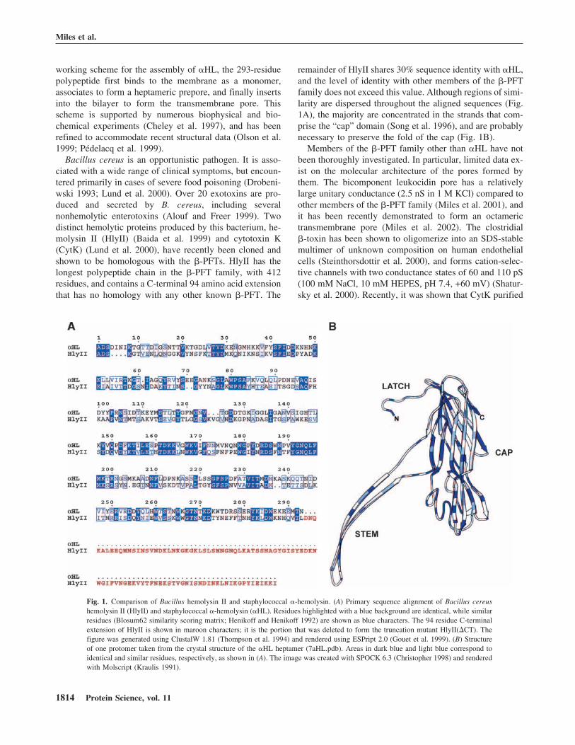
remainder of HlyII shares 30% sequence identity with �HL,and the level of identity with other members of the �-PFTfamily does not exceed this value. Although regions of simi-larity are dispersed throughout the aligned sequences (Fig.1A), the majority are concentrated in the strands that com-prise the “cap” domain (Song et al. 1996), and are probablynecessary to preserve the fold of the cap (Fig. 1B).
Members of the �-PFT family other than �HL have notbeen thoroughly investigated. In particular, limited data ex-ist on the molecular architecture of the pores formed bythem. The bicomponent leukocidin pore has a relativelylarge unitary conductance (2.5 nS in 1 M KCl) compared toother members of the �-PFT family (Miles et al. 2001), andit has been recently demonstrated to form an octamerictransmembrane pore (Miles et al. 2002). The clostridial�-toxin has been shown to oligomerize into an SDS-stablemultimer of unknown composition on human endothelialcells (Steinthorsdottir et al. 2000), and forms cation-selec-tive channels with two conductance states of 60 and 110 pS(100 mM NaCl, 10 mM HEPES, pH 7.4, +60 mV) (Shatur-sky et al. 2000). Recently, it was shown that CytK purified
Fig. 1. Comparison of Bacillus hemolysin II and staphylococcal �-hemolysin. (A) Primary sequence alignment of Bacillus cereushemolysin II (HlyII) and staphylococcal �-hemolysin (�HL). Residues highlighted with a blue background are identical, while similarresidues (Blosum62 similarity scoring matrix; Henikoff and Henikoff 1992) are shown as blue characters. The 94 residue C-terminalextension of HlyII is shown in maroon characters; it is the portion that was deleted to form the truncation mutant HlyII(�CT). Thefigure was generated using ClustalW 1.81 (Thompson et al. 1994) and rendered using ESPript 2.0 (Gouet et al. 1999). (B) Structureof one protomer taken from the crystal structure of the �HL heptamer (7aHL.pdb). Areas in dark blue and light blue correspond toidentical and similar residues, respectively, as shown in (A). The image was created with SPOCK 6.3 (Christopher 1998) and renderedwith Molscript (Kraulis 1991).
Miles et al.
1814 Protein Science, vol. 11
from B. cereus supernatants forms pores in planar lipidbilayers, and is cytotoxic towards intestinal epithelial cells(Hardy et al. 2001). The subunit stoichiometries of the Ba-cillus toxins, HlyII and CytK, have not been determined.
In this study, we demonstrate that HlyII, produced by invitro transcription and translation, forms a heptameric trans-membrane pore in red cell membranes, which is resistant toSDS. In planar lipid bilayers, the pores are rectifying andlack voltage-induced gating. HlyII with the C-terminal ex-tension removed, HlyII(�CT), has similar properties.Knowledge of the subunit stoichiometry and channel prop-erties of this relative of �HL adds to the understanding ofthe �-PFTs family, and will be helpful in protein engineer-ing aimed at the construction of pore-forming proteins withnew properties (Bayley 1999; Bayley and Cremer 2001).
Results and Discussion
HlyII and HlyII(�CT) form SDS-resistant oligomers onred cell membranes and liposomes
HlyII includes a 94-amino acid C-terminal extension, whichis not homologous with any known �-PFT. The two most
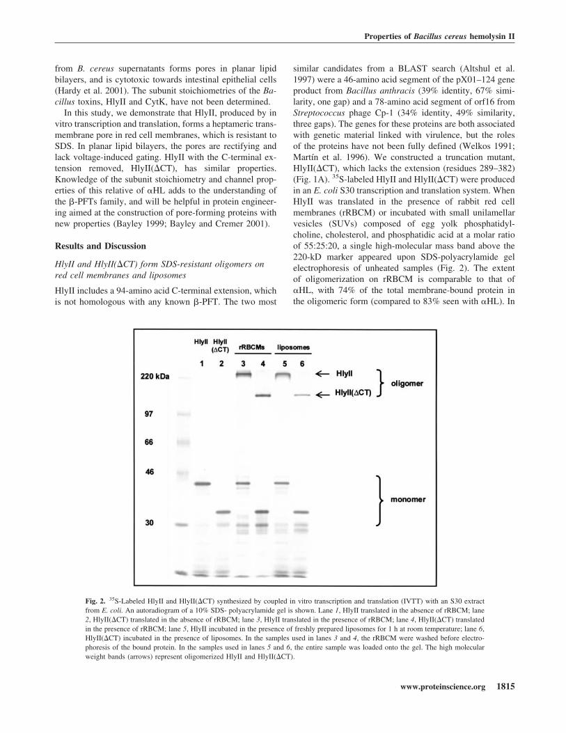
similar candidates from a BLAST search (Altshul et al.1997) were a 46-amino acid segment of the pX01–124 geneproduct from Bacillus anthracis (39% identity, 67% simi-larity, one gap) and a 78-amino acid segment of orf16 fromStreptococcus phage Cp-1 (34% identity, 49% similarity,three gaps). The genes for these proteins are both associatedwith genetic material linked with virulence, but the rolesof the proteins have not been fully defined (Welkos 1991;Martín et al. 1996). We constructed a truncation mutant,HlyII(�CT), which lacks the extension (residues 289–382)(Fig. 1A). 35S-labeled HlyII and HlyII(�CT) were producedin an E. coli S30 transcription and translation system. WhenHlyII was translated in the presence of rabbit red cellmembranes (rRBCM) or incubated with small unilamellarvesicles (SUVs) composed of egg yolk phosphatidyl-choline, cholesterol, and phosphatidic acid at a molar ratioof 55:25:20, a single high-molecular mass band above the220-kD marker appeared upon SDS-polyacrylamide gelelectrophoresis of unheated samples (Fig. 2). The extentof oligomerization on rRBCM is comparable to that of�HL, with 74% of the total membrane-bound protein inthe oligomeric form (compared to 83% seen with �HL). In
Fig. 2. 35S-Labeled HlyII and HlyII(�CT) synthesized by coupled in vitro transcription and translation (IVTT) with an S30 extractfrom E. coli. An autoradiogram of a 10% SDS- polyacrylamide gel is shown. Lane 1, HlyII translated in the absence of rRBCM; lane2, HlyII(�CT) translated in the absence of rRBCM; lane 3, HlyII translated in the presence of rRBCM; lane 4, HlyII(�CT) translatedin the presence of rRBCM; lane 5, HlyII incubated in the presence of freshly prepared liposomes for 1 h at room temperature; lane 6,HlyII(�CT) incubated in the presence of liposomes. In the samples used in lanes 3 and 4, the rRBCM were washed before electro-phoresis of the bound protein. In the samples used in lanes 5 and 6, the entire sample was loaded onto the gel. The high molecularweight bands (arrows) represent oligomerized HlyII and HlyII(�CT).
Properties of Bacillus cereus hemolysin II
www.proteinscience.org 1815
the case of HlyII(�CT), the extent of oligomerization isreduced to 49%. Oligomers formed by HlyII and HlyII-(�CT) are stable in 2.3% SDS (1× Laemmli sample buffer)at room temperature and dissociate at 82°C and 78°C, re-spectively. �HL dissociates at 70°C in the sample buffer(data not shown).
A deletion variant lacking the extension (and two addi-tional amino acids) was previously shown to retain somehemolytic activity towards human red blood cells (Baida etal. 1999). The removal of the two additional amino acids isexpected to lead to a reduction in activity. We found earlierthat the removal of three amino acids from the C terminusof �HL causes a dramatic reduction in hemolytic activitythat is associated with an almost complete loss of ability toform SDS-stable oligomers (Walker et al. 1992a).
Examination of the HlyII oligomer by limitedproteolysis suggests structural similarity to �HL
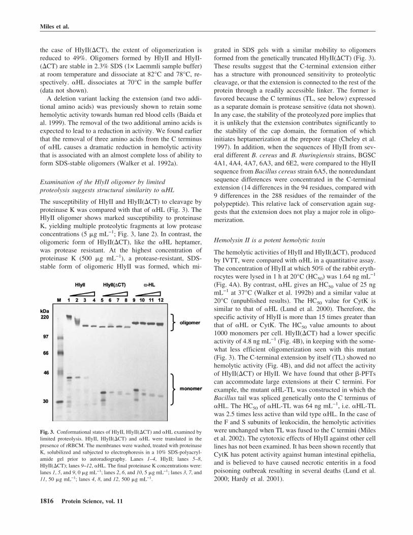
The susceptibility of HlyII and HlyII(�CT) to cleavage byproteinase K was compared with that of �HL (Fig. 3). TheHlyII oligomer shows marked susceptibility to proteinaseK, yielding multiple proteolytic fragments at low proteaseconcentrations (5 �g mL−1; Fig. 3, lane 2). In contrast, theoligomeric form of HlyII(�CT), like the �HL heptamer,was protease resistant. At the highest concentration ofproteinase K (500 �g mL−1), a protease-resistant, SDS-stable form of oligomeric HlyII was formed, which mi-
grated in SDS gels with a similar mobility to oligomersformed from the genetically truncated HlyII(�CT) (Fig. 3).These results suggest that the C-terminal extension eitherhas a structure with pronounced sensitivity to proteolyticcleavage, or that the extension is connected to the rest of theprotein through a readily accessible linker. The former isfavored because the C terminus (TL, see below) expressedas a separate domain is protease sensitive (data not shown).In any case, the stability of the proteolyzed pore implies thatit is unlikely that the extension contributes significantly tothe stability of the cap domain, the formation of whichinitiates heptamerization at the prepore stage (Cheley et al.1997). In addition, when the sequences of HlyII from sev-eral different B. cereus and B. thuringiensis strains, BGSC4A1, 4A4, 4A7, 6A3, and 6E2, were compared to the HlyIIsequence from Bacillus cereus strain 6A5, the nonredundantsequence differences were concentrated in the C-terminalextension (14 differences in the 94 residues, compared with9 differences in the 288 residues of the remainder of thepolypeptide). This relative lack of conservation again sug-gests that the extension does not play a major role in oligo-merization.
Hemolysin II is a potent hemolytic toxin
The hemolytic activities of HlyII and HlyII(�CT), producedby IVTT, were compared with �HL in a quantitative assay.The concentration of HlyII at which 50% of the rabbit eryth-rocytes were lysed in 1 h at 20°C (HC50) was 1.64 ng mL−1
(Fig. 4A). By contrast, �HL gives an HC50 value of 25 ngmL−1 at 37°C (Walker et al. 1992b) and a similar value at20°C (unpublished results). The HC50 value for CytK issimilar to that of �HL (Lund et al. 2000). Therefore, thespecific activity of HlyII is more than 15 times greater thanthat of �HL or CytK. The HC50 value amounts to about1000 monomers per cell. HlyII(�CT) had a lower specificactivity of 4.8 ng mL−1 (Fig. 4B), in keeping with the some-what less efficient oligomerization seen with this mutant(Fig. 3). The C-terminal extension by itself (TL) showed nohemolytic activity (Fig. 4B), and did not affect the activityof HlyII(�CT) or HlyII. We have found that other �-PFTscan accommodate large extensions at their C termini. Forexample, the mutant �HL-TL was constructed in which theBacillus tail was spliced genetically onto the C terminus of�HL. The HC50 of �HL-TL was 64 ng mL−1, i.e. �HL-TLwas 2.5 times less active than wild type �HL. In the case ofthe F and S subunits of leukocidin, the hemolytic activitieswere unchanged when TL was fused to the C termini (Mileset al. 2002). The cytotoxic effects of HlyII against other celllines has not been examined. It has been shown recently thatCytK has potent activity against human intestinal epithelia,and is believed to have caused necrotic enteritis in a foodpoisoning outbreak resulting in several deaths (Lund et al.2000; Hardy et al. 2001).
Fig. 3. Conformational states of HlyII, HlyII(�CT) and �HL examined bylimited proteolysis. HlyII, HlyII(�CT) and �HL were translated in thepresence of rRBCM. The membranes were washed, treated with proteinaseK, solubilized and subjected to electrophoresis in a 10% SDS-polyacryl-amide gel prior to autoradiography. Lanes 1–4, HlyII; lanes 5–8,HlyII(�CT); lanes 9–12, �HL. The final proteinase K concentrations were:lanes 1, 5, and 9, 0 �g mL−1; lanes 2, 6, and 10, 5 �g mL−1; lanes 3, 7, and11, 50 �g mL−1; lanes 4, 8, and 12, 500 �g mL−1.
Miles et al.
1816 Protein Science, vol. 11
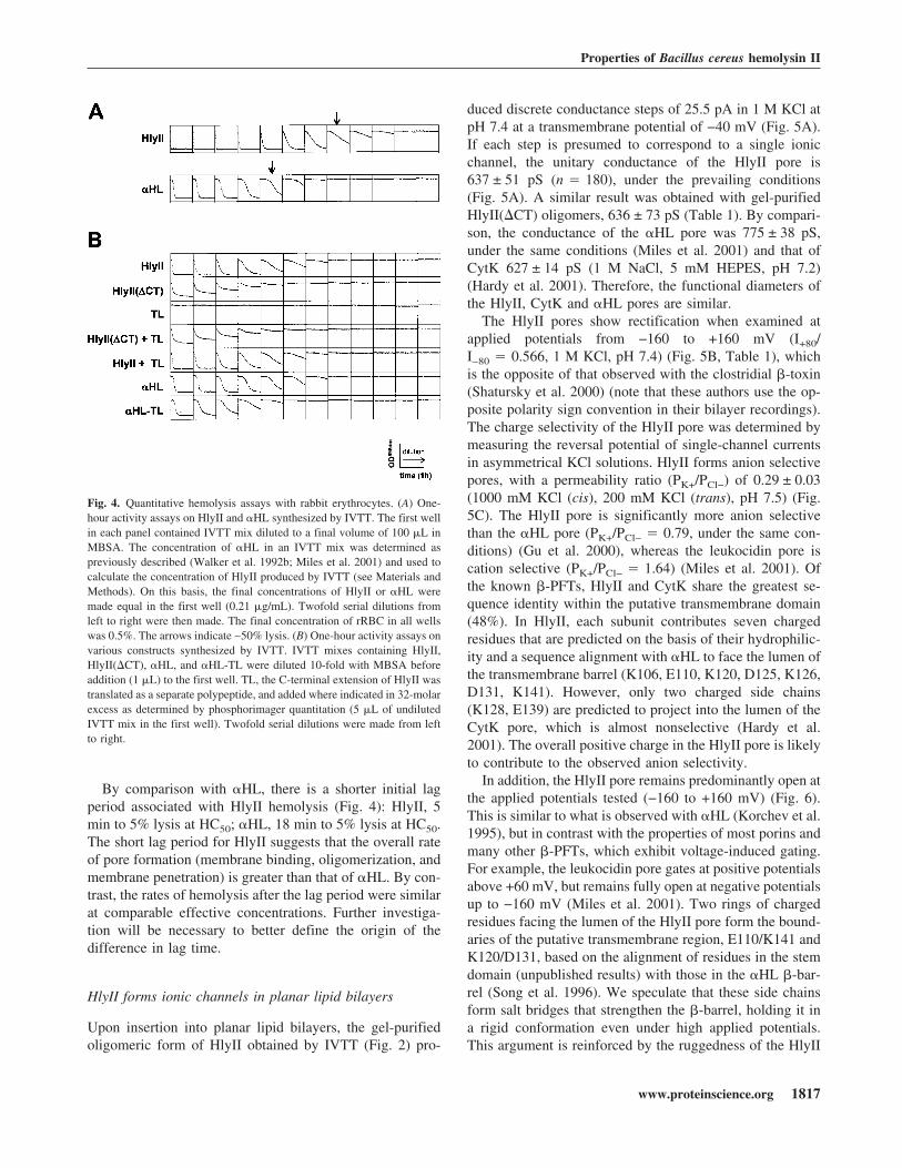
By comparison with �HL, there is a shorter initial lagperiod associated with HlyII hemolysis (Fig. 4): HlyII, 5min to 5% lysis at HC50; �HL, 18 min to 5% lysis at HC50.The short lag period for HlyII suggests that the overall rateof pore formation (membrane binding, oligomerization, andmembrane penetration) is greater than that of �HL. By con-trast, the rates of hemolysis after the lag period were similarat comparable effective concentrations. Further investiga-tion will be necessary to better define the origin of thedifference in lag time.
HlyII forms ionic channels in planar lipid bilayers
Upon insertion into planar lipid bilayers, the gel-purifiedoligomeric form of HlyII obtained by IVTT (Fig. 2) pro-
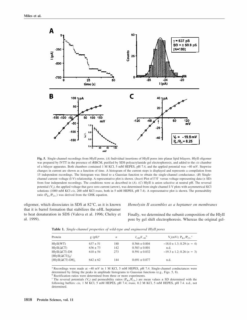
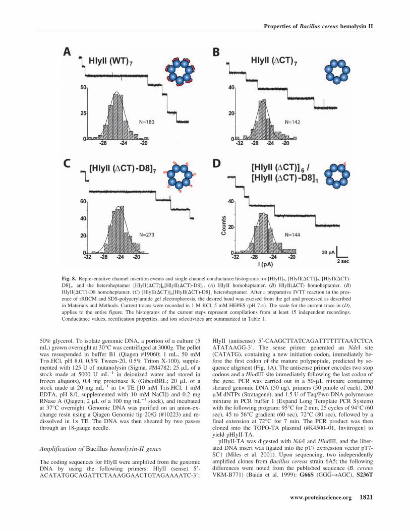
duced discrete conductance steps of 25.5 pA in 1 M KCl atpH 7.4 at a transmembrane potential of −40 mV (Fig. 5A).If each step is presumed to correspond to a single ionicchannel, the unitary conductance of the HlyII pore is637 ± 51 pS (n � 180), under the prevailing conditions(Fig. 5A). A similar result was obtained with gel-purifiedHlyII(�CT) oligomers, 636 ± 73 pS (Table 1). By compari-son, the conductance of the �HL pore was 775 ± 38 pS,under the same conditions (Miles et al. 2001) and that ofCytK 627 ± 14 pS (1 M NaCl, 5 mM HEPES, pH 7.2)(Hardy et al. 2001). Therefore, the functional diameters ofthe HlyII, CytK and �HL pores are similar.
The HlyII pores show rectification when examined atapplied potentials from −160 to +160 mV (I+80/I−80 � 0.566, 1 M KCl, pH 7.4) (Fig. 5B, Table 1), whichis the opposite of that observed with the clostridial �-toxin(Shatursky et al. 2000) (note that these authors use the op-posite polarity sign convention in their bilayer recordings).The charge selectivity of the HlyII pore was determined bymeasuring the reversal potential of single-channel currentsin asymmetrical KCl solutions. HlyII forms anion selectivepores, with a permeability ratio (PK+/PCl−) of 0.29 ± 0.03(1000 mM KCl (cis), 200 mM KCl (trans), pH 7.5) (Fig.5C). The HlyII pore is significantly more anion selectivethan the �HL pore (PK+/PCl− � 0.79, under the same con-ditions) (Gu et al. 2000), whereas the leukocidin pore iscation selective (PK+/PCl− � 1.64) (Miles et al. 2001). Ofthe known �-PFTs, HlyII and CytK share the greatest se-quence identity within the putative transmembrane domain(48%). In HlyII, each subunit contributes seven chargedresidues that are predicted on the basis of their hydrophilic-ity and a sequence alignment with �HL to face the lumen ofthe transmembrane barrel (K106, E110, K120, D125, K126,D131, K141). However, only two charged side chains(K128, E139) are predicted to project into the lumen of theCytK pore, which is almost nonselective (Hardy et al.2001). The overall positive charge in the HlyII pore is likelyto contribute to the observed anion selectivity.
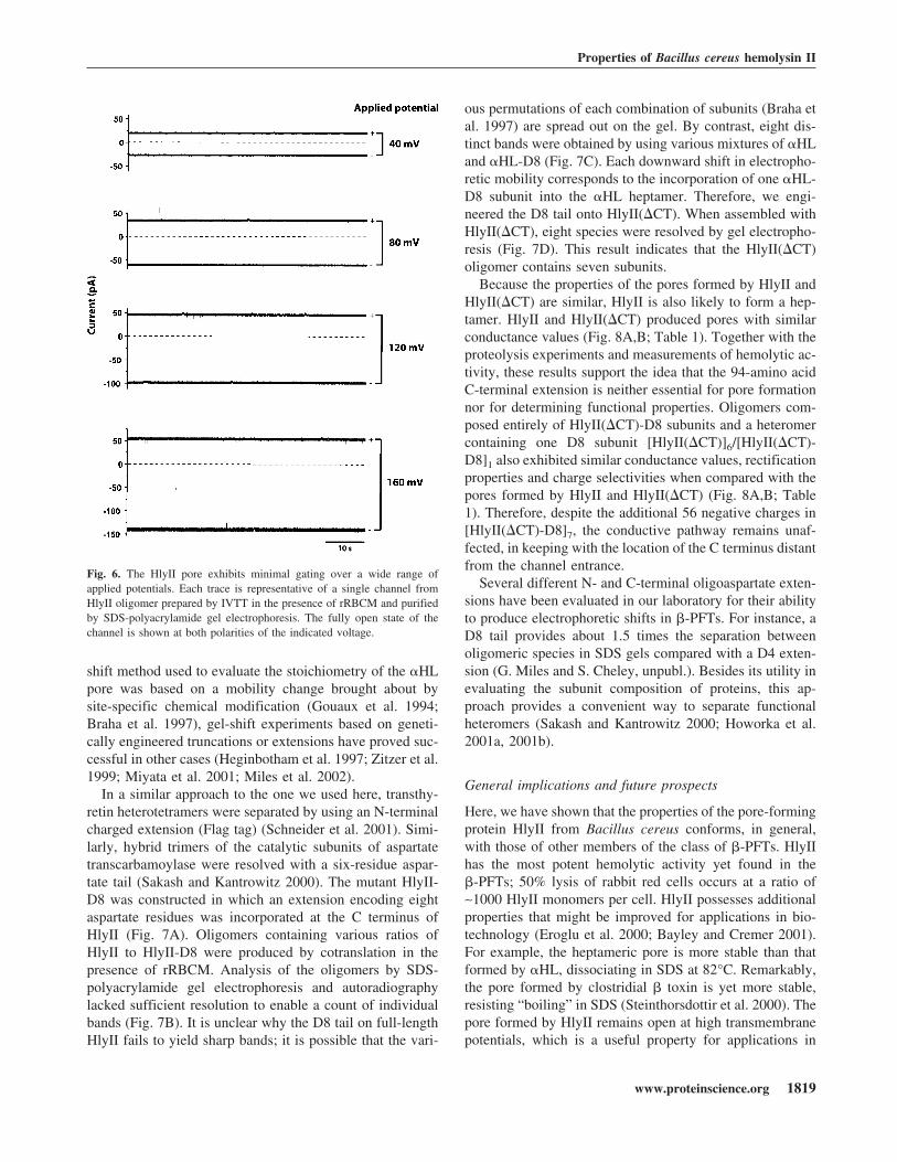
In addition, the HlyII pore remains predominantly open atthe applied potentials tested (−160 to +160 mV) (Fig. 6).This is similar to what is observed with �HL (Korchev et al.1995), but in contrast with the properties of most porins andmany other �-PFTs, which exhibit voltage-induced gating.For example, the leukocidin pore gates at positive potentialsabove +60 mV, but remains fully open at negative potentialsup to −160 mV (Miles et al. 2001). Two rings of chargedresidues facing the lumen of the HlyII pore form the bound-aries of the putative transmembrane region, E110/K141 andK120/D131, based on the alignment of residues in the stemdomain (unpublished results) with those in the �HL �-bar-rel (Song et al. 1996). We speculate that these side chainsform salt bridges that strengthen the �-barrel, holding it ina rigid conformation even under high applied potentials.This argument is reinforced by the ruggedness of the HlyII
Fig. 4. Quantitative hemolysis assays with rabbit erythrocytes. (A) One-hour activity assays on HlyII and �HL synthesized by IVTT. The first wellin each panel contained IVTT mix diluted to a final volume of 100 �L inMBSA. The concentration of �HL in an IVTT mix was determined aspreviously described (Walker et al. 1992b; Miles et al. 2001) and used tocalculate the concentration of HlyII produced by IVTT (see Materials andMethods). On this basis, the final concentrations of HlyII or �HL weremade equal in the first well (0.21 �g/mL). Twofold serial dilutions fromleft to right were then made. The final concentration of rRBC in all wellswas 0.5%. The arrows indicate ∼50% lysis. (B) One-hour activity assays onvarious constructs synthesized by IVTT. IVTT mixes containing HlyII,HlyII(�CT), �HL, and �HL-TL were diluted 10-fold with MBSA beforeaddition (1 �L) to the first well. TL, the C-terminal extension of HlyII wastranslated as a separate polypeptide, and added where indicated in 32-molarexcess as determined by phosphorimager quantitation (5 �L of undilutedIVTT mix in the first well). Twofold serial dilutions were made from leftto right.
Properties of Bacillus cereus hemolysin II
www.proteinscience.org 1817
oligomer, which dissociates in SDS at 82°C, as it is knownthat it is barrel formation that stabilizes the �HL heptamerto heat denaturation in SDS (Valeva et al. 1996; Cheley etal. 1999).
Hemolysin II assembles as a heptamer on membranes
Finally, we determined the subunit composition of the HlyIIpore by gel shift electrophoresis. Whereas the original gel-
Table 1. Single-channel properties of wild-type and engineered HlyII pores
Protein g (pS)a n I+80/I−80b Vr(mV); PK+/PCl−
c
HlyII(WT) 637 ± 51 180 0.566 ± 0.004 −18.0 ± 1.3; 0.29 (n � 4)HlyII(�CT) 636 ± 73 142 0.583 ± 0.001 n.d.HlyII(�CT)-D8 610 ± 54 273 0.591 ± 0.032 −19.3 ± 1.2; 0.26 (n � 3)[HlyII(�CT)]6/[HlyII(�CT)-D8]1 642 ± 62 144 0.691 ± 0.077 n.d.
a Recordings were made at −40 mV in 1 M KCl, 5 mM HEPES, pH 7.4. Single-channel conductances weredetermined by fitting the peaks in amplitude histograms to Gaussian functions (e.g., Figs. 5, 8).b Rectification ratios were determined from three or more experiments.c The reversal potentials (Vr) and permeability ratios (PK+/PCl−) are mean values ± SD determined with thefollowing buffers: cis, 1 M KCl, 5 mM HEPES, pH 7.4; trans, 0.2 M KCl, 5 mM HEPES, pH 7.4. n.d., notdetermined.
Fig. 5. Single-channel recordings from HlyII pores. (A) Individual insertions of HlyII pores into planar lipid bilayers. HlyII oligomerwas prepared by IVTT in the presence of rRBCM, purified by SDS-polyacrylamide gel electrophoresis, and added to the cis chamberof a bilayer apparatus. Both chambers contained 1 M KCl, 5 mM HEPES, pH 7.4, and the applied potential was −40 mV. Stepwisechanges in current are shown as a function of time. A histogram of the current steps is displayed and represents a compilation from15 independent recordings. The histogram was fitted to a Gaussian function to obtain the single-channel conductance. (B) Single-channel current–voltage (I-V) relationship. A representative plot is shown. (Inset) Plot of I+/I− versus voltage representing data (± SD)from four independent recordings. The conditions were as described in (A). (C) HlyII is anion selective at neutral pH. The reversalpotential (Vr), the applied voltage that gave zero current (arrow), was determined from single channel I-V plots with asymmetrical KClsolutions (1000 mM KCl cis, 200 mM KCl trans, both in 5 mM HEPES, pH 7.4). A representative plot is shown. The permeabilityratio (PK+/PCl−) was derived from the GHK equation.
Miles et al.
1818 Protein Science, vol. 11
shift method used to evaluate the stoichiometry of the �HLpore was based on a mobility change brought about bysite-specific chemical modification (Gouaux et al. 1994;Braha et al. 1997), gel-shift experiments based on geneti-cally engineered truncations or extensions have proved suc-cessful in other cases (Heginbotham et al. 1997; Zitzer et al.1999; Miyata et al. 2001; Miles et al. 2002).
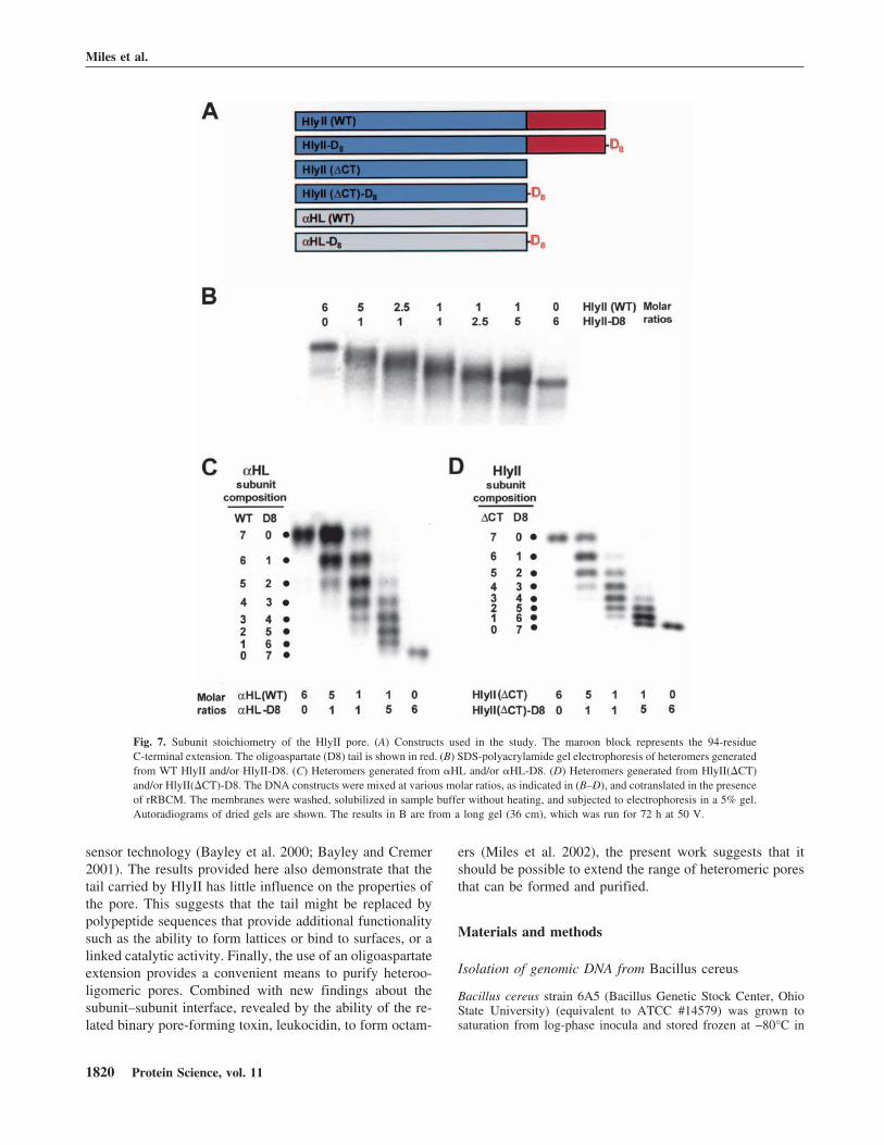
In a similar approach to the one we used here, transthy-retin heterotetramers were separated by using an N-terminalcharged extension (Flag tag) (Schneider et al. 2001). Simi-larly, hybrid trimers of the catalytic subunits of aspartatetranscarbamoylase were resolved with a six-residue aspar-tate tail (Sakash and Kantrowitz 2000). The mutant HlyII-D8 was constructed in which an extension encoding eightaspartate residues was incorporated at the C terminus ofHlyII (Fig. 7A). Oligomers containing various ratios ofHlyII to HlyII-D8 were produced by cotranslation in thepresence of rRBCM. Analysis of the oligomers by SDS-polyacrylamide gel electrophoresis and autoradiographylacked sufficient resolution to enable a count of individualbands (Fig. 7B). It is unclear why the D8 tail on full-lengthHlyII fails to yield sharp bands; it is possible that the vari-
ous permutations of each combination of subunits (Braha etal. 1997) are spread out on the gel. By contrast, eight dis-tinct bands were obtained by using various mixtures of �HLand �HL-D8 (Fig. 7C). Each downward shift in electropho-retic mobility corresponds to the incorporation of one �HL-D8 subunit into the �HL heptamer. Therefore, we engi-neered the D8 tail onto HlyII(�CT). When assembled withHlyII(�CT), eight species were resolved by gel electropho-resis (Fig. 7D). This result indicates that the HlyII(�CT)oligomer contains seven subunits.
Because the properties of the pores formed by HlyII andHlyII(�CT) are similar, HlyII is also likely to form a hep-tamer. HlyII and HlyII(�CT) produced pores with similarconductance values (Fig. 8A,B; Table 1). Together with theproteolysis experiments and measurements of hemolytic ac-tivity, these results support the idea that the 94-amino acidC-terminal extension is neither essential for pore formationnor for determining functional properties. Oligomers com-posed entirely of HlyII(�CT)-D8 subunits and a heteromercontaining one D8 subunit [HlyII(�CT)]6/[HlyII(�CT)-D8]1 also exhibited similar conductance values, rectificationproperties and charge selectivities when compared with thepores formed by HlyII and HlyII(�CT) (Fig. 8A,B; Table1). Therefore, despite the additional 56 negative charges in[HlyII(�CT)-D8]7, the conductive pathway remains unaf-fected, in keeping with the location of the C terminus distantfrom the channel entrance.
Several different N- and C-terminal oligoaspartate exten-sions have been evaluated in our laboratory for their abilityto produce electrophoretic shifts in �-PFTs. For instance, aD8 tail provides about 1.5 times the separation betweenoligomeric species in SDS gels compared with a D4 exten-sion (G. Miles and S. Cheley, unpubl.). Besides its utility inevaluating the subunit composition of proteins, this ap-proach provides a convenient way to separate functionalheteromers (Sakash and Kantrowitz 2000; Howorka et al.2001a, 2001b).
General implications and future prospects
Here, we have shown that the properties of the pore-formingprotein HlyII from Bacillus cereus conforms, in general,with those of other members of the class of �-PFTs. HlyIIhas the most potent hemolytic activity yet found in the�-PFTs; 50% lysis of rabbit red cells occurs at a ratio of∼1000 HlyII monomers per cell. HlyII possesses additionalproperties that might be improved for applications in bio-technology (Eroglu et al. 2000; Bayley and Cremer 2001).For example, the heptameric pore is more stable than thatformed by �HL, dissociating in SDS at 82°C. Remarkably,the pore formed by clostridial � toxin is yet more stable,resisting “boiling” in SDS (Steinthorsdottir et al. 2000). Thepore formed by HlyII remains open at high transmembranepotentials, which is a useful property for applications in
Fig. 6. The HlyII pore exhibits minimal gating over a wide range ofapplied potentials. Each trace is representative of a single channel fromHlyII oligomer prepared by IVTT in the presence of rRBCM and purifiedby SDS-polyacrylamide gel electrophoresis. The fully open state of thechannel is shown at both polarities of the indicated voltage.
Properties of Bacillus cereus hemolysin II
www.proteinscience.org 1819
sensor technology (Bayley et al. 2000; Bayley and Cremer2001). The results provided here also demonstrate that thetail carried by HlyII has little influence on the properties ofthe pore. This suggests that the tail might be replaced bypolypeptide sequences that provide additional functionalitysuch as the ability to form lattices or bind to surfaces, or alinked catalytic activity. Finally, the use of an oligoaspartateextension provides a convenient means to purify heteroo-ligomeric pores. Combined with new findings about thesubunit–subunit interface, revealed by the ability of the re-lated binary pore-forming toxin, leukocidin, to form octam-
ers (Miles et al. 2002), the present work suggests that itshould be possible to extend the range of heteromeric poresthat can be formed and purified.
Materials and methods
Isolation of genomic DNA from Bacillus cereus
Bacillus cereus strain 6A5 (Bacillus Genetic Stock Center, OhioState University) (equivalent to ATCC #14579) was grown tosaturation from log-phase inocula and stored frozen at −80°C in
Fig. 7. Subunit stoichiometry of the HlyII pore. (A) Constructs used in the study. The maroon block represents the 94-residueC-terminal extension. The oligoaspartate (D8) tail is shown in red. (B) SDS-polyacrylamide gel electrophoresis of heteromers generatedfrom WT HlyII and/or HlyII-D8. (C) Heteromers generated from �HL and/or �HL-D8. (D) Heteromers generated from HlyII(�CT)and/or HlyII(�CT)-D8. The DNA constructs were mixed at various molar ratios, as indicated in (B–D), and cotranslated in the presenceof rRBCM. The membranes were washed, solubilized in sample buffer without heating, and subjected to electrophoresis in a 5% gel.Autoradiograms of dried gels are shown. The results in B are from a long gel (36 cm), which was run for 72 h at 50 V.
Miles et al.
1820 Protein Science, vol. 11
50% glycerol. To isolate genomic DNA, a portion of a culture (5mL) grown overnight at 30°C was centrifuged at 3000g. The pelletwas resuspended in buffer B1 (Qiagen #19060; 1 mL, 50 mMTris.HCl, pH 8.0, 0.5% Tween-20, 0.5% Triton X-100), supple-mented with 125 U of mutanolysin (Sigma. #M4782; 25 �L of astock made at 5000 U mL−1 in deionized water and stored infrozen aliquots), 0.4 mg proteinase K (GibcoBRL; 20 �L of astock made at 20 mg mL−1 in 1× TE [10 mM Tris.HCl, 1 mMEDTA, pH 8.0, supplemented with 10 mM NaCl]) and 0.2 mgRNase A (Qiagen; 2 �L of a 100 mg mL−1 stock), and incubatedat 37°C overnight. Genomic DNA was purified on an anion-ex-change resin using a Qiagen Genomic tip 20/G (#10223) and re-dissolved in 1× TE. The DNA was then sheared by two passesthrough an 18-gauge needle.
Amplification of Bacillus hemolysin-II genes
The coding sequences for HlyII were amplified from the genomicDNA by using the following primers: HlyII (sense) 5�-ACATATGGCAGATTCTAAAGGAACTGTAGAAAATC-3�;
HlyII (antisense) 5�-CAAGCTTATCAGATTTTTTTAATCTCAATATAAGG-3�. The sense primer generated an NdeI site(CATATG), containing a new initiation codon, immediately be-fore the first codon of the mature polypeptide, predicted by se-quence aligment (Fig. 1A). The antisense primer encodes two stopcodons and a HindIII site immediately following the last codon ofthe gene. PCR was carried out in a 50-�L mixture containingsheared genomic DNA (50 ng), primers (50 pmole of each), 200�M dNTPs (Stratagene), and 1.5 U of Taq/Pwo DNA polymerasemixture in PCR buffer 1 (Expand Long Template PCR System)with the following program: 95°C for 2 min, 25 cycles of 94°C (60sec), 45 to 56°C gradient (60 sec), 72°C (80 sec), followed by afinal extension at 72°C for 7 min. The PCR product was thencloned into the TOPO-TA plasmid (#K4500–01, Invitrogen) toyield pHlyII-TA.
pHlyII-TA was digested with NdeI and HindIII, and the liber-ated DNA insert was ligated into the pT7 expression vector pT7-SC1 (Miles et al. 2001). Upon sequencing, two independentlyamplified clones from Bacillus cereus strain 6A5; the followingdifferences were noted from the published sequence (B. cereusVKM-B771) (Baida et al. 1999): G66S (GGG→AGC), S236T
Fig. 8. Representative channel insertion events and single channel conductance histograms for [HlyII]7, [HlyII(�CT)]7, [HlyII(�CT)-D8]7, and the heteroheptamer [HlyII(�CT)]6[HlyII(�CT)-D8]1. (A) HlyII homoheptamer. (B) HlyII(�CT) homoheptamer. (B)HlyII(�CT)-D8 homoheptamer. (C) [HlyII(�CT)]6[HlyII(�CT)-D8]1 heteroheptamer. After a preparative IVTT reaction in the pres-ence of rRBCM and SDS-polyacrylamide gel electrophoresis, the desired band was excised from the gel and processed as describedin Materials and Methods. Current traces were recorded in 1 M KCl, 5 mM HEPES (pH 7.4). The scale for the current trace in (D),applies to the entire figure. The histograms of the current steps represent compilations from at least 15 independent recordings.Conductance values, rectification properties, and ion selectivities are summarized in Table 1.
Properties of Bacillus cereus hemolysin II
www.proteinscience.org 1821
(TCT→ACT), N276H (AAC→CAC), P294L (CCT→CTT), I299N(ATT→AAT), G300S (GGT→AGT), N303S (AAC→AGC),N306D (AAC→GAT), Q307K (CAG→AAA), F317L (TTT→CTT),T358S (ACA→TCA). With the exception of three residues (inbold), the variations noted occurred in the C-terminal extension. Inaddition, there were 35 silent changes throughout the sequence.The DNA sequence from strain 6A5 has been deposited in Gen-Bank with the accession number AF448485.
Hemolysin-II C-terminal truncation
Unless otherwise noted, the constructs used in this study werecreated by PCR and cloned into the TOPO-TA plasmid prior tobeing subcloned into pT7-SC1 by ligation. Each construct wasverified by DNA sequencing.
HlyII(�CT), a mutant of HlyII truncated at the C terminus, wasconstructed by removing the sequence encoding the last 94 aminoacids of HlyII (residues 289–382). PCR was carried out on linear-ized pT7-HlyII(6A5) using the forward primer SC001 5�-CACTATAGGGAGACCACAACGG-3� and the reverse primer BAC6A5TRN1 5�-TAAGCTTCATTAAAGAGTAACTTGATG-3�. Thelatter encodes two stop codons and a HindIII site immediately afterthe Leu-288 codon in the HlyII gene.
The C-terminal extension (TL) was cloned separately into thepT7 vector by using PCR. The sense primer BAC6A5TAILBEGIN5�-ACATATGGATAACCAAAAAGCCCTT-3� generated an NdeIsite (CATATG), containing a new initiation codon, immediatelybefore the first codon of the C-terminal extension (Asp–289). Theantisense primer was SC011 5�-CCCCTCAAGACCCGTTTAGAGGC-3�, which hybridized at a site in the vector downstream ofthe stop codons.
Construction of mutants with C-terminal(oligo)–aspartic acid extensions
We sought to develop a genetic alternative to the original gel shiftmethod for counting subunits, which was based on a mobilitychange brought about by site-specific chemical modification(Gouaux et al. 1994). The mutants HlyII-D8, HlyII(�CT)-D8, and�HL-D8 were constructed with an extension encoding eight C-terminal aspartate residues (Fig. 7A). The “D8 tail” was expectedto change the electrophoretic mobility of the assembled pore basedon charge. PCR was carried out with SC001 as the sense primer,using each of the following reverse primers: HlyII-D8, 5�-AAGCTTATCAATCGTCATCGTCATCGTCATCGTCGATTTTTTTAATCTCAA-3�; HlyII(�CT)-D8, 5�-AAGCTTATCAATCGTCATCGTCATCGTCGTCGTCAAGAGT AACAAGATGGTT-3�; �HLD8,5�-AAGCTTATCAATCATCGTCGTCATCATCGTCATCATTTGTCATTTCTTCTTTTTCCC-3�. These electrophoreticallypurified antisense oligonucleotides incorporated two stop codons(bold) after the D8 tail codons (underscored) followed by a HindIIIsite (italics).
Construction of the fusion protein, �HL-TL
In vivo recombination (Howorka and Bayley 1998) was used tofuse a 3� extension directly to the last codon of the �HL gene(Jones 1995). The extension encoded the 94 amino acids of theBacillus hemolysin II C-terminal tail (residues 289–382). Thefused gene (�HL-TL) was generated in pT7-SC1 by cotransform-ing Escherichia coli XL-10 Gold cells with two PCR productsencompassing the Bacillus tail and the �HL gene. The primer sets
for the two amplification reactions were: (1) nonmutagenic primer(F-NM) (sense), 5�-GTATTCAACATTTCCGTGTCGCCCTTATTC-3�; �HL-TAIL-�HL, (antisense), 5�-AAGGGCTTTAAGGTTATCATTTGTCATTTCTTCTTT-3�; and (2) �HL-TAIL-TL (sense),5�-AAAGAAGAAATGACAAATGATAACCAAAAAGCCCTT-3�; nonmutagenic primer (R-NM) (antisense) 5�-GAATAAGGGCGACACGGAAATGTTGAATAC-3�. The underlined 18-nt se-quences form the overlap for recombination between the two PCRproducts.
In vitro transcription and translation (IVTT)
Polypeptides were synthesized in a cell-free in vitro transcriptionand translation (IVTT) system by using an S30 extract from E. coli(T7 S30 No. L114A, Promega) supplemented with rifampicin (20�g mL−1) (Walker et al. 1992b). Radiolabeled polypeptides weresynthesized by IVTT with the complete amino acid mix supple-mented with [35S]methionine (ICN Biomedicals, Inc., 1175 Cimmole−1, 10 �Ci per 25 �L translation). The concentrations of thetranslated polypeptides were determined by phosphorimageranalysis, by comparison with �HL standards radiolabeled in par-allel IVTT reactions (Miles et al. 2001). The specific radioactivityof the �HL, in phosphorimager units, was determined after deduc-ing the concentration of the protein from its hemolytic activity,assuming a specific hemolytic activity of 25 ng mL−1 (Walker etal. 1992b). The specific radioactivity was normalized to the num-ber of Met residues in the polypeptide chain (assuming that theN-terminal Met is intact). The concentration of HlyII, and its vari-ants, in a translation mix could then be determined from thestrengths of the phosphorimager signals and the number of Metresidues in the polypeptide chains.
Quantitative hemolysis assay
HlyII proteins, synthesized by IVTT with the complete amino acidmix, were diluted into MBSA (10 mM 3-[N-morpholino]propanesulfonic acid; MOPS, Cat. No. AB1270, American Bioanalytical,150 mM NaCl, pH 7.4, containing 1 mg mL−1 bovine serum al-bumin, Cat. No. 4503, Sigma) and subjected to 12 twofold serialdilutions in the same buffer in microtiter wells (final volume 50�L). An equal volume of 1% washed rabbit erythrocytes (rRBC)in MBSA was quickly added to each well, beginning with the mostdiluted sample. Hemolysis was recorded for 1 h at 20°C by moni-toring the decrease in light scattering at 595 nm with a Bio-Radmicroplate reader (Model 3550-UV) and using the MicroplateManager 4.0 software.
HlyII oligomer formationon rabbit erythrocyte membranes
Radiolabeled HlyII oligomers were prepared by IVTT in the pres-ence of [35S]methionine and rRBCM (5 �L, 3.0 mg protein mL−1)(Cheley et al. 1999) in a total reaction volume of 25 �L. After 1h at 30°C, the mixture was centrifuged and the supernatant dis-carded. The membrane pellet was washed and resuspended inMBSA (80 �L) prior to solubilization by the addition of 5×Laemmli sample buffer (20 �L; Laemmli 1970). A portion (20�L) was subjected to electrophoresis in a 10% SDS-polyacryl-amide gel. The gel was fixed for 1 h prior to drying and autora-diography.
Miles et al.
1822 Protein Science, vol. 11
Oligomerization of HlyII on liposomes
Egg yolk phosphatidylcholine, cholesterol, and phosphatidic acidin chloroform were mixed in the desired molar ratio of 55:25:20.After drying under vacuum, the lipid film was resuspended inbuffer (10 mM Tris, pH 8.0, 150 mM NaCl, 1 mM EDTA) to atotal lipid concentration of 5 mg mL−1. Liposomes were preparedby ultrasonication for 30 min on ice using a probe sonicator (Dy-natech Sonic Dismembrator Model 150) (relative output set to50% power), followed by a brief centrifugation (30 sec, 16,000g)to remove titanium particles. [35S]Methionine-labeled HlyII orHlyII(�CT) (2 �L of an IVTT reaction mix) was incubated withfreshly prepared liposomes (8 �L) for 1 h at room temperature.After solubilization in Laemmli sample buffer, the mixture wassubjected to SDS polyacrylamide gel electrophoresis.
Proteinase K treatment of HlyII, HlyII(�CT) and �HLpolypeptides on membranes
Proteinase K (Sigma, #P-0390) solutions (5.0, 0.5, and 0.05 mgmL−1 in water) were prepared by dilution of an enzyme stock (10mg mL−1 in water) and used immediately. Limited proteolysis wasperformed on HlyII, HlyII(�CT) and �HL bound to rRBCM. Themembranes were resuspended in MBSA at 0.19 mg membraneprotein mL−1 and divided into four tubes (18 �L in each). Pro-teinase K or water (2 �L) was added to each tube. After 5 min atroom temperature, the reactions were stopped by treatment withPMSF (9 mM final, added in 2 �L of isopropanol, 5 min, roomtemperature), followed by the addition of 2× Laemmli loadingbuffer. The samples were subjected to electrophoresis in 10%SDS-polyacrylamide gels (unheated samples) or 12% gels (heatedsamples: 95°C, 5 min).
Gel purification of HlyII oligomers
HlyII, HlyII(�CT) and HlyII(�CT)-D8, and were prepared bytranslation in the presence of rRBCM as described above, but inpreparative amounts (75 �L IVTT reaction). To obtain,HlyII(�CT)6(�CT)-D81, HlyII(�CT) and HlyII(�CT)-D8 weretranslated using the corresponding plasmids at a ratio of 5:1. Theoligomers were purified by SDS-polyacrylamide gel electrophore-sis in an 8% gel in the presence of 0.1 mM sodium thioglycolate(Movileanu et al. 2001; Miles et al. 2001), stored at −80°C in 10mM Tris.HCl, pH 7.5, and used for bilayer recordings withoutfurther treatment.
Hetero-oligomer formation for the determinationof stoichiometry
Hetero-oligomers of hemolysin II subunits containing HlyII and/orHlyII-D8, and HlyII(�CT) and/or HlyII(�CT)-D8 were preparedby mixing the corresponding plasmids in the desired molar ratios(see Fig. 7 legend) prior to IVTT in the presence of rRBCM. Toobtain �HL heteromers, �HL and/or �HL-D8 were used. Thewashed membrane pellets were solubilized with Laemmli samplebuffer and subjected, without heating, to electrophoresis in 5%SDS-polyacrylamide gels. Autoradiographs were made of thedried gels.
Planar lipid bilayer recordings
All measurements were performed at 25°C. Numerical values aregiven as the mean ± SD (�n−1). Planar lipid bilayer membranes
were formed with 1,2-diphytanoyl-sn-glycero-3-phosphocholine(Avanti Polar Lipids) on a 150–160 �m- diameter aperture in a 25�m-thick Teflon film (Goodfellow Corporation) separating the cisand trans compartments (2 mL each) of a bilayer apparatus (Mon-tal and Mueller 1972). Prior to forming the lipid bilayer, the orificewas pretreated with 10% (v/v) hexadecane (#29,631-7; Aldrich) inn-pentane (Burdick & Jackson) and allowed to dry thoroughly. Formost measurements, the cis and trans chambers contained 1 MKCl, 5 mM HEPES, pH 7.4. Various concentrations of KCl in 5mM HEPES, pH 7.4 were used for ion selectivity measurements.Protein samples were added to the cis chamber, which was atground.
Currents were recorded by using a Dagan 3900A patch clampamplifier (Dagan Corporation) with a 3910 expander and a built-inlow-pass four-pole Bessel filter set at 5 kHz. Data were stored ondigital audio tape with a DAS-75 data recorder (Dagan Corpora-tion). Prior to analysis, the signal was low-pass filtered at 1 kHzwith an eight-pole Bessel filter (Model 902, Frequency Devices)and acquired with a Digidata 1200A A/D board with a samplingtime interval of 200 �sec. Data were acquired and analyzed withpClamp 8.0 software (Axon Instruments). Single-channel conduc-tance values were determined by fitting the peaks in amplitudehistograms to Gaussian functions. Current–voltage (I-V) relation-ships for single channels were determined by recording the cur-rents obtained after stepwise changes in applied potential. Thepermeability ratios (PK+/PCl−) were calculated from experimen-tally determined reversal potentials (Vr) by using the Goldman-Hodgkin-Katz (GHK) equation (Hille 2001) and the appropriateactivity coefficients for KCl solutions (Zemaitis et al. 1986). Inthese measurements, the cis compartment contained 1000 mMKCl, while the other chamber contained 200 mM KCl. Any elec-trode DC offset was balanced prior to the addition of protein to thecis chamber. The applied voltage that gave zero current was noted.In addition, the reversal potential was more accurately determinedby polynomial fits to current–voltage (I-V) data. Symmetrical so-lutions were then reestablished to evaluate whether or not any DCoffset had built up during the course of the experiment. It all cases,the offset was less than 1 mV.
Acknowledgments
This work was supported by grants from the DOE and NIH. G.M.holds an MD-PhD training fellowship at The Texas A&M Uni-versity System Health Science Center, College of Medicine, andwas the recipient of an ASSERT (ARO) award. The authors thankDaniel Zeigler, BGSC Director, Ohio State University, for gra-ciously supplying Bacillus strains; Orit Braha and Li-Qun Gu fortheir advice on ion selectivity; Brian Lauman for technical help;Michael Palmer for advice on liposomes; and Sean Conlan forguidance on using Spock.
The publication costs of this article were defrayed in part bypayment of page charges. This article must therefore be herebymarked “advertisement” in accordance with 18 USC section 1734solely to indicate this fact.
References
Alouf, J.E. and Freer, J.H. 1999. The comprehensive sourcebook of bacterialprotein toxins. Academic Press, New York.
Altshul, S.F., Madden, T.L., Schaffer, A.A., Zhang, J., Zhang, Z., Miller, W.,and Lipman, D.J. 1997. Gapped BLAST and PSI-BLAST; A new genera-tion of protein database search programs. Nucleic Acids Res. 25: 3389–3402.
Baida, G., Budarina, Z.I., Kuzmin, N.P., and Solonin, A.S. 1999. Complete
Properties of Bacillus cereus hemolysin II
www.proteinscience.org 1823
nucleotide sequence and molecular characterization of hemolysin II genefrom Bacillus cereus. FEMS Microbiol. Lett. 180: 7–14.
Bayley, H. 1999. Designed membrane channels and pores. Curr. Opin. Biotech-nol. 10: 94–103.
Bayley, H. and Cremer, P.S. 2001. Stochastic sensors inspired by biology.Nature 413: 226–230.
Bayley, H., Braha, O., and Gu, L.-Q. 2000. Stochastic sensing with proteinpores. Adv. Mater. 12: 139–142.
Bhakdi, S., Walev, I., Palmer, M., and Valeva, A. 2000. Staphylococcal � toxin.In Bacterial protein toxins (eds. K. Aktories and I. Just), pp. 509–527.Springer, Berlin.
Braha, O., Walker, B., Cheley, S., Kasianowicz, J.J., Song, L., Gouaux, J.E., andBayley, H. 1997. Designed protein pores as components for biosensors.Chem. Biol. 4: 497–505.
Cheley, S., Braha, O., Lu, X., Conlan, S., and Bayley, H. 1999. A functionalprotein pore with a “retro” transmembrane domain. Protein Sci. 8: 1257–1267.
Cheley, S., Malghani, M.S., Song, L., Hobaugh, M., Gouaux, J.E., Yang, J., andBayley, H. 1997. Spontaneous oligomerization of a staphylococcal �-he-molysin conformationally constrained by removal of residues that form thetransmembrane � barrel. Protein Eng. 10: 1433–1443.
Christopher, J.A. 1998. SPOCK: The structural properties observation and cal-culation kit (program manual). Center for Macromolecular Design, TexasA&M University, College Station, TX.
Drobeniwski, F.A. 1993. Bacillus cereus and related species. Clin. Microbiol.Rev. 6: 324–338.
Eroglu, A., Russo, M.J., Bieganski, R., Fowler, A., Cheley, S., Bayley, H., andToner, M. 2000. Intracellular trehalose improves the survival of cryopre-served mammalian cells. Nat Biotechnol 18: 163–167.
Fang, Y., Cheley, S., Bayley, H., and Yang, J. 1997. The heptameric prepore ofa staphylococcal �-hemolysin mutant in lipid bilayers imaged by atomicforce microscopy. Biochemistry 36: 9518–9522.
Gouaux, J.E., Braha, O., Hobaugh, M.R., Song, L., Cheley, S., Shustak, C., andBayley, H. 1994. Subunit stoichiometry of staphylococcal �-hemolysin incrystals and on membranes: A heptameric transmembrane pore. Proc. Natl.Acad. Sci. 91: 12828–12831.
Gouaux, E., Hobaugh, M., and Song, L. 1997. �-Hemolysin, �-hemolysin andleukocidin from Staphylococcus aureus: Distant in sequence but similar instructure. Protein Sci. 6: 2631–2635.
Gouet, P., Courcelle, E., Stuart, D.I., and Metoz, F. 1999. ESPript: Analysis ofmultiple sequence alignments in PostScript. Bioinformatics 15: 305–308.
Gu, L.-Q., Dalla Serra, M., Vincent, J.B., Vigh, G., Cheley, S., Braha, O., andBayley, H. 2000. Reversal of charge selectivity in transmembrane proteinpores by using non-covalent molecular adapters. Proc. Natl. Acad. Sci. 97:3959–3964.
Hardy, S.P., Lund, T., and Granum, P.E. 2001. CytK toxin of Bacillus cereusforms pores in planar lipid bilayers and is cytotoxic to intestinal epithelia.FEMS Microbiol. Lett. 197: 47–51.
Heginbotham, L., Odessey, E., and Miller, C. 1997. Tetrameric structure of aprokaryotic K+ channel. Biochemistry 36: 10335–10342.
Henikoff, S. and Henikoff, J.G. 1992. Amino acid substitution matrices fromprotein blocks. Proc. Natl. Acad. Sci. 89: 10915–10919.
Hille, B. 2001. Ion channels of excitable membranes. Sinauer, Sunderland, MA.Howorka, S. and Bayley, H. 1998. Improved protocol for high-throughput cys-
teine scanning mutagenesis. Biotechniques 25: 766–772.Howorka, S., Cheley, S., and Bayley, H. 2001a. Sequence-specific detection of
individual DNA strands using engineered nanopores. Nat Biotechnol 19:636–639.
Howorka, S., Movileanu, L., Braha, O., and Bayley, H. 2001b. Kinetics ofduplex formation for individual DNA strands within a single proteinnanopore. Proc. Natl. Acad. Sci. 98: 12996–13001.
Jones, D.H. 1995. PCR mutagenesis and recombination in vivo. In PCR primer:A laboratory manual (eds. C.W. Dieffenbach and G.S. Dveksler), pp. 591–601. Cold Spring Harbor Laboratory Press, Cold Spring Harbor, NY.
Korchev, Y.E., Alder, G.M., Bakhramov, A., Bashford, C.L., Joomun, B.S.,Sviderskaya, E.V., Usherwood, P.N.R., and Pasternak, C.A. 1995. Staphy-lococcus aureus alpha-toxin-induced pores: Channel-like behavior in lipidbilayers and patch clamped cells. J. Membr. Biol. 143: 143–151.
Krasilnikov, O.V., Merzlyak, P.G., Yuldasheva, L.N., Rodrigues, C.G., Bhakdi,S., and Valeva, A. 2000. Electrophysiological evidence for heptameric stoi-chiometry of ion channels formed by Staphylococcus aureus alpha-toxin inplanar lipid bilayers. Mol. Microbiol. 37: 1372–1378.
Kraulis, P.J. 1991. MOLSCRIPT: A program to produce both detailed andschematic plots of protein structure. J. Appl. Crystallogr. 24: 946–949.
Laemmli, U.K. 1970. Cleavage of structural proteins during the assembly of thehead of bacteriophage T4. Nature 227: 680–685.
Lund, T., De Buyser, M.L., and Granum, P.E. 2000. A new cytotoxin fromBacillus cereus that may cause necrotic enteritis. Mol. Microbiol. 38: 254–261.
Martín, A.C., López, R., and García, P. 1996. Analysis of the complete nucleo-tide sequence and functional organization of the genome of the Streptococ-cus pneumoniae bacteriophage Cp-1. J. Virol. 70: 3678–3687.
Menestrina, G., Dalla Serra, M., and Prévost, G. 2001. Mode of action of �barrel pore-forming toxins of the staphylococcal �-hemolysin family. Toxi-con 39: 1661–1672.
Miles, G., Cheley, S., Braha, O., and Bayley, H. 2001. The staphylococcalleukocidin bicomponent toxin forms large ionic channels. Biochemistry 40:8514–8522.
Miles, G., Movileanu, L., and Bayley, H. 2002. Subunit composition of abicomponent toxin: Staphylococcal leukocidin forms an octameric trans-membrane pore. Protein Sci. 11: 894–902.
Miyata, S., Matsushita, O., Minami, J., Katayama, S., Shimamoto, S., andOkabe, A. 2001. Cleavage of a C-terminal peptide is essential for heptamer-ization of Clostridium perfringens �-toxin in the synaptosomal membrane.J. Biol. Chem. 276: 13778–13783.
Montal, M. and Mueller, P. 1972. Formation of bimolecular membranes fromlipid monolayers and study of their electrical properties. Proc. Natl. Acad.Sci. 69: 3561–3566.
Movileanu, L., Cheley, S., Howorka, S., Braha, O., and Bayley, H. 2001. Lo-cation of a constriction in the lumen of a transmembrane pore by targetedcovalent attachment of polymer molecules. J. Gen. Physiol. 117: 239–251.
Olson, R., Nariya, H., Yokota, K., Kamio, Y., and Gouaux, E. 1999. Crystalstructure of staphylococcal LukF delineates conformational changes accom-panying formation of a transmembrane channel. Nat. Struct. Biol. 6: 134–140.
Pédelacq, J.-D., Maveyraud, L., Prévost, G., Baba-Moussa, L., González, A.,Courcelle, E., Shepard, W., Monteil, H., Samama, J.-P., and Mourey, L.1999. The structure of Staphylococcus aureus leukocidin component (LukF-PV) reveals the fold of the water-soluble species of a family of transmem-brane pore-forming toxins. Structure 7: 277–288.
Sakash, J.B. and Kantrowitz, E.R. 2000. The contribution of individual inter-chain interactions to the stabilization of the T and R states of Escherichiacoli aspartate transcarbamoylase. J. Biol. Chem. 275: 28701–28707.
Schneider, F., Hammarström, P., and Kelly, J.W. 2001. Transthyretin slowlyexchanges subunits under physiological conditions: A convenient chromato-graphic method to study subunit exchange in oligomeric proteins. ProteinSci. 10: 1606–1613.
Shatursky, O., Bayles, R., Rogers, M., Jost, B.H., Songer, J.G., and Tweten,R.K. 2000. Clostridium perfringens beta-toxin forms potential-dependent,cation-selective channels in lipid bilayers. Infect. Immun. 68: 5546–5551.
Song, L., Hobaugh, M.R., Shustak, C., Cheley, S., Bayley, H., and Gouaux, J.E.1996. Structure of staphylococcal �-hemolysin, a heptameric transmem-brane pore. Science 274: 1859–1865.
Steinthorsdottir, V., Halldorsson, H., and Andresson, O.S. 2000. Clostridiumperfringens beta-toxin forms multimeric transmembrane pores in humanendothelial cells. Microb. Pathog. 28: 45–50.
Thompson, J.D., Higgins, D.G., and Gibson, T.J. 1994. CLUSTAL W: Improv-ing the sensitivity of progressive multiple sequence alignment through se-quence weighting, positions-specific gap penalties and weight matrixchoice. Nucleic Acids Res. 22: 4673–4680.
Valeva, A., Weisser, A., Walker, B., Kehoe, M., Bayley, H., Bhakdi, S., andPalmer, M. 1996. Molecular architecture of a toxin pore: A 15-residuesequence lines the transmembrane channel of staphylococcal alpha-toxin.EMBO J. 15: 1857–1864.
Walker, B.J., Krishnasastry, M., Zorn, L., and Bayley, H. 1992a. Assembly ofthe oligomeric membrane pore formed by staphylococcal �-hemolysin ex-amined by truncation mutagenesis. J. Biol. Chem. 267: 21782–21786.
Walker, B.J., Krishnasastry, M., Zorn, L., Kasianowicz, J.J., and Bayley, H.1992b. Functional expression of the �-hemolysin of Staphylococcus aureusin intact Escherichia coli and in cell lysates. J. Biol. Chem. 267: 10902–10909.
Welkos, S.L. 1991. Plasmid-associated virulence factors of non-toxigenic(pX01–) Bacillus anthracis. Microb. Pathog. 10: 183–198.
Zemaitis, J.F., D.M. Clark, M. Rafal, and N. Scriver. 1986. Handbook of aque-ous electrolyte thermodynamics: Theory and application. American Instituteof Chemical Engineers, New York.
Zitzer, A., Zitzer, O., Bhakdi, S., and Palmer, M. 1999. Oligomerization ofVibrio cholera cytolysin yields a pentameric pore and has a dual specificityfor cholesterol and sphingolipids in the target membrane. J. Biol. Chem.274: 1375–1380.
Miles et al.
1824 Protein Science, vol. 11