Embed Size (px)
Citation preview
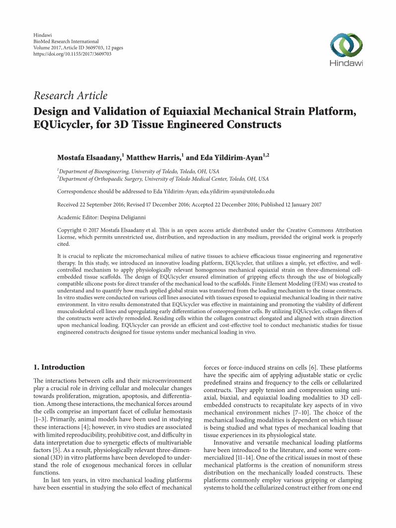
Research ArticleDesign and Validation of Equiaxial Mechanical Strain Platform,EQUicycler, for 3D Tissue Engineered Constructs
Mostafa Elsaadany,1 Matthew Harris,1 and Eda Yildirim-Ayan1,2
1Department of Bioengineering, University of Toledo, Toledo, OH, USA2Department of Orthopaedic Surgery, University of Toledo Medical Center, Toledo, OH, USA
Correspondence should be addressed to Eda Yildirim-Ayan; [email protected]
Received 22 September 2016; Revised 17 December 2016; Accepted 22 December 2016; Published 12 January 2017
Academic Editor: Despina Deligianni
Copyright © 2017 Mostafa Elsaadany et al. This is an open access article distributed under the Creative Commons AttributionLicense, which permits unrestricted use, distribution, and reproduction in any medium, provided the original work is properlycited.
It is crucial to replicate the micromechanical milieu of native tissues to achieve efficacious tissue engineering and regenerativetherapy. In this study, we introduced an innovative loading platform, EQUicycler, that utilizes a simple, yet effective, and well-controlled mechanism to apply physiologically relevant homogenous mechanical equiaxial strain on three-dimensional cell-embedded tissue scaffolds. The design of EQUicycler ensured elimination of gripping effects through the use of biologicallycompatible silicone posts for direct transfer of the mechanical load to the scaffolds. Finite Element Modeling (FEM) was created tounderstand and to quantify how much applied global strain was transferred from the loading mechanism to the tissue constructs.In vitro studies were conducted on various cell lines associated with tissues exposed to equiaxial mechanical loading in their nativeenvironment. In vitro results demonstrated that EQUicycler was effective in maintaining and promoting the viability of differentmusculoskeletal cell lines and upregulating early differentiation of osteoprogenitor cells. By utilizing EQUicycler, collagen fibers ofthe constructs were actively remodeled. Residing cells within the collagen construct elongated and aligned with strain directionupon mechanical loading. EQUicycler can provide an efficient and cost-effective tool to conduct mechanistic studies for tissueengineered constructs designed for tissue systems under mechanical loading in vivo.
1. Introduction
The interactions between cells and their microenvironmentplay a crucial role in driving cellular and molecular changestowards proliferation, migration, apoptosis, and differentia-tion. Among these interactions, themechanical forces aroundthe cells comprise an important facet of cellular hemostasis[1–3]. Primarily, animal models have been used in studyingthese interactions [4]; however, in vivo studies are associatedwith limited reproducibility, prohibitive cost, and difficulty indata interpretation due to synergetic effects of multivariablefactors [5]. As a result, physiologically relevant three-dimen-sional (3D) in vitro platforms have been developed to under-stand the role of exogenous mechanical forces in cellularfunctions.
In last ten years, in vitro mechanical loading platformshave been essential in studying the solo effect of mechanical
forces or force-induced strains on cells [6]. These platformshave the specific aim of applying adjustable static or cyclicpredefined strains and frequency to the cells or cellularizedconstructs. They apply tension and compression using uni-axial, biaxial, and equiaxial loading modalities to 3D cell-embedded constructs to recapitulate key aspects of in vivomechanical environment niches [7–10]. The choice of themechanical loading modalities is dependent on which tissueis being studied and what types of mechanical loading thattissue experiences in its physiological state.
Innovative and versatile mechanical loading platformshave been introduced to the literature, and some were com-mercialized [11–14]. One of the critical issues in most of thesemechanical platforms is the creation of nonuniform stressdistribution on the mechanically loaded constructs. Theseplatforms commonly employ various gripping or clampingsystems to hold the cellularized construct either fromone end
HindawiBioMed Research InternationalVolume 2017, Article ID 3609703, 12 pageshttps://doi.org/10.1155/2017/3609703
2 BioMed Research International
Mot
or
Cam
Cam Ca
m
Cam
Fixed plate Fixed plate
Base Base
Unstrainedposition
Guidingrods
Movingplate
Rotatingshaft
Strainedposition
(a)
Unstrained
Deformablesilicone post
Cell-embeddedcollagenconstruct
Cross-section ofconstruct(b)
(c) (d)
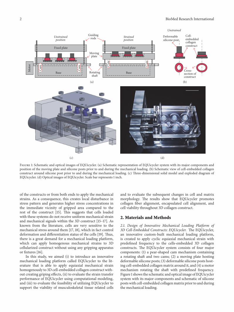
Figure 1: Schematic and optical images of EQUicycler. (a) Schematic representation of EQUicycler system with its major components andposition of the moving plate and silicone posts prior to and during the mechanical loading. (b) Schematic view of cell-embedded collagenconstruct around silicone post prior to and during the mechanical loading. (c) Three-dimensional solid model and exploded diagram ofEQUicycler. (d) Optical images of EQUicycler. Scale bar represents 1 inch.
of the constructs or from both ends to apply the mechanicalstrains. As a consequence, this creates local disturbance instress pattern and generates higher stress concentrations inthe immediate vicinity of gripped area compared to therest of the construct [15]. This suggests that cells loadedwith these systems do not receive uniform mechanical strainand mechanical signals within the 3D construct [15–17]. Asknown from the literature, cells are very sensitive to themechanical stress around them [17, 18], which in fact controldeformation and differentiation status of the cells [19]. Thus,there is a great demand for a mechanical loading platform,which can apply homogenous mechanical strains to 3Dcellularized construct without using any gripping apparatusor fixtures [16].
In this study, we aimed (i) to introduce an innovativemechanical loading platform called EQUicycler to the lit-erature that is able to apply equiaxial mechanical strainhomogenously to 3D cell-embedded collagen construct with-out creating griping effects, (ii) to evaluate the strain transferperformance of EQUicycler using computational modeling,and (iii) to evaluate the feasibility of utilizing EQUicycler tosupport the viability of musculoskeletal tissue related cells
and to evaluate the subsequent changes in cell and matrixmorphology. The results show that EQUicycler promotescollagen fiber alignment, encapsulated cell alignment, andcell viability throughout 3D collagen construct.
2. Materials and Methods
2.1. Design of Innovative Mechanical Loading Platform of3D Cell-Embedded Constructs: EQUicycler. The EQUicycler,an innovative custom-built mechanical loading platform,is created to apply cyclic equiaxial mechanical strain withpredefined frequency to the cells-embedded 3D collagenconstructs. The EQUicycler system consists of four majorcomponents: (1) a pear-shaped cam mechanism containinga rotating shaft and two cams; (2) a moving plate hostingdeformable silicone posts; (3) deformable silicone posts host-ing cell-embedded collagenmatrix around it, and (4) amotormechanism rotating the shaft with predefined frequency.Figure 1 shows the schematic and optical image of EQUicyclersystem with its major components and schematic of siliconeposts with cell-embedded collagenmatrix prior to and duringthe mechanical loading.
BioMed Research International 3
The EQUicycler’s working mechanism is based on cre-ating a mechanical strain on silicone post hosting cell-embedded collagen ring around it.Themotor with adjustablefrequency rotates the shaft with pear-shaped cams. As thecams rotate, the moving plate, which is placed on the cams,moves up and down. The displacement distance of themoving plate is defined by the geometry of the cams. Duringthis reciprocating motion, the displacement of the movingplate against the fixed plate causes compression of the siliconepost. This compression further creates mechanical strainin the cell-embedded collagen ring, which surrounds thedeformable silicone post.
The EQUicycler eliminates the problems associated withthe gripping effects as experienced in themajority ofmechan-ical loading platforms.The grips used at either end of the cell-embedded construct to mount the construct to the platforms[20–25] create stress/strain spike in the vicinity of the grippedarea that leads to a nonhomogenous stress/strain distributioncompared to the areas in the middle of the sample [26–28].The EQUicycler is designed such that the cell-embedded col-lagen construct is integrated into the platform by depositingit around a deformable silicone post rather than using grips.The compression of the silicone post produces mechanicalstrain in the cell-embedded collagen ring, which surroundsthe post.Therefore, homogeneous strain throughout the cell-embedded collagen construct can be created without creatinga stress concentration.
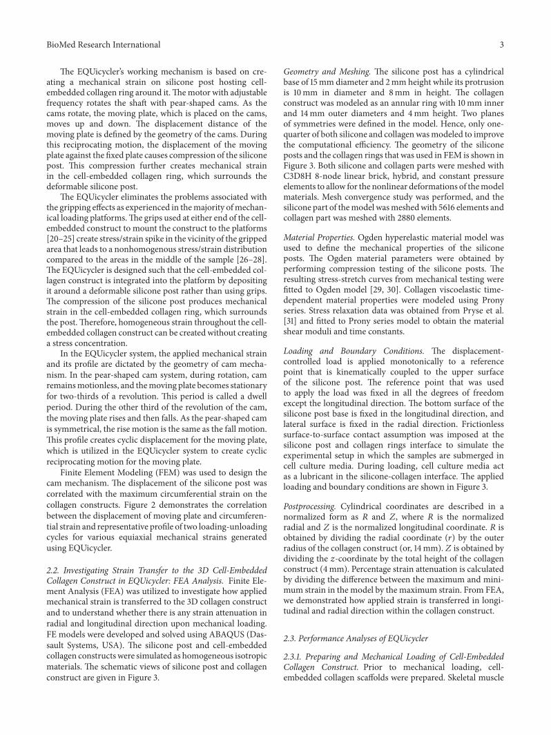
In the EQUicycler system, the applied mechanical strainand its profile are dictated by the geometry of cam mecha-nism. In the pear-shaped cam system, during rotation, camremainsmotionless, and themoving plate becomes stationaryfor two-thirds of a revolution. This period is called a dwellperiod. During the other third of the revolution of the cam,the moving plate rises and then falls. As the pear-shaped camis symmetrical, the rise motion is the same as the fall motion.This profile creates cyclic displacement for the moving plate,which is utilized in the EQUicycler system to create cyclicreciprocating motion for the moving plate.
Finite Element Modeling (FEM) was used to design thecam mechanism. The displacement of the silicone post wascorrelated with the maximum circumferential strain on thecollagen constructs. Figure 2 demonstrates the correlationbetween the displacement of moving plate and circumferen-tial strain and representative profile of two loading-unloadingcycles for various equiaxial mechanical strains generatedusing EQUicycler.
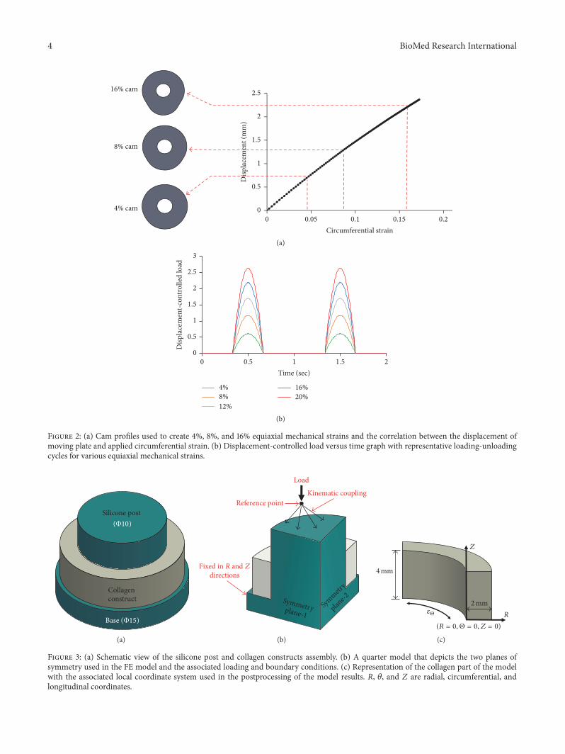
2.2. Investigating Strain Transfer to the 3D Cell-EmbeddedCollagen Construct in EQUicycler: FEA Analysis. Finite Ele-ment Analysis (FEA) was utilized to investigate how appliedmechanical strain is transferred to the 3D collagen constructand to understand whether there is any strain attenuation inradial and longitudinal direction upon mechanical loading.FE models were developed and solved using ABAQUS (Das-sault Systems, USA). The silicone post and cell-embeddedcollagen constructswere simulated as homogeneous isotropicmaterials. The schematic views of silicone post and collagenconstruct are given in Figure 3.
Geometry and Meshing. The silicone post has a cylindricalbase of 15mm diameter and 2mm height while its protrusionis 10mm in diameter and 8mm in height. The collagenconstruct was modeled as an annular ring with 10mm innerand 14mm outer diameters and 4mm height. Two planesof symmetries were defined in the model. Hence, only one-quarter of both silicone and collagenwasmodeled to improvethe computational efficiency. The geometry of the siliconeposts and the collagen rings that was used in FEM is shown inFigure 3. Both silicone and collagen parts were meshed withC3D8H 8-node linear brick, hybrid, and constant pressureelements to allow for the nonlinear deformations of themodelmaterials. Mesh convergence study was performed, and thesilicone part of themodel wasmeshedwith 5616 elements andcollagen part was meshed with 2880 elements.
Material Properties. Ogden hyperelastic material model wasused to define the mechanical properties of the siliconeposts. The Ogden material parameters were obtained byperforming compression testing of the silicone posts. Theresulting stress-stretch curves from mechanical testing werefitted to Ogden model [29, 30]. Collagen viscoelastic time-dependent material properties were modeled using Pronyseries. Stress relaxation data was obtained from Pryse et al.[31] and fitted to Prony series model to obtain the materialshear moduli and time constants.
Loading and Boundary Conditions. The displacement-controlled load is applied monotonically to a referencepoint that is kinematically coupled to the upper surfaceof the silicone post. The reference point that was usedto apply the load was fixed in all the degrees of freedomexcept the longitudinal direction. The bottom surface of thesilicone post base is fixed in the longitudinal direction, andlateral surface is fixed in the radial direction. Frictionlesssurface-to-surface contact assumption was imposed at thesilicone post and collagen rings interface to simulate theexperimental setup in which the samples are submerged incell culture media. During loading, cell culture media actas a lubricant in the silicone-collagen interface. The appliedloading and boundary conditions are shown in Figure 3.
Postprocessing. Cylindrical coordinates are described in anormalized form as 𝑅 and 𝑍, where 𝑅 is the normalizedradial and 𝑍 is the normalized longitudinal coordinate. 𝑅 isobtained by dividing the radial coordinate (𝑟) by the outerradius of the collagen construct (or, 14mm). 𝑍 is obtained bydividing the 𝑧-coordinate by the total height of the collagenconstruct (4mm). Percentage strain attenuation is calculatedby dividing the difference between the maximum and mini-mum strain in the model by the maximum strain. From FEA,we demonstrated how applied strain is transferred in longi-tudinal and radial direction within the collagen construct.
2.3. Performance Analyses of EQUicycler
2.3.1. Preparing and Mechanical Loading of Cell-EmbeddedCollagen Construct. Prior to mechanical loading, cell-embedded collagen scaffolds were prepared. Skeletal muscle
4 BioMed Research International
Disp
lace
men
t (m
m)
2.5
2
1.5
1
0.5
0
0 0.05 0.1 0.15 0.2
Circumferential strain
16% cam
8% cam
4% cam
(a)
0
0.5
1
1.5
2
2.5
3
0 0.5 1 1.5 2Time (sec)
Disp
lace
men
t-con
trolle
d lo
ad
4%8%12%
16%20%
(b)
Figure 2: (a) Cam profiles used to create 4%, 8%, and 16% equiaxial mechanical strains and the correlation between the displacement ofmoving plate and applied circumferential strain. (b) Displacement-controlled load versus time graph with representative loading-unloadingcycles for various equiaxial mechanical strains.
Collagen construct
Silicone post (Φ10)
Base (Φ15)
(a)
Reference point
Load
directions
Kinematic coupling
Symmetryplane-1Sym
metry
plane-2
Fixed in R and Z
(b)
Z
R
4mm
2mm𝜀Θ
(R = 0, Θ = 0, Z = 0)
(c)
Figure 3: (a) Schematic view of the silicone post and collagen constructs assembly. (b) A quarter model that depicts the two planes ofsymmetry used in the FE model and the associated loading and boundary conditions. (c) Representation of the collagen part of the modelwith the associated local coordinate system used in the postprocessing of the model results. 𝑅, 𝜃, and 𝑍 are radial, circumferential, andlongitudinal coordinates.

BioMed Research International 5
cells (C2C12) and osteoprogenitor cells (MC3T3-E1) were uti-lized to analyze EQUicycler’s capacity with different cell lines.C2C12 (ATCC, USA) cells were cultured in Dulbecco’s Low-Glucose Modified Eagle’s Medium (DMEM) (ThermoFisher,USA) supplemented with 10% Fetal Bovine Serum (FBS)(Gibco, USA) and 1% penicillin/streptomycin (Life Tech-nologies, USA). MC3T3-E1 (ATCC, USA) were maintainedin complete media comprising Alpha-Minimum EssentialMedium (𝛼-MEM) (Life Technologies, USA) supplementedwith 10% FBS (Gibco, USA) and 1% penicillin/streptomycin(Life Technologies, USA). Upon confluence, cells wereharvested and encapsulated within the collagen type-Isolution with 106 cells/mL seeding density. For the cellularstudy, 2.5mg/mL collagen type-I (Corning, USA) solutionwas prepared from stock collagen type-I solution. Briefly,collagen type-I stock solution at 3.7mg/mL concentrationwith pH ∼ 3-4 was diluted to 2.5mg/mL and neutralized withchilled 1NNaOH, phosphate buffer saline (PBS), and deion-ized water according to manufacturer’s protocol. C2C12 cellsor MC3T3-E1 cells-embedded collagen constructs were thendeposited around the silicone posts and incubated overnight.Next day, scaffolds were exposed to various equiaxialstrains (4% and 8%) with 1Hz frequency using EQUicycler.Unstrained (0%) scaffolds were used as control group.
2.3.2. Assessing Cell Proliferation, Viability, and Differentia-tion. The proliferation of the encapsulated cells in collagenwas examined using nondestructive alamarBlue (aB) assay(Biosource International, USA). Four samples (𝑛 = 4) wereused for each of the characterization and control groups.On the day of characterization, the samples were incubatedwith 10% (v/v) of aB assay solution in cell culture mediafor 6 hours. After the incubation period, the reduced colorof the cell culture media was quantified by measuring thefluorescence intensity using amicroplate fluorometer (Wallac1420, USA) at 545/585 nm excitation/emission wavelengths.
The viability of the encapsulated cells at day 7was assessedusing live/dead assay (Life Technologies, USA). Upon incu-bation with live/dead assay reagents, confocal images of liveand dead cells within 3D scaffolds were obtained. On thecharacterization day, the samples were first rinsed twice withPBS for 5min and then incubated for 30 minutes with a1 : 4 concentration ratio of Calcein-AM for labeling live cells(green) and ethidium homodimer-1 for labeling dead cells(red). After incubation, the samples were fixed using 4%paraformaldehyde (Sigma-Aldrich, USA) in PBS for 30 min-utes and washed thrice with PBS for 15 minutes and stored at4∘C until they were imaged using a Leica TCS SPE confocalmicroscope. Cells stained with Calcein-AM were visualizedusing a 488 nm excitation and an emission spectrum of491 nm to 545 nm wavelength. Cells stained with ethidiumhomodimer-1 were visualized using a 568 nm excitation andan emission spectrum of 590 nm to 685 nm wavelength.
The cells alkaline phosphatase (ALP) activity was eval-uated at day 14 and day 21 using ALP colorimetric assay(Abcam,USA). Before conducting the assay, the encapsulatedcells within the collagen scaffolds were liberated using 0.2%(w/v) collagenase type-I solution (Life Technologies, USA)
in complete media. The samples were incubated in thecollagenase solution for 1 h at 37∘C in shaking water bath.After collagenase digestion, the solution was centrifugedto isolate the cell pellet. The ALP assay was performedbased on the manufacturer’s protocol (Abcam, USA). Briefly,the amount of p-nitrophenol (pNP) converted from p-nitrophenyl phosphate (pNPP) substrate in the presence ofALP was quantified by measuring the absorbance at 405 nmusing a microplate reader (SOFTmax Pro). A calibrationcurve obtained using a serial dilution of pNP was used toconvert the measured absorbance to pNP value.
2.3.3. Changes in Collagen Fiber Morphology and Alignment.The morphological changes on 3D collagen scaffold wereexamined by Scanning Electron Microscopy (SEM) (Quanta3D FEG) and histological analyses. For SEM examination,first, control and mechanically strained collagen constructswere fixed with 4% paraformaldehyde solution (Sigma-Aldrich, USA) for 1 h. In order to preserve the collagenfibrils and improve the image quality the samples were, then,postfixed in 1% (v/v) osmium tetroxide (Across Organics,USA) diluted with Sorensen’s buffer. After fixation, thesamples were washed twice with PBS and subsequentlydehydrated in increasing concentrations of ethanol from 30%to 100% and ethanol/hexamethyldisilazane (HMDS) (FisherScientific, USA) solutions from 30% to 100% for 15 min each.Additionally, the samples were air-dried overnight to evapo-rate the residual organic solvents. Before SEM examination,the dried samples were gold sputter-coated to avoid chargeaccumulation.
The changes in collagen fiber morphology and alignmentin mechanically strained samples compared to control werevisualized using histological analysis and Mason’s Trichromestaining. On the characterization day, cell-encapsulated con-structs were fixed in 10% formalin for 24 h, dehydratedusing an ethanol gradient, and incubated in xylene beforeembedding them in paraffin. The embedded samples weresectioned at 20𝜇m thickness and mounted on microscopeslides. The rehydrated sections were incubated in Mason’sTrichrome stain, washed, and viewed under a bright fieldmicroscope to observe collagen fibers and cells.
2.4. Statistical Analysis. One-way Analysis of Variance testswere performed using SPSS Statistics Package (IBM, USA)to determine if there is a significant difference between themeans of the experimental and control groups. Fisher’s LSDpost hocwas used formultiple comparisons between differentgroups. Statistical significance was defined as 𝑝 < 0.05.
3. Results
3.1. Finite Element Analysis (FEA) of the Strain Transfer in theCollagen Constructs. Representative circumferential straincontours for the silicone-collagen assembly and the collagenconstruct alone are shown in Figure 4. The displacement-controlled loading pertinent to the cam profiles utilized inEQUicycler resulted in the compression of silicone posts in
6 BioMed Research International
LE, LE22(Avg: 75%)
0.222
0.204
0.185
0.167
0.148
0.130
0.112
0.093
0.075
0.056
0.038
0.019
0.001
(a)
LE, LE22(Avg: 75%)
0.200
0.193
0.186
0.178
0.171
0.163
0.156
0.149
0.141
0.134
0.127
0.119
0.112
(b)
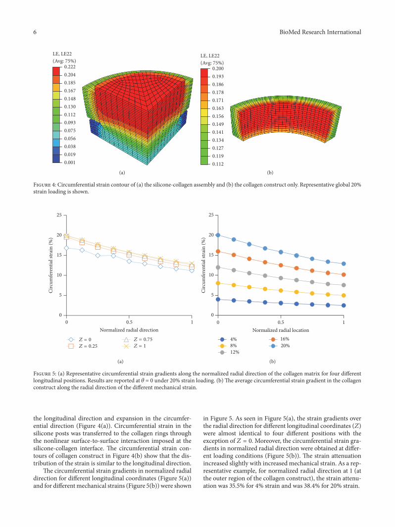
Figure 4: Circumferential strain contour of (a) the silicone-collagen assembly and (b) the collagen construct only. Representative global 20%strain loading is shown.
0
5
10
15
20
25
0 0.5 1Normalized radial direction
Circ
umfe
rent
ial s
trai
n (%
)
Z = 0
Z = 0.25
Z = 0.75
Z = 1
(a)
0
5
10
15
20
25
0 0.5 1
Circ
umfe
rent
ial s
trai
n (%
)
Normalized radial location
4%8%12%
16%20%
(b)
Figure 5: (a) Representative circumferential strain gradients along the normalized radial direction of the collagen matrix for four differentlongitudinal positions. Results are reported at 𝜃 = 0 under 20% strain loading. (b)The average circumferential strain gradient in the collagenconstruct along the radial direction of the different mechanical strain.
the longitudinal direction and expansion in the circumfer-ential direction (Figure 4(a)). Circumferential strain in thesilicone posts was transferred to the collagen rings throughthe nonlinear surface-to-surface interaction imposed at thesilicone-collagen interface. The circumferential strain con-tours of collagen construct in Figure 4(b) show that the dis-tribution of the strain is similar to the longitudinal direction.
The circumferential strain gradients in normalized radialdirection for different longitudinal coordinates (Figure 5(a))and for differentmechanical strains (Figure 5(b)) were shown
in Figure 5. As seen in Figure 5(a), the strain gradients overthe radial direction for different longitudinal coordinates (𝑍)were almost identical to four different positions with theexception of 𝑍 = 0. Moreover, the circumferential strain gra-dients in normalized radial direction were obtained at differ-ent loading conditions (Figure 5(b)). The strain attenuationincreased slightly with increased mechanical strain. As a rep-resentative example, for normalized radial direction at 1 (atthe outer region of the collagen construct), the strain attenu-ation was 35.5% for 4% strain and was 38.4% for 20% strain.
BioMed Research International 7
0
1
2
3
4
5
6
Day 1 Day 3Days in culture
Nor
mal
ized
cell
num
ber
4%8%
∗
Figure 6: Normalized C2C12 cell number within 4% and 8%mechanically strained collagen. Normalized cell number is the ratioof cell number in mechanical strain group over cell number in thecontrol group on culture day. ∗ indicates significant difference fromother groups.
3.2. Effect ofMechanical Loading on Cells and CollagenMatrix
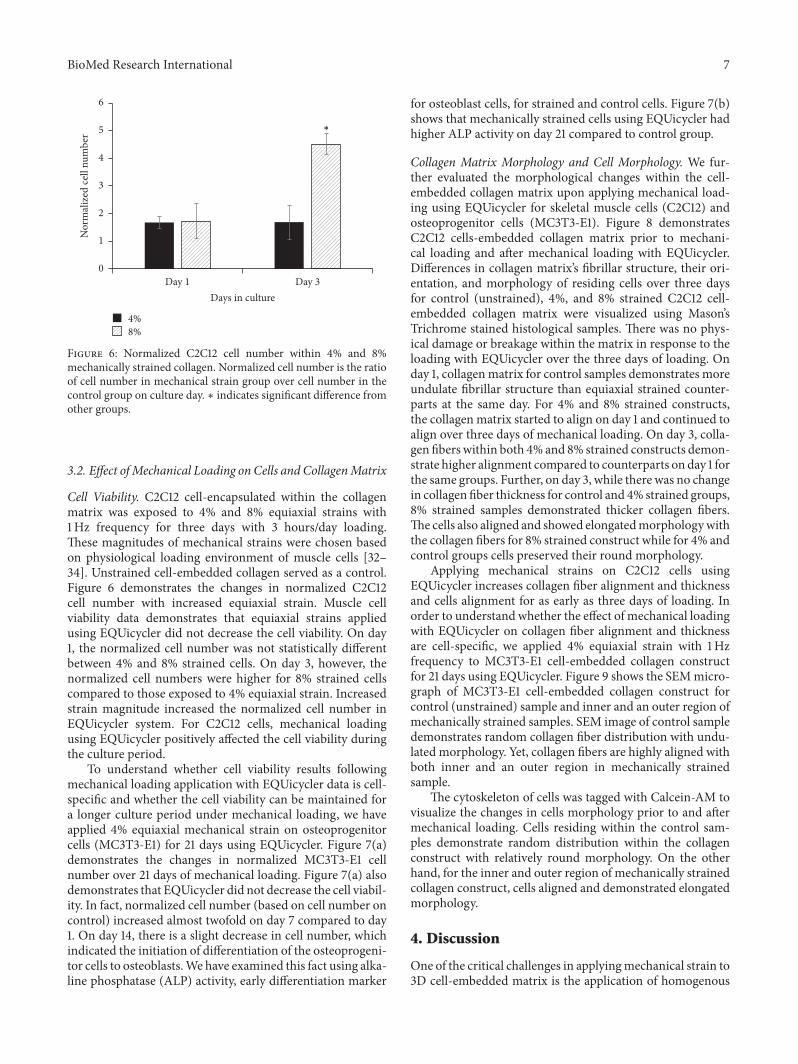
Cell Viability. C2C12 cell-encapsulated within the collagenmatrix was exposed to 4% and 8% equiaxial strains with1Hz frequency for three days with 3 hours/day loading.These magnitudes of mechanical strains were chosen basedon physiological loading environment of muscle cells [32–34]. Unstrained cell-embedded collagen served as a control.Figure 6 demonstrates the changes in normalized C2C12cell number with increased equiaxial strain. Muscle cellviability data demonstrates that equiaxial strains appliedusing EQUicycler did not decrease the cell viability. On day1, the normalized cell number was not statistically differentbetween 4% and 8% strained cells. On day 3, however, thenormalized cell numbers were higher for 8% strained cellscompared to those exposed to 4% equiaxial strain. Increasedstrain magnitude increased the normalized cell number inEQUicycler system. For C2C12 cells, mechanical loadingusing EQUicycler positively affected the cell viability duringthe culture period.
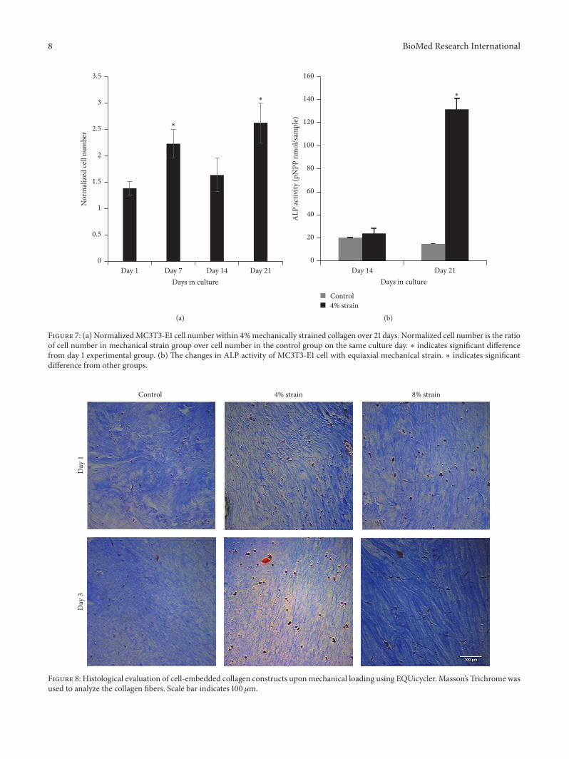
To understand whether cell viability results followingmechanical loading application with EQUicycler data is cell-specific and whether the cell viability can be maintained fora longer culture period under mechanical loading, we haveapplied 4% equiaxial mechanical strain on osteoprogenitorcells (MC3T3-E1) for 21 days using EQUicycler. Figure 7(a)demonstrates the changes in normalized MC3T3-E1 cellnumber over 21 days of mechanical loading. Figure 7(a) alsodemonstrates that EQUicycler did not decrease the cell viabil-ity. In fact, normalized cell number (based on cell number oncontrol) increased almost twofold on day 7 compared to day1. On day 14, there is a slight decrease in cell number, whichindicated the initiation of differentiation of the osteoprogeni-tor cells to osteoblasts.We have examined this fact using alka-line phosphatase (ALP) activity, early differentiation marker
for osteoblast cells, for strained and control cells. Figure 7(b)shows that mechanically strained cells using EQUicycler hadhigher ALP activity on day 21 compared to control group.
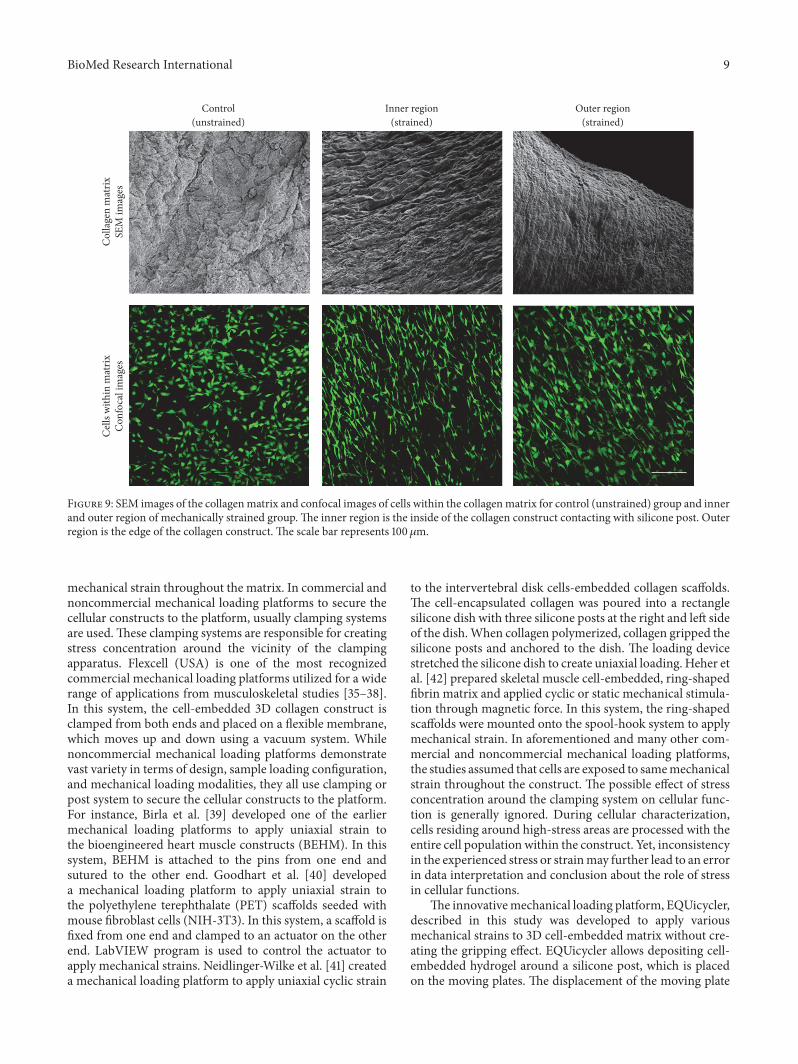
Collagen Matrix Morphology and Cell Morphology. We fur-ther evaluated the morphological changes within the cell-embedded collagen matrix upon applying mechanical load-ing using EQUicycler for skeletal muscle cells (C2C12) andosteoprogenitor cells (MC3T3-E1). Figure 8 demonstratesC2C12 cells-embedded collagen matrix prior to mechani-cal loading and after mechanical loading with EQUicycler.Differences in collagen matrix’s fibrillar structure, their ori-entation, and morphology of residing cells over three daysfor control (unstrained), 4%, and 8% strained C2C12 cell-embedded collagen matrix were visualized using Mason’sTrichrome stained histological samples. There was no phys-ical damage or breakage within the matrix in response to theloading with EQUicycler over the three days of loading. Onday 1, collagenmatrix for control samples demonstrates moreundulate fibrillar structure than equiaxial strained counter-parts at the same day. For 4% and 8% strained constructs,the collagen matrix started to align on day 1 and continued toalign over three days of mechanical loading. On day 3, colla-gen fiberswithin both 4% and 8% strained constructs demon-strate higher alignment compared to counterparts on day 1 forthe same groups. Further, on day 3, while there was no changein collagen fiber thickness for control and 4% strained groups,8% strained samples demonstrated thicker collagen fibers.The cells also aligned and showed elongatedmorphologywiththe collagen fibers for 8% strained construct while for 4% andcontrol groups cells preserved their round morphology.
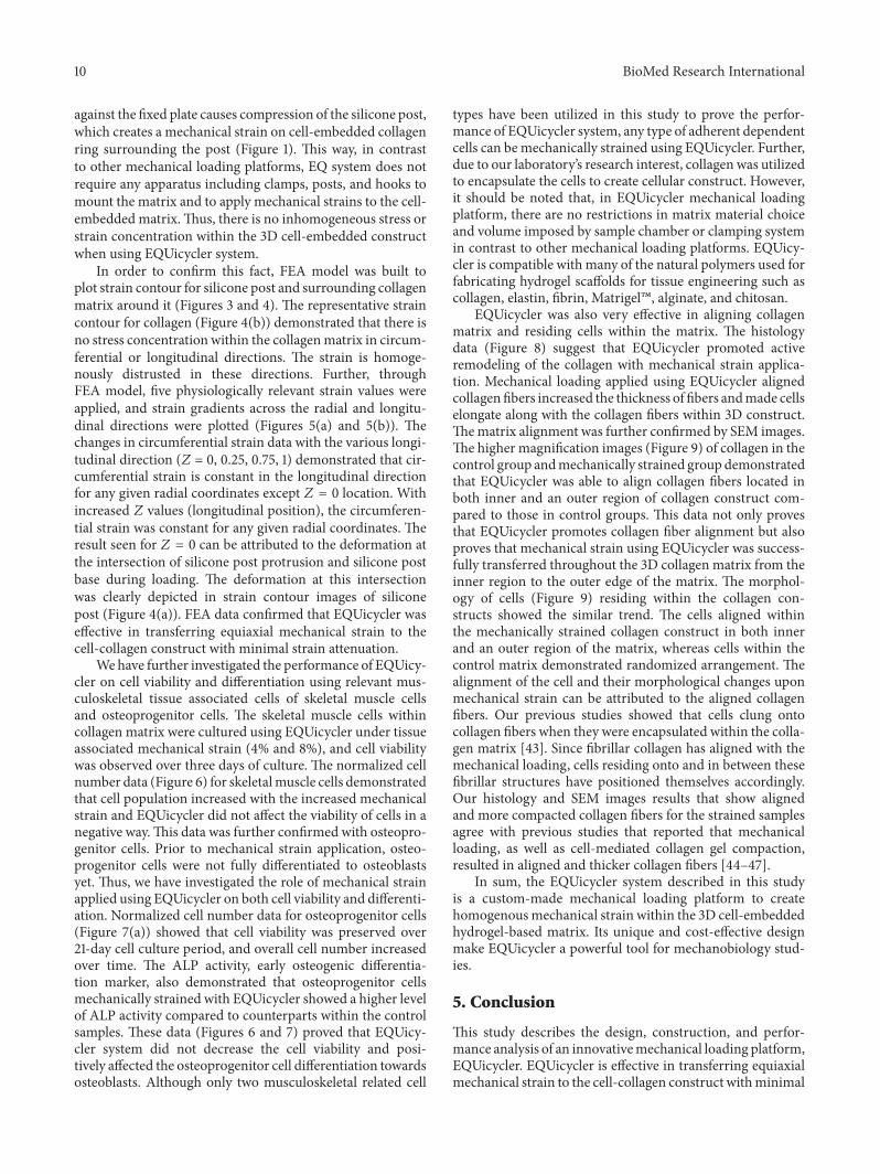
Applying mechanical strains on C2C12 cells usingEQUicycler increases collagen fiber alignment and thicknessand cells alignment for as early as three days of loading. Inorder to understand whether the effect of mechanical loadingwith EQUicycler on collagen fiber alignment and thicknessare cell-specific, we applied 4% equiaxial strain with 1Hzfrequency to MC3T3-E1 cell-embedded collagen constructfor 21 days using EQUicycler. Figure 9 shows the SEMmicro-graph of MC3T3-E1 cell-embedded collagen construct forcontrol (unstrained) sample and inner and an outer region ofmechanically strained samples. SEM image of control sampledemonstrates random collagen fiber distribution with undu-lated morphology. Yet, collagen fibers are highly aligned withboth inner and an outer region in mechanically strainedsample.
The cytoskeleton of cells was tagged with Calcein-AM tovisualize the changes in cells morphology prior to and aftermechanical loading. Cells residing within the control sam-ples demonstrate random distribution within the collagenconstruct with relatively round morphology. On the otherhand, for the inner and outer region of mechanically strainedcollagen construct, cells aligned and demonstrated elongatedmorphology.
4. Discussion
One of the critical challenges in applyingmechanical strain to3D cell-embedded matrix is the application of homogenous
8 BioMed Research International
0
0.5
1
1.5
2
2.5
3
3.5
Day 1 Day 7 Day 14 Day 21Days in culture
Nor
mal
ized
cell
num
ber
∗
∗
(a)
0
20
40
60
80
100
120
140
160
Day 14 Day 21
ALP
activ
ity (p
NPP
nm
ol/s
ampl
e)Control4% strain
Days in culture
∗
(b)
Figure 7: (a) NormalizedMC3T3-E1 cell number within 4%mechanically strained collagen over 21 days. Normalized cell number is the ratioof cell number in mechanical strain group over cell number in the control group on the same culture day. ∗ indicates significant differencefrom day 1 experimental group. (b) The changes in ALP activity of MC3T3-E1 cell with equiaxial mechanical strain. ∗ indicates significantdifference from other groups.
Control
Day
3D
ay 1
4% strain 8% strain
Figure 8: Histological evaluation of cell-embedded collagen constructs uponmechanical loading using EQUicycler. Masson’s Trichrome wasused to analyze the collagen fibers. Scale bar indicates 100 𝜇m.
BioMed Research International 9
Control(unstrained)
Col
lage
n m
atrix
SEM
imag
esC
ells
with
in m
atrix
Con
foca
l im
ages
Inner region(strained)
Outer region(strained)
Figure 9: SEM images of the collagenmatrix and confocal images of cells within the collagenmatrix for control (unstrained) group and innerand outer region of mechanically strained group.The inner region is the inside of the collagen construct contacting with silicone post. Outerregion is the edge of the collagen construct. The scale bar represents 100 𝜇m.
mechanical strain throughout the matrix. In commercial andnoncommercial mechanical loading platforms to secure thecellular constructs to the platform, usually clamping systemsare used.These clamping systems are responsible for creatingstress concentration around the vicinity of the clampingapparatus. Flexcell (USA) is one of the most recognizedcommercial mechanical loading platforms utilized for a widerange of applications from musculoskeletal studies [35–38].In this system, the cell-embedded 3D collagen construct isclamped from both ends and placed on a flexible membrane,which moves up and down using a vacuum system. Whilenoncommercial mechanical loading platforms demonstratevast variety in terms of design, sample loading configuration,and mechanical loading modalities, they all use clamping orpost system to secure the cellular constructs to the platform.For instance, Birla et al. [39] developed one of the earliermechanical loading platforms to apply uniaxial strain tothe bioengineered heart muscle constructs (BEHM). In thissystem, BEHM is attached to the pins from one end andsutured to the other end. Goodhart et al. [40] developeda mechanical loading platform to apply uniaxial strain tothe polyethylene terephthalate (PET) scaffolds seeded withmouse fibroblast cells (NIH-3T3). In this system, a scaffold isfixed from one end and clamped to an actuator on the otherend. LabVIEW program is used to control the actuator toapply mechanical strains. Neidlinger-Wilke et al. [41] createda mechanical loading platform to apply uniaxial cyclic strain
to the intervertebral disk cells-embedded collagen scaffolds.The cell-encapsulated collagen was poured into a rectanglesilicone dish with three silicone posts at the right and left sideof the dish.When collagen polymerized, collagen gripped thesilicone posts and anchored to the dish. The loading devicestretched the silicone dish to create uniaxial loading. Heher etal. [42] prepared skeletal muscle cell-embedded, ring-shapedfibrin matrix and applied cyclic or static mechanical stimula-tion through magnetic force. In this system, the ring-shapedscaffolds were mounted onto the spool-hook system to applymechanical strain. In aforementioned and many other com-mercial and noncommercial mechanical loading platforms,the studies assumed that cells are exposed to samemechanicalstrain throughout the construct. The possible effect of stressconcentration around the clamping system on cellular func-tion is generally ignored. During cellular characterization,cells residing around high-stress areas are processed with theentire cell population within the construct. Yet, inconsistencyin the experienced stress or strainmay further lead to an errorin data interpretation and conclusion about the role of stressin cellular functions.
The innovativemechanical loading platform, EQUicycler,described in this study was developed to apply variousmechanical strains to 3D cell-embedded matrix without cre-ating the gripping effect. EQUicycler allows depositing cell-embedded hydrogel around a silicone post, which is placedon the moving plates. The displacement of the moving plate
10 BioMed Research International
against the fixed plate causes compression of the silicone post,which creates a mechanical strain on cell-embedded collagenring surrounding the post (Figure 1). This way, in contrastto other mechanical loading platforms, EQ system does notrequire any apparatus including clamps, posts, and hooks tomount the matrix and to apply mechanical strains to the cell-embedded matrix.Thus, there is no inhomogeneous stress orstrain concentration within the 3D cell-embedded constructwhen using EQUicycler system.
In order to confirm this fact, FEA model was built toplot strain contour for silicone post and surrounding collagenmatrix around it (Figures 3 and 4). The representative straincontour for collagen (Figure 4(b)) demonstrated that there isno stress concentration within the collagenmatrix in circum-ferential or longitudinal directions. The strain is homoge-nously distrusted in these directions. Further, throughFEA model, five physiologically relevant strain values wereapplied, and strain gradients across the radial and longitu-dinal directions were plotted (Figures 5(a) and 5(b)). Thechanges in circumferential strain data with the various longi-tudinal direction (𝑍 = 0, 0.25, 0.75, 1) demonstrated that cir-cumferential strain is constant in the longitudinal directionfor any given radial coordinates except 𝑍 = 0 location. Withincreased 𝑍 values (longitudinal position), the circumferen-tial strain was constant for any given radial coordinates. Theresult seen for 𝑍 = 0 can be attributed to the deformation atthe intersection of silicone post protrusion and silicone postbase during loading. The deformation at this intersectionwas clearly depicted in strain contour images of siliconepost (Figure 4(a)). FEA data confirmed that EQUicycler waseffective in transferring equiaxial mechanical strain to thecell-collagen construct with minimal strain attenuation.
We have further investigated the performance of EQUicy-cler on cell viability and differentiation using relevant mus-culoskeletal tissue associated cells of skeletal muscle cellsand osteoprogenitor cells. The skeletal muscle cells withincollagen matrix were cultured using EQUicycler under tissueassociated mechanical strain (4% and 8%), and cell viabilitywas observed over three days of culture. The normalized cellnumber data (Figure 6) for skeletalmuscle cells demonstratedthat cell population increased with the increased mechanicalstrain and EQUicycler did not affect the viability of cells in anegative way.This data was further confirmed with osteopro-genitor cells. Prior to mechanical strain application, osteo-progenitor cells were not fully differentiated to osteoblastsyet. Thus, we have investigated the role of mechanical strainapplied using EQUicycler on both cell viability and differenti-ation. Normalized cell number data for osteoprogenitor cells(Figure 7(a)) showed that cell viability was preserved over21-day cell culture period, and overall cell number increasedover time. The ALP activity, early osteogenic differentia-tion marker, also demonstrated that osteoprogenitor cellsmechanically strained with EQUicycler showed a higher levelof ALP activity compared to counterparts within the controlsamples. These data (Figures 6 and 7) proved that EQUicy-cler system did not decrease the cell viability and posi-tively affected the osteoprogenitor cell differentiation towardsosteoblasts. Although only two musculoskeletal related cell
types have been utilized in this study to prove the perfor-mance of EQUicycler system, any type of adherent dependentcells can be mechanically strained using EQUicycler. Further,due to our laboratory’s research interest, collagen was utilizedto encapsulate the cells to create cellular construct. However,it should be noted that, in EQUicycler mechanical loadingplatform, there are no restrictions in matrix material choiceand volume imposed by sample chamber or clamping systemin contrast to other mechanical loading platforms. EQUicy-cler is compatible with many of the natural polymers used forfabricating hydrogel scaffolds for tissue engineering such ascollagen, elastin, fibrin, Matrigel�, alginate, and chitosan.
EQUicycler was also very effective in aligning collagenmatrix and residing cells within the matrix. The histologydata (Figure 8) suggest that EQUicycler promoted activeremodeling of the collagen with mechanical strain applica-tion. Mechanical loading applied using EQUicycler alignedcollagen fibers increased the thickness of fibers andmade cellselongate along with the collagen fibers within 3D construct.Thematrix alignment was further confirmed by SEM images.The higher magnification images (Figure 9) of collagen in thecontrol group andmechanically strained group demonstratedthat EQUicycler was able to align collagen fibers located inboth inner and an outer region of collagen construct com-pared to those in control groups. This data not only provesthat EQUicycler promotes collagen fiber alignment but alsoproves that mechanical strain using EQUicycler was success-fully transferred throughout the 3D collagen matrix from theinner region to the outer edge of the matrix. The morphol-ogy of cells (Figure 9) residing within the collagen con-structs showed the similar trend. The cells aligned withinthe mechanically strained collagen construct in both innerand an outer region of the matrix, whereas cells within thecontrol matrix demonstrated randomized arrangement. Thealignment of the cell and their morphological changes uponmechanical strain can be attributed to the aligned collagenfibers. Our previous studies showed that cells clung ontocollagen fibers when they were encapsulated within the colla-gen matrix [43]. Since fibrillar collagen has aligned with themechanical loading, cells residing onto and in between thesefibrillar structures have positioned themselves accordingly.Our histology and SEM images results that show alignedand more compacted collagen fibers for the strained samplesagree with previous studies that reported that mechanicalloading, as well as cell-mediated collagen gel compaction,resulted in aligned and thicker collagen fibers [44–47].
In sum, the EQUicycler system described in this studyis a custom-made mechanical loading platform to createhomogenousmechanical strain within the 3D cell-embeddedhydrogel-based matrix. Its unique and cost-effective designmake EQUicycler a powerful tool for mechanobiology stud-ies.
5. Conclusion
This study describes the design, construction, and perfor-mance analysis of an innovativemechanical loading platform,EQUicycler. EQUicycler is effective in transferring equiaxialmechanical strain to the cell-collagen construct withminimal
BioMed Research International 11
strain attenuation. The performance of EQUicycler was eval-uated using Finite ElementAnalysis and experimental studiesusing various musculoskeletal tissue associated cell lines.The mechanical loading platform was proved to maintainand promote cell viability over extended periods of culture.Also, the early osteogenic differentiation marker of MC3T3-E1 cells, ALP, was upregulated due to mechanical stimulationperformed using EQUicycler. Additionally, the matrix wasactively remodeled due tomechanical strains. Collagen fiberswere successfully aligned to the direction of the appliedtensile equiaxial strain. Morphological changes were noticedfor both C2C12 andMC3T3-E1 cells as they were also alignedand elongated in the direction of the applied strain.
Competing Interests
The authors declare that there are no competing interestsregarding the publication of this paper.
Acknowledgments
The authors would like to thank Dr. Malathi Krishnamurthyfrom the Department of Biological Sciences at the Universityof Toledo for providing the C2C12 cells. This work is sup-ported by the deArce-Koch Memorial Endowment Fund inSupport of Medical Research and Development.
References
[1] X. Cheng, U. A. Gurkan, C. J. Dehen et al., “An electrochemicalfabrication process for the assembly of anisotropically orientedcollagen bundles,” Biomaterials, vol. 29, no. 22, pp. 3278–3288,2008.
[2] N. S. Hwang, S. Varghese, and J. Elisseeff, “Controlled differen-tiation of stem cells,” Advanced Drug Delivery Reviews, vol. 60,no. 2, pp. 199–214, 2008.
[3] R. T. Tranquillo, “Self-organization of tissue-equivalents: thenature and role of contact guidance,” Biochemical Society Sym-posium, vol. 65, pp. 27–42, 1999.
[4] J. Halpern, C. C. Lynch, J. Fleming et al., “The application of amurine bone bioreactor as a model of tumor: bone interaction,”Clinical & ExperimentalMetastasis, vol. 23, no. 7-8, pp. 345–356,2006.
[5] J. C. Reichert, V. M. C. Quent, L. J. Burke, S. H. Stansfield, J. A.Clements, and D. W. Hutmacher, “Mineralized human primaryosteoblast matrices as a model system to analyse interactionsof prostate cancer cells with the bone microenvironment,”Biomaterials, vol. 31, no. 31, pp. 7928–7936, 2010.
[6] A. Trumbull, G. Subramanian, and E. Yildirim-Ayan,“Mechanoresponsive musculoskeletal tissue differentiation ofadipose-derived stem cells,” BioMedical Engineering Online,vol. 15, article 43, 2016.
[7] S. Amin, S. E. Banijamali, M. Tafazoli-Shadpour et al., “Com-paring the effect of equiaxial cyclic mechanical stimulationon GATA4 expression in adipose-derived and bone marrow-derivedmesenchymal stem cells,”Cell Biology International, vol.38, no. 2, pp. 219–227, 2014.
[8] S. F. Carroll, C. T. Buckley, and D. J. Kelly, “Cyclic hydrostaticpressure promotes a stable cartilage phenotype and enhancesthe functional development of cartilaginous grafts engineered
using multipotent stromal cells isolated from bone marrow andinfrapatellar fat pad,” Journal of Biomechanics, vol. 47, no. 9, pp.2115–2121, 2014.
[9] J. Li, Q. Zhao, E. Wang, C. Zhang, G. Wang, and Q. Yuan,“Dynamic compression of rabbit adipose-derived stem cellstransfected with insulin-like growth factor 1 in chitosan/gelatinscaffolds induces chondrogenesis and matrix biosynthesis,”Journal of Cellular Physiology, vol. 227, no. 5, pp. 2003–2012,2012.
[10] A. Charoenpanich, M. E. Wall, C. J. Tucker, D. M. K. Andrews,D. S. Lalush, and E. G. Loboa, “Microarray analysis of humanadipose-derived stem cells in three-dimensional collagen cul-ture: osteogenesis inhibits bone morphogenic protein and wntsignaling pathways, and cyclic tensile strain causes upregula-tion of proinflammatory cytokine regulators and angiogenicfactors,” Tissue Engineering A, vol. 17, no. 21-22, pp. 2615–2627,2011.
[11] V. F. Shimko andW. C. Claycomb, “Effect ofmechanical loadingon three-dimensional cultures of embryonic stem cell-derivedcardiomyocytes,” Tissue Engineering Part A, vol. 14, no. 1, pp.49–58, 2008.
[12] M. E. Wall, P. S. Weinhold, T. Siu, T. D. Brown, and A. J. Banes,“Comparison of cellular strain with applied substrate strain invitro,” Journal of Biomechanics, vol. 40, no. 1, pp. 173–181, 2007.
[13] B. Lohberger, H. Kaltenegger, N. Stuendl, B. Rinner, A. Leithner,and P. Sadoghi, “Impact of cyclic mechanical stimulation on theexpression of extracellular matrix proteins in human primaryrotator cuff fibroblasts,” Knee Surgery, Sports Traumatology,Arthroscopy, vol. 24, no. 12, pp. 3884–3891, 2016.
[14] E. Azizi and T. J. Roberts, “Biaxial strain and variable stiffnessin aponeuroses,” The Journal of Physiology, vol. 587, no. 17, pp.4309–4318, 2009.
[15] B. D. Riehl, J.-H. Park, I. K. Kwon, and J. Y. Lim, “Mechanicalstretching for tissue engineering: two-dimensional and three-dimensional constructs,” Tissue Engineering—Part B: Reviews,vol. 18, no. 4, pp. 288–300, 2012.
[16] T. Wang, B. S. Gardiner, Z. Lin et al., “Bioreactor designfor tendon/ligament engineering,” Tissue Engineering Part B:Reviews, vol. 19, no. 2, pp. 133–146, 2013.
[17] H. Kamble, M. J. Barton, M. Jun, S. Park, and N. Nguyen, “Cellstretching devices as research tools: engineering and biologicalconsiderations,” Lab Chip, vol. 16, no. 17, pp. 3193–3203, 2016.
[18] C. A. Cook, P. Y. Huri, B. P. Ginn et al., “Characterizationof a novel bioreactor system for 3D cellular mechanobiologystudies,” Biotechnology and Bioengineering, vol. 113, no. 8, pp.1825–1837, 2016.
[19] G. H. Altman, R. L. Horan, I. Martin et al., “Cell differentiationbymechanical stress,”TheFASEB Journal, vol. 16, no. 2, pp. 270–272, 2002.
[20] M. Eastwood, V. C. Mudera, D. A. McGrouther, and R. A.Brown, “Effect of precise mechanical loading on fibroblast pop-ulated collagen lattices: morphological changes,” Cell Motilityand the Cytoskeleton, vol. 40, no. 1, pp. 13–21, 1998.
[21] E. Langelier, D. Rancourt, S. Bouchard et al., “Cyclic tractionmachine for long-term culture of fibroblast-populated collagengels,” Annals of Biomedical Engineering, vol. 27, no. 1, pp. 67–72,1999.
[22] S. M. Tanaka, “A new mechanical stimulator for cultured bonecells using piezoelectric actuator,” Journal of Biomechanics, vol.32, no. 4, pp. 427–430, 1999.
12 BioMed Research International
[23] T. D. Brown, “Techniques for mechanical stimulation of cells invitro: a review,” Journal of Biomechanics, vol. 33, no. 1, pp. 3–14,2000.
[24] D. Kaspar, W. Seidl, C. Neidlinger-Wilke, A. Ignatius, and L.Claes, “Dynamic cell stretching increases human osteoblastproliferation and CICP synthesis but decreases osteocalcinsynthesis and alkaline phosphatase activity,” Journal of Biome-chanics, vol. 33, no. 1, pp. 45–51, 2000.
[25] C. L. Keefer and J. P. Desai, “Mechanical phenotyping of stemcells,”Theriogenology, vol. 75, no. 8, pp. 1426–1430, 2011.
[26] L. A. Matheson, N. J. Fairbank, G. N. Maksym, J. P. Santerre,and R. S. Labow, “Characterization of the Flexcell� Uniflex�cyclic strain culture system with U937 macrophage-like cells,”Biomaterials, vol. 27, no. 2, pp. 226–233, 2006.
[27] N. Yusoff, N. A. AbuOsman, and B. Pingguan-Murphy, “Designand validation of a bi-axial loading bioreactor for mechanicalstimulation of engineered cartilage,” Medical Engineering &Physics, vol. 33, no. 6, pp. 782–788, 2011.
[28] K. Y. Xie, L. Yang, K. Chen, and Q. Li, “In vitro study ofthe effect of cyclic strains on the dermal fibroblast (GM3384)morphology—mapping of cell responses to strain field,”MedicalEngineering and Physics, vol. 34, no. 7, pp. 826–831, 2012.
[29] T. C. Gasser, R. W. Ogden, and G. A. Holzapfel, “Hyperelasticmodelling of arterial layers with distributed collagen fibreorientations,” Journal of the Royal Society Interface, vol. 3, no.6, pp. 15–35, 2006.
[30] G. A. Holzapfel, T. C. Gasser, and R. W. Ogden, “A new consti-tutive framework for arterial wall mechanics and a comparativestudy of material models,” Journal of Elasticity. The Physical andMathematical Science of Solids, vol. 61, no. 1-3, pp. 1–48, 2000.
[31] K. M. Pryse, A. Nekouzadeh, G. M. Genin, E. L. Elson,and G. I. Zahalak, “Incremental mechanics of collagen gels:new experiments and a new viscoelastic model,” Annals ofBiomedical Engineering, vol. 31, no. 10, pp. 1287–1296, 2003.
[32] T. Zhang, L. Q. Wan, Z. Xiong et al., “Channelled scaffolds forengineeringmyocardiumwithmechanical stimulation,” Journalof Tissue Engineering and RegenerativeMedicine, vol. 6, no. 9, pp.748–756, 2012.
[33] I. V. Ogneva, “Cell mechanosensitivity: mechanical propertiesand interaction with gravitational field,” BioMed Research Inter-national, vol. 2013, Article ID 598461, 17 pages, 2013.
[34] K. C. Clause, J. P. Tinney, L. J. Liu, B. B. Keller, and K.Tobita, “Engineered early embryonic cardiac tissue increasescardiomyocyte proliferation by cyclic mechanical stretch viap38-MAPkinase phosphorylation,”Tissue Engineering—Part A,vol. 15, no. 6, pp. 1373–1380, 2009.
[35] F. Eckstein, B. Lemberger, T. Stammberger, K. H. Englmeier,andM. Reiser, “Patellar cartilage deformation in vivo after staticversus dynamic loading,” Journal of Biomechanics, vol. 33, no. 7,pp. 819–825, 2000.
[36] C. Neidlinger-Wilke, K.Wurtz, J. P. G. Urban et al., “Regulationof gene expression in intervertebral disc cells by low andhigh hydrostatic pressure,” European Spine Journal, vol. 15,supplement 3, pp. S372–S378, 2006.
[37] S. L. Bevill, P. L. Briant, M. E. Levenston, and T. P. Andri-acchi, “Central and peripheral region tibial plateau chondro-cytes respond differently to in vitro dynamic compression,”Osteoarthritis and Cartilage, vol. 17, no. 8, pp. 980–987, 2009.
[38] D. J. Huey andK.A.Athanasiou, “Tension-compression loadingwith chemical stimulation results in additive increases tofunctional properties of anatomic meniscal constructs,” PLOSONE, vol. 6, no. 11, Article ID e27857, 2011.
[39] R. K. Birla, Y. C. Huang, and R. G. Dennis, “Developmentof a novel bioreactor for the mechanical loading of tissue-engineered heart muscle,” Tissue Engineering, vol. 13, no. 9, pp.2239–2248, 2007.
[40] J. M. Goodhart, J. Cooper, R. Smith, J. Williams, W. Haggard,and J. Bumgardner, “Design and validation of a cyclic strainbioreactor to condition spatially-selective scaffolds in dualstrain regimes,” Processes, vol. 2, no. 2, pp. 345–360, 2014.
[41] C. Neidlinger-Wilke, K. Wurtz, A. Liedert et al., “A three-dimensional collagen matrix as a suitable culture system for thecomparison of cyclic strain and hydrostatic pressure effects onintervertebral disc cells,” Journal of Neurosurgery. Spine, vol. 2,no. 4, pp. 457–465, 2005.
[42] P. Heher, B. Maleiner, J. Pruller et al., “A novel bioreactor for thegeneration of highly aligned 3D skeletal muscle-like constructsthrough orientation of fibrin via application of static strain,”Acta Biomaterialia, vol. 24, pp. 251–265, 2015.
[43] N. Baylan, S. Bhat, M. Ditto, J. G. Lawrence, B. Lecka-Czernik, and E. Yildirim-Ayan, “Polycaprolactone nanofiberinterspersed collagen type-I scaffold for bone regeneration: aunique injectable osteogenic scaffold,” Biomedical Materials,vol. 8, no. 4, Article ID 045011, 2013.
[44] E. Sander andV. Barocas, “Biomimetic collagen tissues: collage-nous tissue engineering and other applications,” inCollagen, pp.475–504, Springer, New York, NY, USA, 2008.
[45] B.C. Isenberg andR. T. Tranquillo, “Long-termcyclic distentionenhances the mechanical properties of collagen-based media-equivalents,”Annals of Biomedical Engineering, vol. 31, no. 8, pp.937–949, 2003.
[46] B. A. Roeder, K. Kokini, J. E. Sturgis, J. P. Robinson, andS. L. Voytik-Harbin, “Tensile mechanical properties of three-dimensional type I collagen extracellular matrices with variedmicrostructure,” Journal of Biomechanical Engineering, vol. 124,no. 2, pp. 214–222, 2002.
[47] K. A. Garvin, J. Vanderburgh, D. C. Hocking, and D.Dalecki, “Controlling collagen fiber microstructure in three-dimensional hydrogels using ultrasound,” The Journal of theAcoustical Society of America, vol. 134, no. 2, pp. 1491–1502, 2013.

Submit your manuscripts athttps://www.hindawi.com
Stem CellsInternational
Hindawi Publishing Corporationhttp://www.hindawi.com Volume 2014
Hindawi Publishing Corporationhttp://www.hindawi.com Volume 2014
MEDIATORSINFLAMMATION
of
Hindawi Publishing Corporationhttp://www.hindawi.com Volume 2014
Behavioural Neurology
EndocrinologyInternational Journal of
Hindawi Publishing Corporationhttp://www.hindawi.com Volume 2014
Hindawi Publishing Corporationhttp://www.hindawi.com Volume 2014
Disease Markers
Hindawi Publishing Corporationhttp://www.hindawi.com Volume 2014
BioMed Research International
OncologyJournal of
Hindawi Publishing Corporationhttp://www.hindawi.com Volume 2014
Hindawi Publishing Corporationhttp://www.hindawi.com Volume 2014
Oxidative Medicine and Cellular Longevity
Hindawi Publishing Corporationhttp://www.hindawi.com Volume 2014
PPAR Research
The Scientific World JournalHindawi Publishing Corporation http://www.hindawi.com Volume 2014
Immunology ResearchHindawi Publishing Corporationhttp://www.hindawi.com Volume 2014
Journal of
ObesityJournal of
Hindawi Publishing Corporationhttp://www.hindawi.com Volume 2014
Hindawi Publishing Corporationhttp://www.hindawi.com Volume 2014
Computational and Mathematical Methods in Medicine
OphthalmologyJournal of
Hindawi Publishing Corporationhttp://www.hindawi.com Volume 2014
Diabetes ResearchJournal of
Hindawi Publishing Corporationhttp://www.hindawi.com Volume 2014
Hindawi Publishing Corporationhttp://www.hindawi.com Volume 2014
Research and TreatmentAIDS
Hindawi Publishing Corporationhttp://www.hindawi.com Volume 2014
Gastroenterology Research and Practice
Hindawi Publishing Corporationhttp://www.hindawi.com Volume 2014
Parkinson’s Disease
Evidence-Based Complementary and Alternative Medicine
Volume 2014Hindawi Publishing Corporationhttp://www.hindawi.com