Embed Size (px)
Citation preview
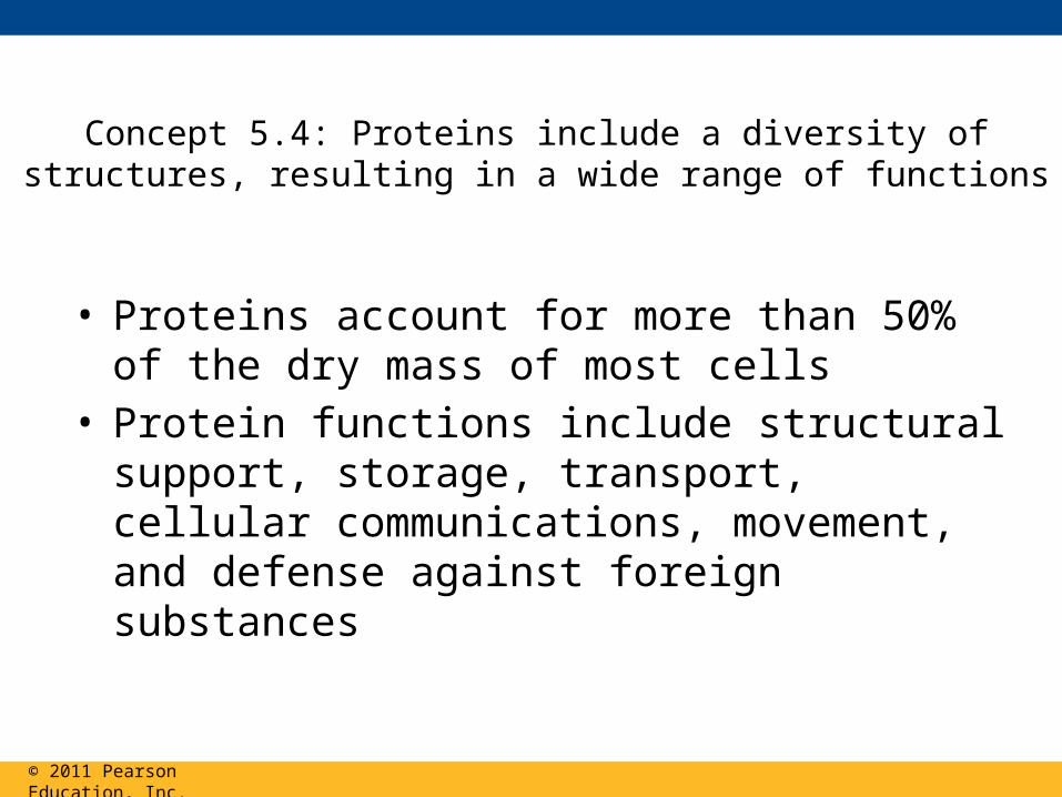
Concept 5.4: Proteins include a diversity of structures, resulting in a wide range of functions
• Proteins account for more than 50% of the dry mass of most cells
• Protein functions include structural support, storage, transport, cellular communications, movement, and defense against foreign substances
© 2011 Pearson Education, Inc.
Polypeptides
• Polypeptides are unbranched polymers built from the same set of 20 amino acids
• A protein is a biologically functional molecule that consists of one or more polypeptides
© 2011 Pearson Education, Inc.
Amino Acid Monomers
• Amino acids are organic molecules with carboxyl and amino groups
• Amino acids differ in their properties due to differing side chains, called R groups
© 2011 Pearson Education, Inc.
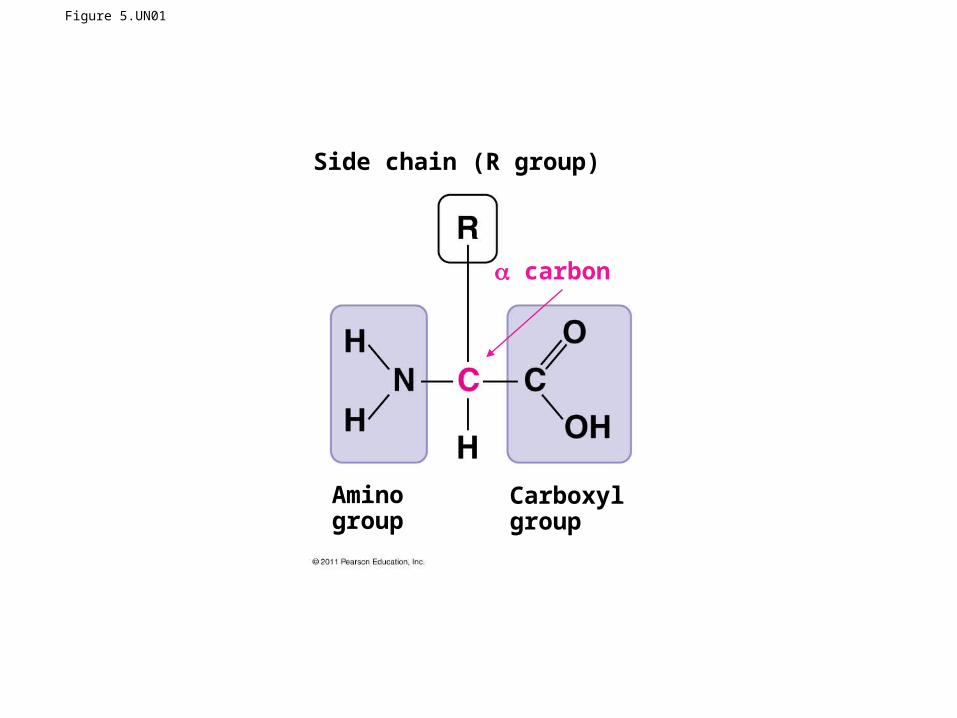
Figure 5.UN01
Side chain (R group)
Aminogroup
Carboxylgroup
carbon
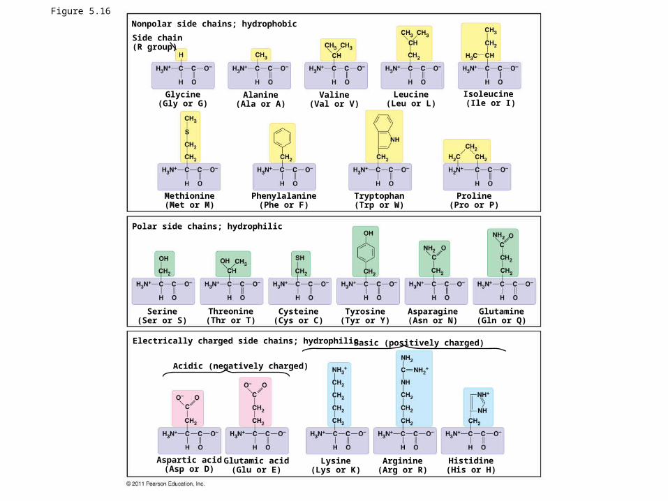
Figure 5.16Nonpolar side chains; hydrophobic
Side chain(R group)
Glycine(Gly or G)
Alanine(Ala or A)
Valine(Val or V)
Leucine(Leu or L)
Isoleucine (Ile or I)
Methionine(Met or M)
Phenylalanine(Phe or F)
Tryptophan(Trp or W)
Proline(Pro or P)
Polar side chains; hydrophilic
Serine(Ser or S)
Threonine(Thr or T)
Cysteine(Cys or C)
Tyrosine(Tyr or Y)
Asparagine(Asn or N)
Glutamine(Gln or Q)
Electrically charged side chains; hydrophilic
Acidic (negatively charged)
Basic (positively charged)
Aspartic acid(Asp or D)
Glutamic acid(Glu or E)
Lysine(Lys or K)
Arginine(Arg or R)
Histidine(His or H)
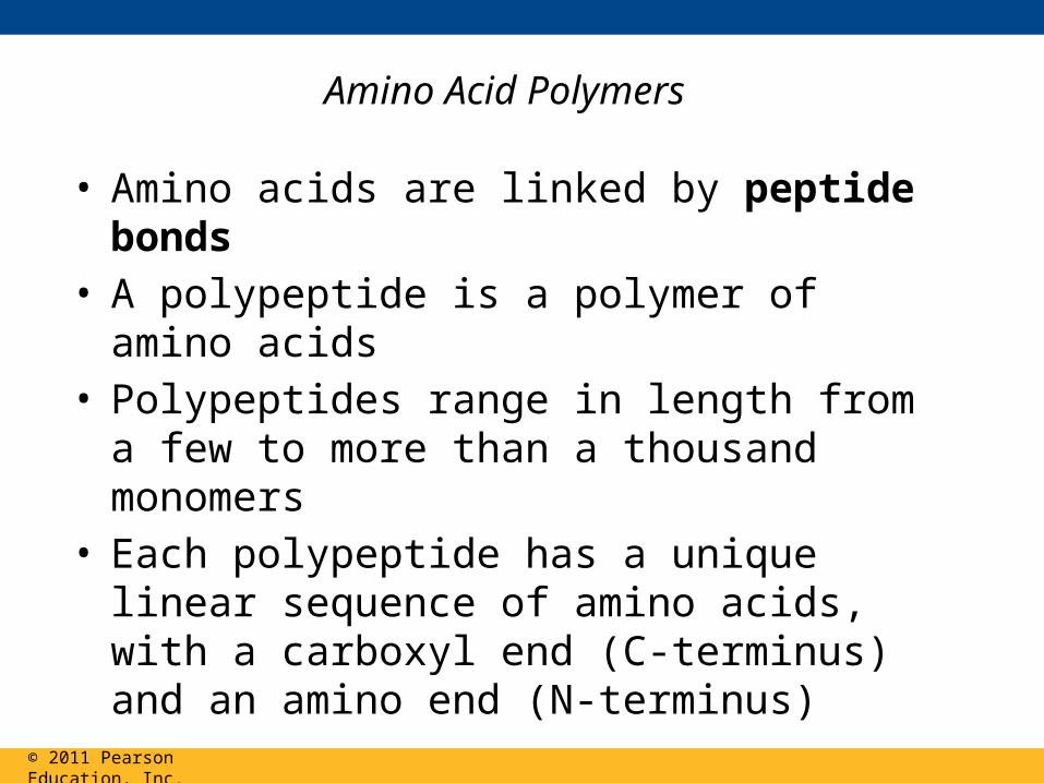
Amino Acid Polymers
• Amino acids are linked by peptide bonds• A polypeptide is a polymer of amino acids• Polypeptides range in length from a few to more
than a thousand monomers • Each polypeptide has a unique linear sequence of
amino acids, with a carboxyl end (C-terminus) and an amino end (N-terminus)
© 2011 Pearson Education, Inc.
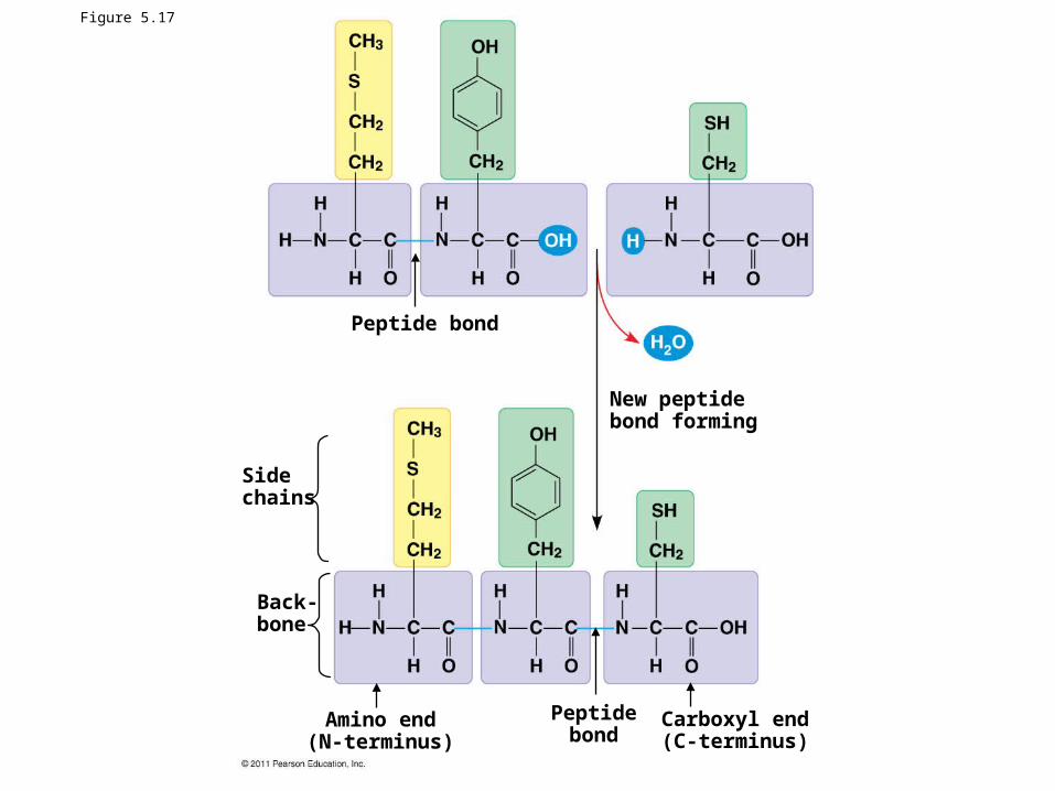
Figure 5.17
Peptide bond
New peptidebond forming
Sidechains
Back-bone
Amino end(N-terminus)
Peptidebond
Carboxyl end(C-terminus)
Protein Structure and Function
• A functional protein consists of one or more polypeptides precisely twisted, folded, and coiled into a unique shape
© 2011 Pearson Education, Inc.
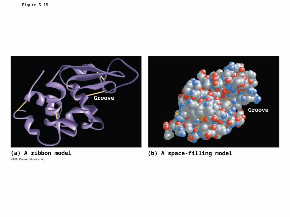
Figure 5.18
(a) A ribbon model (b) A space-filling model
Groove
Groove
• The sequence of amino acids determines a protein’s three-dimensional structure
• A protein’s structure determines its function
© 2011 Pearson Education, Inc.
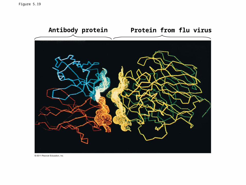
Figure 5.19
Antibody protein Protein from flu virus
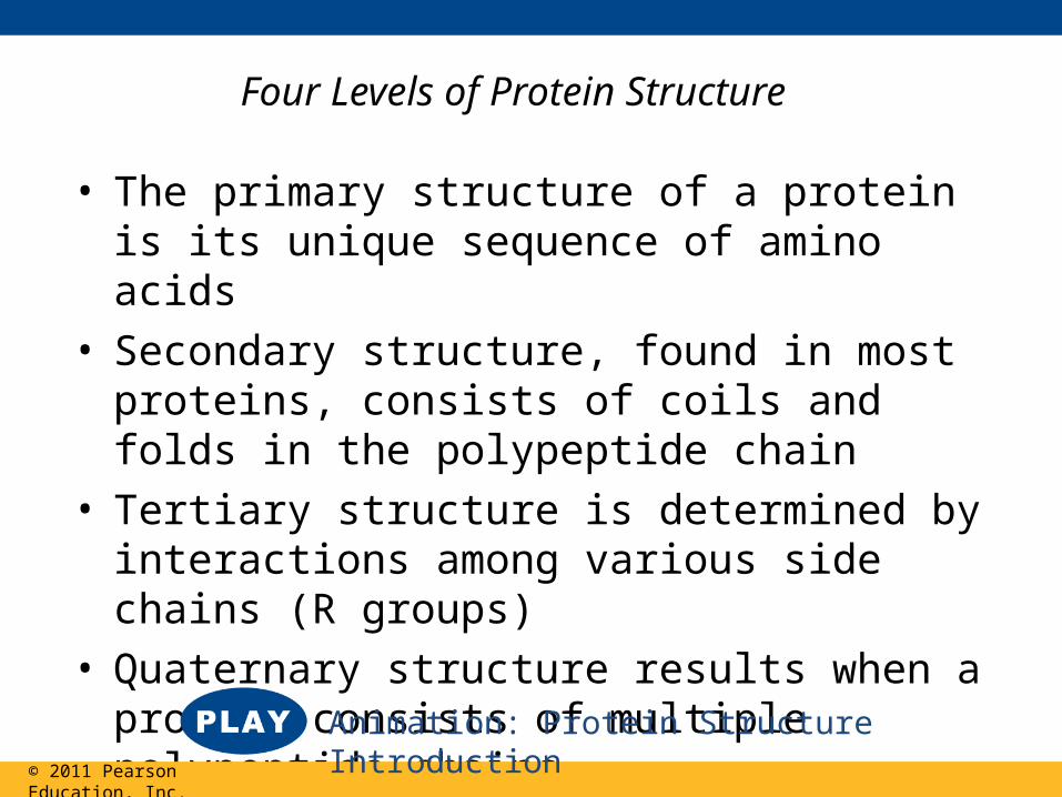
Four Levels of Protein Structure
• The primary structure of a protein is its unique sequence of amino acids
• Secondary structure, found in most proteins, consists of coils and folds in the polypeptide chain
• Tertiary structure is determined by interactions among various side chains (R groups)
• Quaternary structure results when a protein consists of multiple polypeptide chains
© 2011 Pearson Education, Inc.
Animation: Protein Structure Introduction
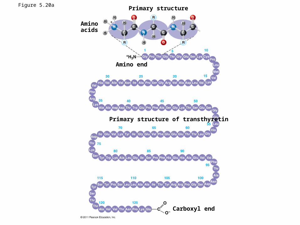
Figure 5.20a Primary structure
Aminoacids
Amino end
Carboxyl end
Primary structure of transthyretin
• Primary structure, the sequence of amino acids in a protein, is like the order of letters in a long word
• Primary structure is determined by inherited genetic information
© 2011 Pearson Education, Inc.
Animation: Primary Protein Structure
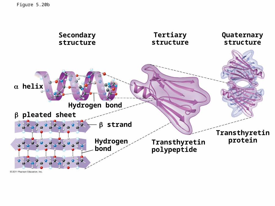
Figure 5.20b
Secondarystructure
Tertiarystructure
Quaternarystructure
Hydrogen bond
helix
pleated sheet strand
Hydrogenbond
Transthyretinpolypeptide
Transthyretinprotein
• The coils and folds of secondary structure result from hydrogen bonds between repeating constituents of the polypeptide backbone
• Typical secondary structures are a coil called an helix and a folded structure called a pleated sheet
© 2011 Pearson Education, Inc.
Animation: Secondary Protein Structure
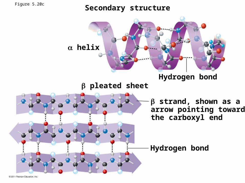
Secondary structure
Hydrogen bond
helix
pleated sheet
strand, shown as a flatarrow pointing towardthe carboxyl end
Hydrogen bond
Figure 5.20c
Figure 5.20d
• Tertiary structure is determined by interactions between R groups, rather than interactions between backbone constituents
• These interactions between R groups include hydrogen bonds, ionic bonds, hydrophobic interactions, and van der Waals interactions
• Strong covalent bonds called disulfide bridges may reinforce the protein’s structure
© 2011 Pearson Education, Inc.
Animation: Tertiary Protein Structure
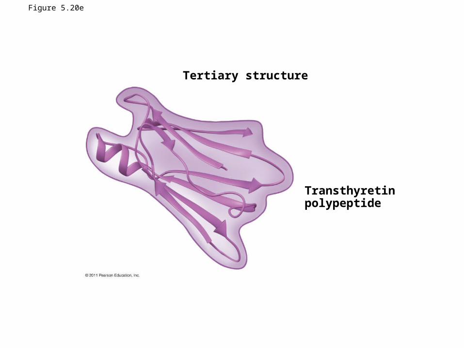
Figure 5.20e
Tertiary structure
Transthyretinpolypeptide
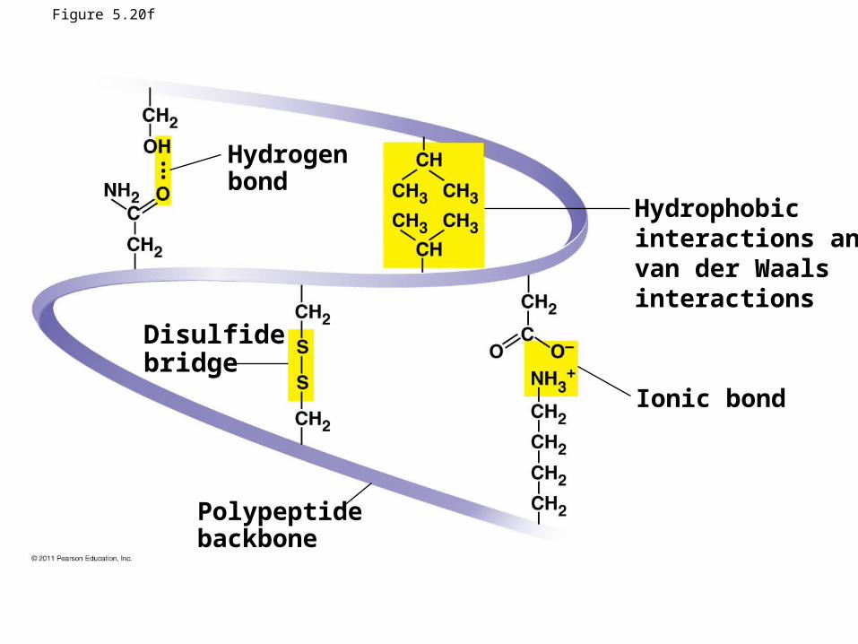
Figure 5.20f
Hydrogenbond
Disulfidebridge
Polypeptidebackbone
Ionic bond
Hydrophobicinteractions andvan der Waalsinteractions
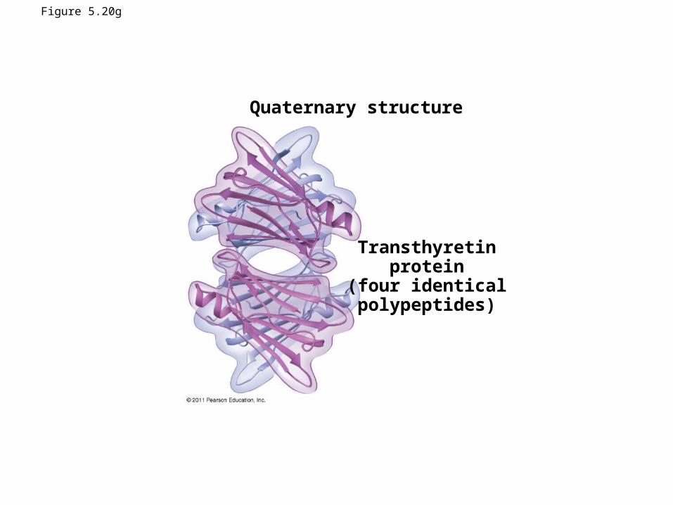
Figure 5.20g
Quaternary structure
Transthyretinprotein
(four identicalpolypeptides)
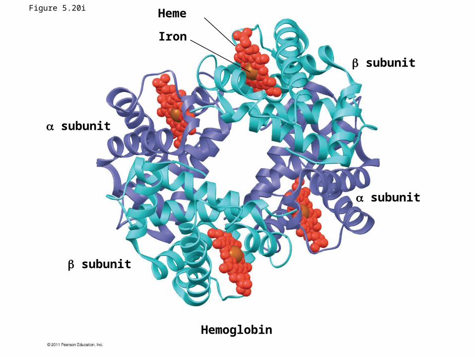
Hemoglobin
Heme
Iron
subunit
subunit
subunit
subunit
Figure 5.20i
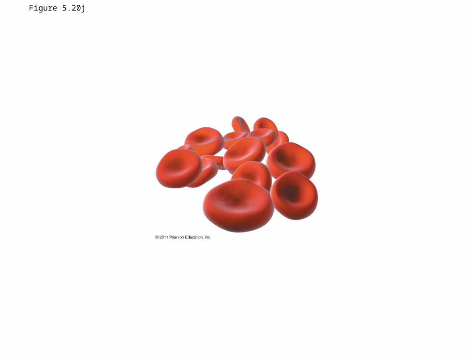
Figure 5.20j
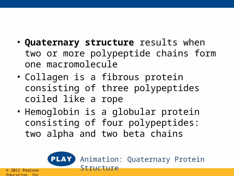
• Quaternary structure results when two or more polypeptide chains form one macromolecule
• Collagen is a fibrous protein consisting of three polypeptides coiled like a rope
• Hemoglobin is a globular protein consisting of four polypeptides: two alpha and two beta chains
© 2011 Pearson Education, Inc.
Animation: Quaternary Protein Structure
Sickle-Cell Disease: A Change in Primary Structure
• A slight change in primary structure can affect a protein’s structure and ability to function
• Sickle-cell disease, an inherited blood disorder, results from a single amino acid substitution in the protein hemoglobin
© 2011 Pearson Education, Inc.
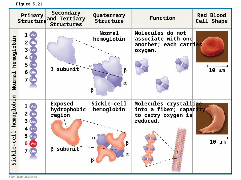
Figure 5.21
PrimaryStructure
Secondaryand TertiaryStructures
QuaternaryStructure Function Red Blood
Cell Shape
subunit
subunit
Exposedhydrophobicregion
Molecules do notassociate with oneanother; each carriesoxygen.
Molecules crystallizeinto a fiber; capacityto carry oxygen isreduced.
Sickle-cellhemoglobin
Normalhemoglobin
10 m
10 m
Sick
le-c
ell h
emog
lobi
nN
orm
al h
emog
lobi
n
1234567
1234567
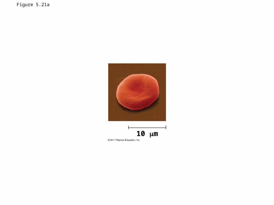
Figure 5.21a
10 m
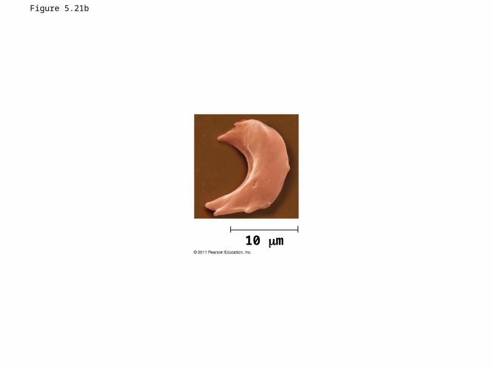
Figure 5.21b
10 m
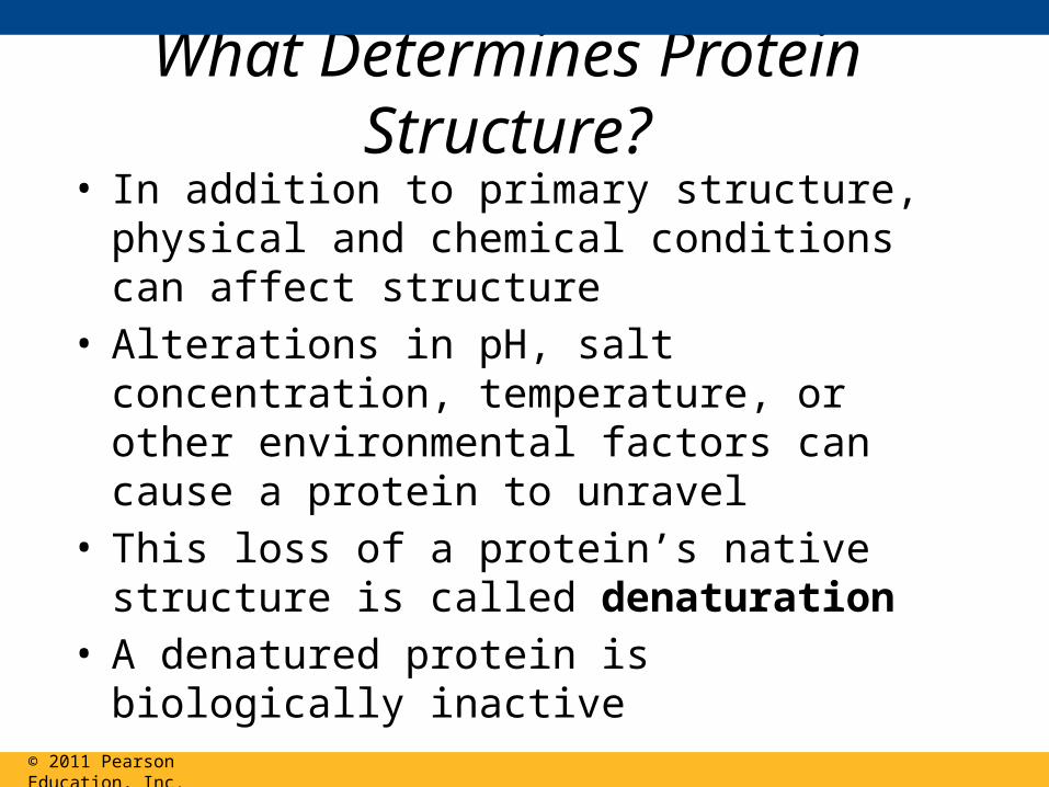
What Determines Protein Structure?• In addition to primary structure, physical and
chemical conditions can affect structure• Alterations in pH, salt concentration, temperature,
or other environmental factors can cause a protein to unravel
• This loss of a protein’s native structure is called denaturation
• A denatured protein is biologically inactive
© 2011 Pearson Education, Inc.
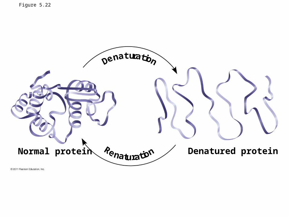
Figure 5.22
Normal protein Denatured protein
Den tur t on
Re n t r t on
a a i
a u a i
Protein Folding in the Cell• It is hard to predict a protein’s structure from its
primary structure• Most proteins probably go through several stages
on their way to a stable structure• Chaperonins are protein molecules that assist the
proper folding of other proteins• Diseases such as Alzheimer’s, Parkinson’s, and
mad cow disease are associated with misfolded proteins
© 2011 Pearson Education, Inc.