Embed Size (px)
Citation preview
© 2014 Pearson Education, Inc.
Concept 5.4: Proteins include a diversity of structures, resulting in a wide range of functions
Proteins account for more than 50% of the dry
mass of most cells
Some proteins speed up chemical reactions
Other protein functions include defense, storage,
transport, cellular communication, movement, or
structural support
© 2014 Pearson Education, Inc.
Figure 5.13a
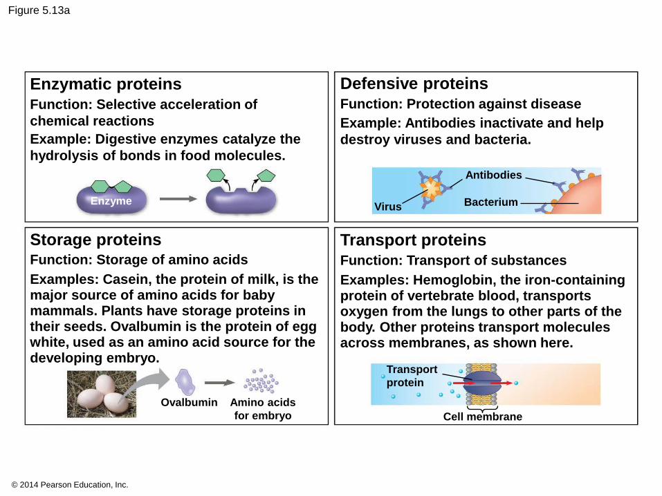
Enzymatic proteins
Function: Selective acceleration of
chemical reactions
Example: Digestive enzymes catalyze the
hydrolysis of bonds in food molecules.
Enzyme
Storage proteins Function: Storage of amino acids
Examples: Casein, the protein of milk, is the major source of amino acids for baby mammals. Plants have storage proteins in their seeds. Ovalbumin is the protein of egg white, used as an amino acid source for the developing embryo.
Ovalbumin Amino acids
for embryo
Defensive proteins
Function: Protection against disease
Example: Antibodies inactivate and help
destroy viruses and bacteria.
Virus
Antibodies
Bacterium
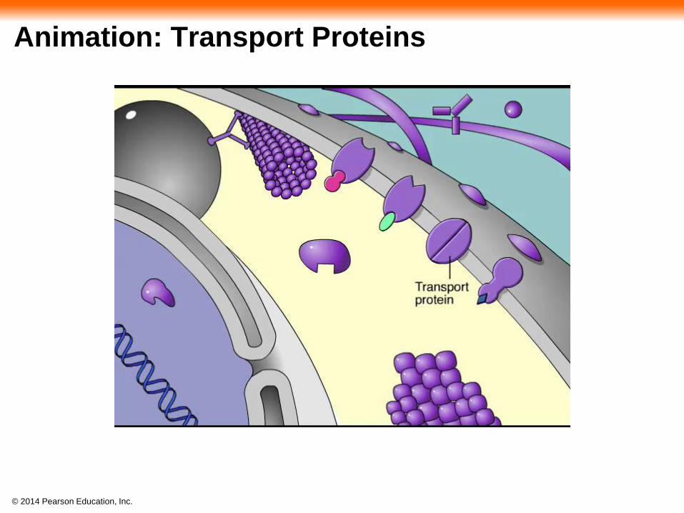
Transport proteins
Function: Transport of substances
Examples: Hemoglobin, the iron-containing protein of vertebrate blood, transports oxygen from the lungs to other parts of the body. Other proteins transport molecules across membranes, as shown here.
Transport
protein
Cell membrane
© 2014 Pearson Education, Inc.
Figure 5.13ac
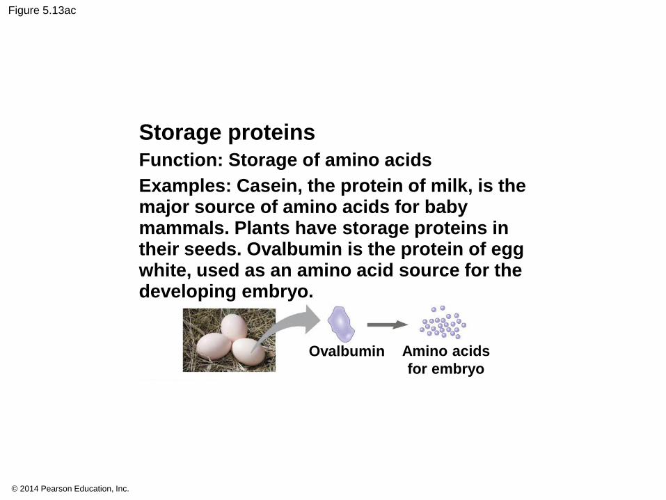
Storage proteins
Function: Storage of amino acids
Examples: Casein, the protein of milk, is the major source of amino acids for baby mammals. Plants have storage proteins in their seeds. Ovalbumin is the protein of egg white, used as an amino acid source for the developing embryo.
Ovalbumin Amino acids
for embryo
© 2014 Pearson Education, Inc.
Figure 5.13b
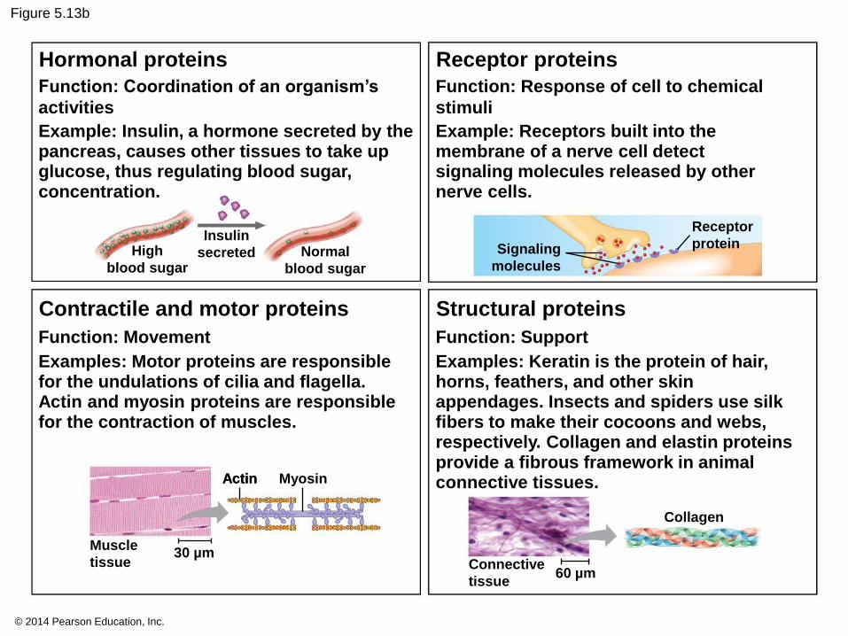
Function: Coordination of an organism’s
activities
Example: Insulin, a hormone secreted by the pancreas, causes other tissues to take up glucose, thus regulating blood sugar, concentration.
High
blood sugar
Insulin
secreted
Hormonal proteins
Normal
blood sugar
Function: Response of cell to chemical
stimuli
Example: Receptors built into the membrane of a nerve cell detect signaling molecules released by other nerve cells.
Receptor proteins
Signaling
molecules
Receptor
protein
Function: Movement
Examples: Motor proteins are responsible for the undulations of cilia and flagella. Actin and myosin proteins are responsible for the contraction of muscles.
Contractile and motor proteins
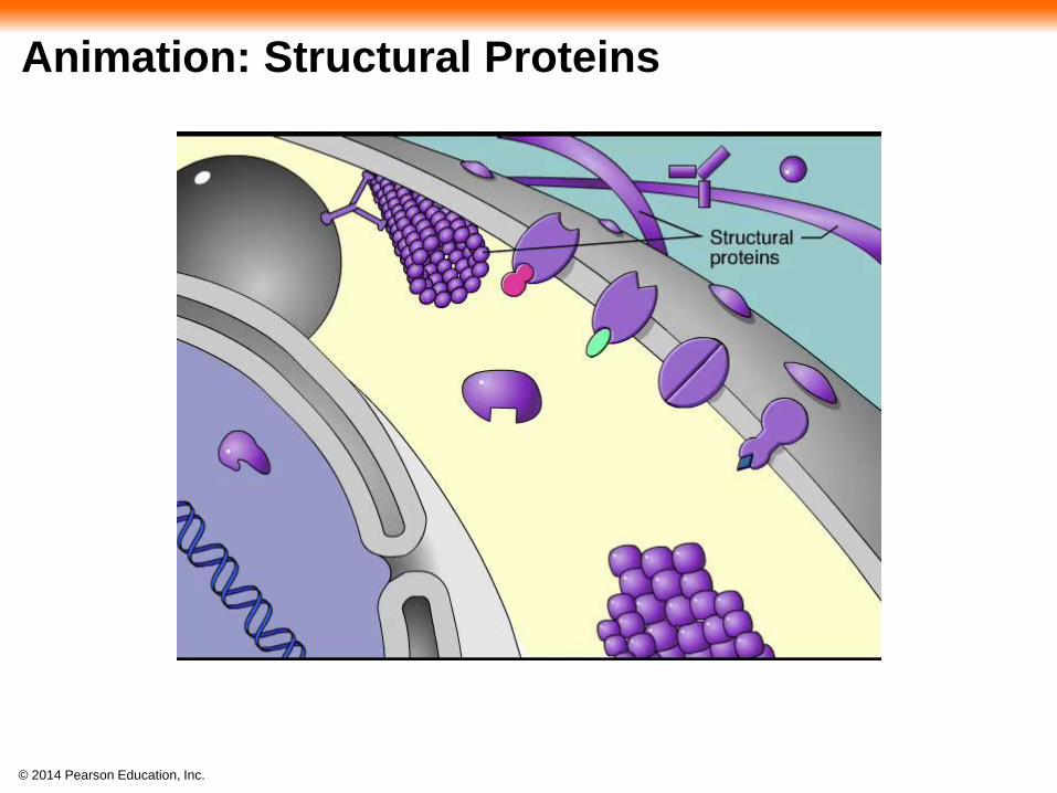
Function: Support
Examples: Keratin is the protein of hair, horns, feathers, and other skin appendages. Insects and spiders use silk fibers to make their cocoons and webs, respectively. Collagen and elastin proteins provide a fibrous framework in animal connective tissues.
Structural proteins
Muscle
tissue 30 µm
Actin Myosin Actin
60 µm Connective
tissue
Collagen

© 2014 Pearson Education, Inc.
Animation: Contractile Proteins

© 2014 Pearson Education, Inc.
Animation: Defensive Proteins
© 2014 Pearson Education, Inc.
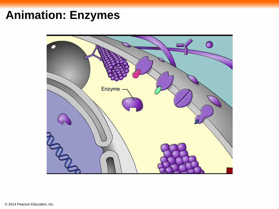
Animation: Enzymes
© 2014 Pearson Education, Inc.
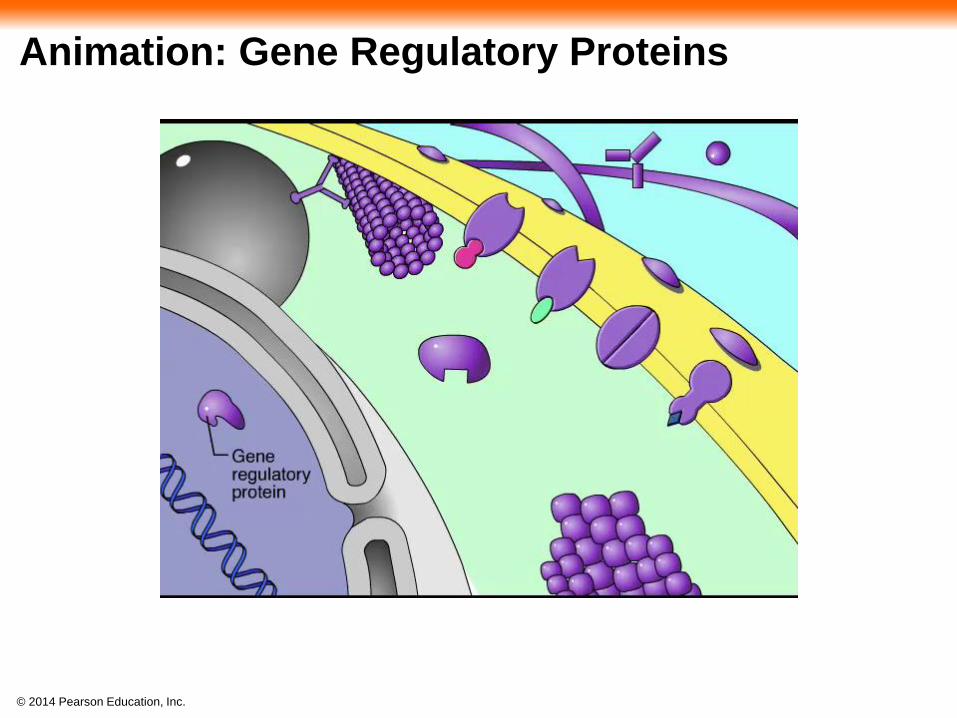
Animation: Gene Regulatory Proteins

© 2014 Pearson Education, Inc.
Animation: Hormonal Proteins

© 2014 Pearson Education, Inc.
Animation: Receptor Proteins

© 2014 Pearson Education, Inc.
Animation: Sensory Proteins

© 2014 Pearson Education, Inc.
Animation: Storage Proteins
© 2014 Pearson Education, Inc.
Animation: Structural Proteins
© 2014 Pearson Education, Inc.
Animation: Transport Proteins
© 2014 Pearson Education, Inc.
Enzymes are proteins that act as catalysts to
speed up chemical reactions
Enzymes can perform their functions repeatedly,
functioning as workhorses that carry out the
processes of life
© 2014 Pearson Education, Inc.
Proteins are all constructed from the same set of
20 amino acids
Polypeptides are unbranched polymers built from
these amino acids
A protein is a biologically functional molecule that
consists of one or more polypeptides

© 2014 Pearson Education, Inc.
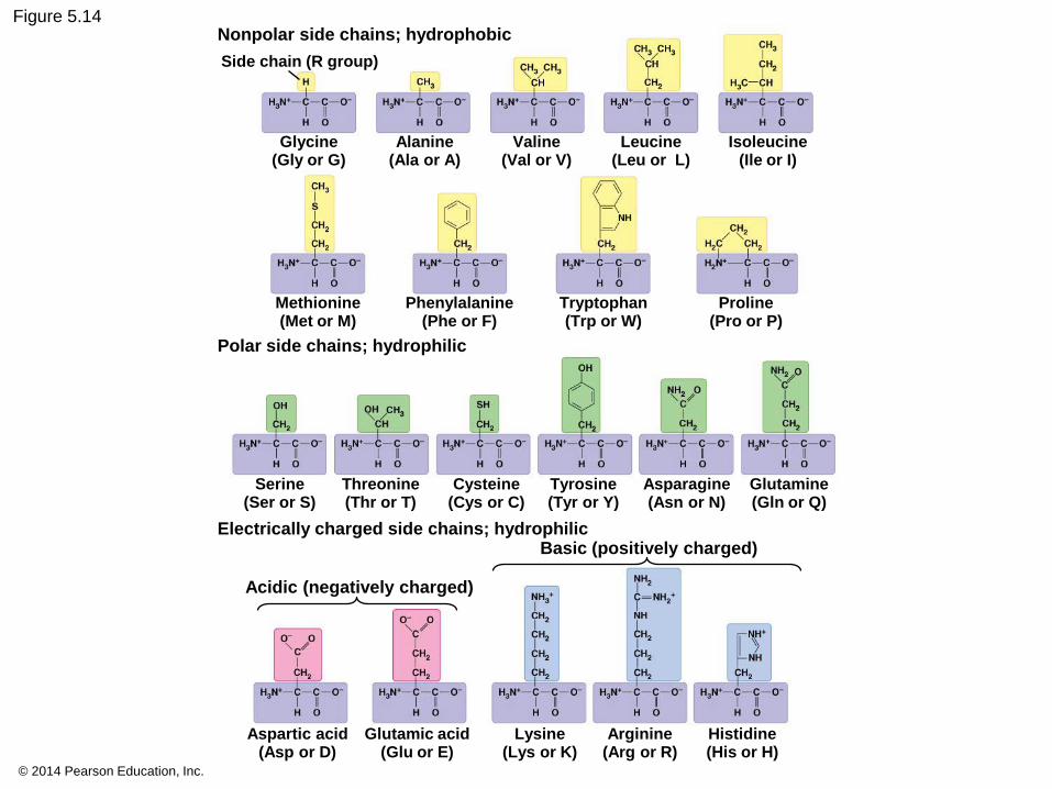
Amino Acid Monomers
Amino acids are organic molecules with amino
and carboxyl groups
Amino acids differ in their properties due to
differing side chains, called R groups

© 2014 Pearson Education, Inc.
Figure 5.UN01
Side chain (R group)
Amino group
Carboxyl group
𝛂 carbon
© 2014 Pearson Education, Inc.
Figure 5.14 Nonpolar side chains; hydrophobic
Side chain (R group)
Glycine (Gly or G)
Alanine (Ala or A)
Valine (Val or V)
Leucine (Leu or L)
Isoleucine (Ile or I)
Proline (Pro or P)
Tryptophan (Trp or W)
Phenylalanine (Phe or F)
Methionine (Met or M)
Polar side chains; hydrophilic
Electrically charged side chains; hydrophilic
Aspartic acid (Asp or D)
Glutamic acid (Glu or E)
Lysine (Lys or K)
Arginine (Arg or R)
Histidine (His or H)
Glutamine (Gln or Q)
Acidic (negatively charged)
Basic (positively charged)
Asparagine (Asn or N)
Tyrosine (Tyr or Y)
Cysteine (Cys or C)
Threonine (Thr or T)
Serine (Ser or S)

© 2014 Pearson Education, Inc.
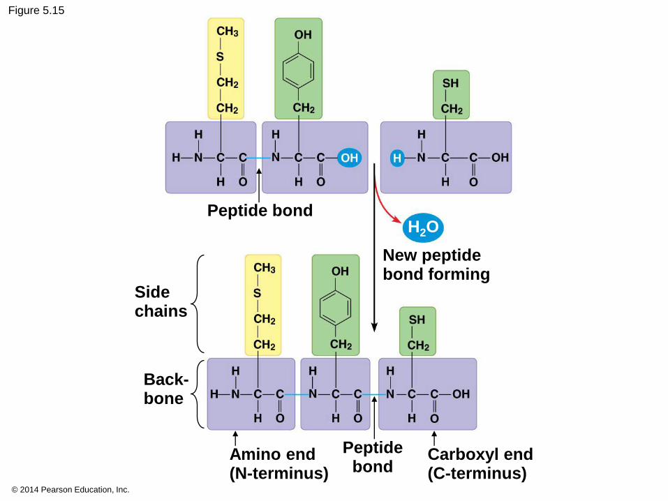
Polypeptides (Amino Acid Polymers)
Amino acids are linked by covalent bonds called
peptide bonds
A polypeptide is a polymer of amino acids
Polypeptides range in length from a few to more
than a thousand monomers
Each polypeptide has a unique linear sequence of
amino acids, with a carboxyl end (C-terminus) and
an amino end (N-terminus)
© 2014 Pearson Education, Inc.
Figure 5.15
Peptide bond
New peptide bond forming
H2O
Side chains
Back- bone
Amino end (N-terminus)
Peptide bond
Carboxyl end (C-terminus)
© 2014 Pearson Education, Inc.
Protein Structure and Function
The specific activities of proteins result from their
intricate three-dimensional architecture
A functional protein consists of one or more
polypeptides precisely twisted, folded, and coiled
into a unique shape

© 2014 Pearson Education, Inc.
Figure 5.16
(a) A ribbon model (b) A space-filling model (c) A wireframe model
Groove Groove
Target molecule
© 2014 Pearson Education, Inc.
Animation: Protein Structure Introduction

© 2014 Pearson Education, Inc.
The sequence of amino acids determines a
protein’s three-dimensional structure
A protein’s structure determines how it works
The function of a protein usually depends on
its ability to recognize and bind to some other
molecule

© 2014 Pearson Education, Inc.
Figure 5.17
Antibody protein Protein from flu virus

© 2014 Pearson Education, Inc.
Four Levels of Protein Structure
The primary structure of a protein is its unique
sequence of amino acids
Secondary structure, found in most proteins,
consists of coils and folds in the polypeptide chain
Tertiary structure is determined by interactions
among various side chains (R groups)
Quaternary structure results when a protein
consists of multiple polypeptide chains
© 2014 Pearson Education, Inc.
Sickle-Cell Disease: A Change in Primary Structure
A slight change in primary structure can affect a
protein’s structure and ability to function
Sickle-cell disease, an inherited blood disorder,
results from a single amino acid substitution in the
protein hemoglobin
© 2014 Pearson Education, Inc.
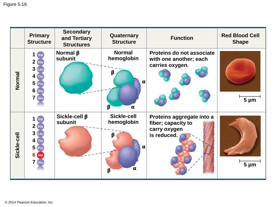
Figure 5.19
Primary
Structure
Secondary
and Tertiary
Structures
Quaternary
Structure Function
Red Blood Cell
Shape
5 µm
Proteins do not associate with one another; each carries oxygen.
Proteins aggregate into a fiber; capacity to carry oxygen is reduced.
5 µm
Normal hemoglobin
Normal 𝛃 subunit
𝛃
𝛃 𝛂
𝛂
𝛂
𝛂
𝛃
𝛃
Sickle-cell hemoglobin
Sickle-cell 𝛃 subunit
Sic
kle
-cell
No
rma
l
1
2
3
4
5
6
7
1
2
3
4
5
6
7
© 2014 Pearson Education, Inc.
Secondary and Tertiary Structures
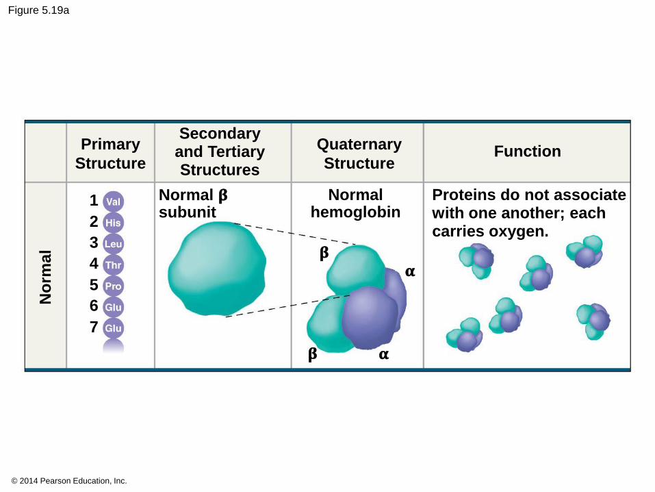
Figure 5.19a
Primary
Structure
Quaternary
Structure Function
Proteins do not associate with one another; each carries oxygen.
Normal hemoglobin
Normal 𝛃 subunit
𝛃
𝛂
No
rma
l
1
2
3
4
5
6
7
𝛃
𝛂
© 2014 Pearson Education, Inc.
What Determines Protein Structure?
In addition to primary structure, physical and
chemical conditions can affect structure
Alterations in pH, salt concentration,
temperature, or other environmental factors
can cause a protein to unravel
This loss of a protein’s native structure is
called denaturation
A denatured protein is biologically inactive
© 2014 Pearson Education, Inc.
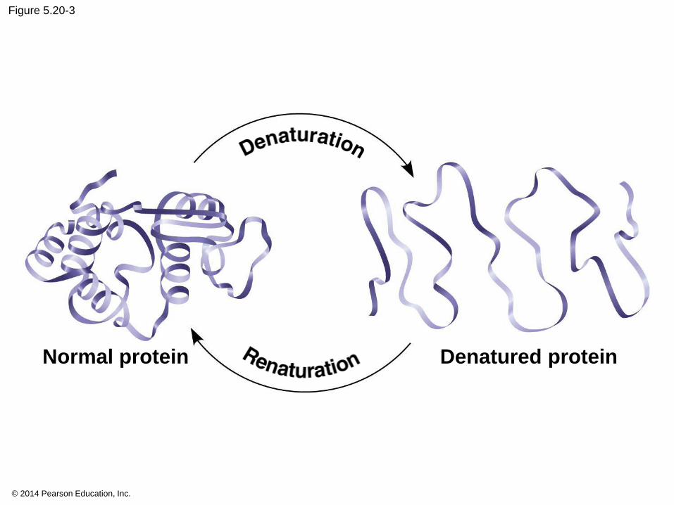
Figure 5.20-3
Normal protein Denatured protein

© 2014 Pearson Education, Inc.
Protein Folding in the Cell
It is hard to predict a protein’s structure from its
primary structure
Most proteins probably go through several stages
on their way to a stable structure
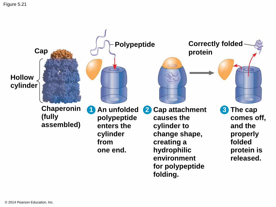
Chaperonins are protein molecules that assist
the proper folding of other proteins
Diseases such as Alzheimer’s, Parkinson’s,
and mad cow disease are associated with
misfolded proteins
© 2014 Pearson Education, Inc.
Figure 5.21
Hollow cylinder
Cap
Chaperonin (fully assembled)
Polypeptide
1 An unfolded polypeptide enters the cylinder from one end.
2 Cap attachment causes the cylinder to change shape, creating a hydrophilic environment for polypeptide folding.
3 The cap comes off, and the properly folded protein is released.
Correctly folded
protein
© 2014 Pearson Education, Inc.
Concept 5.5: Nucleic acids store, transmit, and help express hereditary information
The amino acid sequence of a polypeptide is
programmed by a unit of inheritance called
a gene
Genes consist of DNA, a nucleic acid made of
monomers called nucleotides

© 2014 Pearson Education, Inc.
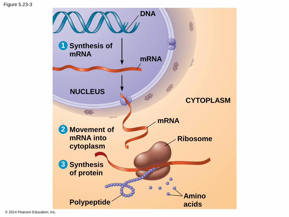
The Roles of Nucleic Acids
There are two types of nucleic acids
Deoxyribonucleic acid (DNA)
Ribonucleic acid (RNA)
DNA provides directions for its own replication
DNA directs synthesis of messenger RNA (mRNA)
and, through mRNA, controls protein synthesis
This process is called gene expression
© 2014 Pearson Education, Inc.
Figure 5.23-3
DNA
1
mRNA
Synthesis of mRNA
2 Movement of mRNA into cytoplasm
3 Synthesis of protein
NUCLEUS
CYTOPLASM
mRNA
Ribosome
Polypeptide Amino acids
© 2014 Pearson Education, Inc.
Each gene along a DNA molecule directs
synthesis of a messenger RNA (mRNA)
The mRNA molecule interacts with the cell’s
protein-synthesizing machinery to direct
production of a polypeptide
The flow of genetic information can be
summarized as DNA → RNA → protein

© 2014 Pearson Education, Inc.
The Components of Nucleic Acids
Nucleic acids are polymers called
polynucleotides
Each polynucleotide is made of monomers called
nucleotides
Each nucleotide consists of a nitrogenous base, a
pentose sugar, and one or more phosphate
groups
The portion of a nucleotide without the phosphate
group is called a nucleoside
© 2014 Pearson Education, Inc.
Animation: DNA and RNA Structure
© 2014 Pearson Education, Inc.
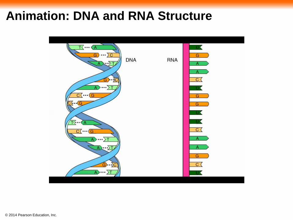
The Structures of DNA and RNA Molecules
DNA molecules have two polynucleotides spiraling
around an imaginary axis, forming a double helix
The backbones run in opposite 5 → 3 directions
from each other, an arrangement referred to as
antiparallel
One DNA molecule includes many genes
© 2014 Pearson Education, Inc.
Only certain bases in DNA pair up and form
hydrogen bonds: adenine (A) always with thymine
(T), and guanine (G) always with cytosine (C)
This is called complementary base pairing
This feature of DNA structure makes it possible
to generate two identical copies of each DNA
molecule in a cell preparing to divide

© 2014 Pearson Education, Inc.
RNA, in contrast to DNA, is single stranded
Complementary pairing can also occur between
two RNA molecules or between parts of the
same molecule
In RNA, thymine is replaced by uracil (U) so
A and U pair
While DNA always exists as a double helix,
RNA molecules are more variable in form
© 2014 Pearson Education, Inc.
Animation: DNA Double Helix