Embed Size (px)
Citation preview
1
13-1
Chapter 14
The Cardiovascular System: The Heart
• Heart pumps over
2.6 million gallons
per year
• Over 60,000 miles
of blood vessels
13-2
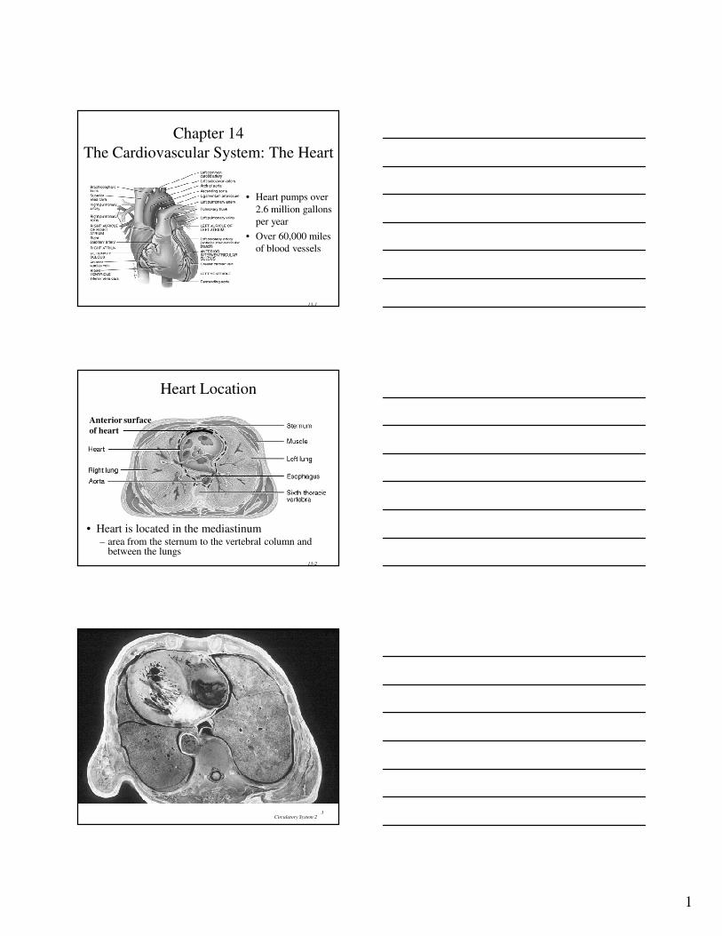
Heart Location
• Heart is located in the mediastinum– area from the sternum to the vertebral column and
between the lungs
Anterior surface
of heart
Circulatory System 23
2
13-4
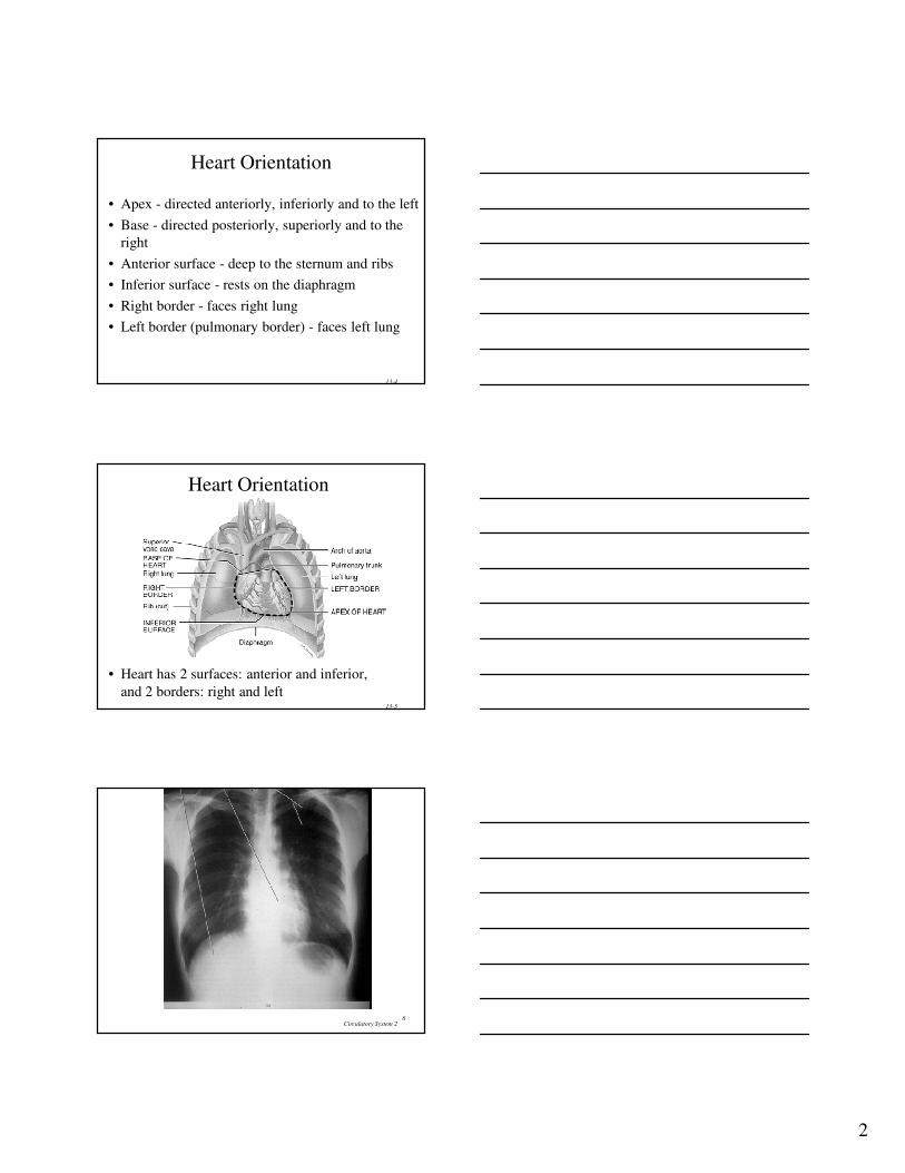
Heart Orientation
• Apex - directed anteriorly, inferiorly and to the left
• Base - directed posteriorly, superiorly and to the
right
• Anterior surface - deep to the sternum and ribs
• Inferior surface - rests on the diaphragm
• Right border - faces right lung
• Left border (pulmonary border) - faces left lung
13-5
Heart Orientation
• Heart has 2 surfaces: anterior and inferior,
and 2 borders: right and left
Circulatory System 26
3
13-7
Surface Projection of the Heart
• Superior right point at the superior border of the 3rd right costal cartilage
• Superior left point at the inferior border of the 2nd left costal cartilage 3cm to the left of midline
• Inferior left point at the 5th intercostal space, 9 cm from the midline
• Inferior right point at superior border of the 6th right costal cartilage, 3 cm from the midline
13-8
Pericardium
• Fibrous pericardium
– dense irregular CT
– protects and anchors the heart, prevents overstretching
• Serous pericardium
– thin delicate membrane
– contains
• parietal layer-outer layer
• pericardial cavity with pericardial fluid
• visceral layer (epicardium)
13-9
Layers of Heart Wall
• Epicardium
– visceral layer of
serous pericardium
• Myocardium
– cardiac muscle layer is the bulk of the heart
• Endocardium
– chamber lining &
valves
4
Circulatory System 210
13-11
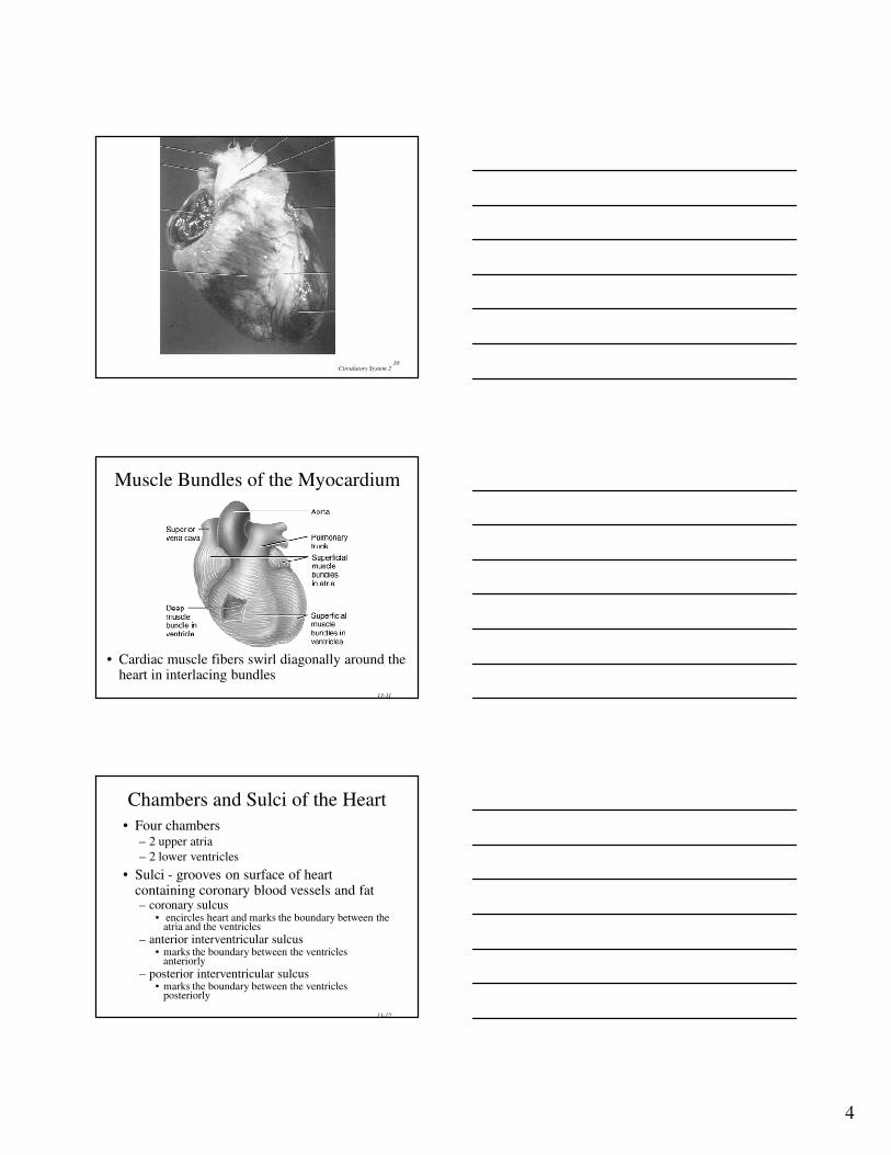
Muscle Bundles of the Myocardium
• Cardiac muscle fibers swirl diagonally around the heart in interlacing bundles
13-12
Chambers and Sulci of the Heart
• Four chambers – 2 upper atria
– 2 lower ventricles
• Sulci - grooves on surface of heart containing coronary blood vessels and fat– coronary sulcus
• encircles heart and marks the boundary between the atria and the ventricles
– anterior interventricular sulcus • marks the boundary between the ventricles
anteriorly
– posterior interventricular sulcus • marks the boundary between the ventricles
posteriorly
5
13-13
Chambers and Sulci
Anterior View
13-14
Posterior View
Chambers and Sulci
13-15
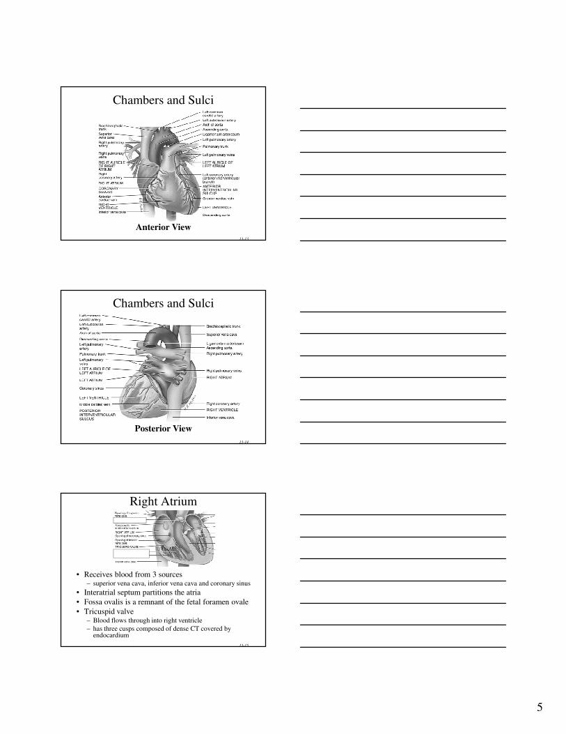
Right Atrium
• Receives blood from 3 sources– superior vena cava, inferior vena cava and coronary sinus
• Interatrial septum partitions the atria
• Fossa ovalis is a remnant of the fetal foramen ovale
• Tricuspid valve– Blood flows through into right ventricle
– has three cusps composed of dense CT covered by endocardium
6
13-16
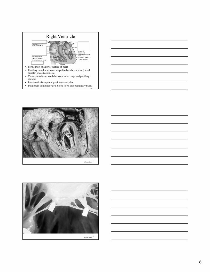
Right Ventricle
• Forms most of anterior surface of heart
• Papillary muscles are cone shaped trabeculae carneae (raised bundles of cardiac muscle)
• Chordae tendineae: cords between valve cusps and papillary muscles
• Interventricular septum: partitions ventricles
• Pulmonary semilunar valve: blood flows into pulmonary trunk
Circulation 617
Circulation 618
7
13-19
Left Atrium
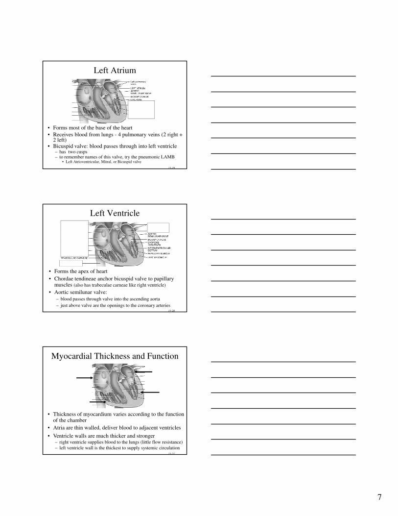
• Forms most of the base of the heart
• Receives blood from lungs - 4 pulmonary veins (2 right + 2 left)
• Bicuspid valve: blood passes through into left ventricle– has two cusps– to remember names of this valve, try the pneumonic LAMB
• Left Atrioventricular, Mitral, or Bicuspid valve
13-20
Left Ventricle
• Forms the apex of heart
• Chordae tendineae anchor bicuspid valve to papillary muscles (also has trabeculae carneae like right ventricle)
• Aortic semilunar valve:
– blood passes through valve into the ascending aorta
– just above valve are the openings to the coronary arteries
13-21
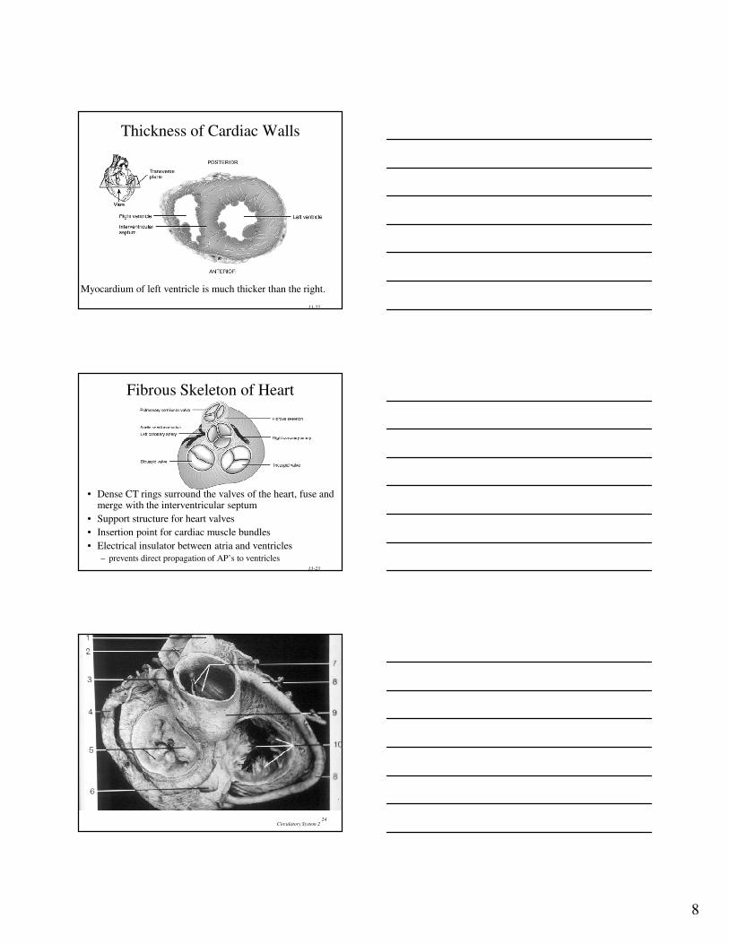
Myocardial Thickness and Function
• Thickness of myocardium varies according to the function of the chamber
• Atria are thin walled, deliver blood to adjacent ventricles
• Ventricle walls are much thicker and stronger– right ventricle supplies blood to the lungs (little flow resistance)
– left ventricle wall is the thickest to supply systemic circulation
8
13-22
Thickness of Cardiac Walls
Myocardium of left ventricle is much thicker than the right.
13-23
Fibrous Skeleton of Heart
• Dense CT rings surround the valves of the heart, fuse and merge with the interventricular septum
• Support structure for heart valves
• Insertion point for cardiac muscle bundles
• Electrical insulator between atria and ventricles
– prevents direct propagation of AP’s to ventricles
Circulatory System 224
9
13-25
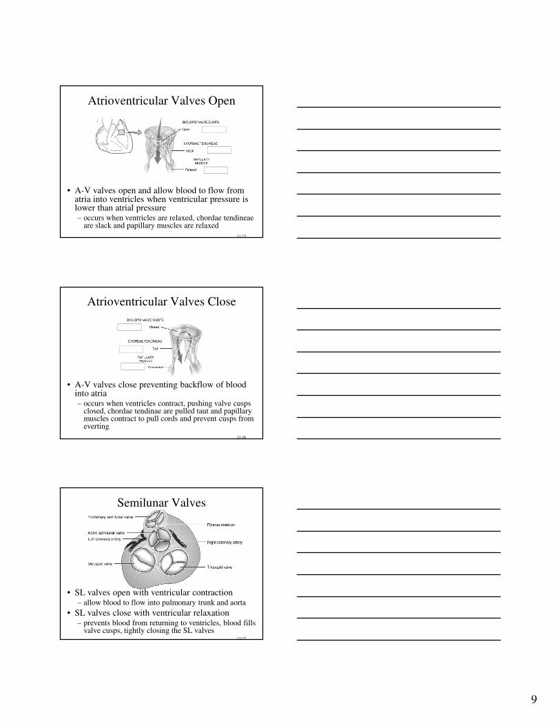
• A-V valves open and allow blood to flow from atria into ventricles when ventricular pressure is lower than atrial pressure– occurs when ventricles are relaxed, chordae tendineae
are slack and papillary muscles are relaxed
Atrioventricular Valves Open
13-26
• A-V valves close preventing backflow of blood into atria – occurs when ventricles contract, pushing valve cusps
closed, chordae tendinae are pulled taut and papillary muscles contract to pull cords and prevent cusps from everting
Atrioventricular Valves Close
13-27
Semilunar Valves
• SL valves open with ventricular contraction– allow blood to flow into pulmonary trunk and aorta
• SL valves close with ventricular relaxation– prevents blood from returning to ventricles, blood fills
valve cusps, tightly closing the SL valves
10
13-28
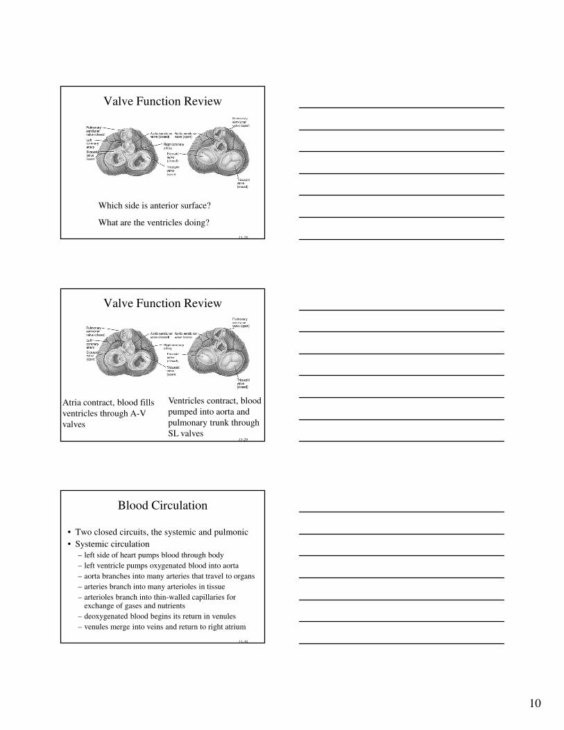
Which side is anterior surface?
What are the ventricles doing?
Valve Function Review
13-29
Atria contract, blood fills
ventricles through A-V
valves
Ventricles contract, blood
pumped into aorta and
pulmonary trunk through
SL valves
Valve Function Review
13-30
• Two closed circuits, the systemic and pulmonic
• Systemic circulation
– left side of heart pumps blood through body
– left ventricle pumps oxygenated blood into aorta
– aorta branches into many arteries that travel to organs
– arteries branch into many arterioles in tissue
– arterioles branch into thin-walled capillaries for exchange of gases and nutrients
– deoxygenated blood begins its return in venules
– venules merge into veins and return to right atrium
Blood Circulation
11
13-31
• Pulmonary circulation
– right side of heart pumps deoxygenated blood to lungs
– right ventricle pumps blood to pulmonary trunk
– pulmonary trunk branches into pulmonary arteries
– pulmonary arteries carry blood to lungs for exchange
of gases
– oxygenated blood returns to heart in pulmonary veins
Blood Circulation (cont.)
13-32
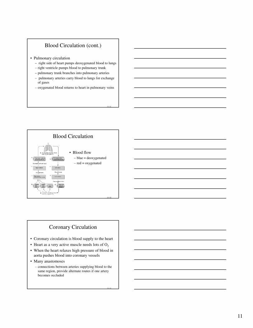
Blood Circulation
• Blood flow
– blue = deoxygenated
– red = oxygenated
13-33
Coronary Circulation
• Coronary circulation is blood supply to the heart
• Heart as a very active muscle needs lots of O2
• When the heart relaxes high pressure of blood in
aorta pushes blood into coronary vessels
• Many anastomoses
– connections between arteries supplying blood to the
same region, provide alternate routes if one artery
becomes occluded
12
13-34
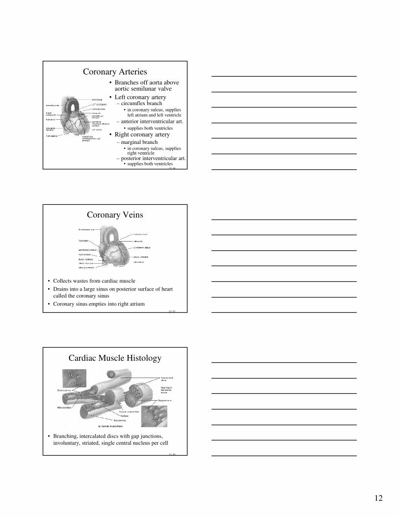
Coronary Arteries
• Branches off aorta above aortic semilunar valve
• Left coronary artery– circumflex branch
• in coronary sulcus, supplies left atrium and left ventricle
– anterior interventricular art.
• supplies both ventricles
• Right coronary artery
– marginal branch• in coronary sulcus, supplies
right ventricle– posterior interventricular art.
• supplies both ventricles
13-35
Coronary Veins
• Collects wastes from cardiac muscle
• Drains into a large sinus on posterior surface of heart
called the coronary sinus
• Coronary sinus empties into right atrium
13-36
Cardiac Muscle Histology
• Branching, intercalated discs with gap junctions,
involuntary, striated, single central nucleus per cell
13
13-37
Cardiac Myofibril
13-38
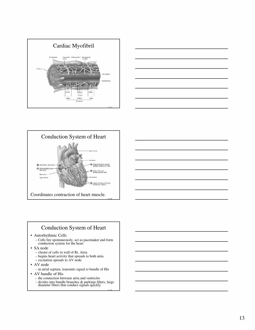
Conduction System of Heart
Coordinates contraction of heart muscle.
13-39
• Autorhythmic Cells– Cells fire spontaneously, act as pacemaker and form
conduction system for the heart
• SA node– cluster of cells in wall of Rt. Atria– begins heart activity that spreads to both atria– excitation spreads to AV node
• AV node– in atrial septum, transmits signal to bundle of His
• AV bundle of His – the connection between atria and ventricles– divides into bundle branches & purkinje fibers, large
diameter fibers that conduct signals quickly
Conduction System of Heart
14
13-40
Rhythm of Conduction System
• SA node fires spontaneously 90-100 times per
minute
• AV node fires at 40-50 times per minute
• If both nodes are suppressed fibers in ventricles by
themselves fire only 20-40 times per minute
• Artificial pacemaker needed if pace is too slow
• Extra beats forming at other sites are called
ectopic pacemakers
– caffeine & nicotine increase activity
13-41
Timing of Atrial &
Ventricular Excitation
• SA node setting pace since is the fastest
• In 50 msec excitation spreads through both atria
and down to AV node
• 100 msec delay at AV node due to smaller
diameter fibers- allows atria to fully contract filling
ventricles before ventricles contract
• In 50 msec excitation spreads through both
ventricles simultaneously
13-42
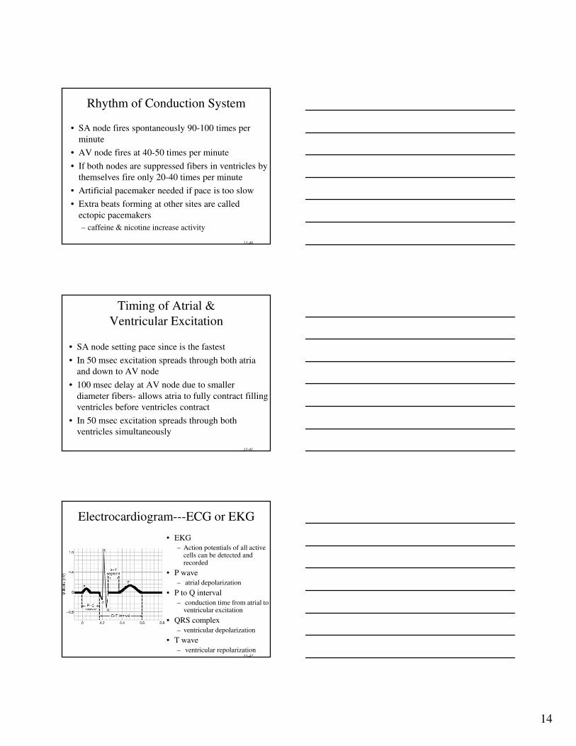
Electrocardiogram---ECG or EKG
• EKG
– Action potentials of all active cells can be detected and recorded
• P wave
– atrial depolarization
• P to Q interval
– conduction time from atrial to ventricular excitation
• QRS complex
– ventricular depolarization
• T wave
– ventricular repolarization
15
13-43
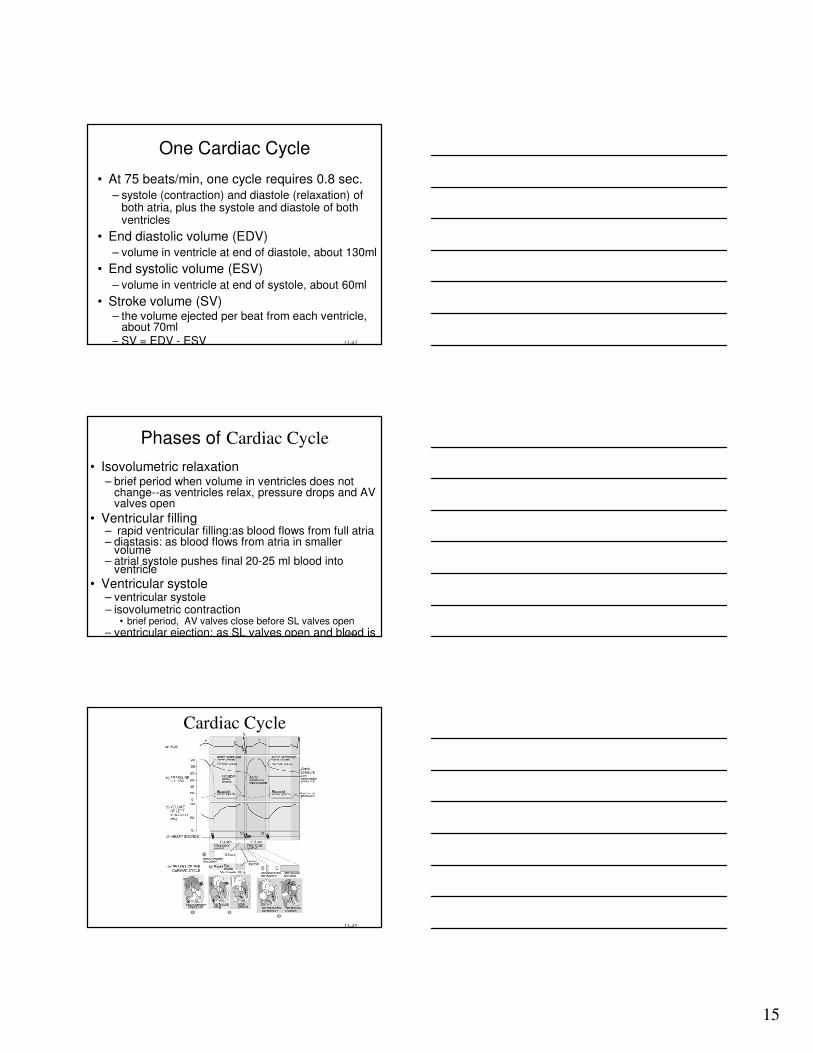
One Cardiac Cycle
• At 75 beats/min, one cycle requires 0.8 sec.– systole (contraction) and diastole (relaxation) of
both atria, plus the systole and diastole of both ventricles
• End diastolic volume (EDV)– volume in ventricle at end of diastole, about 130ml
• End systolic volume (ESV)– volume in ventricle at end of systole, about 60ml
• Stroke volume (SV)– the volume ejected per beat from each ventricle,
about 70ml– SV = EDV - ESV
13-44
Phases of Cardiac Cycle
• Isovolumetric relaxation– brief period when volume in ventricles does not
change--as ventricles relax, pressure drops and AV valves open
• Ventricular filling– rapid ventricular filling:as blood flows from full atria– diastasis: as blood flows from atria in smaller
volume– atrial systole pushes final 20-25 ml blood into
ventricle
• Ventricular systole– ventricular systole– isovolumetric contraction
• brief period, AV valves close before SL valves open
– ventricular ejection: as SL valves open and blood is ejected
13-45
Cardiac Cycle
16
13-46
Auscultation
• Stethoscope
• Sounds of heartbeat are from turbulence in
blood flow caused by valve closure
– first heart sound (lubb) is created with the closing of the atrioventricular valves
– second heart sound (dupp) is created with the closing of semilunar valves
13-47
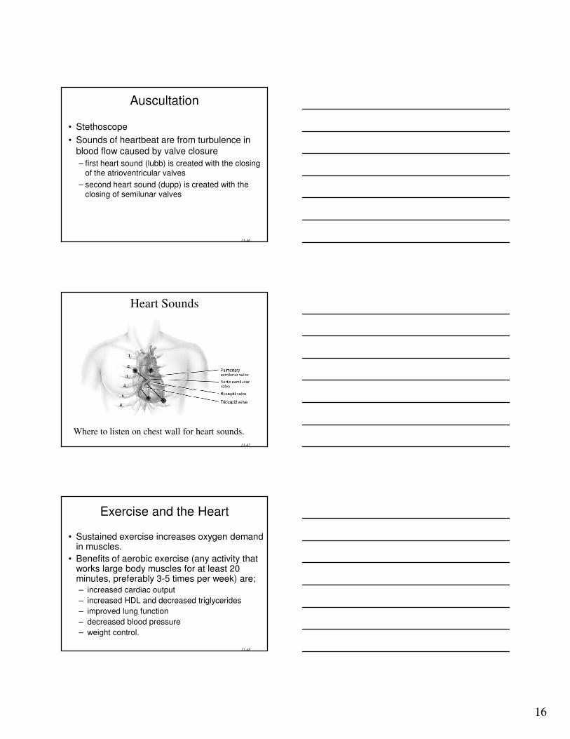
Heart Sounds
Where to listen on chest wall for heart sounds.
13-48
Exercise and the Heart
• Sustained exercise increases oxygen demand in muscles.
• Benefits of aerobic exercise (any activity that works large body muscles for at least 20 minutes, preferably 3-5 times per week) are;– increased cardiac output
– increased HDL and decreased triglycerides
– improved lung function
– decreased blood pressure
– weight control.
17
13-49
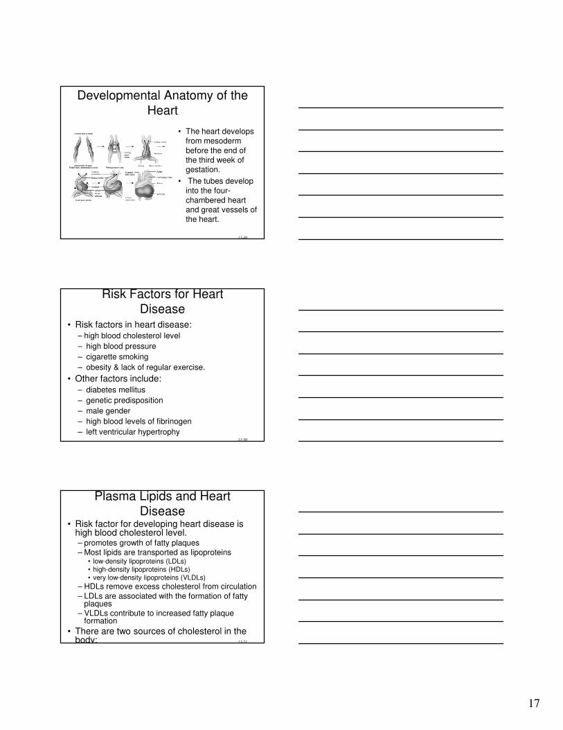
Developmental Anatomy of the
Heart
• The heart develops from mesoderm
before the end of the third week of gestation.
• The tubes develop into the four-
chambered heart and great vessels of the heart.
13-50
Risk Factors for Heart
Disease• Risk factors in heart disease:
– high blood cholesterol level
– high blood pressure
– cigarette smoking
– obesity & lack of regular exercise.
• Other factors include:– diabetes mellitus
– genetic predisposition
– male gender
– high blood levels of fibrinogen
– left ventricular hypertrophy
13-51
Plasma Lipids and Heart
Disease• Risk factor for developing heart disease is
high blood cholesterol level.– promotes growth of fatty plaques – Most lipids are transported as lipoproteins
• low-density lipoproteins (LDLs)• high-density lipoproteins (HDLs)• very low-density lipoproteins (VLDLs)
– HDLs remove excess cholesterol from circulation
– LDLs are associated with the formation of fatty plaques
– VLDLs contribute to increased fatty plaque formation
• There are two sources of cholesterol in the body:
18
13-52
Desirable Levels of Blood
Cholesterol for Adults
• TC (total cholesterol) under 200 mg/dl
• LDL under 130 mg/dl
• HDL over 40 mg/dl
• Normally, triglycerides are in the range of 10-
190 mg/dl.
• Among the therapies used to reduce blood
cholesterol level are exercise, diet, and drugs.
13-53
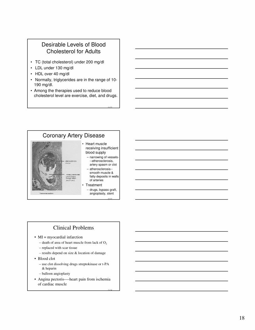
Coronary Artery Disease
• Heart muscle receiving insufficient
blood supply
– narrowing of vessels---atherosclerosis, artery spasm or clot
– atherosclerosis--smooth muscle & fatty deposits in walls of arteries
• Treatment
– drugs, bypass graft, angioplasty, stent
13-54
Clinical Problems
• MI = myocardial infarction
– death of area of heart muscle from lack of O2
– replaced with scar tissue
– results depend on size & location of damage
• Blood clot
– use clot dissolving drugs streptokinase or t-PA
& heparin
– balloon angioplasty
• Angina pectoris----heart pain from ischemia
of cardiac muscle
19
13-55
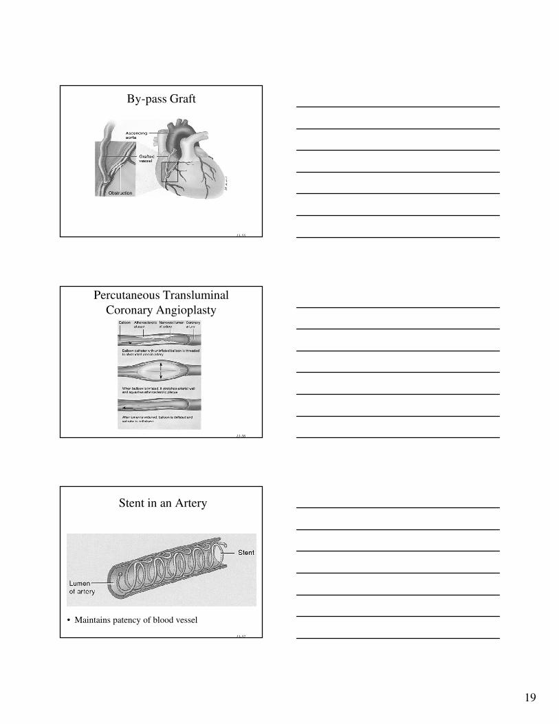
By-pass Graft
13-56
Percutaneous Transluminal
Coronary Angioplasty
13-57
Stent in an Artery
• Maintains patency of blood vessel
20
13-58
• Periacarditis
• Cardiac Tamponade
• Rheumatic Fever
• Arrhythmias
• Congenital Heart Defects