Embed Size (px)
Citation preview
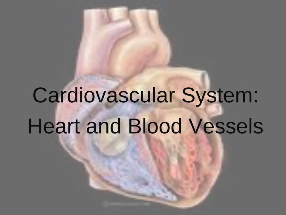
Cardiovascular System:
Heart and Blood Vessels
What to Know
• What are the functions of the cardiovascular system?
• What is the anatomy of the heart and blood vessels (veins and arteries)?
• How does blood flow through the heart?
• How is the heart beat regulated?
• What is blood pressure?
• What are common cardiovascular diseases and how might you prevent them?

The Circulatory System
• The cardiovascular system
consists of the blood vessels and
the heart
• The blood vessels conduct blood
in continuous loops
• The heart is a muscular pump
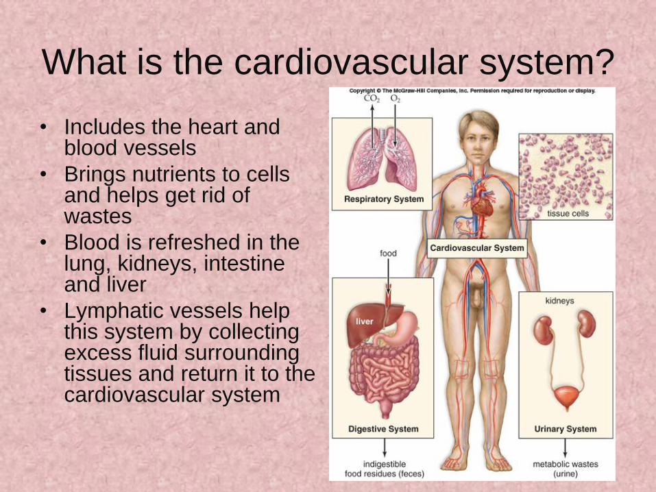
What is the cardiovascular system?
• Includes the heart and blood vessels
• Brings nutrients to cells and helps get rid of wastes
• Blood is refreshed in the lung, kidneys, intestine and liver
• Lymphatic vessels help this system by collecting excess fluid surrounding tissues and return it to the cardiovascular system

What are the functions of the
cardiovascular system?
1. Generate blood pressure
2. Transport blood
3. Exchange of nutrients and
wastes at the capillaries
4. Regulate blood flow as needed

What is the main pathway of blood in
the body?
Arteries
Arterioles
Capillaries
Venules
Veins
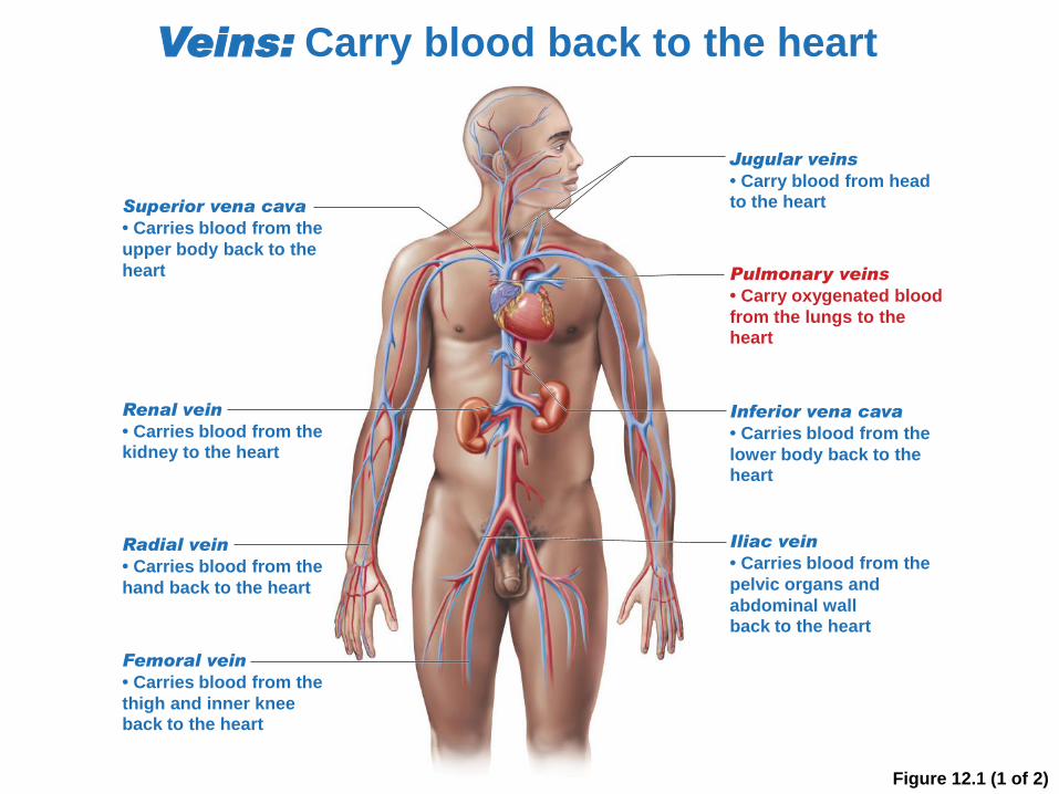
Figure 12.1 (1 of 2)
Veins: Carry blood back to the heart
Jugular veins
• Carry blood from headto the heart
Renal vein
• Carries blood from thekidney to the heart
Pulmonary veins
• Carry oxygenated blood
from the lungs to theheart
Inferior vena cava
• Carries blood from the
lower body back to theheart
Superior vena cava
• Carries blood from the
upper body back to the
heart
Iliac vein
• Carries blood from the
pelvic organs and
abdominal wallback to the heart
Radial vein
• Carries blood from the
hand back to the heart
Femoral vein
• Carries blood from the
thigh and inner kneeback to the heart
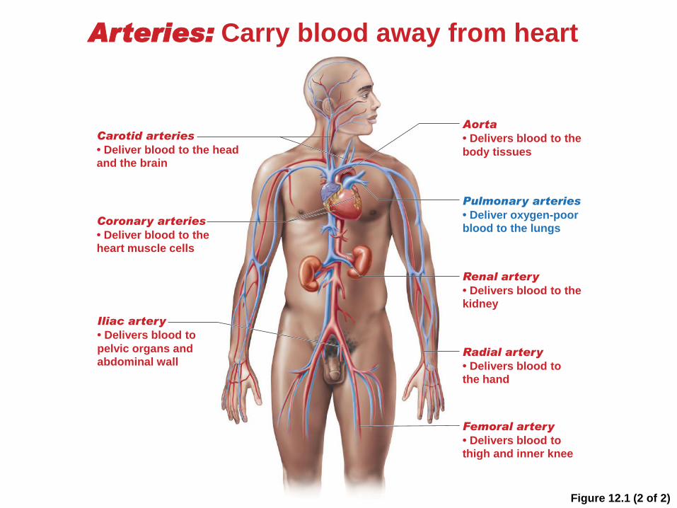
Figure 12.1 (2 of 2)
Arteries: Carry blood away from heart
Carotid arteries
• Deliver blood to the headand the brain
Aorta
• Delivers blood to the
body tissues
Pulmonary arteries
• Deliver oxygen-poorblood to the lungs
Coronary arteries
• Deliver blood to theheart muscle cells
Renal artery
• Delivers blood to thekidney
Iliac artery
• Delivers blood to
pelvic organs andabdominal wall
Radial artery
• Delivers blood tothe hand
Femoral artery
• Delivers blood tothigh and inner knee

Arteries• Carry blood away from the heart
• Their walls have 3 layers:
– Thin inner epithelium-tunica intima
– Thick smooth muscle layer-tunica media
– Outer connective tissue- tunica adventitia
• The elasticity of the arteries maintains pressure on the
blood between heartbeats to keep it flowing through the
vessels
• As the heart pumps blood into the arteries, they expand
such that one is able to feel a pulse
• The pulse rate is the same as the heart rate

Arterioles• are small arteries that regulate blood
pressure
• serve as gatekeepers to the capillary
networks keeping them open or closed
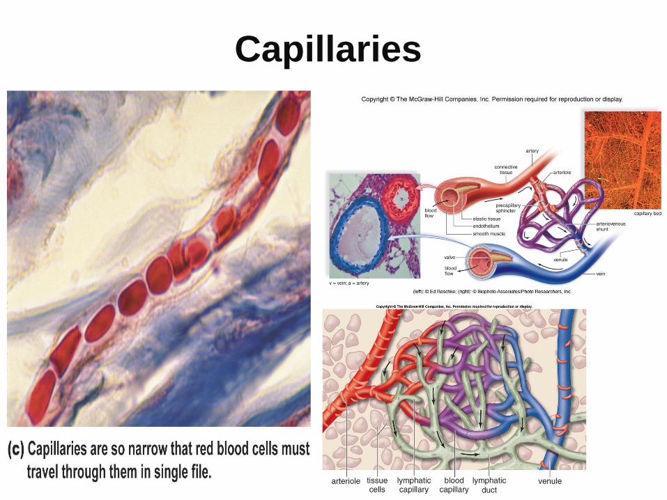
Capillaries

Capillaries• Microscopic vessels between arterioles
and venules
• Made of one layer of epithelial tissueallow for the exchange of materials between the blood and tissues
• Combined large surface area– Blood flows more slowly due to the large surface area
– Provides more time for the exchange of materials

The Blood Vessels Conduct Blood
in Continuous Loops
Figure 12.3b

Capillary Exchange
• At arterial end bp (30) is higher than (21)
osmotic pressure which is created by salts and
proteins so H20 leaves the bloodstream
• At midsection O2 and CO2 follow their
concentration gradients- O, a.a., glucose-out
CO2 and wastes in
• At venous end osmotic pressure is higher than
bp so H2O enters blood
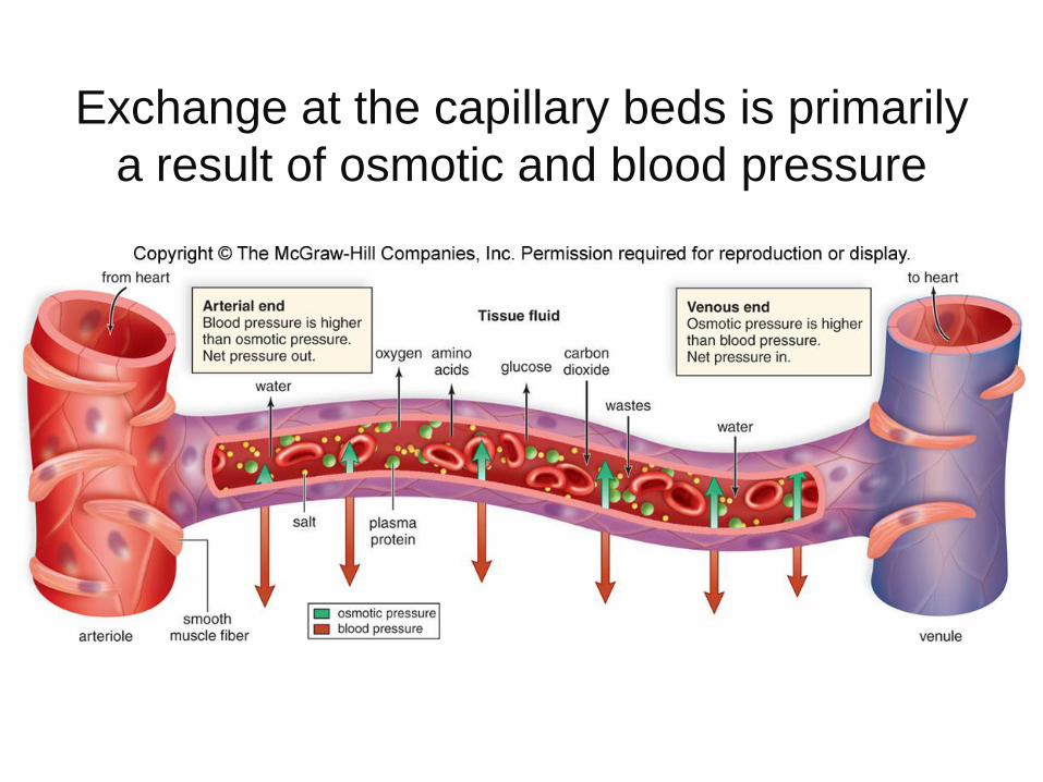
Exchange at the capillary beds is primarily
a result of osmotic and blood pressure
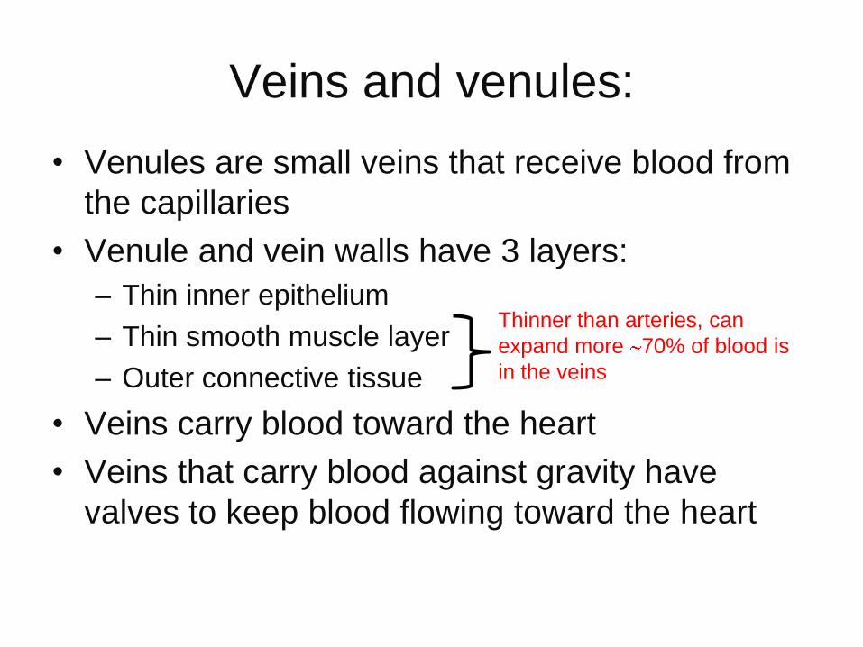
Veins and venules:
• Venules are small veins that receive blood from
the capillaries
• Venule and vein walls have 3 layers:
– Thin inner epithelium
– Thin smooth muscle layer
– Outer connective tissue
• Veins carry blood toward the heart
• Veins that carry blood against gravity have
valves to keep blood flowing toward the heart
Thinner than arteries, can
expand more 70% of blood is
in the veins
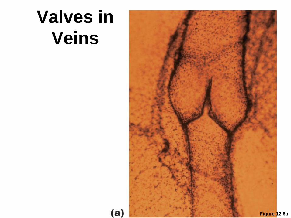
Valves in
Veins
Figure 12.6a

Figure 12.6b
Valve
closed
Valve
open
Relaxed calf
muscles
Skeletal muscles
relax, and blood
fills the valves
and closes them.
Muscle contraction
squeezes the vein,
pushing blood
through the open
valve toward the
heart.
Valve
closed
Contracted calf
muscles
(b)

Know how you can tell the difference
between an artery and vein?

Anatomy of the Heart
• A large, muscular organ consisting of mostly cardiac tissue called the myocardium
• It is surrounded by a sac called the pericardium
• Consists of two sides, right and left, separated by a septum
• Consists of 4 chambers: 2 atria and 2 ventricles
• 2 sets of valves: semilunar valves and atrioventricular valves (AV valves)
• The valves give the resulting “lub” and “dub” sound of the heart Heart Sounds
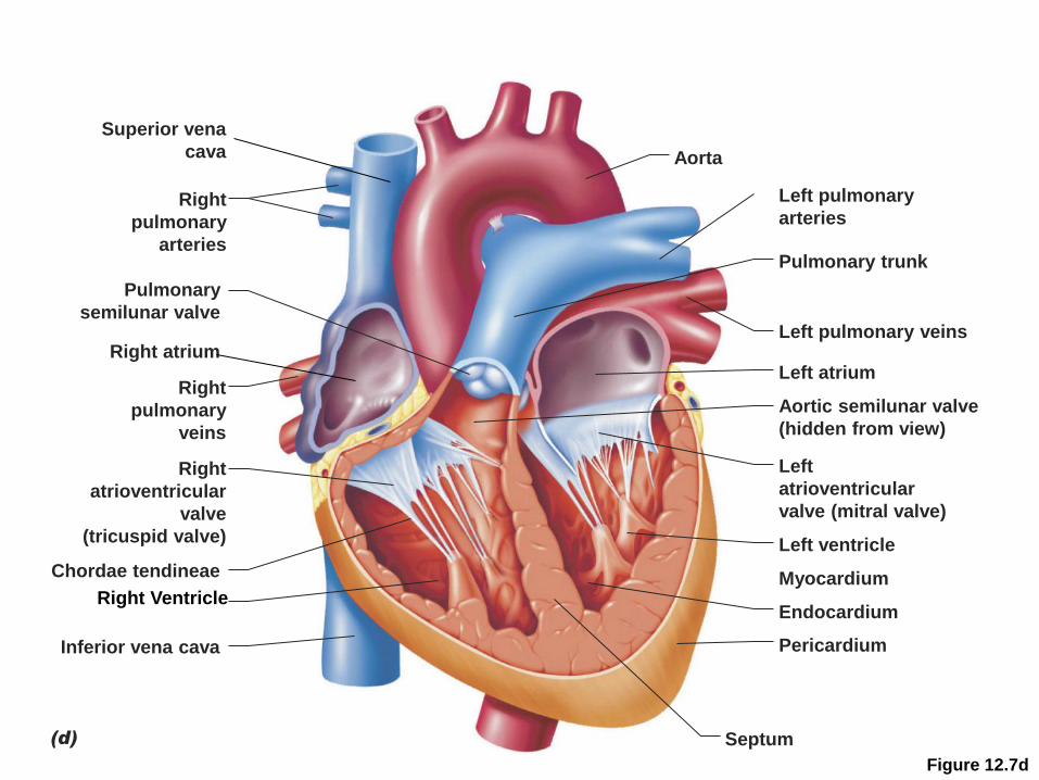
Figure 12.7d
Right
ventricle
Superior vena
cava Aorta
Right
pulmonary
arteries
Pulmonary
semilunar valve
Right atrium
Right
pulmonary
veins
Right
atrioventricular
valve
(tricuspid valve)
Chordae tendineae
Inferior vena cava
Left pulmonary
arteries
Pulmonary trunk
Left pulmonary veins
Left atrium
Aortic semilunar valve
(hidden from view)
Left
atrioventricular
valve (mitral valve)
Left ventricle
Myocardium
Endocardium
Pericardium
Septum(d)
Right Ventricle
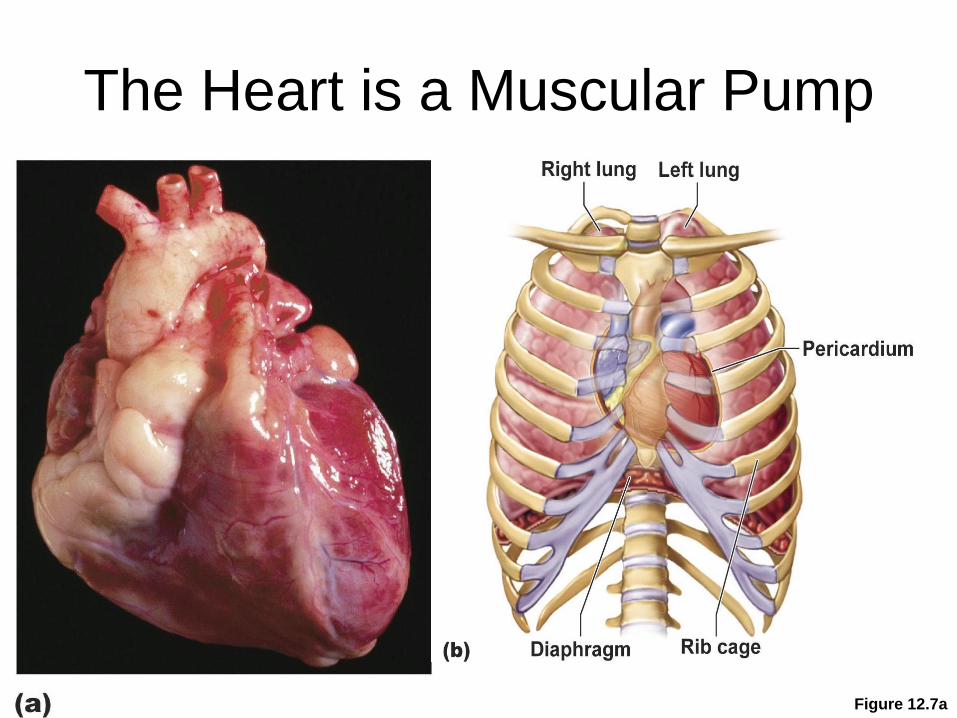
The Heart is a Muscular Pump
Figure 12.7a
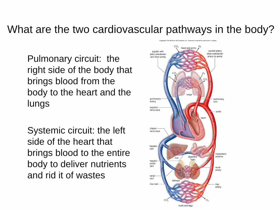
What are the two cardiovascular pathways in the body?
• Pulmonary circuit: the
right side of the body that
brings blood from the
body to the heart and the
lungs
• Systemic circuit: the left
side of the heart that
brings blood to the entire
body to deliver nutrients
and rid it of wastes

How does blood flow through the
heart?• Inferior and superior vena cava (1) dump blood into the
right atrium (2)
• Right ventricle (3)
• 2 pulmonary arteries (4) that lead to the lungs (5) where blood becomes oxygenated
• Pulmonary veins (6) bring blood from the lungs back the left atrium (7)
• Left ventricle (8) is large and muscular to pump blood into the aorta (9) and to the rest of the body (10)
• Eventually blood will be pumped back to each vena cava(1)

Visualizing blood flow through the heart
Hyperlinks to Watch
• Animated tutorial of the Cardiac Cycle
• How the Heart Works
How do the structure of the vessels and heart
match their functions?
• The left ventricle is much more muscular than
the right ventricle because it must pump blood to
the entire body
• The arteries are more muscular than veins to
withstand the higher pressure exerted on them
• The veins have a thinner wall and a larger
center to store blood
How does the heartbeat occur?
• During systole the atria contract together followed by the ventricles contracting together
• This is followed by diastole, a rest phase, when the chambers relax
• This cardiac cycle, heartbeat, on average occurs 70 times/minute
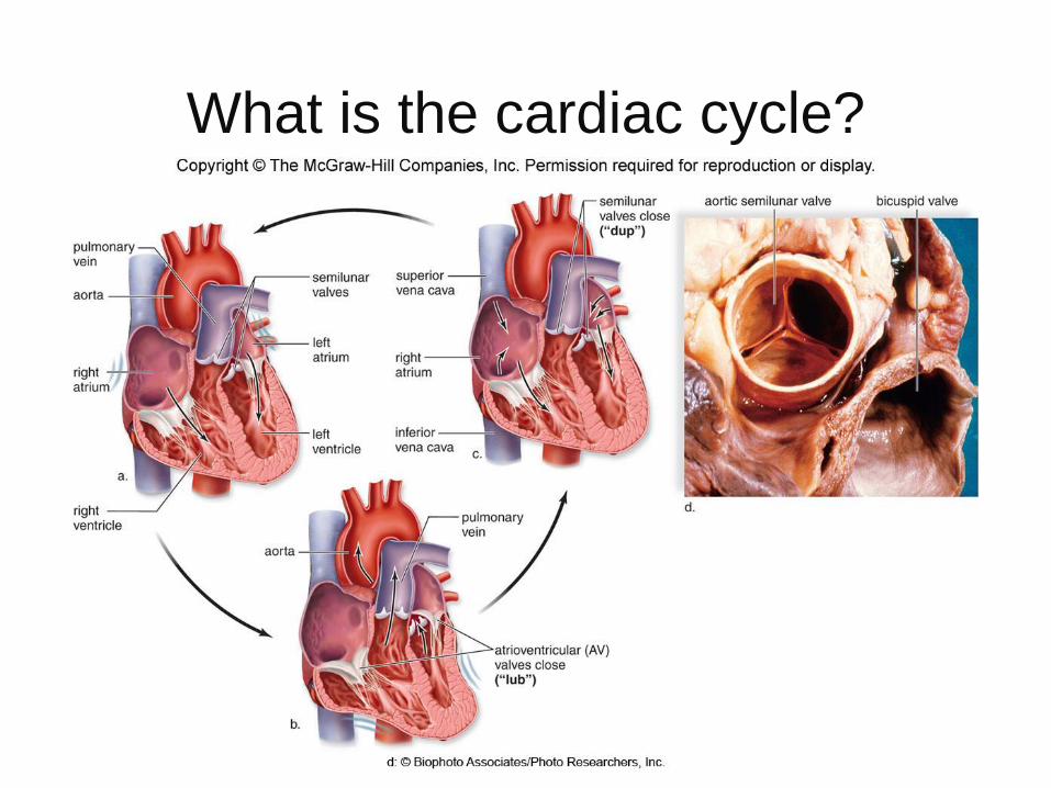
What is the cardiac cycle?
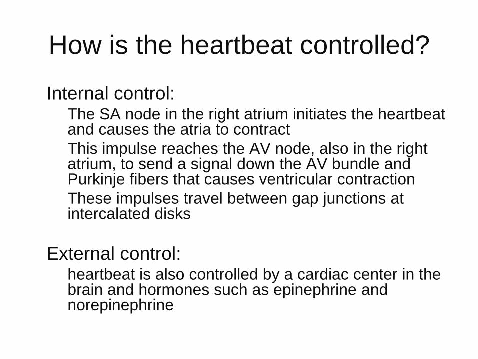
How is the heartbeat controlled?
• Internal control:– The SA node in the right atrium initiates the heartbeat
and causes the atria to contract
– This impulse reaches the AV node, also in the right atrium, to send a signal down the AV bundle and Purkinje fibers that causes ventricular contraction
– These impulses travel between gap junctions at intercalated disks
• External control:– heartbeat is also controlled by a cardiac center in the
brain and hormones such as epinephrine and norepinephrine

Visualizing the heartbeat
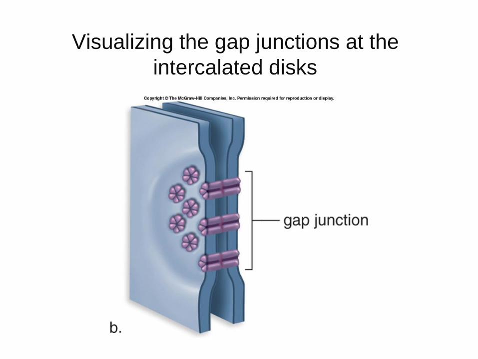
Visualizing the gap junctions at the
intercalated disks

• An electrocardiogram
(ECG/EKG)
– Recording of the
electrical events
associated with the
heartbeat
– A powerful diagnostic
tool
• Abnormal patterns can
indicate heart problems
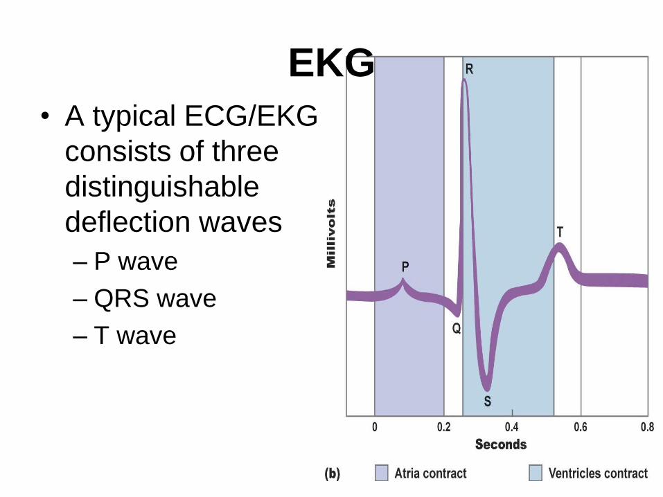
EKG
• A typical ECG/EKG
consists of three
distinguishable
deflection waves
– P wave
– QRS wave
– T wave

What is an electrocardiogram (ECG)?
• A record of the electrical changes in the heart muscle during a cardiac cycle
• The atria produce an electrical current when stimulated by the SA node called the P wave
• The contraction of the ventricles is the QRS complex
• The recovery of the ventricles is called the T wave
• Looking at these electrical changes allows doctors to detect abnormalities

How does a “normal” and abnormal
ECG compare?
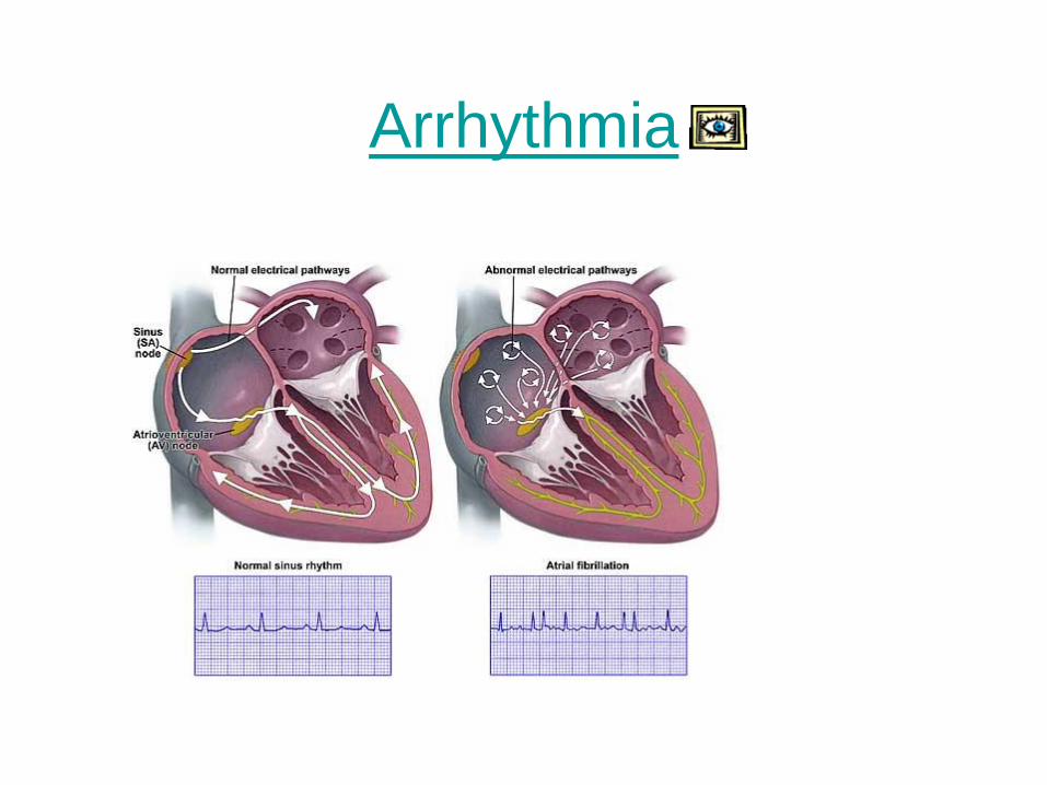
Arrhythmia
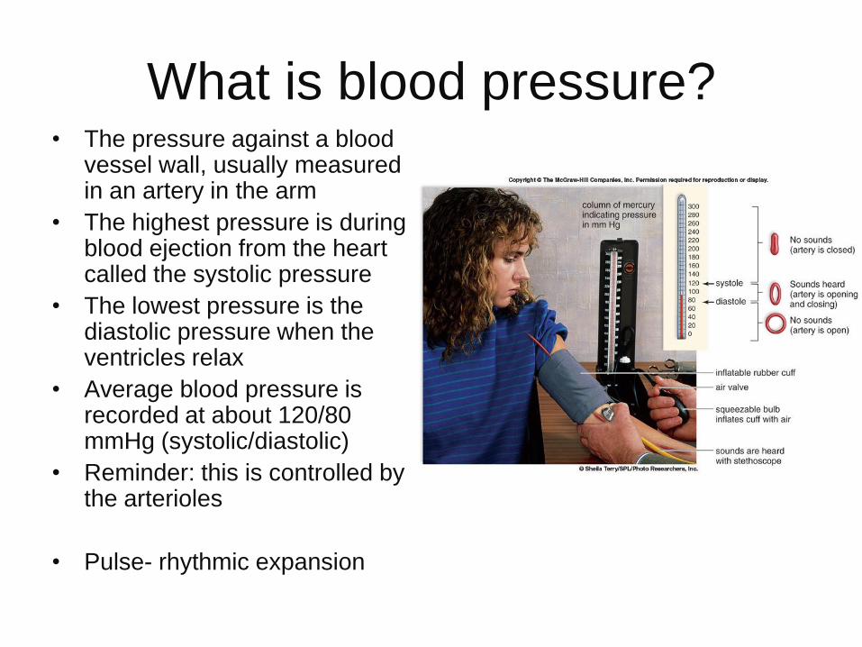
What is blood pressure?• The pressure against a blood
vessel wall, usually measured in an artery in the arm
• The highest pressure is during blood ejection from the heart called the systolic pressure
• The lowest pressure is the diastolic pressure when the ventricles relax
• Average blood pressure is recorded at about 120/80 mmHg (systolic/diastolic)
• Reminder: this is controlled by the arterioles
• Pulse- rhythmic expansion
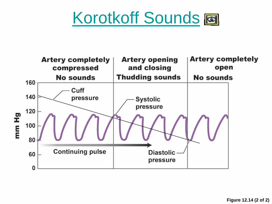
Korotkoff Sounds
Figure 12.14 (2 of 2)
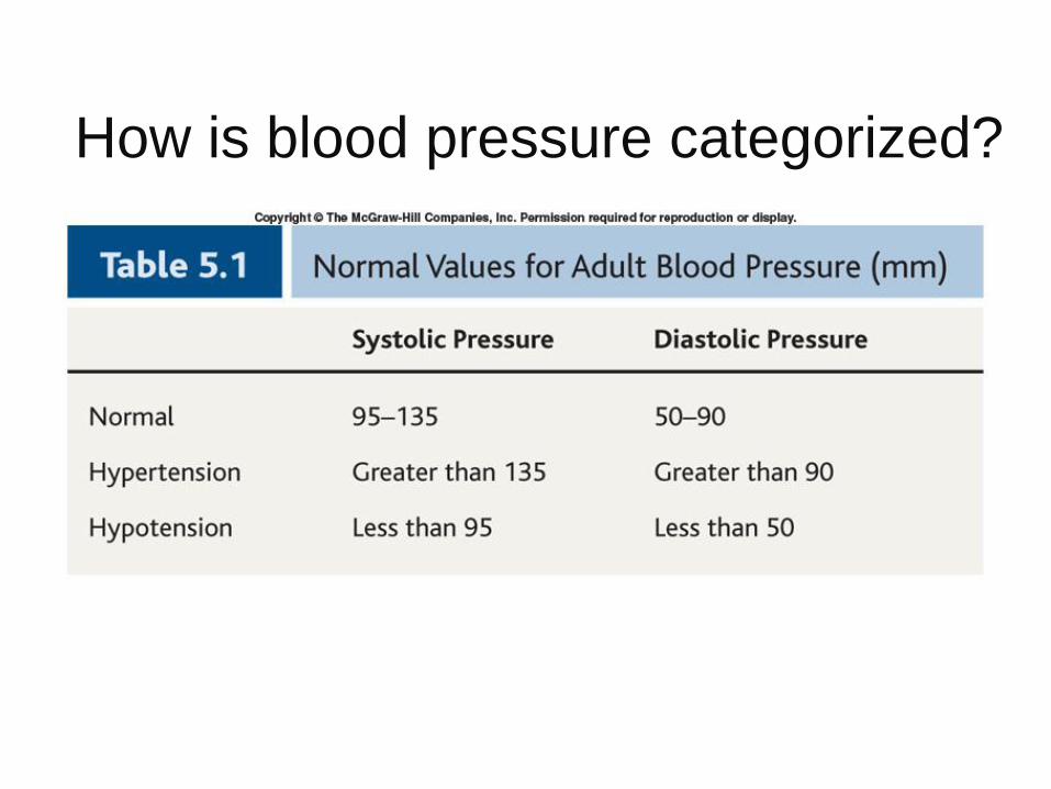
How is blood pressure categorized?

What is important about blood
flow?
• Blood flow is under the
highest pressure in the
arteries but remember the
thick, muscular walls
• Blood flow is slower in the
capillaries which is
important to allow time for
exchange between cells
• Blood pressure is minimal
in the veins and venules
but blood flow increases
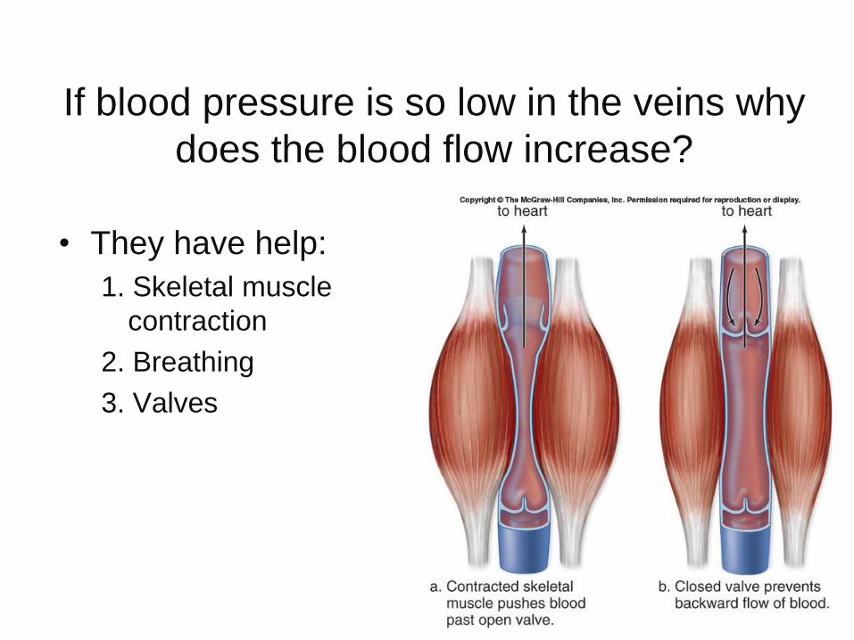
If blood pressure is so low in the veins why
does the blood flow increase?
• They have help:
1. Skeletal muscle
contraction
2. Breathing
3. Valves
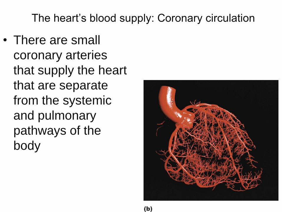
The heart’s blood supply: Coronary circulation
• There are small
coronary arteries
that supply the heart
that are separate
from the systemic
and pulmonary
pathways of the
body

Figure 12.10a
Inferior
vena
cava
Superior
vena cava
Pulmonary
trunk
Aorta
Right
coronary
vein
Right
coronary
artery
Left coronary
artery
Left
coronary
vein
(a)
Pulmonary
veins

What is the hepatic portal system?
• A system that brings blood from the digestive tract rich in amino acids and glucose to the liver
• The liver synthesizes blood proteins and stores the glucose as glycogen
• The liver also plays a role in purifying blood from the digestive tract
• Finally, the blood will return to the heart via the inferior vena cava

Why should we care about cardiovascular
disease?
• Cardiovascular disease (CVD) is the most
common cause of death in the western
world

Disorders of the blood vessels:
• Hypertension/high blood pressure
• Atherosclerosis
• Stroke
• Heart attack
• Aneurysm
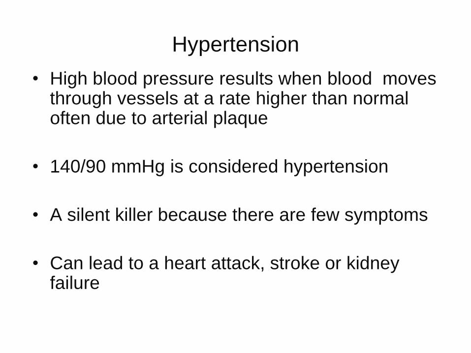
Hypertension
• High blood pressure results when blood moves through vessels at a rate higher than normal often due to arterial plaque
• 140/90 mmHg is considered hypertension
• A silent killer because there are few symptoms
• Can lead to a heart attack, stroke or kidney failure

Atherosclerosis
• A build up of plaque in blood vessels
• Plaque that is stationary is called a thrombus and an embolus when it detaches and can move to distant sites
• Associated with a stroke, heart attack and aneurysm

Stroke
• Also known as a cerebrovascular accident (CVA)
• Usually occurs when a cranial artery is blocked or bursts
• Part of the brain dies dues to lack of oxygen
• Symptoms may occur including numbness of hands or face, difficulty speaking and inability to see in one eye

Heart attack
• Also known as a myocardial infarction (MI)
• Part of the heart dies due to lack of
oxygen
• Can begin with angina pectoris, a pain that
radiates down the left arm due to a
blockage of a coronary artery

Aneurysm
• A ballooning of a blood vessel
• Atherosclerosis and hypertension can
weaken a vessel and cause ballooning
• The most commonly affected is the
abdominal artery or the arteries leading to
the brain

How are disorders of the blood vessels
treated?
• Dissolving blood clots: – t-PA is a drug that dissolves clots
• Treating clogged arteries:– Bypass surgery: usually a vein from the leg is taken
and used to bypass a clogged artery
– Stents: wire mesh cylinder inserted into a clogged artery to hold it open
– Angioplasty: a tube with a balloon is inserted into the clogged area and the balloon is then inflated to open the vessel
– A stent and angioplasty may be used in combination
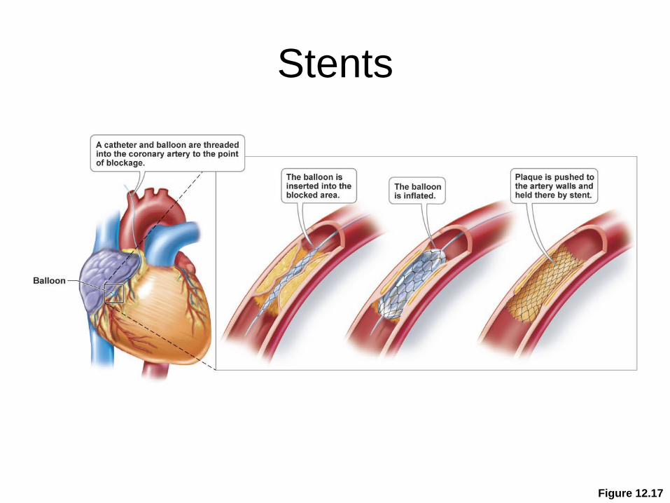
Stents
Figure 12.17

Figure 12.18

Cardiovascular Disease
• Heart muscle dies because of an
insufficient blood supply during a heart
attack (myocardial infarction) and is
gradually replaced by scar tissue
• Scar tissue cannot contract, so part of the
heart permanently loses its pumping ability

Figure 12.19

Disorders of the heart and its treatment
• Disorders:
– Heart failure is when
the heart no longer
pumps properly
• Treatments:
– Left ventricular assist
device(LVAD)
– Heart transplant either
natural or artificial
Health Focus: The do’s and don’ts for
prevention of cardiovascular disease?
• Do not smoke
• Do not abuse drugs
• Keep your weight down to decrease chances of
hypertension and Type II diabetes
• Eat a healthy diet
– Low in saturated and trans fats
– Low in cholesterol
• Know your blood cholesterol
• Exercise