Embed Size (px)
Citation preview
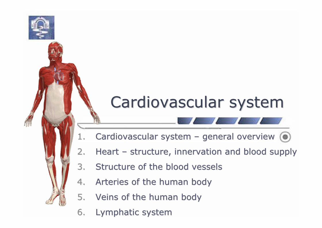
Cardiovascular systemCardiovascular system
1.1. Cardiovascular systemCardiovascular system –– general overviewgeneral overview
2.2. HeartHeart –– structurestructure, , innervation and blood supplyinnervation and blood supply
3.3. Structure of the blood vesselsStructure of the blood vessels
4.4. Arteries of the human bodyArteries of the human body
5.5. Veins of the human bodyVeins of the human body
6.6. Lymphatic systemLymphatic system
Cardiovascular systemCardiovascular system
22
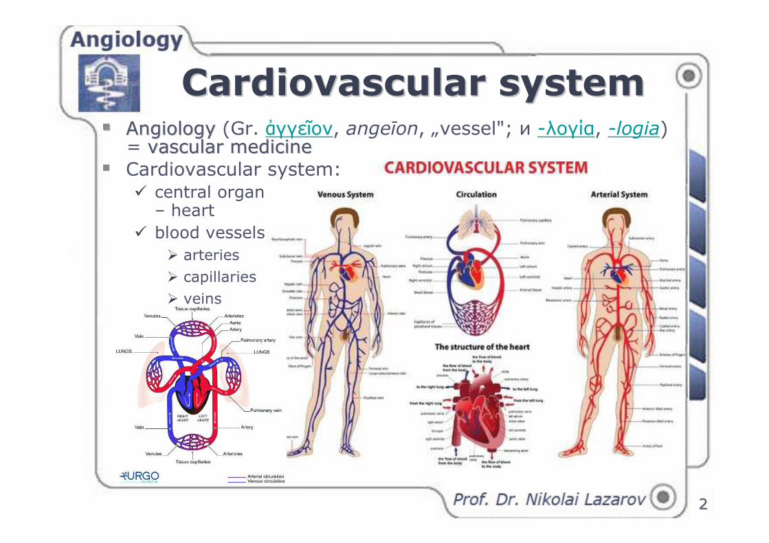
�� AngiologyAngiology (Gr. ἀγγεῖον, angeīon, „vessel"; и -λογία, -logia)= vascular medicinevascular medicine
� Cardiovascular system:� central organ
– heart
� blood vessels
� arteries
� capillaries
� veins
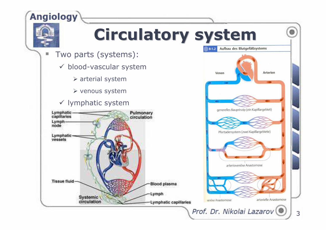
Circulatory systemCirculatory system� Two parts (systems):
� blood-vascular system
� arterial system
� venous system
� lymphatic system
33
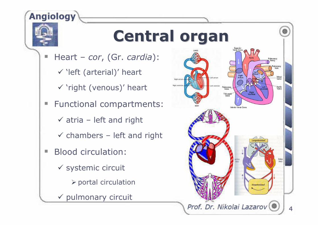
Central organCentral organ
44
� Heart – cor, (Gr. cardia):
� ‘left (arterial)’ heart
� ‘right (venous)’ heart
� Functional compartments:
� atria – left and right
� chambers – left and right
� Blood circulation:
� systemic circuit
� portal circulation
� pulmonary circuit
55
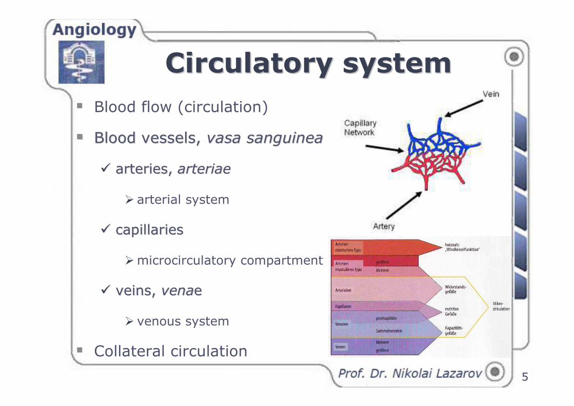
� Blood flow (circulation)
�� Blood vesselsBlood vessels, , vasa sanguineavasa sanguinea
�� arteries, arteries, arteriaearteriae
� arterial system
�� capillaries capillaries
�microcirculatory compartment
�� veinsveins, , venavenaee
� venous system
� Collateral circulation
Circulatory systemCirculatory system
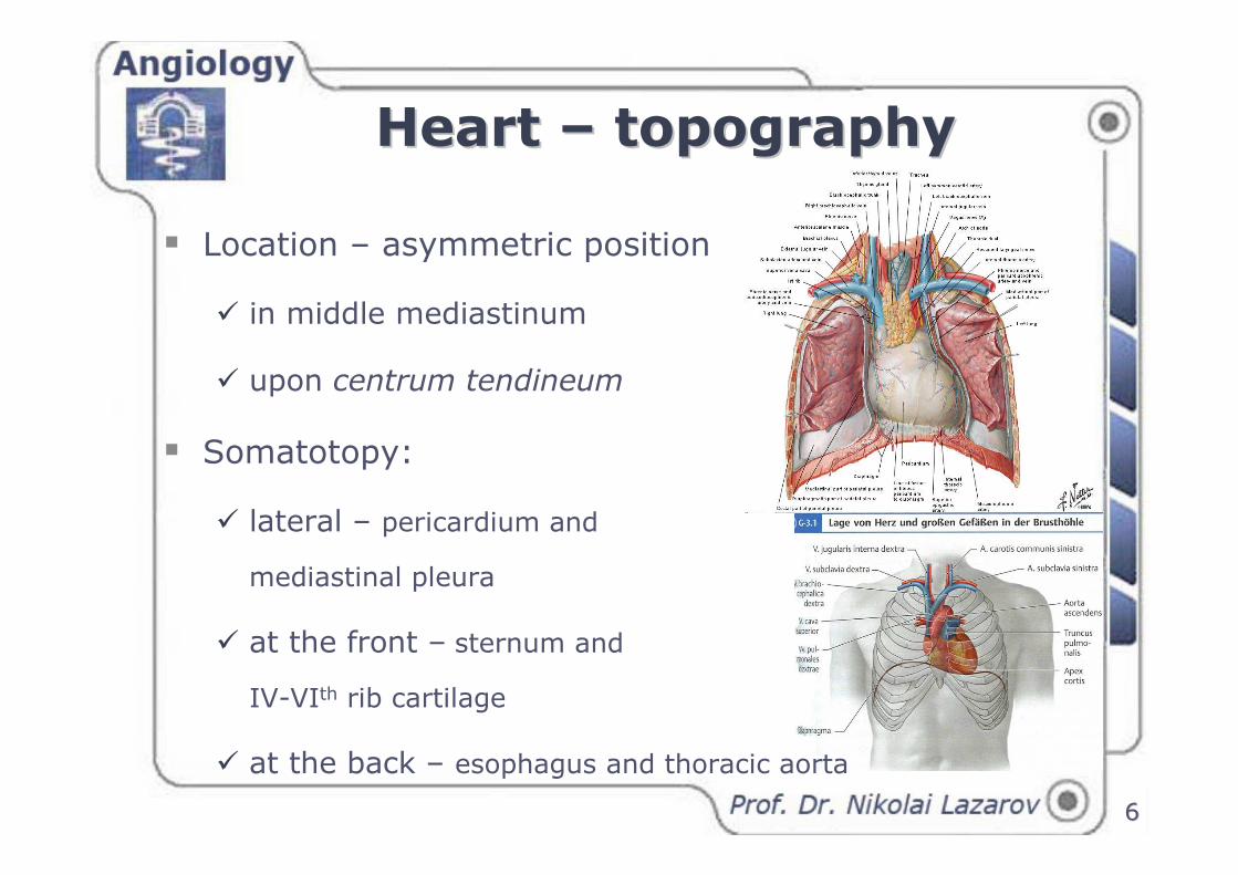
Heart Heart –– topographytopography
66
� Location – asymmetric position
� in middle mediastinum
� upon centrum tendineum
� Somatotopy:
� lateral – pericardium and
mediastinal pleura
� at the front – sternum and
IV-VIth rib cartilage
� at the back – esophagus and thoracic aorta
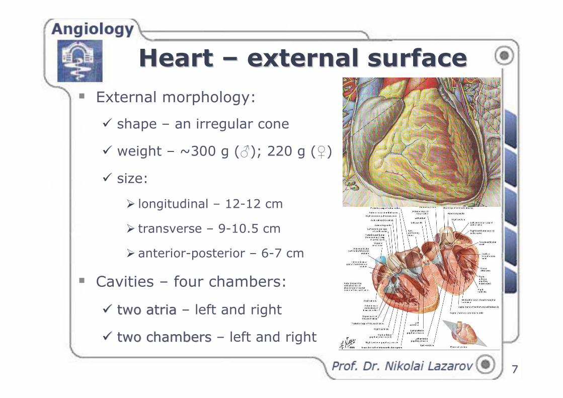
Heart Heart –– external surfaceexternal surface
� External morphology:
� shape – an irregular cone
� weight – ~300 g (♂); 220 g (♀)
� size:
� longitudinal – 12-12 cm
� transverse – 9-10.5 cm
� anterior-posterior – 6-7 cm
� Cavities – four chambers:
�� two atria two atria – left and right
�� two chambers two chambers – left and right
77
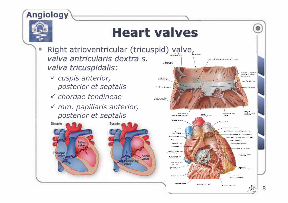
Heart valvesHeart valves
�� Right atrioventricular (tricuspid) valveRight atrioventricular (tricuspid) valve, , valva antricularis dextra s. valva antricularis dextra s.
valva tricuspidalisvalva tricuspidalis::
� cuspis anterior,
posterior et septalis
� chordae tendineae
� mm. papillaris anterior,
posterior et septalis
88
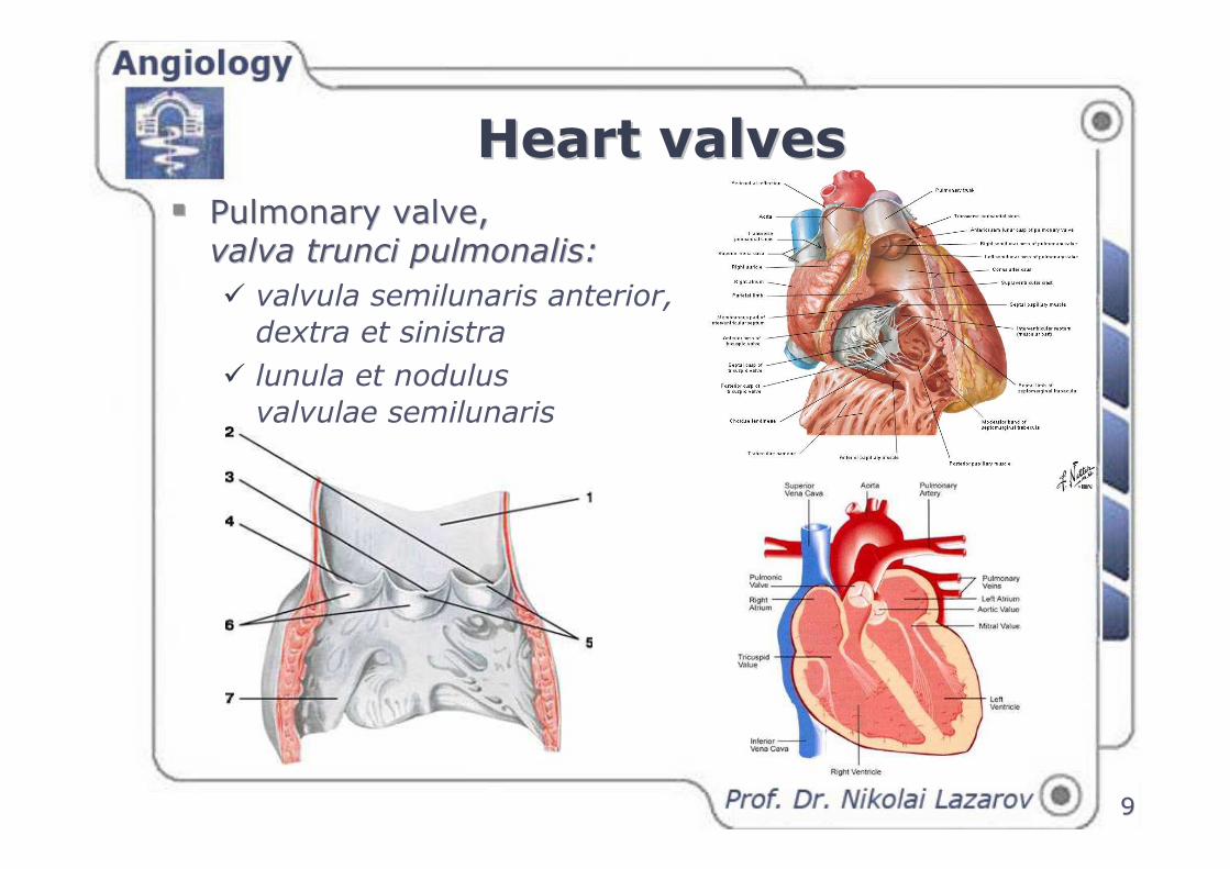
Heart valvesHeart valves
�� Pulmonary valvePulmonary valve, , valva trunci pulmonalisvalva trunci pulmonalis::
� valvula semilunaris anterior,
dextra et sinistra
� lunula et nodulus
valvulae semilunaris
99
Heart valvesHeart valves
1010
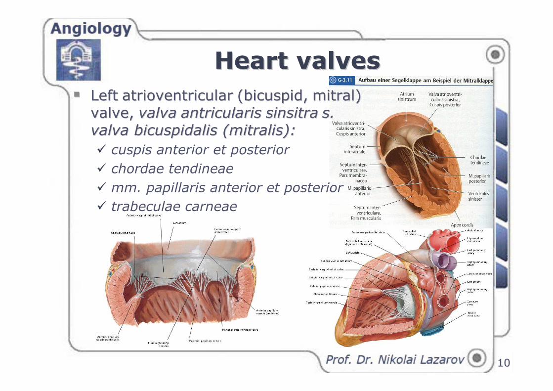
�� LeftLeft atrioventricularatrioventricular (bicuspid,(bicuspid, mitral) mitral) valve,valve, valvavalva antricularisantricularis sinsitrasinsitra s. s.
valva bicuspidalis (mitralis)valva bicuspidalis (mitralis)::
� cuspis anterior et posterior
� chordae tendineae
� mm. papillaris anterior et posterior
� trabeculae carneae
Heart valvesHeart valves
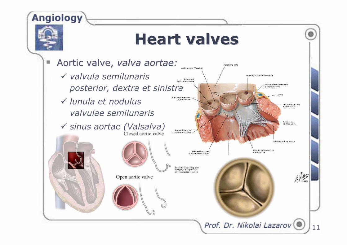
�� Aortic valveAortic valve, , valva aortaevalva aortae::
� valvula semilunaris
posterior, dextra et sinistra
� lunula et nodulus
valvulae semilunaris
� sinus aortae (Valsalva)
1111
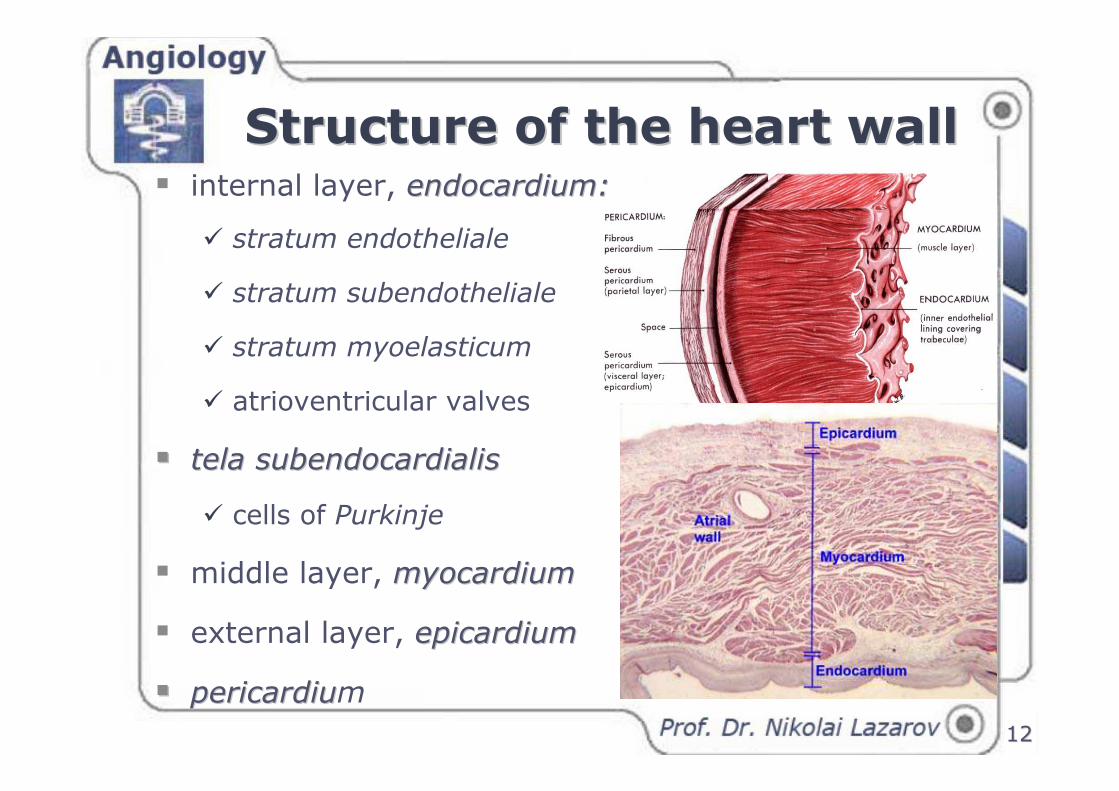
Structure of the heart wallStructure of the heart wall� internal layer, endocardiumendocardium::
� stratum endotheliale
� stratum subendotheliale
� stratum myoelasticum
� atrioventricular valves
�� tela subendocardialistela subendocardialis
� cells of Purkinje
� middle layer, myocardiummyocardium
� external layer, epicardiumepicardium
�� pericardiupericardium
1212
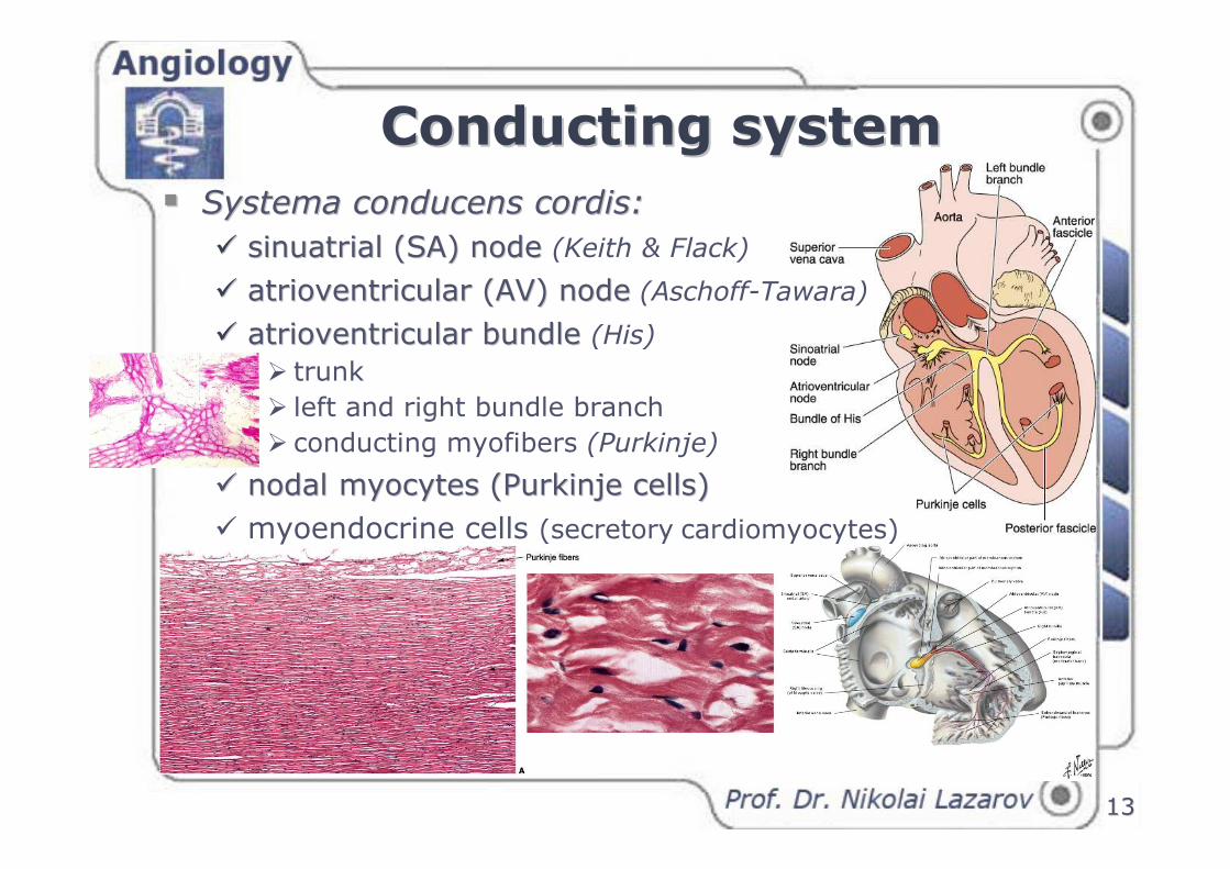
Conducting systemConducting system
1313
�� Systema conducens cordisSystema conducens cordis::
�� sinuatrial (SA) node sinuatrial (SA) node (Keith & Flack)
�� atrioventricular (AV) nodeatrioventricular (AV) node (Aschoff-Tawara)
�� atrioventricular bundle atrioventricular bundle (His)
� trunk
� left and right bundle branch
� conducting myofibers (Purkinje)
�� nodalnodal myocytes (Purkinje cells)myocytes (Purkinje cells)
� myoendocrine cells (secretory cardiomyocytes)
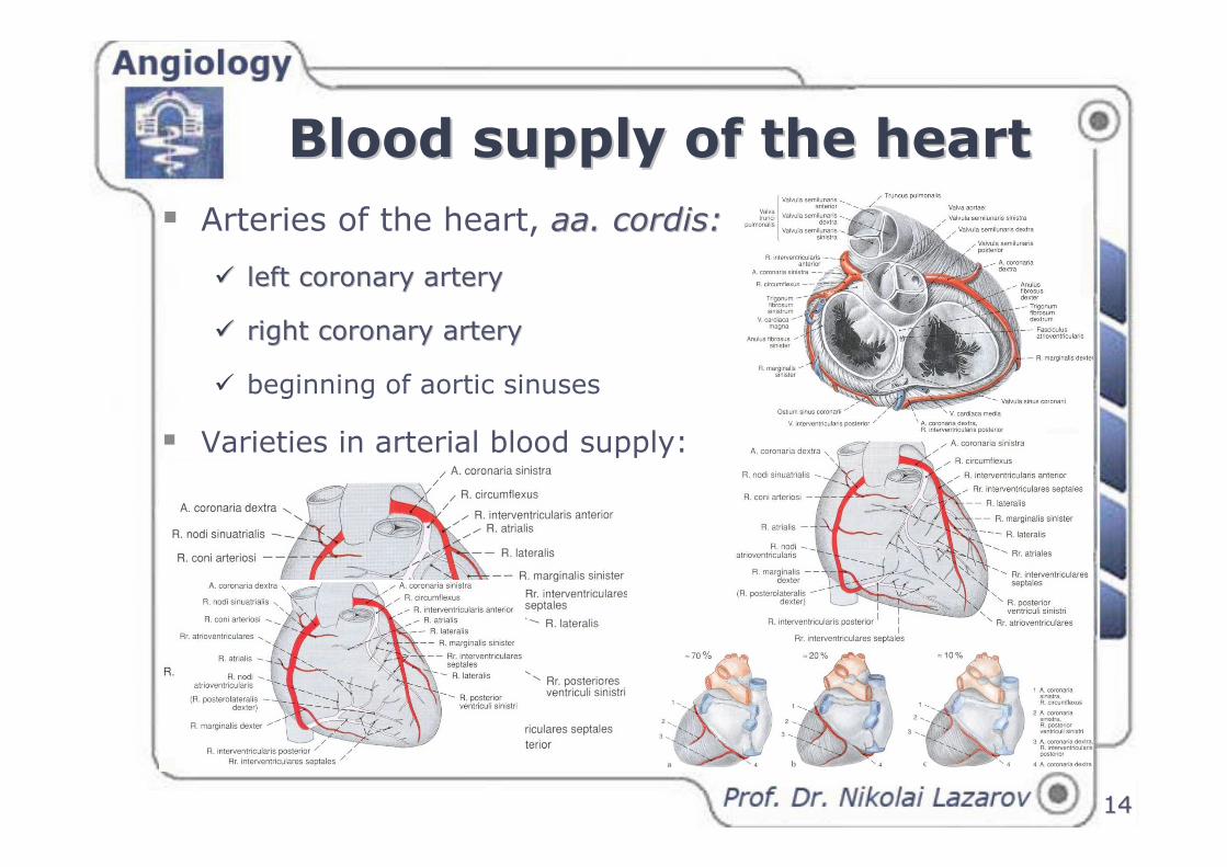
Blood supply of the heartBlood supply of the heart
1414
� Arteries of the heart, aa. cordisaa. cordis::
�� left coronary arteryleft coronary artery
�� right coronary arteryright coronary artery
� beginning of aortic sinuses
� Varieties in arterial blood supply:
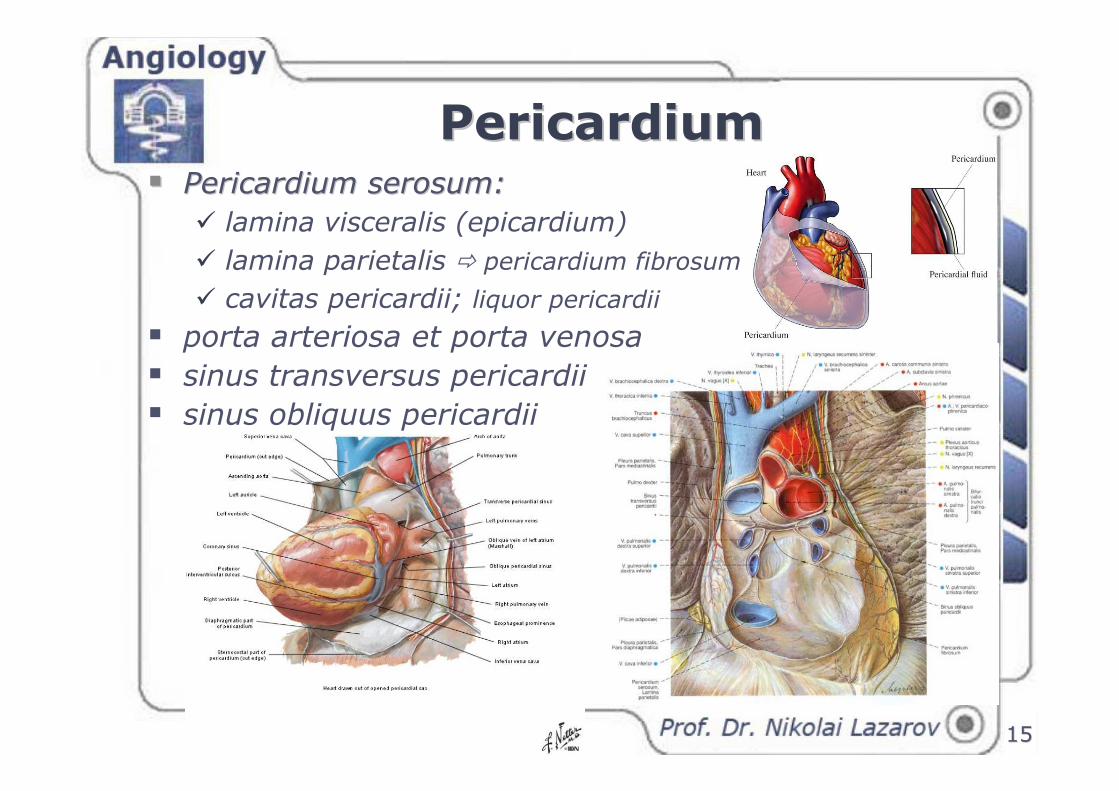
PericardiumPericardium
1515
�� Pericardium serosumPericardium serosum::
� lamina visceralis (epicardium)
� lamina parietalis � pericardium fibrosum
� cavitas pericardii; liquor pericardii
� porta arteriosa et porta venosa
� sinus transversus pericardii
� sinus obliquus pericardii
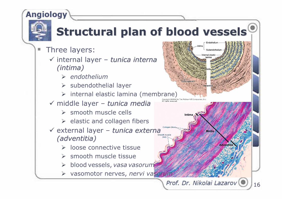
Structural plan of blood vesselsStructural plan of blood vessels
1616
� Three layers:
� internal layer – tunica interna tunica interna
(intima)(intima)
� endothelium
� subendothelial layer
� internal elastic lamina (membrane)
� middle layer – tunica mediatunica media
� smooth muscle cells
� elastic and collagen fibers
� external layer – tunica externa tunica externa
(adventitia)(adventitia)
� loose connective tissue
� smooth muscle tissue
� blood vessels, vasa vasorum
� vasomotor nerves, nervi vasorum
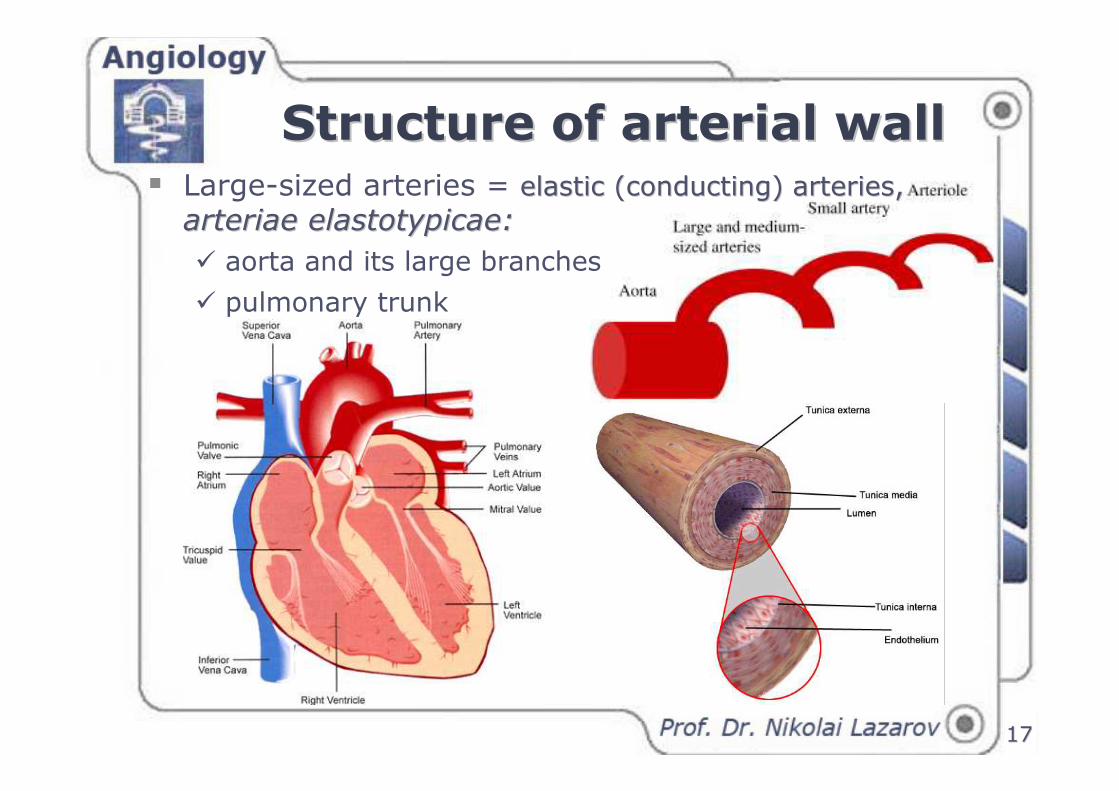
Structure of arterial wallStructure of arterial wall
1717
� Large-sized arteries = elastic elastic ((conductingconducting) ) arteries, arteries,
arteriae elastotypicaearteriae elastotypicae: :
� aorta and its large branches
� pulmonary trunk
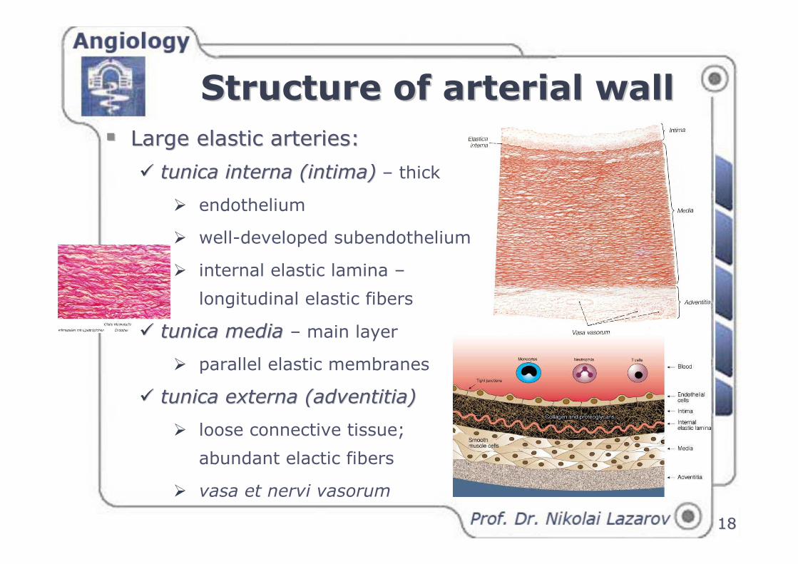
1818
�� Large elastic arteries:Large elastic arteries:
�� tunica interna (intima)tunica interna (intima) – thick
� endothelium
� well-developed subendothelium
� internal elastic lamina –
longitudinal elastic fibers
�� tunica mediatunica media – main layer
� parallel elastic membranes
�� tunica externa (adventitia)tunica externa (adventitia)
� loose connective tissue;
abundant elactic fibers
� vasa et nervi vasorum
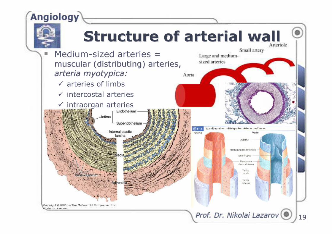
Structure of arterial wallStructure of arterial wall
1919
� Medium-sized arteries = muscularmuscular ((distributingdistributing) ) arteries, arteries, arteria myotypicaarteria myotypica: :
� arteries of limbs
� intercostal arteries
� intraorgan arteries
Structure of arterial wallStructure of arterial wall
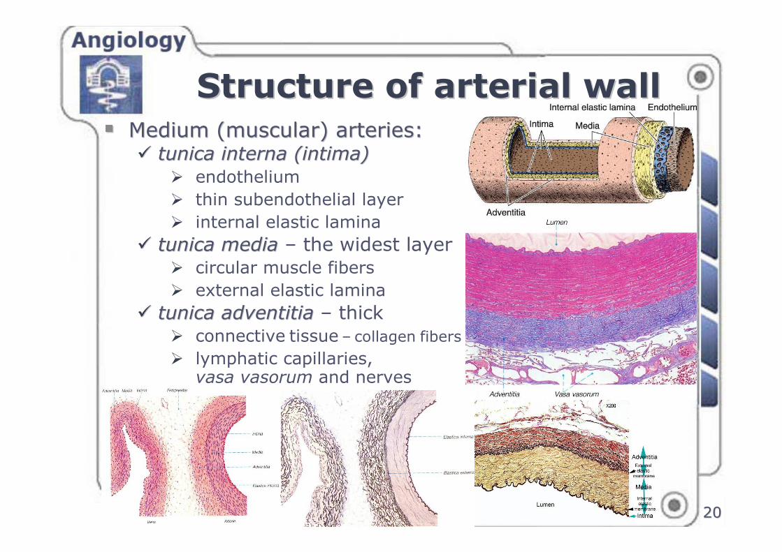
2020
�� Medium (muscular) arteries:Medium (muscular) arteries:�� tunica interna (intima)tunica interna (intima)
� endothelium
� thin subendothelial layer
� internal elastic lamina
�� tunica mediatunica media – the widest layer� circular muscle fibers
� external elastic lamina
�� tunica adventitiatunica adventitia – thick� connective tissue – collagen fibers
� lymphatic capillaries, vasa vasorum and nerves
Structure of arterial wallStructure of arterial wall
2121
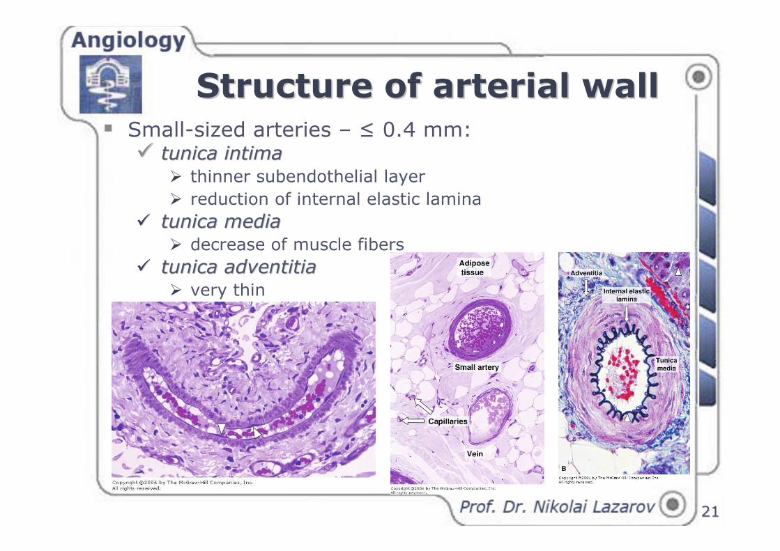
� Small-sized arteries – ≤ 0.4 mm:�� tunica intimatunica intima
� thinner subendothelial layer
� reduction of internal elastic lamina
�� tunica mediatunica media� decrease of muscle fibers
�� tunica adventitiatunica adventitia� very thin
Structure of arterial wallStructure of arterial wall
2222
Structure of arterial wallStructure of arterial wall
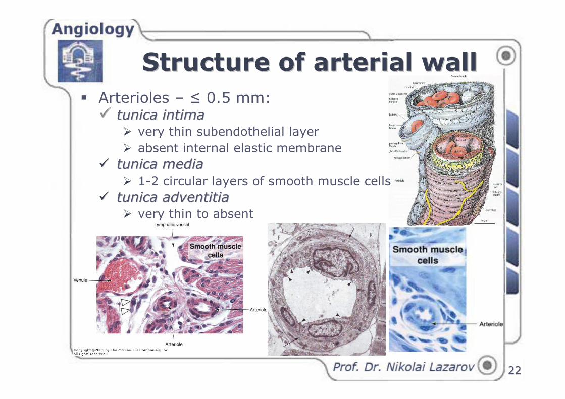
� Arterioles – ≤ 0.5 mm:�� tunica intimatunica intima
� very thin subendothelial layer
� absent internal elastic membrane
�� tunica mediatunica media� 1-2 circular layers of smooth muscle cells
�� tunica adventitiatunica adventitia� very thin to absent
Structure of capillariesStructure of capillaries
2323
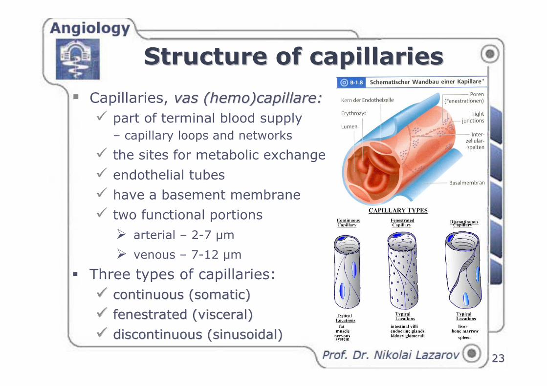
� Capillaries, vas (hemo)capillare:vas (hemo)capillare:
� part of terminal blood supply
– capillary loops and networks
� the sites for metabolic exchange
� endothelial tubes
� have a basement membrane
� two functional portions
� arterial – 2-7 µm
� venous – 7-12 µm
� Three types of capillaries:
�� continuous (somatic)continuous (somatic)
�� fenestrated (visceral)fenestrated (visceral)
�� discontinuous discontinuous ((sinusoidalsinusoidal))
2424
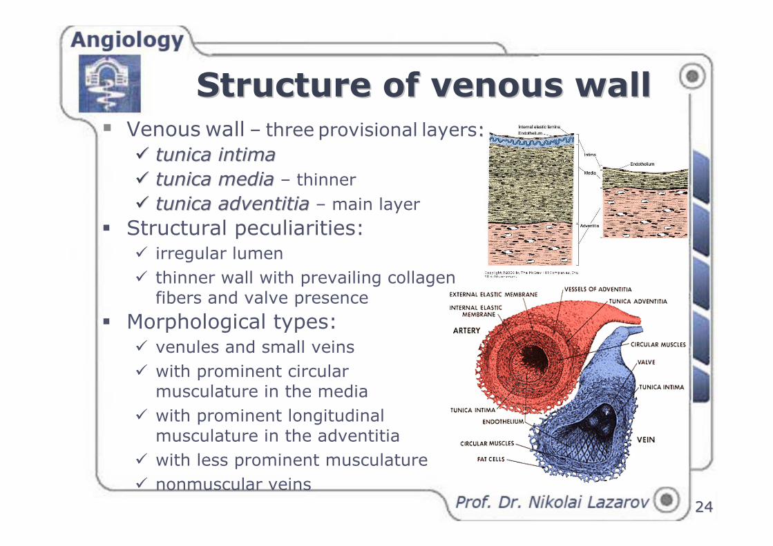
Structure of venous wallStructure of venous wall� Venous wall – three provisional layers: :
�� tunica intimatunica intima
�� tunica mediatunica media – thinner
�� tunica adventitiatunica adventitia – main layer
� Structural peculiarities:� irregular lumen
� thinner wall with prevailing collagen fibers and valve presence
� Morphological types:� venules and small veins
� with prominent circular musculature in the media
� with prominent longitudinal musculature in the adventitia
� with less prominent musculature
� nonmuscular veins
2525
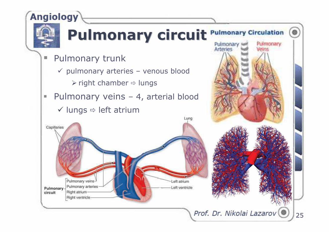
� Pulmonary trunk
� pulmonary arteries – venous blood
� right chamber � lungs
� Pulmonary veins – 4, arterial blood
� lungs � left atrium
Pulmonary circuitPulmonary circuit

2626
� Systemic circulation – course � arteries
� capillaries
� veins
� coronary vessels
� portal veins
Systemic circuitSystemic circuit

2727
Venous systemVenous system� system of superior vena cava
� system of inferior vena cava
� hepatic portal system
2828
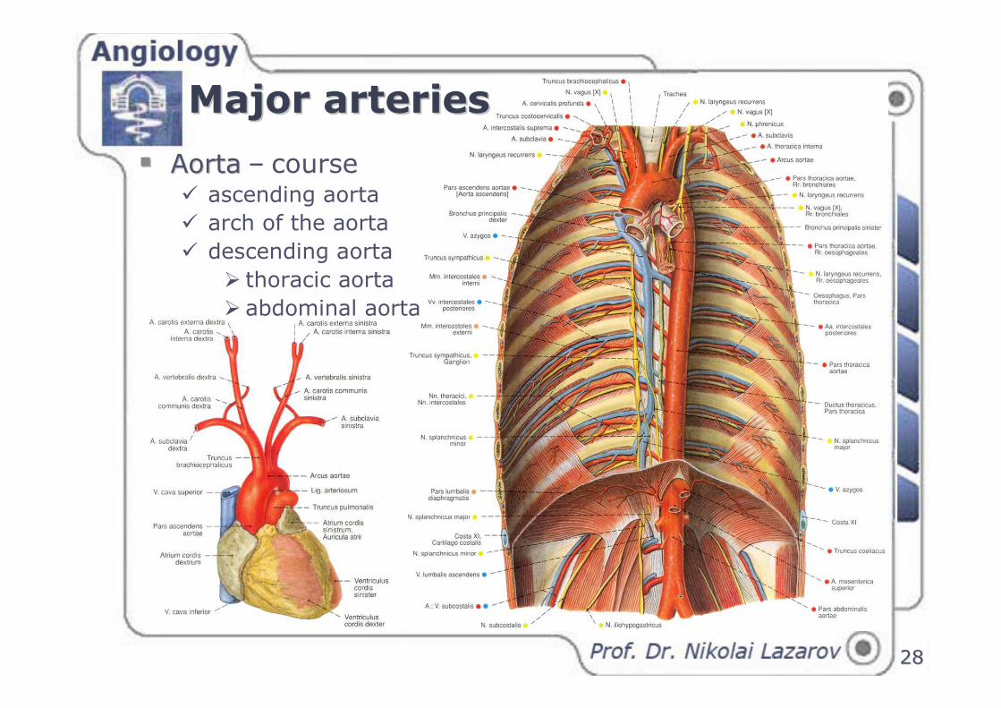
Major arteriesMajor arteries
�� AortaAorta – course � ascending aorta
� arch of the aorta
� descending aorta
� thoracic aorta
� abdominal aorta
2929
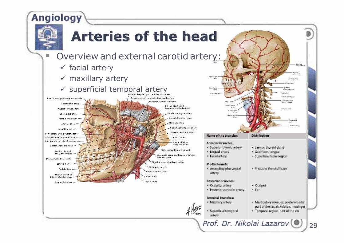
� Overview and external carotid artery:� facial artery
� maxillary artery
� superficial temporal artery
Arteries of the headArteries of the head
3030
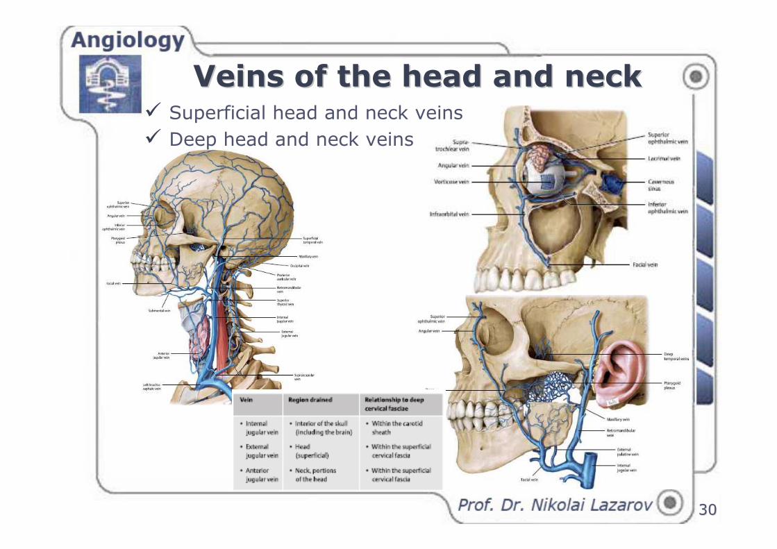
Veins of the head and neck Veins of the head and neck � Superficial head and neck veins
� Deep head and neck veins
3131
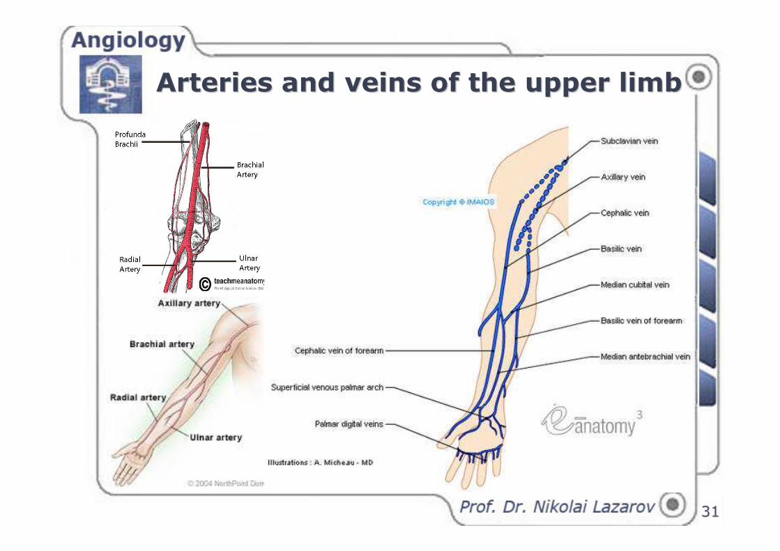
Arteries and veins of the upper limbArteries and veins of the upper limb
3232
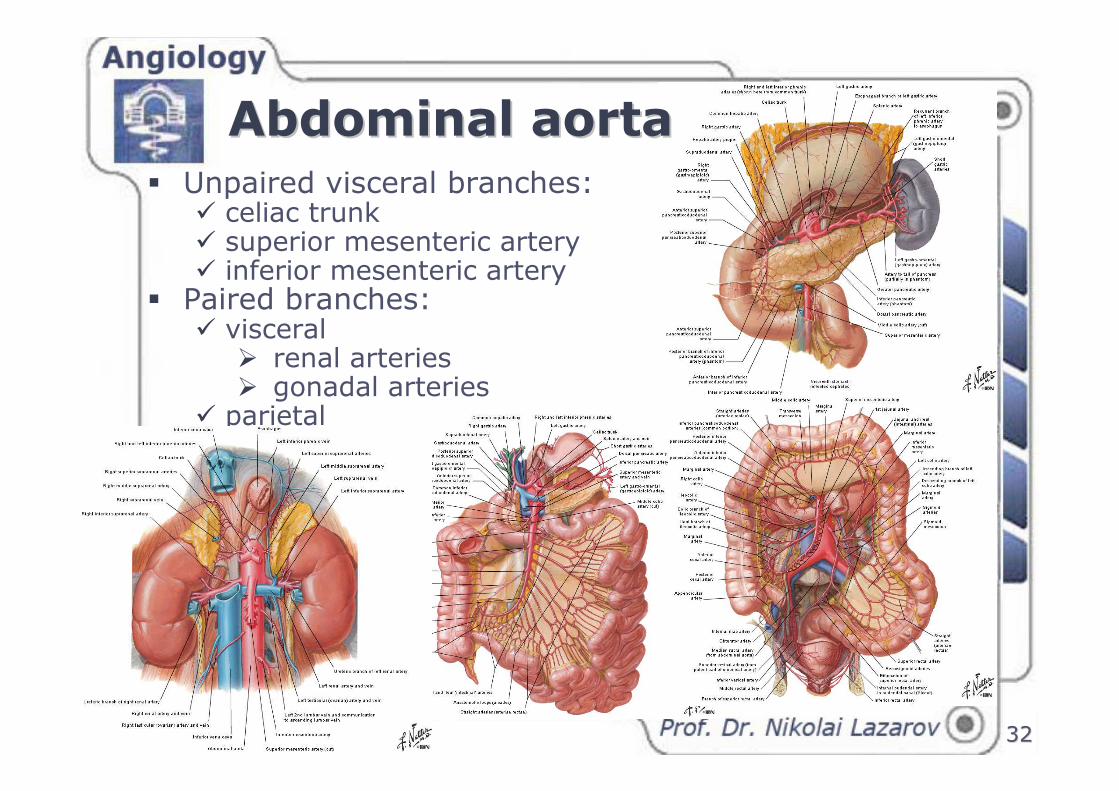
� Unpaired visceral branches:� celiac trunk� superior mesenteric artery� inferior mesenteric artery
� Paired branches:� visceral
� renal arteries� gonadal arteries
� parietal
Abdominal aortaAbdominal aorta
3333
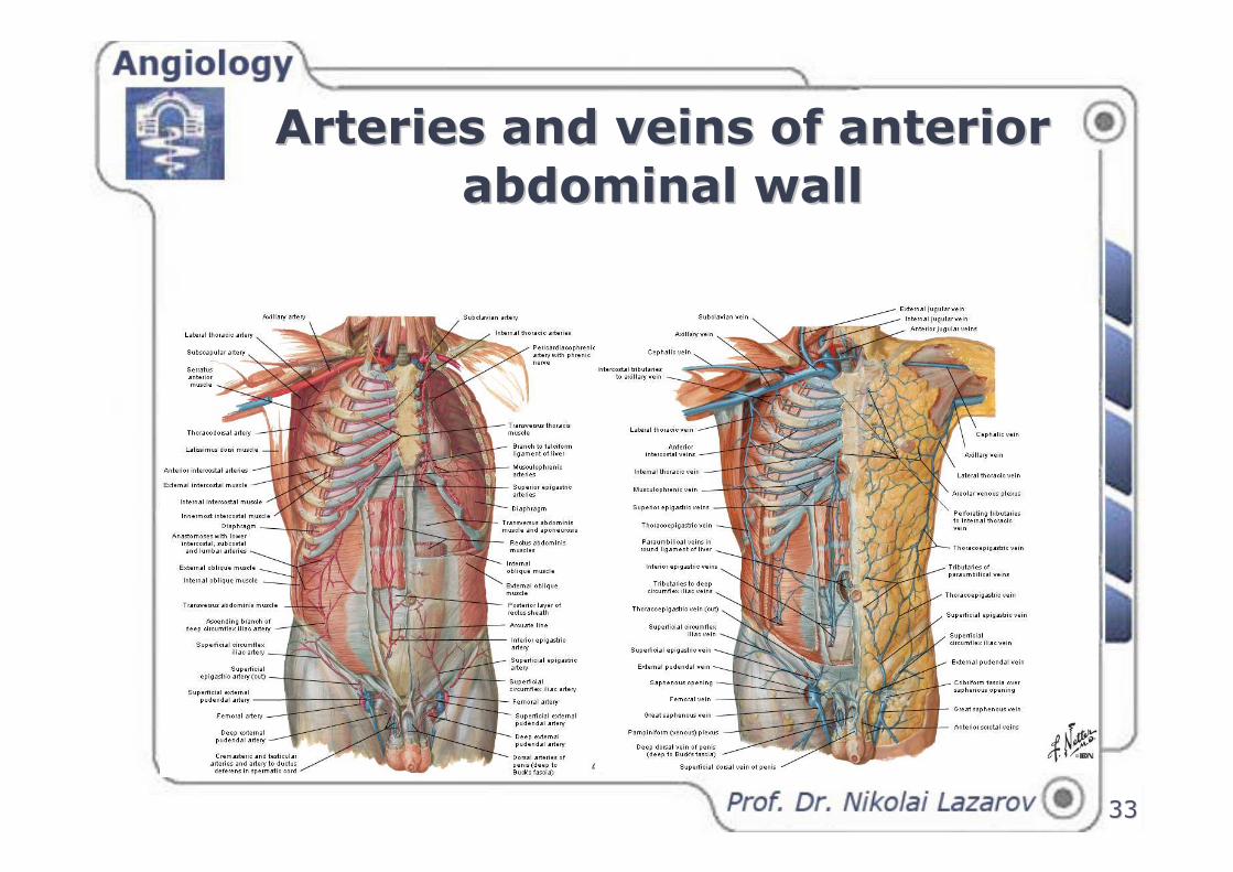
Arteries and veins of anterior Arteries and veins of anterior
abdominal wallabdominal wall
3434
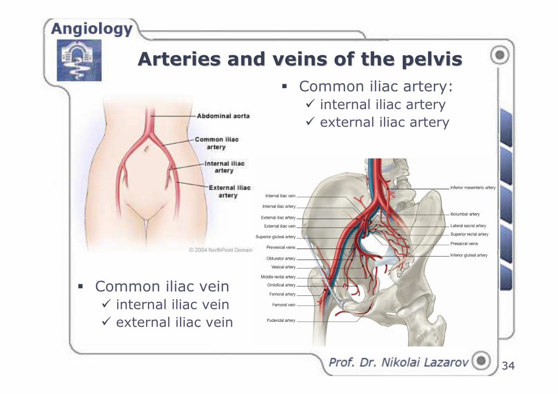
Arteries and veins of the pelvisArteries and veins of the pelvis
� Common iliac artery:� internal iliac artery
� external iliac artery
� Common iliac vein� internal iliac vein
� external iliac vein
3535
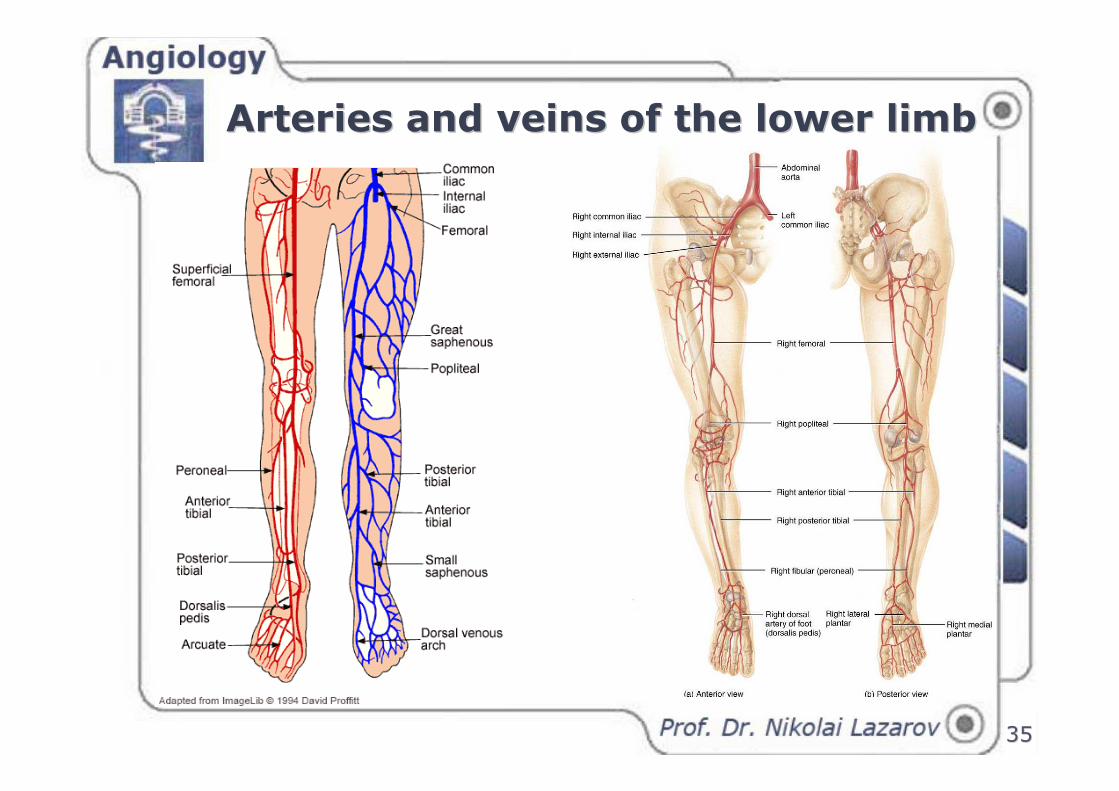
Arteries and veins of the lower limbArteries and veins of the lower limb

3636
PrePre-- and postnatal circulationand postnatal circulation
3737
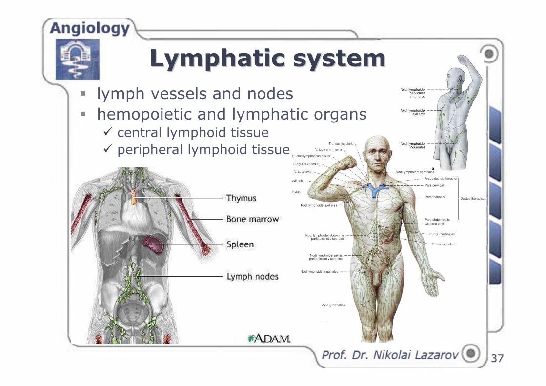
Lymphatic systemLymphatic system
� lymph vessels and nodes
� hemopoietic and lymphatic organs � central lymphoid tissue
� peripheral lymphoid tissue
3838
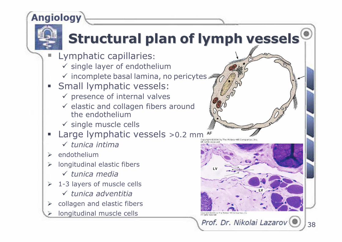
� Lymphatic capillaries: :
� single layer of endothelium
� incomplete basal lamina, no pericytes
� Small lymphatic vessels:� presence of internal valves
� elastic and collagen fibers around the endothelium
� single muscle cells
� Large lymphatic vessels >0.2 mm
� tunica intima
� endothelium
� longitudinal elastic fibers
� tunica media
� 1-3 layers of muscle cells
� tunica adventitia
� collagen and elastic fibers
� longitudinal muscle cells
StructuralStructural planplan ofof lymphlymph vesselsvessels
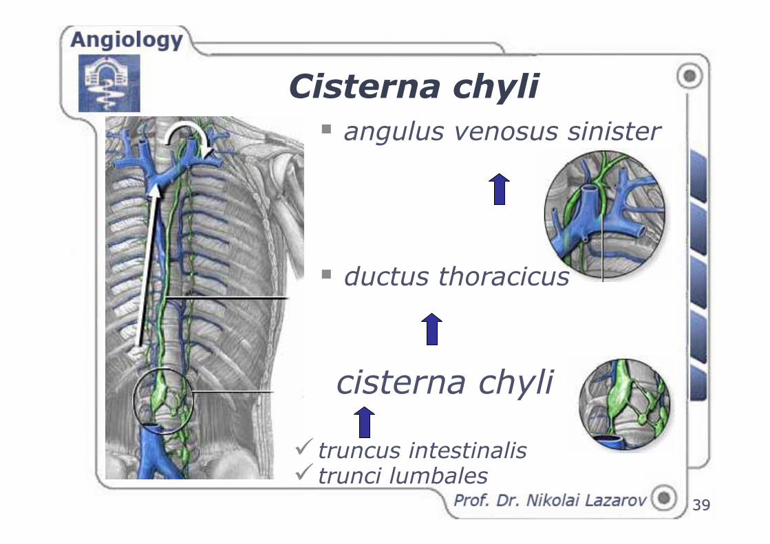
Cisterna chyli
� truncus intestinalis� trunci lumbales
cisterna chyli
� angulus venosus sinister
� ductus thoracicus
3939
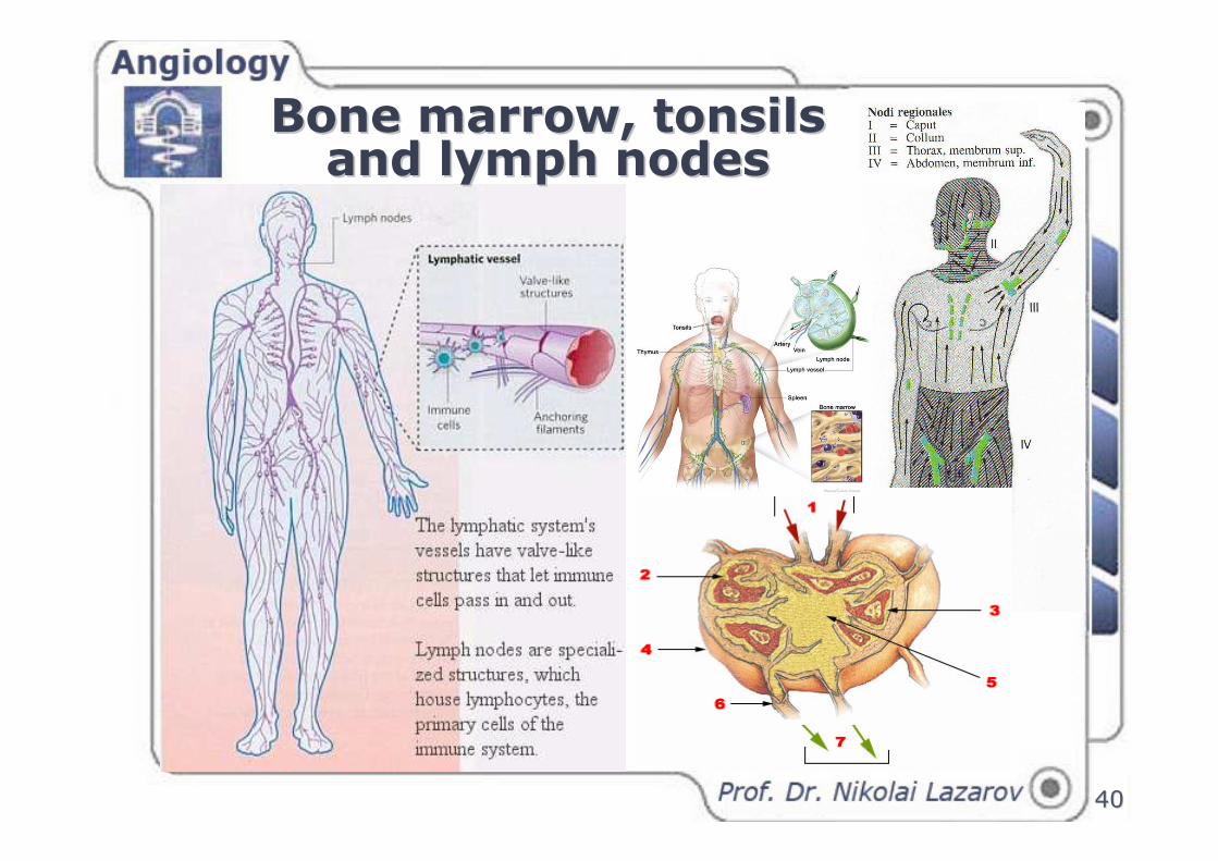
Bone marrow, tonsils Bone marrow, tonsils and lymph nodesand lymph nodes
4040
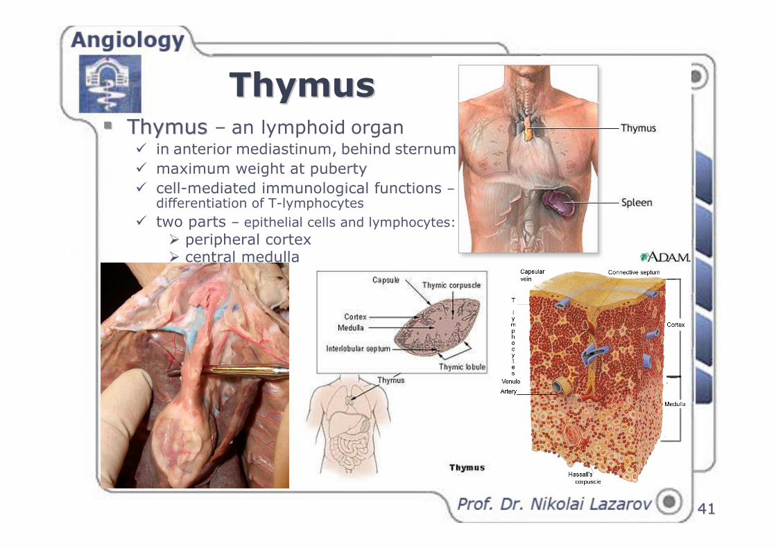
ThymusThymus�� Thymus Thymus – an lymphoid organ
� in anterior mediastinum, behind sternum
� maximum weight at puberty
� cell-mediated immunological functions –differentiation of T-lymphocytes
� two parts – epithelial cells and lymphocytes:
� peripheral cortex� central medulla
4141
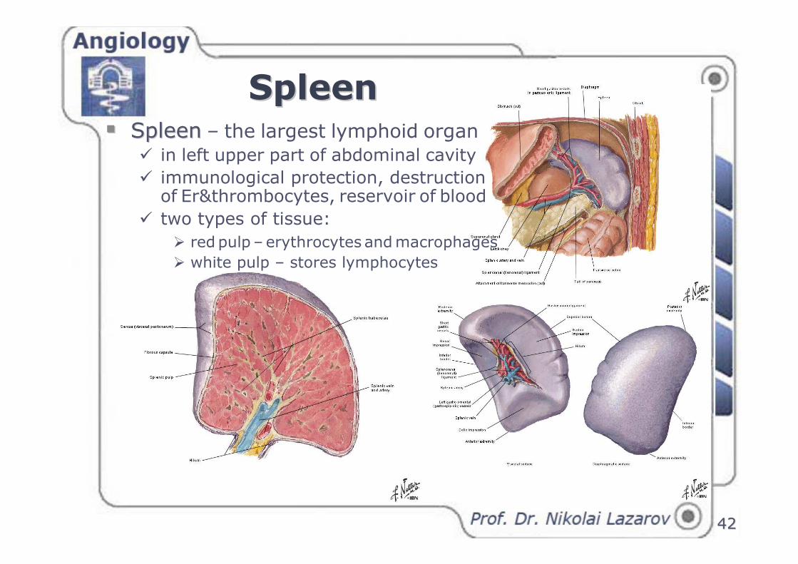
SpleenSpleen�� SpleenSpleen – the largest lymphoid organ
� in left upper part of abdominal cavity
� immunological protection, destruction of Er&thrombocytes, reservoir of blood
� two types of tissue:
� red pulp – erythrocytes and macrophages
� white pulp – stores lymphocytes
4242
Doctor FunDoctor FunDoctor FunDoctor FunDoctor FunDoctor FunDoctor FunDoctor Fun
Thank youThank youThank youThank youThank youThank youThank youThank you ........................