Embed Size (px)
Citation preview
1
Manual of Structural Kinesiology The Ankle and Foot Joints 11-1
Chapter 11The Ankle and Foot JointsManual of Structural Kinesiology
R.T. Floyd, Ed.D, ATC, CSCS
Copyright © The McGraw-Hill Companies, Inc. Reprinted by permission.
Manual of Structural Kinesiology The Ankle and Foot Joints 11-2
The Ankle and Foot Joint
• 26 bones• 19 large muscles• Many small (intrinsic) muscles• More than 100 ligaments• Support & propulsion
– Foot trouble - common ailment– Poor foot mechanics leads to foot discomfort– No substitute for adequate muscular development,
strength, & proper foot mechanics
Manual of Structural Kinesiology The Ankle and Foot Joints 11-3
The Ankle and Foot Joint
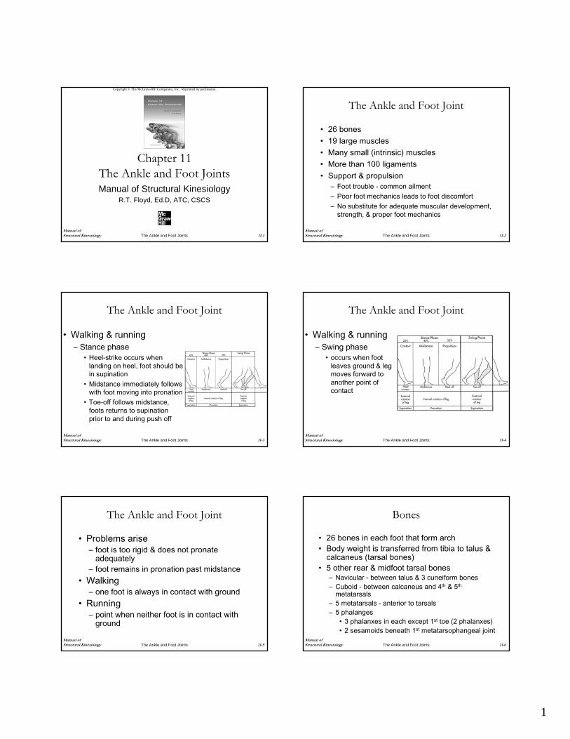
• Walking & running– Stance phase
• Heel-strike occurs when landing on heel, foot should be in supination
• Midstance immediately follows with foot moving into pronation
• Toe-off follows midstance, foots returns to supinationprior to and during push off
Manual of Structural Kinesiology The Ankle and Foot Joints 11-4
The Ankle and Foot Joint
• Walking & running– Swing phase
• occurs when foot leaves ground & leg moves forward to another point of contact
Manual of Structural Kinesiology The Ankle and Foot Joints 11-5
The Ankle and Foot Joint
• Problems arise– foot is too rigid & does not pronate
adequately– foot remains in pronation past midstance
• Walking– one foot is always in contact with ground
• Running– point when neither foot is in contact with
ground
Manual of Structural Kinesiology The Ankle and Foot Joints 11-6
Bones
• 26 bones in each foot that form arch• Body weight is transferred from tibia to talus &
calcaneus (tarsal bones)• 5 other rear & midfoot tarsal bones
– Navicular - between talus & 3 cuneiform bones– Cuboid - between calcaneus and 4th & 5th
metatarsals– 5 metatarsals - anterior to tarsals– 5 phalanges
• 3 phalanxes in each except 1st toe (2 phalanxes)• 2 sesamoids beneath 1st metatarsophangeal joint
2
Manual of Structural Kinesiology The Ankle and Foot Joints 11-7
Bones
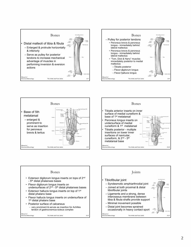
• Distal malleoli of tibia & fibula– Enlarged & protrude horizontally
& inferiorly– Serve as pulley for posterior
tendons to increase mechanical advantage of muscles in performing inversion & eversionactions
Manual of Structural Kinesiology The Ankle and Foot Joints 11-8
Bones– Pulley for posterior tendons
• Peroneus brevis & peroneuslongus - immediately behind lateral malleolus
• Peroneus brevis & peroneuslongus - immediately behind lateral malleolus
• “Tom, Dick & Harry” muscles immediately posterior to medial malleolus
– Tibialis posterior– Flexor digitorum longus– Flexor hallucis longus
Manual of Structural Kinesiology The Ankle and Foot Joints 11-9
Bones
• Base of 5th metatarsal – enlarged &
prominent to serve as insertion for peroneusbrevis & tertius
Manual of Structural Kinesiology The Ankle and Foot Joints 11-10
Bones
• Tibialis anterior inserts on inner surface of medial cuneiform & base of 1st metatarsal
• Peroneus longus inserts on undersurface of medial cuneiform & 1st metatarsal
• Tibialis posterior - multiple insertions on lower inner surfaces of navicular, cuneiform, & 2nd - 5th
metatarsal base
Manual of Structural Kinesiology The Ankle and Foot Joints 11-11
Bones• Extensor digitorum longus inserts on tops of 2nd
- 5th distal phalanxes bases• Flexor digitorum longus inserts on
undersurfaces of 2nd - 5th distal phalanxes bases• Extensor hallucis longus inserts on top of 1st
distal phalanx base• Flexor hallucis longus inserts on undersurface of
1st distal phalanx base• Posterior surface of calcaneus
– very prominent & serves as insertion for Achilles tendon of gastrocnemius-soleus complex
Manual of Structural Kinesiology The Ankle and Foot Joints 11-12
Joints
• Tibiofibular joint– Syndesmotic amphiarthrodial joint– Joined at both proximal & distal
tibiofibular joints– Ligaments and a strong, dense
interosseus membrane between tibia & fibula shafts provide support
– Minimal movement possible– Distal joint becomes sprained
occasionally in heavy contact sport
3
Manual of Structural Kinesiology The Ankle and Foot Joints 11-13
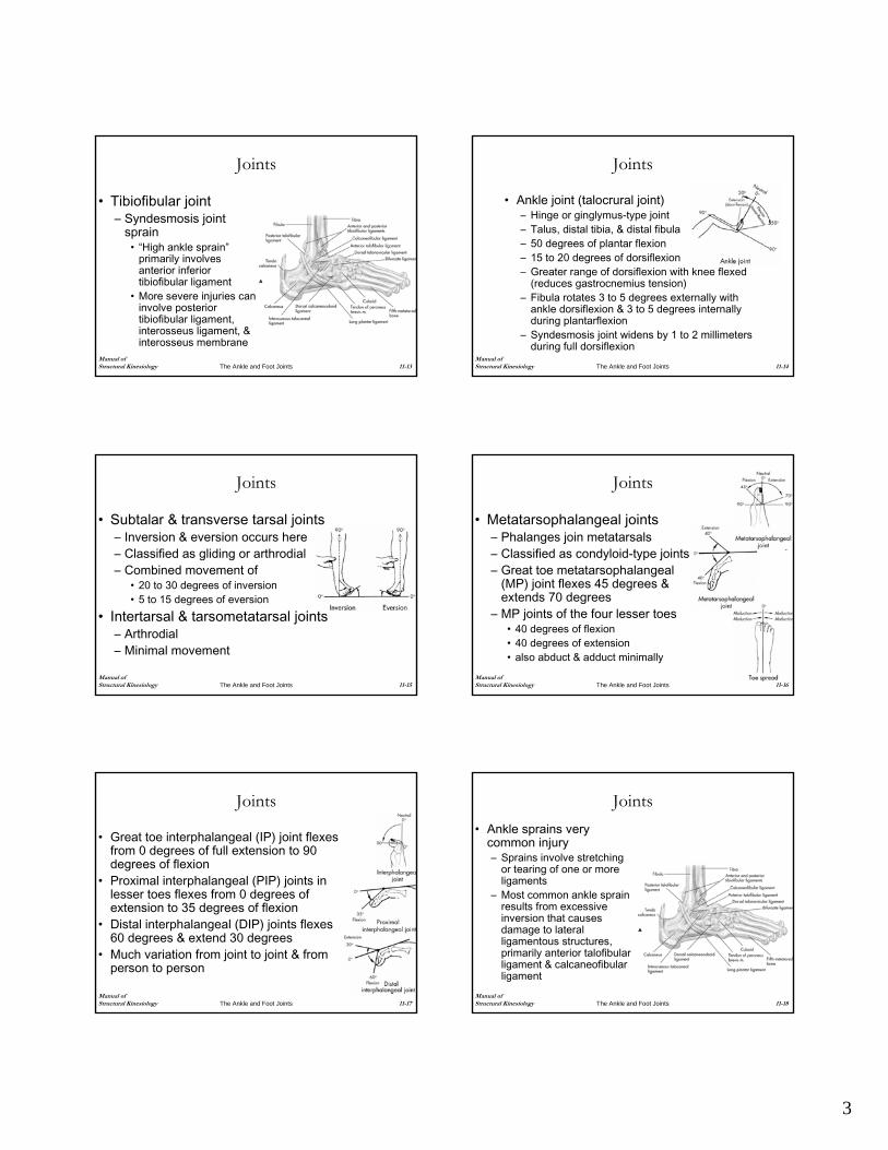
Joints
• Tibiofibular joint– Syndesmosis joint
sprain• “High ankle sprain”
primarily involves anterior inferior tibiofibular ligament
• More severe injuries can involve posterior tibiofibular ligament, interosseus ligament, & interosseus membrane
Manual of Structural Kinesiology The Ankle and Foot Joints 11-14
Joints
• Ankle joint (talocrural joint)– Hinge or ginglymus-type joint– Talus, distal tibia, & distal fibula– 50 degrees of plantar flexion– 15 to 20 degrees of dorsiflexion– Greater range of dorsiflexion with knee flexed
(reduces gastrocnemius tension)– Fibula rotates 3 to 5 degrees externally with
ankle dorsiflexion & 3 to 5 degrees internally during plantarflexion
– Syndesmosis joint widens by 1 to 2 millimeters during full dorsiflexion
Manual of Structural Kinesiology The Ankle and Foot Joints 11-15
Joints
• Subtalar & transverse tarsal joints– Inversion & eversion occurs here – Classified as gliding or arthrodial– Combined movement of
• 20 to 30 degrees of inversion• 5 to 15 degrees of eversion
• Intertarsal & tarsometatarsal joints– Arthrodial– Minimal movement
Manual of Structural Kinesiology The Ankle and Foot Joints 11-16
Joints
• Metatarsophalangeal joints– Phalanges join metatarsals– Classified as condyloid-type joints– Great toe metatarsophalangeal
(MP) joint flexes 45 degrees & extends 70 degrees
– MP joints of the four lesser toes• 40 degrees of flexion• 40 degrees of extension• also abduct & adduct minimally
Manual of Structural Kinesiology The Ankle and Foot Joints 11-17
Joints
• Great toe interphalangeal (IP) joint flexes from 0 degrees of full extension to 90 degrees of flexion
• Proximal interphalangeal (PIP) joints in lesser toes flexes from 0 degrees of extension to 35 degrees of flexion
• Distal interphalangeal (DIP) joints flexes 60 degrees & extend 30 degrees
• Much variation from joint to joint & from person to person
Manual of Structural Kinesiology The Ankle and Foot Joints 11-18
Joints• Ankle sprains very
common injury– Sprains involve stretching
or tearing of one or more ligaments
– Most common ankle sprain results from excessive inversion that causes damage to lateral ligamentous structures, primarily anterior talofibularligament & calcaneofibularligament
4
Manual of Structural Kinesiology The Ankle and Foot Joints 11-19
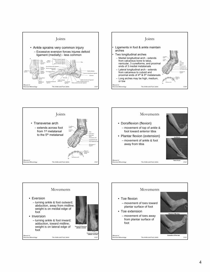
Joints
• Ankle sprains very common injury– Excessive eversion forces injures deltoid
ligament (medially) - less common
Manual of Structural Kinesiology The Ankle and Foot Joints 11-20
Joints
• Ligaments in foot & ankle maintain arches
• Two longitudinal arches– Medial longitudinal arch - extends
from calcaneus bone to talus, navicular, 3 cuneiforms, and proximal ends of 3 medial metatarsals
– Lateral longitudinal arch - extends from calcaneus to cuboid and proximal ends of 4th & 5th metatarsals
– Long arches may be high, medium, or low
Manual of Structural Kinesiology The Ankle and Foot Joints 11-21
Joints
• Transverse arch– extends across foot
from 1st metatarsal to the 5th metatarsal
Manual of Structural Kinesiology The Ankle and Foot Joints 11-22
Movements
• Dorsiflexion (flexion)– movement of top of ankle &
foot toward anterior tibia• Plantar flexion (extension)
– movement of ankle & foot away from tibia
Manual of Structural Kinesiology The Ankle and Foot Joints 11-23
Movements
• Eversion– turning ankle & foot outward;
abduction, away from midline; weight is on medial edge of foot
• Inversion– turning ankle & foot inward;
adduction, toward midline; weight is on lateral edge of foot
Manual of Structural Kinesiology The Ankle and Foot Joints 11-24
Movements
• Toe flexion– movement of toes toward
plantar surface of foot• Toe extension
– movement of toes away from plantar surface of foot
5
Manual of Structural Kinesiology The Ankle and Foot Joints 11-25
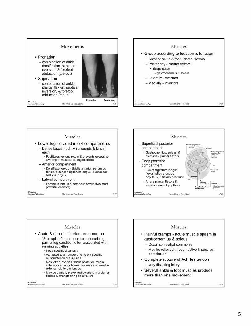
Movements
• Pronation– combination of ankle
dorsiflexion, subtalareversion, & forefoot abduction (toe-out)
• Supination– combination of ankle
plantar flexion, subtalarinversion, & forefoot adduction (toe-in)
Manual of Structural Kinesiology The Ankle and Foot Joints 11-26
Muscles• Group according to location & function
– Anterior ankle & foot - dorsal flexors– Posteriorly - plantar flexors
• triceps surae– gastrocnemius & soleus
– Laterally - evertors– Medially - invertors
Manual of Structural Kinesiology The Ankle and Foot Joints 11-27
Muscles• Lower leg - divided into 4 compartments
– Dense fascia - tightly surrounds & binds each
• Facilitates venous return & prevents excessive swelling of muscles during exercise
– Anterior compartment• Dorsiflexor group - tibialis anterior, peroneus
tertius, extensor digitorum longus, & extensor hallucis longus
– Lateral compartment• Peroneus longus & peroneus brevis (two most
powerful evertors)Manual of Structural Kinesiology The Ankle and Foot Joints 11-28
Muscles– Superficial posterior
compartment• Gastrocnemius, soleus, &
plantaris - plantar flexors– Deep posterior
compartment• Flexor digitorum longus,
flexor hallucis longus, popliteus, & tibialis posterior
• All are plantar flexors & invertors except popliteus
Manual of Structural Kinesiology The Ankle and Foot Joints 11-29
Muscles• Acute & chronic injuries are common
– “Shin splints” - common term describing painful leg condition often associated with running activities
• Not a specific diagnosis• Attributed to a number of different specific
musculotendinous injuries• Most often involves tibialis posterior, medial
soleus, or anterior tibialis, but may also involve extensor digitorum longus
• May be partially prevented by stretching plantar flexors & strengthening dorsiflexors
Manual of Structural Kinesiology The Ankle and Foot Joints 11-30
Muscles• Painful cramps - acute muscle spasm in
gastrocnemius & soleus– Occur somewhat commonly– May be relieved through active & passive
dorsiflexion• Complete rupture of Achilles tendon
– very disabling injury• Several ankle & foot muscles produce
more than one movement
6
Manual of Structural Kinesiology The Ankle and Foot Joints 11-31
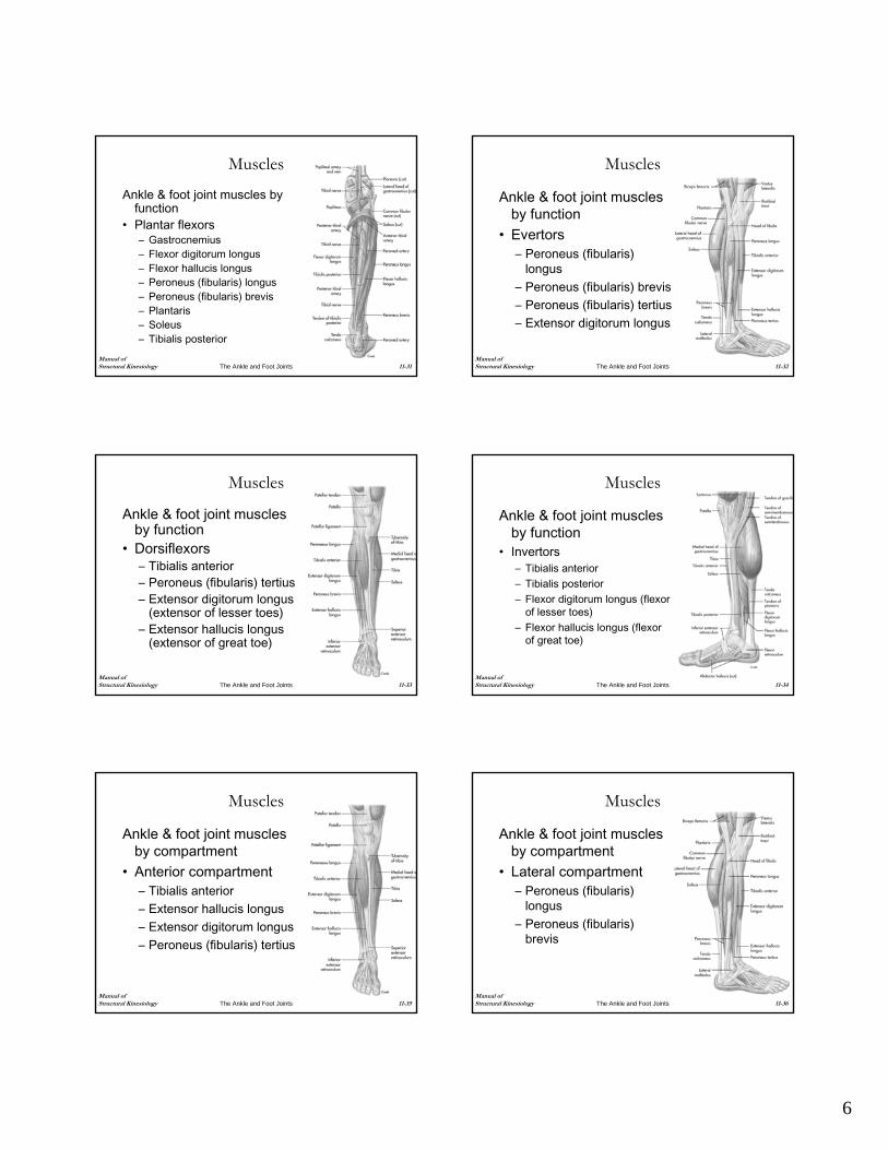
Muscles
Ankle & foot joint muscles by function
• Plantar flexors– Gastrocnemius– Flexor digitorum longus– Flexor hallucis longus– Peroneus (fibularis) longus– Peroneus (fibularis) brevis– Plantaris– Soleus– Tibialis posterior
Manual of Structural Kinesiology The Ankle and Foot Joints 11-32
Muscles
Ankle & foot joint muscles by function
• Evertors– Peroneus (fibularis)
longus– Peroneus (fibularis) brevis– Peroneus (fibularis) tertius– Extensor digitorum longus
Manual of Structural Kinesiology The Ankle and Foot Joints 11-33
Muscles
Ankle & foot joint muscles by function
• Dorsiflexors– Tibialis anterior– Peroneus (fibularis) tertius– Extensor digitorum longus
(extensor of lesser toes)– Extensor hallucis longus
(extensor of great toe)
Manual of Structural Kinesiology The Ankle and Foot Joints 11-34
Muscles
Ankle & foot joint muscles by function
• Invertors– Tibialis anterior– Tibialis posterior– Flexor digitorum longus (flexor
of lesser toes)– Flexor hallucis longus (flexor
of great toe)
Manual of Structural Kinesiology The Ankle and Foot Joints 11-35
Muscles
Ankle & foot joint muscles by compartment
• Anterior compartment– Tibialis anterior– Extensor hallucis longus– Extensor digitorum longus– Peroneus (fibularis) tertius
Manual of Structural Kinesiology The Ankle and Foot Joints 11-36
Muscles
Ankle & foot joint muscles by compartment
• Lateral compartment– Peroneus (fibularis)
longus– Peroneus (fibularis)
brevis
7
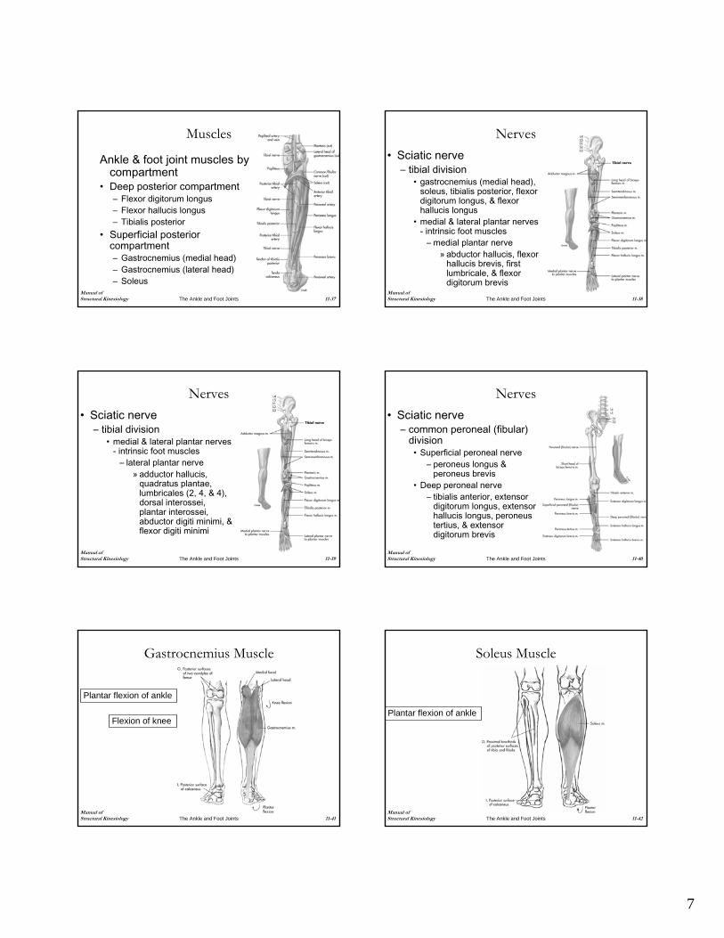
Manual of Structural Kinesiology The Ankle and Foot Joints 11-37
Muscles
Ankle & foot joint muscles by compartment
• Deep posterior compartment– Flexor digitorum longus– Flexor hallucis longus– Tibialis posterior
• Superficial posterior compartment– Gastrocnemius (medial head)– Gastrocnemius (lateral head)– Soleus
Manual of Structural Kinesiology The Ankle and Foot Joints 11-38
Nerves• Sciatic nerve
– tibial division• gastrocnemius (medial head),
soleus, tibialis posterior, flexor digitorum longus, & flexor hallucis longus
• medial & lateral plantar nerves - intrinsic foot muscles
– medial plantar nerve» abductor hallucis, flexor
hallucis brevis, first lumbricale, & flexor digitorum brevis
Manual of Structural Kinesiology The Ankle and Foot Joints 11-39
Nerves• Sciatic nerve
– tibial division• medial & lateral plantar nerves
- intrinsic foot muscles– lateral plantar nerve
» adductor hallucis, quadratus plantae, lumbricales (2, 4, & 4), dorsal interossei, plantar interossei, abductor digiti minimi, & flexor digiti minimi
Manual of Structural Kinesiology The Ankle and Foot Joints 11-40
Nerves• Sciatic nerve
– common peroneal (fibular) division
• Superficial peroneal nerve– peroneus longus &
peroneus brevis• Deep peroneal nerve
– tibialis anterior, extensor digitorum longus, extensor hallucis longus, peroneustertius, & extensor digitorum brevis
Manual of Structural Kinesiology The Ankle and Foot Joints 11-41
Gastrocnemius Muscle
Plantar flexion of ankle
Flexion of knee
Manual of Structural Kinesiology The Ankle and Foot Joints 11-42
Soleus Muscle
Plantar flexion of ankle
8
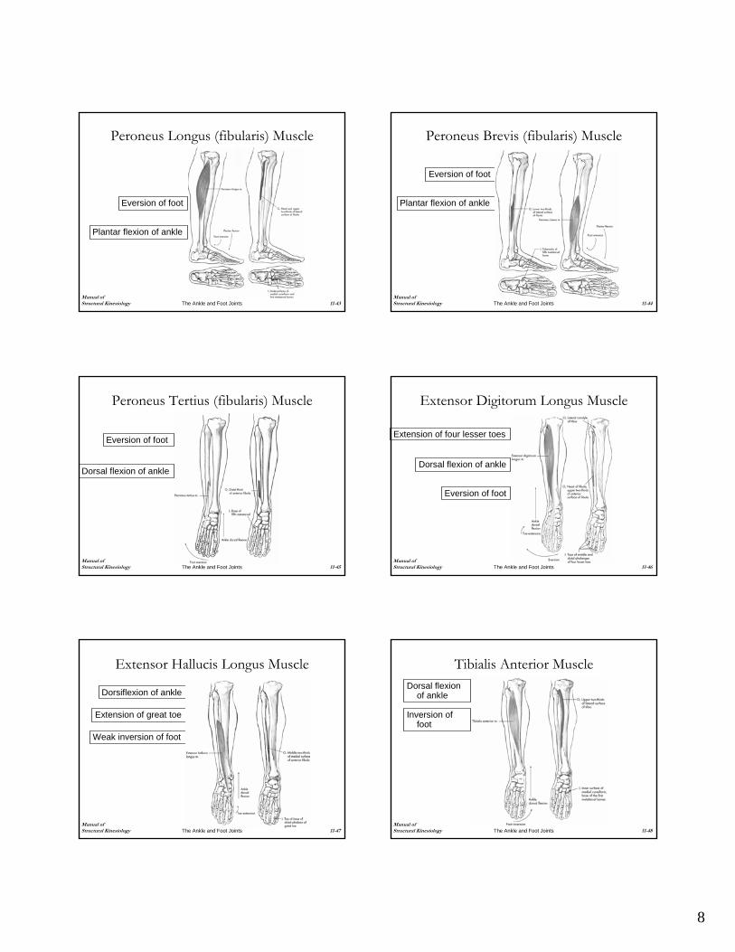
Manual of Structural Kinesiology The Ankle and Foot Joints 11-43
Peroneus Longus (fibularis) Muscle
Eversion of foot
Plantar flexion of ankle
Manual of Structural Kinesiology The Ankle and Foot Joints 11-44
Peroneus Brevis (fibularis) Muscle
Eversion of foot
Plantar flexion of ankle
Manual of Structural Kinesiology The Ankle and Foot Joints 11-45
Peroneus Tertius (fibularis) Muscle
Eversion of foot
Dorsal flexion of ankle
Manual of Structural Kinesiology The Ankle and Foot Joints 11-46
Extensor Digitorum Longus Muscle
Extension of four lesser toes
Dorsal flexion of ankle
Eversion of foot
Manual of Structural Kinesiology The Ankle and Foot Joints 11-47
Extensor Hallucis Longus Muscle
Extension of great toe
Dorsiflexion of ankle
Weak inversion of foot
Manual of Structural Kinesiology The Ankle and Foot Joints 11-48
Tibialis Anterior MuscleDorsal flexion
of ankle
Inversion of foot
9
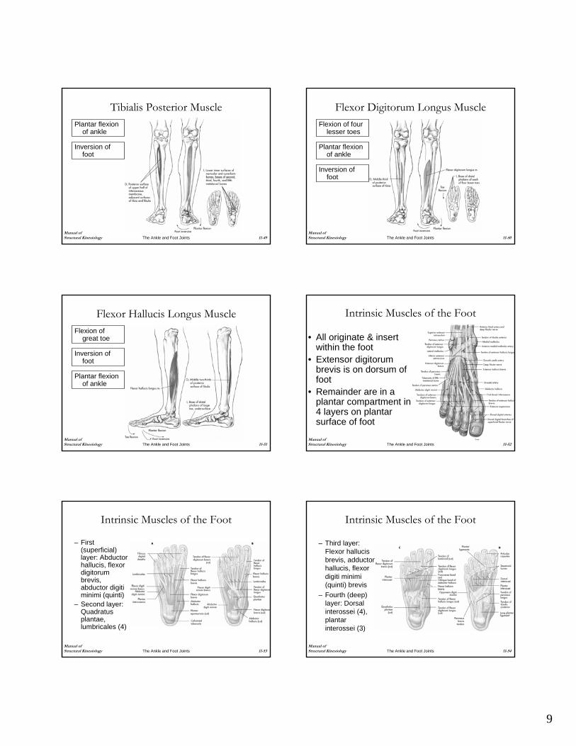
Manual of Structural Kinesiology The Ankle and Foot Joints 11-49
Tibialis Posterior MusclePlantar flexion
of ankle
Inversion of foot
Manual of Structural Kinesiology The Ankle and Foot Joints 11-50
Flexor Digitorum Longus MuscleFlexion of four
lesser toes
Plantar flexion of ankle
Inversion of foot
Manual of Structural Kinesiology The Ankle and Foot Joints 11-51
Flexor Hallucis Longus MuscleFlexion of
great toe
Inversion of foot
Plantar flexion of ankle
Manual of Structural Kinesiology The Ankle and Foot Joints 11-52
Intrinsic Muscles of the Foot
• All originate & insert within the foot
• Extensor digitorumbrevis is on dorsum of foot
• Remainder are in a plantar compartment in 4 layers on plantar surface of foot
Manual of Structural Kinesiology The Ankle and Foot Joints 11-53
Intrinsic Muscles of the Foot
– First (superficial) layer: Abductor hallucis, flexor digitorumbrevis, abductor digitiminimi (quinti)
– Second layer: Quadratusplantae, lumbricales (4)
Manual of Structural Kinesiology The Ankle and Foot Joints 11-54
Intrinsic Muscles of the Foot
– Third layer: Flexor hallucisbrevis, adductor hallucis, flexor digiti minimi(quinti) brevis
– Fourth (deep) layer: Dorsal interossei (4), plantar interossei (3)
10
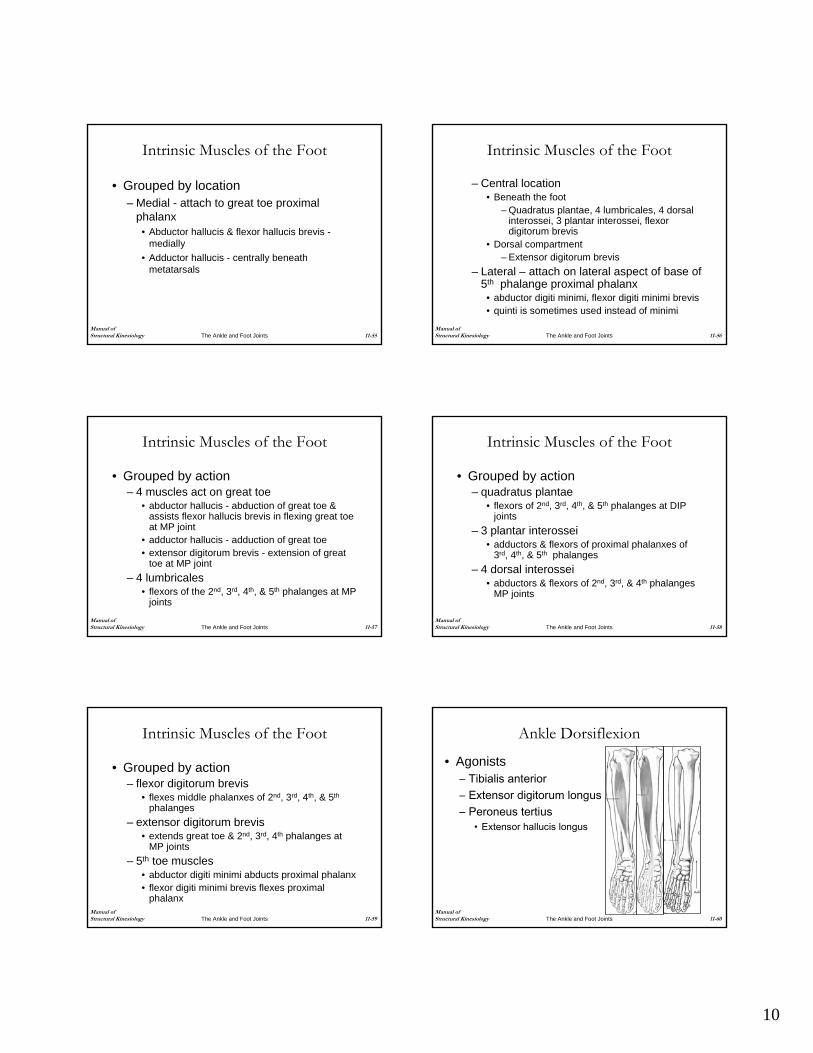
Manual of Structural Kinesiology The Ankle and Foot Joints 11-55
Intrinsic Muscles of the Foot
• Grouped by location – Medial - attach to great toe proximal
phalanx• Abductor hallucis & flexor hallucis brevis -
medially• Adductor hallucis - centrally beneath
metatarsals
Manual of Structural Kinesiology The Ankle and Foot Joints 11-56
Intrinsic Muscles of the Foot
– Central location• Beneath the foot
– Quadratus plantae, 4 lumbricales, 4 dorsal interossei, 3 plantar interossei, flexor digitorum brevis
• Dorsal compartment– Extensor digitorum brevis
– Lateral – attach on lateral aspect of base of 5th phalange proximal phalanx
• abductor digiti minimi, flexor digiti minimi brevis• quinti is sometimes used instead of minimi
Manual of Structural Kinesiology The Ankle and Foot Joints 11-57
Intrinsic Muscles of the Foot
• Grouped by action– 4 muscles act on great toe
• abductor hallucis - abduction of great toe & assists flexor hallucis brevis in flexing great toe at MP joint
• adductor hallucis - adduction of great toe• extensor digitorum brevis - extension of great
toe at MP joint– 4 lumbricales
• flexors of the 2nd, 3rd, 4th, & 5th phalanges at MP joints
Manual of Structural Kinesiology The Ankle and Foot Joints 11-58
Intrinsic Muscles of the Foot
• Grouped by action– quadratus plantae
• flexors of 2nd, 3rd, 4th, & 5th phalanges at DIP joints
– 3 plantar interossei• adductors & flexors of proximal phalanxes of
3rd, 4th, & 5th phalanges– 4 dorsal interossei
• abductors & flexors of 2nd, 3rd, & 4th phalanges MP joints
Manual of Structural Kinesiology The Ankle and Foot Joints 11-59
Intrinsic Muscles of the Foot
• Grouped by action– flexor digitorum brevis
• flexes middle phalanxes of 2nd, 3rd, 4th, & 5th
phalanges– extensor digitorum brevis
• extends great toe & 2nd, 3rd, 4th phalanges at MP joints
– 5th toe muscles• abductor digiti minimi abducts proximal phalanx• flexor digiti minimi brevis flexes proximal
phalanxManual of Structural Kinesiology The Ankle and Foot Joints 11-60
Ankle Dorsiflexion
• Agonists– Tibialis anterior– Extensor digitorum longus– Peroneus tertius
• Extensor hallucis longus
11
Manual of Structural Kinesiology The Ankle and Foot Joints 11-61
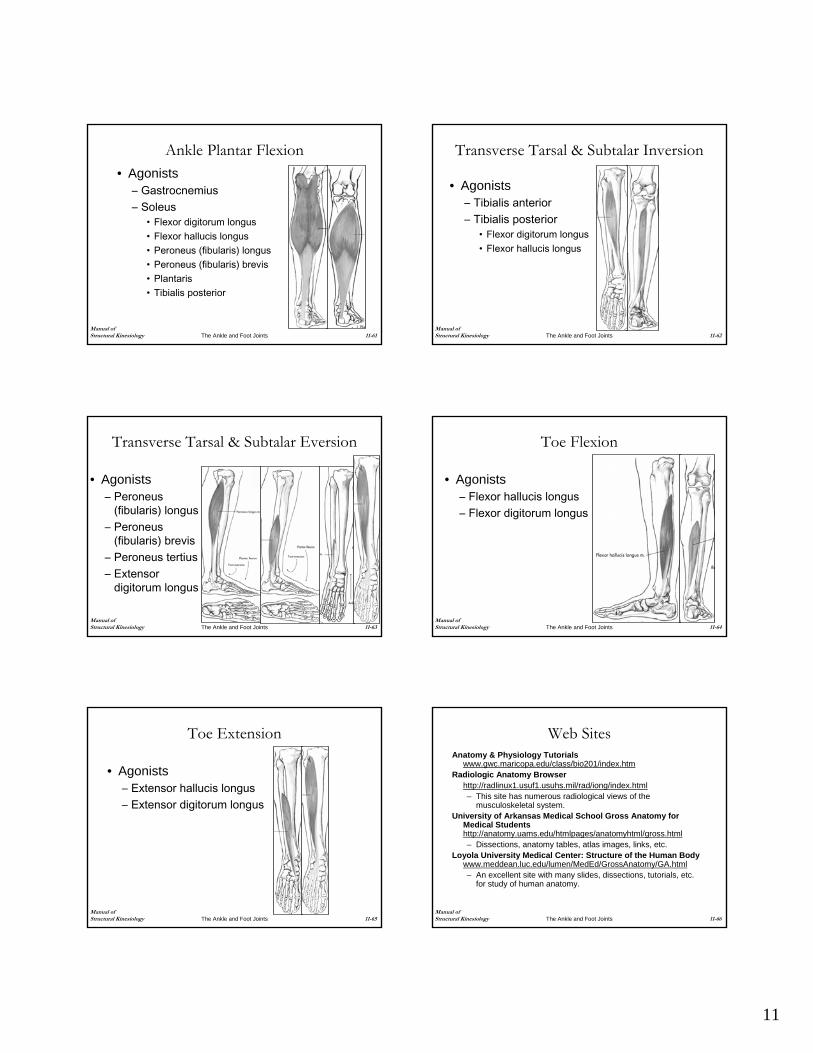
Ankle Plantar Flexion• Agonists
– Gastrocnemius– Soleus
• Flexor digitorum longus• Flexor hallucis longus• Peroneus (fibularis) longus• Peroneus (fibularis) brevis• Plantaris• Tibialis posterior
Manual of Structural Kinesiology The Ankle and Foot Joints 11-62
Transverse Tarsal & Subtalar Inversion
• Agonists– Tibialis anterior– Tibialis posterior
• Flexor digitorum longus• Flexor hallucis longus
Manual of Structural Kinesiology The Ankle and Foot Joints 11-63
Transverse Tarsal & Subtalar Eversion
• Agonists– Peroneus
(fibularis) longus– Peroneus
(fibularis) brevis– Peroneus tertius– Extensor
digitorum longus
Manual of Structural Kinesiology The Ankle and Foot Joints 11-64
Toe Flexion
• Agonists– Flexor hallucis longus– Flexor digitorum longus
Manual of Structural Kinesiology The Ankle and Foot Joints 11-65
Toe Extension
• Agonists– Extensor hallucis longus– Extensor digitorum longus
Manual of Structural Kinesiology The Ankle and Foot Joints 11-66
Web SitesAnatomy & Physiology Tutorials
www.gwc.maricopa.edu/class/bio201/index.htmRadiologic Anatomy Browser
http://radlinux1.usuf1.usuhs.mil/rad/iong/index.html– This site has numerous radiological views of the
musculoskeletal system.University of Arkansas Medical School Gross Anatomy for
Medical Studentshttp://anatomy.uams.edu/htmlpages/anatomyhtml/gross.html– Dissections, anatomy tables, atlas images, links, etc.
Loyola University Medical Center: Structure of the Human Bodywww.meddean.luc.edu/lumen/MedEd/GrossAnatomy/GA.html– An excellent site with many slides, dissections, tutorials, etc.
for study of human anatomy.

12
Manual of Structural Kinesiology The Ankle and Foot Joints 11-67
Web SitesWheeless' Textbook of Orthopaedics
www.ortho-u.net/– This site has an extensive index of links to the fractures,
joints, muscles, nerves, trauma, medications, medical topics, lab tests as well as links to orthopaedic journals, other orthopaedic, and medical news.
Foot and Ankle Web Indexwww.footandankle.com– The foot and ankle link library located at this site is very helpful.
American College of Foot and Ankle Surgeonswww.acfas.org– This site, sponsored by podiatric surgeons and doctors of podiatric
medicine (DPM), has information on topics relating to foot health-foot and ankle deformities and injuries; care of the diabetic foot; foot and ankle disorders caused by arthritis, aging, trauma and sports injuries; and congenital deformities and disease.
Manual of Structural Kinesiology The Ankle and Foot Joints 11-68
Web SitesThe University of Texas MD Anderson Cancer Center
Multimedia and Learning Resourceswww.mdacc.tmc.edu/mmlearn/anatomy.html– This site has numerous cadaveric cuts of the foot, knee, hand,
and elbow; an interactive ankle; and a rotating foot and ankleAmerican Orthopaedic Foot and Ankle Society
www.aofas.org– Numerous patient education brochures regarding foot and
ankle problems are found herePremiere Medical Search Engine
www.medsite.com– This site allows the reader to enter any medical condition and
it will search the net to find relevant articles.
Manual of Structural Kinesiology The Ankle and Foot Joints 11-69
Web SitesVirtual Hospital
www.vh.org– Numerous slides, patient information, etc.
The Dynamic Human version 2.0 CD-ROM: The Visual Guide to Anatomy & Physiologywww.mhhe.com/biosci/ap/dynamichuman2/– Web site that accompanies this CD-ROM