Embed Size (px)
Citation preview
© 2018 Surgical Neurology International | Published by Wolters Kluwer - Medknow
Editor:Ekkehard Kasper, M.D.,Harvard Medical School, Boston, MA, USA
OPEN ACCESSFor entire Editorial Board visit : http://www.surgicalneurologyint.com
SNI: Skull Base
Case Report
Suprasellar keratinous cyst: A case report and review on its radiological features and treatment outcomeC. W. Huo1,2, C. Caputo3, Y. Y. Wang1,2,4
1Department of Neurosurgery, St Vincent’s Hospital Melbourne, Victoria, Australia, 2Department of Surgery, St Vincent’s Hospital, The University of Melbourne, Parkville, Victoria, Australia, 3Department of Endocrinology, St Vincent’s Hospital Melbourne, Victoria, 4Keyhole Neurosurgery, Suite B, Level 2 Healy Wing, 41 Victoria parade, Fitzroy, VIC, Australia
E‑mail: *C. W. Huo ‑ [email protected]; C. Caputo ‑ [email protected]; Y. Y. Wang ‑ [email protected] *Corresponding author
Received: 23 July 17 Accepted: 21 November 17 Published: 22 January 18
AbstractBackground: Keratinous or epidermoid cysts (ECs) are encapsulated lesions lined by squamous cell epithelium. They comprise approximately 1% of intracranial lesions. Contrary to dermoid cysts, they lack dermal elements such as sebaceous or apocrine glands and hair follicles. The sellar region is the second most common intracranial site following the cerebellopontine angle. Here, we report a case of EC in a patient who complained of endocrine disturbances. We also performed a systematic review on previously published cases to analyze clinical and radiological characteristics and report the treatment outcomes of suprasellar ECs.Case Description: A 42‑year‑old woman presented with a one‑year history of amenorrhea, weight gain, severe headache, and visual disturbances for 6 months. Work‑up identified an elevated prolactin level and a temporal field defect of the right eye. Magnetic resonance imaging (MRI) showed a cystic suprasellar lesion pushing on the optic chiasm. She underwent endoscopic trans‑sphenoidal surgery, which confirmed a keratinous cyst on histology. Postoperatively, complete resection was confirmed on imaging. She did well although her hospital stay was prolonged due to diabetes insipidus and hypocortisolism.Conclusion: Chronic endocrine disturbances can be the presenting complaints of a suprasellar EC, whose T1‑weighted MRI appearance can be non‑specific, mimicking other differential diagnoses, such as a Rathke’s cleft cyst. However, the T2‑weighted MRI appearances of ECs are generally hyper‑intense and lesions show diffusion restriction. Treatment is surgical and yields good outcomes in most cases reported.
Key Words: Endoscopic, epidermoid cyst, keratinous cyst, skull base, suprasellar
INTRODUCTION
The suprasellar cistern is a cerebrospinal fluid (CSF)‑filled space below the third ventricle with a floor formed by the dura of the diaphragma sellae. Normally, this space contains the optic nerves and chiasm, the tuber cinereum and floor of the anterior third ventricle, the hypothalamus, and the vessels forming the circle of Willis.[61] Central nervous system (CNS) tumors arising
How to cite this article: Huo CW, Caputo C, Wang YY. Suprasellar keratinous cyst: A case report and review on its radiological features and treatment outcome. Surg Neurol Int 2018;9:15.http://surgicalneurologyint.com/Suprasellar-keratinous-cyst:-A-case-report-and-review-on-its-radiological-features-and-treatment-outcome/
This is an open access article distributed under the terms of the Creative Commons Attribution-NonCommercial-ShareAlike 3.0 License, which allows others to remix, tweak, and build upon the work non-commercially, as long as the author is credited and the new creations are licensed under the identical terms.
For reprints contact: [email protected]
Access this article onlineWebsite:www.surgicalneurologyint.comDOI: 10.4103/sni.sni_269_17Quick Response Code:
Surgical Neurology International 2018, 9:15 http://www.surgicalneurologyint.com/content/9/1/15
in the suprasellar region are diverse in etiology but are predominantly pituitary adenomas, meningiomas, and craniopharyngiomas.[15,28,30] Occasionally, a giant aneurysm of the internal carotid or the basilar artery may project into the suprasellar region.[61] Besides neoplastic inclusion cysts (e.g. dermoid and epidermoid cysts), non‑neoplastic cysts such as Rathke’s cleft cysts and arachnoid cysts have been described in this suprasellar space.[48]
Epidermoid cysts (ECs) are also known as keratinous cysts; when they occur within the CNS, they had been erroneously referred as neurenteric cysts[52] in the past. A distinction should be made between the two as neurenteric cysts are rare CNS lesions of endodermal origins, contrasting the ectodermal features that ECs exhibit on histopathology.[10,11] ECs are rare, representing approximately 0.2–1.8% of all primary CNS tumors[13,35,51,57] and are 4–9 times more common than dermoid cysts.[44] ECs were first described by Cruveilhier,[17] and due to their flaky keratin content as a result of epithelial cell desquamation, they appear pearly.[35,44,57] Later, Muller gave it the term “cholesteatoma” due to its cholesterol content[52] a term that was later abandoned.[36]1 These characteristic cysts were named “epidermoid” by Critchley and Ferguson to differentiate them from dermoid cysts, which are of mesodermal origin and contain skin adnexae.[16] In approximately 50% of the cases, ECs are found as extradural or intracerebral lesions along the cisternal space in the cerebellopontine angle (CPA). The second most common regions are the supra/parasellar area[13,22,31,35,70] (10–15%) and the fourth ventricle[13,22,31,35,44,51,57,70] (10–17%). ECs have also been reported intraventricularly, in the brainstem, cerebral parenchyma, pineal region, and Sylvian fissure.[30,44,57]
Here, we report a suprasellar EC case in a patient presenting with endocrine and visual compromise. Histological examination of the excised tumor confirmed a keratinous cyst. To put this observation into appropriate context, we systematically reviewed all previously published English literature on ECs of the sellar region to assess the radiological features of such lesions and their treatment outcomes.
CASE DESCRIPTION
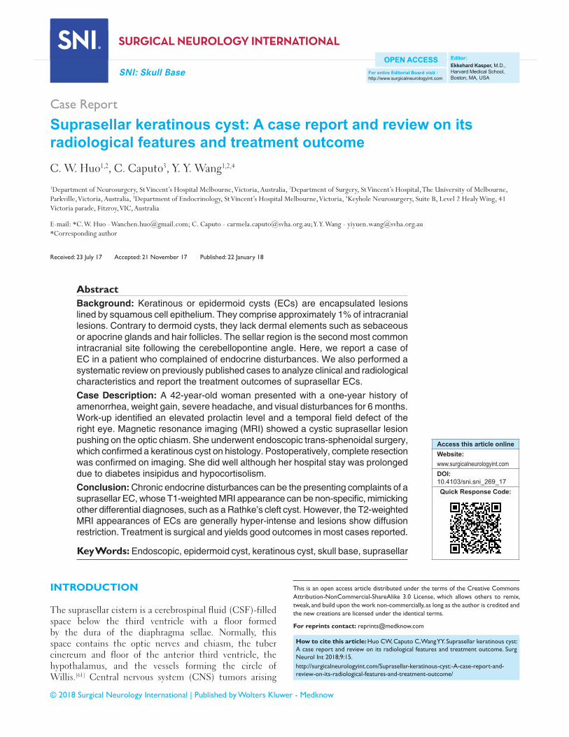
The keywords “epidermoid cyst” OR “keratinous cyst” AND “*sellar” were used to perform an electronic search of the English literature indexed in PubMed and OVID‑Medline databases on human studies (adults and children) without any limited time frame. Collection of case reports and series followed the PRISMA guideline[39] and the algorithm for inclusion of studies was illustrated in
1 In modern days, the term “cholesteatoma” refers to a growth of abnormal keratinizing squamous epithelium most commonly in the middle ear, but can also occur in the mastoid and petrous temporal bone. [Rutkowska J, Ozgirgin N, Olszewska E. Cholesteatoma Definiion and Classification: A Literature Review. J Int Adv Otol 2017; 13(2): 266‑71.]
Figure 1 using the PRISMA flow diagram. The ©Checklist for Case Reports from the Joanna Briggs Institute Critical Appraisal tools[19] was utilized to score published studies [Supplementary Material 1]. Case reports that met at least three out of the eight criteria outlined on the checklist were included in this study [Figure 1].
Regarding the case report, a signed written consent was obtained from the patient for the publication of her case. Information on the presenting history, radiological appearance, surgical management, and pathological reports of her suprasellar lesion were collected from the medical records and directly obtained from the patient.
The patient was a 42‑year‑old administrative officer who initially presented with a one‑year history of amenorrhoea to her family physician. Blood tests revealed normal hormonal functions other than an elevated prolactin level at 875 mIU/L (normal range: 110–560 mIU/L). The mildly increased secretion of prolactin is most likely caused by a disinhibition of lactotropes from impaired dopamine delivery to the pituitary, as a result of the tumor compressing on the pituitary stalk – the Stalk effect; whereas in cases of prolactinomas, the value of prolactin usually exceeds 1,000 mIU/L.[4,60] MRI head showed a cystic lesion compressing the pituitary gland and touching the optic chiasm. Formal visual field testing found a right temporal field defect. On admission, she reported 8/10 headaches for 6 months and a 6‑kilogram weight gain over the preceding 12 months without any increase in appetite. She denied galactorrhoea. On physical examination, she was found to have
Figure 1: Algorithm for epidermoid cyst case study inclusion. *Full‑text articles excluded due to: 1. Studies on dermoid rather than epidermoid cysts;[14,26,62] or 2. Intracranial epidermoid cysts not in the sellar region.[45,46,66]
Surgical Neurology International 2018, 9:15 http://www.surgicalneurologyint.com/content/9/1/15
supraclavicular fat pads, an emerging buffalo hump, moderate central abdominal adiposity, and some striae rubrae. Dexamethasone suppression test and salivary cortisol test yielded normal results. The working diagnosis was a cystic pituitary macroadenoma.
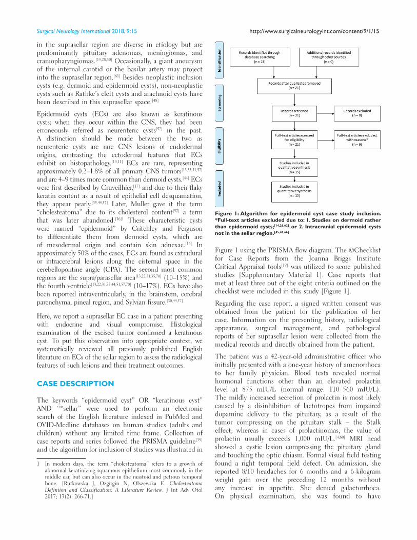
Given her symptoms, the patient was scheduled for endoscopic trans‑sphenoidal surgery. A repeat MRI prior to surgery showed a T1 hypo‑intense, T2 hyper‑intense, peripherally enhancing lesion within the sella extending into the suprasellar space and displacing the optic chiasm. Ill‑defined intrinsic T1 hyper‑intense signal was seen within the lesion [Figure 2]. Rathke’s cleft cyst was suggested as a differential diagnosis. During surgery, the encapsulated tumor had a creamy, cheesy appearance allowing internal debulking [Figure 3]. Its capsule was not adherent to adjacent structures, permitting complete tumor removal. Intraoperative CSF leak prompted repair using a fat and fascial graft obtained from the right thigh. Histology of the surgical specimen confirmed squamous epithelium together with large amounts of amorphous debris consistent with keratin, leading to the pathological diagnosis of a keratinous cyst. There was no evidence of dysplasia or malignancy. The patient’s postoperative clinical course was complicated by diabetes insipidus (DI) and a low cortisol level, which were managed by desmopressin and hydrocortisone. No further liquorrhea was encountered. A follow‑up MRI at 1 month showed postsurgical changes only and no residual. The pituitary gland appeared normal and the displacement of the optic chiasm had resolved. At the outpatient visit, the patient was found to be well, with occasional headaches only. She continued taking oral desmopressin and hydrocortisone.
DISCUSSION
Clinical presentationECs are congenital, indolent tumors that may remain asymptomatic for years prior to detection.[35] Our review
of 23 published cases on sellar ECs is summarized in Table 1. Most patients experienced symptoms related to the tumor for months or years before a diagnosis was made. The most common presenting complaints were headaches and visual disturbances. Headaches were often non‑specific, prompting patients to seek medical help once visual symptoms and, occasionally, symptoms of mass effect had developed [Table 1]. Endocrine dysfunction was uncommon, which were reported in only 4 cases.[40,43,59] The mean age of EC at diagnosis was 38 years, ranging from 3 to 65, with a gender predilection of 1:1.75 (8 females vs. 14 males, see Table 1).
Typically, ECs present in the fourth to fifth decades of life,[53,61] whereas dermoid cysts commonly occur in males of 20–30 years of age.[20,47] Other presentations include aseptic granulomatous meningitis,[1,6,12] obstructive hydrocephalus,[9] and vasospasm[71] due to the spillage or rupture of cyst components into the surrounding arachnoid space. Intraoperative use of copious irrigation with hydrocortisone is considered suitable to prevent aseptic meningitis perioperatively.[18,28,30,70] The use of postoperative steroids was not reported, nevertheless, in our institution oral or intravenous dexamethasone (4 mg four times a day for five days and then tapered gradually) may be used in cases of aseptic meningitis. Some patients remain asymptomatic following cyst ruptures.[6,41]
Radiographic characteristicsReported imaging characteristics of ECs are summarized in Table 2. Because the composition of the EC content may include protein, keratin, epithelial debris, and cholesterol,[12,29] a variety of MRI patterns were seen[24,42,63] [Table 2]. Rupture of the cyst content, intralesional hemorrhage, and subsequent local inflammatory responses may also lead to atypical radiological findings.[12,34] ECs appear hypo‑, iso‑ or hyperintense on T1‑weighted MRI imaging, with or without rim enhancement following contrast administration [Table 2]. On T2‑weighted MRI imaging,
Figure 2: Pre‑ and postoperative MRI images of the suprasellar lesion. Preoperative images show a T1 hypo‑intense and T2 hyper‑intense suprasellar lesion with periphery enhancement post magnevist contrast administration. Postoperative T1‑weighted images demonstrate the fat graft but no residual tumor. White arrows indicate the lesion
Surgical Neurology International 2018, 9:15 http://www.surgicalneurologyint.com/content/9/1/15
however, ECs typically appear hyper‑intense. The most specific MR sequence appears to be diffusion‑weighted imaging where ECs show a restricted pattern.[25,30,44] This allows to differentiate these lesions from other cystic pathologies, including arachnoid cysts, which show isointensity to CSF on all MR sequences without diffusion restriction[5,65] [Table 3].
On CT, ECs most often appear hypo‑dense due to the low absorptive value of their fat content[55] [Table 2]. Occasionally, lesions may present with high density, which is thought to reflect saponification of the keratinized debris with calcium.[3] Comparing the imaging characteristics of ECs with those of other cystic lesions
in the sellar region [Table 3], ECs have the tendency to encase adjacent nerves and vessels, whereas arachnoid cysts tend to displace them.[5]
In comparison to dermoid cysts, the latter typically appear hyper‑intense on T1, with ECs showing hyper‑intense signal on T2;[44,49] however, as illustrated in Table 3, a degree of overlap exists in terms of clinical and imaging features of those lesions, often precluding a definitive diagnosis without a histopathological sample.
Surgical options and outcomesComplete excision of the lesion and its capsule is important for satisfactory long‑term results
Table 1: Summary of patient age, gender and presenting symptoms from published cases
Case Age Gender Presentation (duration, if known)
Huo, 2017 42 F Amenorrhoea, hyperprolactinaemia, and weight gain (1 yr)Headache (6 m)Right temporal field defect
Nakassa, 2017[41] 54 F Progressive vision loss (3 m)Intermittent frontal and occipital headaches, polydipsia, polyuria (7 m)
Prasad, 2017[48] 65 F Occasional headache, progressive vision loss of the left eye (6 m)Polydipsia and polyuria (3 m)
Yanamadala, 2014[69] 3 M Severe vertigoMcCoul, 2011[38] 27 M Episodic headache that was increasing in frequency (3 m)McCoul, 2011 50 M Chronic left occipitoparietal headache (yr)McCoul, 2011 26 M Occipital and temporal headache (2 yr)
Acute onset of bitemporal hemianopsiaSani, 2005[56] 25 M Acute severe headache, diplopia and reduced visual acuity Oge, 1991[43] 36 M Impotence and weakness (8 yr)
Plasma hormone level assays: decreased testosterone and thyroxine levels, increased prolactin levelBollar, 1989[6] 38 M Headache and temporal lobe seizures (1 yr)
Two episodes of severe headaches and stiff necks with spontaneous recoveries in a few daysCambria, 1985[8] 53 F Frontal headache (6 yr)
Apathy, generalised asthenia, ataxia and aboulia developed laterSchubiger, 1983[58] 61 F Headaches radiating into the left eye
Blurred vision → found to have a partial temporal quadrant defect of the left eyeLEWIS, 1983[33] 54 F Depression (3 yr)
Symptoms of obstructive hydrocephalus (acute)Eliash, 1983[18] 5 N/S Diencaphlic syndrome, pituitary insufficiency
Polydipsia, polyuriaReduced visual acuity of the left eye, and Bilateral temporal defects
Sadeh, 1982[55] 27 F Mild headache (1 yr)Progressive vision loss
Mori, 1982[40] 25 F Visual disturbance Mori, 1982 14 M Visual disturbanceMori, 1982 44 M Headache and visual disturbanceMori, 1982 12 M Visual disturbanceMori, 1982 39 M Convulsions, anosmia, and visual disturbances Mori, 1982 64 M Homonymous hemianopia, mental disturbance Rhodes, 1981[52] 38 M Years of hyperphagia and obesity
Died of a cardiac infarct with EC a postmortem finding Selverstone, 1949[59] 22 M Anterorior panhypopituitarism since the age of 13
Progressive visual decline and headache (“pain behind the eyes”) (4 yrs)Homonymous hemianopsia of left eye
EC: Epidermoid cyst, F: Female, m: Month, M: Male, yr: Year
Surgical Neurology International 2018, 9:15 http://www.surgicalneurologyint.com/content/9/1/15
while preserving function and quality‑of‑life of the patient.[22,35,68,70] This requires diligent dissection techniques, whereas, more liquid cysts (such as Rathke’s cleft cysts) can be evacuated by simple aspiration. Because ECs can present as bulky, viscous material that cannot be easily suctioned,[41,43] good exposure is required to achieve gross total resection (GTR) preventing recurrences.[41,43,45] Where the tumor capsule is densely adherent to the
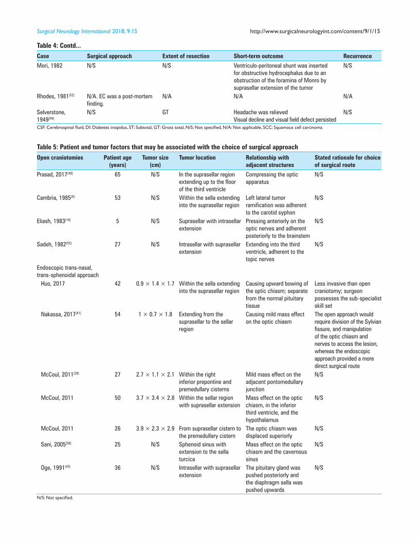
surrounding structures, subtotal resections (STR) have to be performed which can predispose for recurrence.[22,57,68,70] Based on our pooled analysis, 60% were amenable to GTR in cases where extent of resection was reported (10 out of 23 cases reported extent of resection, see Table 4). Long‑term follow‑up outcome was reported in 11 out of 23 cases with a mean follow‑up duration of 20 months (range: 3–60 months). Among these 11 cases, EC spontaneously regressed without any intervention in a 3‑year‑old boy,[69] and in the other 10 no recurrence was found regardless of extent of tumor resection [Table 4]. Gormley et al. reviewed 32 epidermoid and dermoid tumors that were resected at a single institution between 1975 and 1991. While their operative approach was not specified as open or endoscopic, and it was unknown whether the procedures were performed by a single or multiple surgeons, they achieved GTR in 20 (62.5%) cases (epidermoid and dermoid combined), consistent with the percentage found in our analysis. Among their study cohort, 22 were epidermoid tumors of the brain and skull, not limited to the sellar space. They found that the long‑term recurrence rate at a median follow‑up duration of 6.5 years was estimated to be 15.6%.[22] However, subset analysis on epidermoid cysts alone or stratified by tumor location or extent of resection was not performed. Therefore, their result may not accurately reflect the recurrence rate of suprasellar epidermoid cysts. In addition, no adjuvant treatment has been shown to be effective in the treatment of ECs.[50,51,54]
As summarized in Table 4, suprasellar ECs have traditionally been excised via trans‑frontal craniotomies; however, in recent years, a less invasive trans‑nasal endoscopic approach has been increasingly adopted. The first open craniotomy for excision of a suprasellar epidermoid cyst was reported by Sadeh et al.[55] in 1982 where a subtotal resection was achieved. In contrast, the first endoscopic approach was not reported until 1991 when Öge and Özgen described the trans‑nasal and trans‑sphenoidal route with a GTR.[43] As detailed in Table 4, a total of
Table 2: Summary of imaging characteristics of published cases (where reported)
Case Radiological patterns of the lesion
Huo, 2017 (MRI)T1: Homogenous, well‑circumscribed hypointense lesion with rim enhancement post contrastT2: Hyperintense
Nakassa, 2017[41] (MRI)T1: Mixed signalsT2: Mixed signals
Prasad, 2017[48] (MRI)T1:Heterogeneous, well‑circumscribed lobulated lesion with rim enhancement post contrastIsointense to CSF with central hyperintensityT2: Hyperintense with central hypointensityFLAIR: Homogeneously hyperintenseDWI: Heterogeneously diffusion restricted
Yanamadala, 2014[69]
(MRI) a 2 cm nonenhancing lesion with evidence of restricted diffusionT1: hypointenseT2: hyperintense
McCoul, 2011[38] (MRI)T1: Hyperintense with no contrast enhancementT2: N/SFLAIR: Isointense
McCoul, 2011 (MRI)T1: Hyperintense with no contrast enhancementT2: hyperintense
McCoul, 2011 (MRI)T1: hypointenseT2: Hyperintense heterogeneouslyDWI: diffusion restricted
Sani, 2005[56] (MRI) a non‑enhancing lesion post administration of gadoliniumT1: hyperintense mass with an isointense borderT2: isointense mass with a bright signal border
Oge, 1991[43] (CT): hypodense with capsular enhancementBollar, 1989[6] (CT): hypodense with marginal calcificationsCambria, 1985[8] (CT): hypodense with central calcificationsSchubiger, 1983[58] (CT): hyperdense with contrast enhancementSadeh, 1982[55] (CT): hypodense with capsular enhancementLEWIS, 1983[33] (CT): hypodense with no capsular enhancementMori, 1982[40] (CT): heterogeneously hypodense with marginal
calcificationsMori, 1982 (CT): heterogeneously hypodenseCT: Computed tomography; FLAIR: Fluid-attenuation inversion recovery; MRI: Magnetic resonance imaging; N/S: Not specified
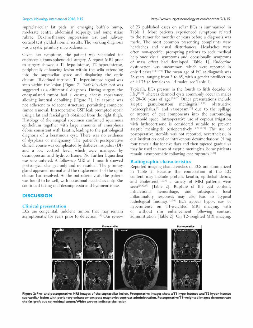
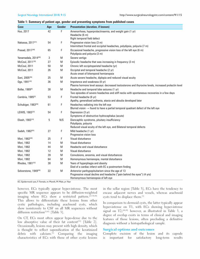
Figure 3: Intraoperative images of the suprasellar lesion (a) Endoscopic pterional view of the laminar terminalis. ICA: internal carotid artery, MCA: middle cerebral artery, R: right, ACA: anterior cerebral artery. (b) A zoomed‑in view of A. L: left, ACOM: anterior communicating artery, RAH: recurrent artery of Heubner
ba
Surgical Neurology International 2018, 9:15 http://www.surgicalneurologyint.com/content/9/1/15
4 cases reported open craniotomies – among which, 1 patient was well postoperatively (25%)[8] and the other three had persisting deficits (vision or hormonal insufficiencies).[18,48,55] By comparison, among the seven cases adopting an endoscopic trans‑nasal approach, four patients improved post‑operatively (57%),[38,43,56] two had persistent deficits[41] (including our case) and the other patient’s recovery was complicated by CSF leakage requiring revision surgery.[38] The rationale behind utilizing an open versus endoscopic surgical route was only explained in two out of the 11 cases (including our case, see Table 5). It is likely that the decision was influenced by the surgeon’s own preference, skill set, and experience. Assessing patient and tumor factors that may potentially dictate surgical approach, the mean age for both open craniotomies and endoscopic cases was 37 years [Table 5]; none of the studies where open resections were performed detailed tumor size, precluding a meaningful comparison with endoscopic cases; tumor location with its extension, adherence to, or local effects on adjacent structures were not associated with worse patient outcomes or a particular
surgical approach [Tables 4 and 5]. Nevertheless, it is worth noting that due to its rare occurrence and the overall low number of published studies, caution is required when drawing any definitive conclusion.
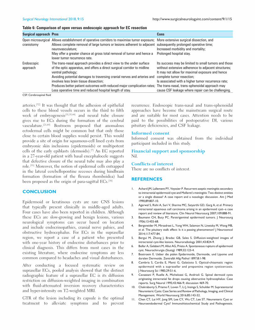
Despite colloid cyst of the anterior third ventricle being a different cystic tumor in a slightly distinct anatomical location compared with EC, due to its more common incidence,[67] two studies have compared open microsurgical versus endoscopic surgical techniques. Grondin et al. conducted a systematic review of the literature between 1980 and 2007, and found that while endoscopic (n = 157) and open microsurgical (n = 551) resections shared similar success rates at complete tumor resection and low recurrence rates, the endoscopic technique was associated with significantly reduced postoperative morbidity and mortality, less operating time, and shorter length of hospital stay than traditional open craniotomies.[23] Margetis and Souweidance in 2013 also conducted a systematic review of case series that included at least 20 patients with a minimum of 36‑month follow‑up durations. They reported that, among a total of 422 patients, although the overall complication rates were comparable between endoscopic and microsurgical open cases, the rate of major complications was significantly higher in open cases than that of endoscopic approach (14.68% vs. 5.45%). Furthermore, the tumor recurrence rate was higher in the endoscopic group compared with that of open microsurgical cases (12.30% vs. 0.85%). However, in keeping with Grondin’s review, endoscopic technique was associated with superior operative time and hospital length of stay.[37] Therefore, in modern neurosurgery, endoscopic technique is increasingly preferred for optimizing patient outcome, whereas open microsurgical approach may be best reserved for large tumors or complex cases. The advantages and disadvantages of both approaches are summarized in Table 6. It is not surprising that the endoscopic trans‑nasal, trans‑sphenoidal surgical corridor has been predominantly used since 1990 for EC resection [Table 4].
Overall, postoperative recovery after EC removal was satisfactory, although persistent DI and other endocrine disturbances had been reported [Table 4]. However, one case showed malignant transformation to squamous cell carcinoma with rapid tumor regrowth.[33] Such malignant transformation of ECs appears exceedingly rare, but confers a poor prognosis.[2,27]
HistogenesisWhile craniopharyngiomas contain cholesterol‑rich fluids, Rathke’s cleft cysts are developmental remnants of the formation of Rathke’s cleft and contain mucoid colloid.[43] This distinguishes EC with its unique feature of keratin‑rich debris, which is ectodermal in origin.
Lepoire and Pertuiset categorized ECs into three groups, retrosellar, suprasellar, and intraventricular based on the embryonic relation to the carotid, basilar, and choroid
Table 3: Radiological diagnostic features of various intracranial cysts in the sellar region
Type of lesion Radiological diagnostic clues
Epidermoid/keratinous cyst
Commonly in the CPA cistern, followed by sellar region and the fourth ventricle;CSF‑like mass that insinuates adjacent neurovasculature;Hyper‑intense to CSF on T2‑weighted MRI images;Displays higher than CSF signal on FLAIR and DWI due to restricted diffusion.
Dermoid cyst Midline homogeneous lesions, however, hair and sebaceous content can give rise to a heterogeneous signal;Commonly found in sellar and frontonasal regions;Resemble fat rather than CSF, hence hyper‑intense on T1 MRI.
Arachnoid cyst Isointense to CSF at all sequences;No DWI restriction;Dark on both FLAIR and DWI, as there is no diffusion restriction;Displaces adjacent structures rather than insinuates them.
Craniopharyngioma Characteristic calcifications on CT;Heterogeneous appearances on MRI due to mixed solid and cystic components;Hyper‑intense on T1 MRI, due to protein, cholesterol, or blood products.
Rathke’s cleft cyst If the protein content is low → isointense with CSF on all sequences on MRI;If protein content is high → hyperintense on T1 MRI;No calcification;May show enhancing rims and nodules just as epidermoid cysts.
CPA: Cerebellopontine angle; CT: Computed tomography; CSF: Cerebrospinal fluid; FLAIR: Fluid-attenuation inversion recovery; MRI: Magnetic resonance imaging
Surgical Neurology International 2018, 9:15 http://www.surgicalneurologyint.com/content/9/1/15
Table 4: Summary of surgical treatment and patient outcomes
Case Surgical approach Extent of resection Short‑term outcome Recurrence
Huo, 2017 Endoscopic trans‑sphenoidal GT DI, and hypocortisolism; otherwise well No at 3 months Nakassa, 2017[41] Endoscopic transnasal
with a combined infrasellar interpituitary and suprasellar approach
GT Vision improved; DI persisted and one year later developed hypothyroidism
No at 1 year
Prasad, 2017[48] Left pterional craniotomy ST DI improved but visual loss persisted N/SYanamadala, 2014[69]
N/A. patient was observed with annual MRI studies for 5 years.
N/A Progressive reduction in tumor size over the next 3 years.
Complete resolution of tumor at 5 years
McCoul, 2011[38] Endoscopic endonasal trans‑sphenoidal trans‑sellar and trans‑tuberculum trans‑planum approach
GT Pt well No at 3 years
McCoul, 2011 Endoscopic endonasal trans‑clival approach, using both trans‑sphenoidal and trans‑nasal corridors
GT CSF rhinorrhoea requiring revision surgery, where a fistula was identified at the margin of the surgical defect. Fat and fascial grafts were used to seal the defect with success
No at 1 year
McCoul, 2011 A combined endoscopic endo‑nasal trans‑sphenoidal trans‑tuberculum trans‑planum approach, trans‑sphenoidal trans‑clival, and trans‑nasal trans‑clival approaches.
ST (95% tumor removed)Small amount encasing the left abducens nerve
Vision and headache improved No at 7 months
Sani, 2005[56] The sublabial trans‑sphenoidal approach
N/S By postoperative day 7 the patient’s visual acuity improved, diplopia resolved, and the third cranial nerve function returned to baseline by 3 months’ follow‑up
No at 3 months
Oge, 1991[43] Trans‑nasal, trans‑sphenoidal route
GT Remained on corticosteroid and thyroid replacement therapy. At 3 months: impotence regressed and weakness diminished; Plasma cortisol and prolactin levels normal.
No at 3 months
Bollar, 1989[6] N/S ST (tumor capsule adherent to the right optic nerve)
Uneventful, no aseptic meningitis, or new neurological deficit
N/S
Cambria, 1985[8] Fronto‑temporal left craniotomy
N/S Preoperative symptoms slowly improved N/S
Schubiger, 1983[58] N/S N/S N/S N/SLEWIS, 1983[33] N/S N/S Patient did well initially, but deteriorated
quickly with tumor recurrence (also malignant transformation of SCC and increasing hydrocephalus)—passed away
N/A
Eliash, 1983[18] Trans‑frontal Subtotal (capsule adherent to surrounding structures, pressing anteriorly onto the optic nerves and adherent posteriorly to the brain stem)
Pituitary insufficiency remained but vision acuity recovered
No at 2 years
Sadeh, 1982[55] Right trans‑frontal craniotomy
N/S Blind for 2 years but vision gradually returned
N/S
Mori, 1982[40] N/S N/S N/S N/SMori, 1982 N/S N/S N/S N/SMori, 1982 N/S N/S N/S N/SMori, 1982 N/S N/S N/S N/SMori, 1982 N/S N/S N/S N/S
Contd...
Surgical Neurology International 2018, 9:15 http://www.surgicalneurologyint.com/content/9/1/15
Table 4: Contd...
Case Surgical approach Extent of resection Short‑term outcome Recurrence
Mori, 1982 N/S N/S Ventriculo‑peritoneal shunt was inserted for obstructive hydrocephalus due to an obstruction of the foramina of Monro by suprasellar extension of the tumor
N/S
Rhodes, 1981[52] N/A. EC was a post‑mortem finding.
N/A N/A N/A
Selverstone, 1949[59]
N/S GT Headache was relievedVisual decline and visual field defect persisted
N/S
CSF: Cerebrospinal fluid, DI: Diabetes insipidus, ST: Subtotal, GT: Gross total, N/S: Not specified, N/A: Not applicable, SCC: Squamous cell carcinoma
Table 5: Patient and tumor factors that may be associated with the choice of surgical approach
Open craniotomies Patient age (years)
Tumor size (cm)
Tumor location Relationship with adjacent structures
Stated rationale for choice of surgical route
Prasad, 2017[48] 65 N/S In the suprasellar region extending up to the floor of the third ventricle
Compressing the optic apparatus
N/S
Cambria, 1985[8] 53 N/S Within the sella extending into the suprasellar region
Left lateral tumor ramification was adherent to the carotid syphon
N/S
Eliash, 1983[18] 5 N/S Suprasellar with intrasellar extension
Pressing anteriorly on the optic nerves and adherent posteriorly to the brainstem
N/S
Sadeh, 1982[55] 27 N/S Intrasellar with suprasellar extension
Extending into the third ventricle, adherent to the topic nerves
N/S
Endoscopic trans‑nasal, trans‑sphenoidal approach
Huo, 2017 42 0.9 × 1.4 × 1.7 Within the sella extending into the suprasellar region
Causing upward bowing of the optic chiasm; separate from the normal pituitary tissue
Less invasive than open craniotomy; surgeon possesses the sub‑specialist skill set
Nakassa, 2017[41] 54 1 × 0.7 × 1.8 Extending from the suprasellar to the sellar region
Causing mild mass effect on the optic chiasm
The open approach would require division of the Sylvian fissure, and manipulation of the optic chiasm and nerves to access the lesion, whereas the endoscopic approach provided a more direct surgical route
McCoul, 2011[38] 27 2.7 × 1.1 × 2.1 Within the right inferior prepontine and premedullary cisterns
Mild mass effect on the adjacent pontomedullary junction
N/S
McCoul, 2011 50 3.7 × 3.4 × 2.8 Within the sellar region with suprasellar extension
Mass effect on the optic chiasm, in the inferior third ventricle, and the hypothalamus
N/S
McCoul, 2011 26 3.9 × 2.3 × 2.9 From suprasellar cistern to the premedullary cistern
The optic chiasm was displaced superiorly
N/S
Sani, 2005[56] 25 N/S Sphenoid sinus with extension to the sella turcica
Mass effect on the optic chiasm and the cavernous sinus
N/S
Oge, 1991[43] 36 N/S Intrasellar with suprasellar extension
The pituitary gland was pushed posteriorly and the diaphragm sella was pushed upwards
N/S
N/S: Not specified.
Surgical Neurology International 2018, 9:15 http://www.surgicalneurologyint.com/content/9/1/15
Table 6: Comparison of open versus endoscopic approach for EC resection
Surgical approach Pros Cons
Open microsurgical craniotomy
Allows establishment of operative corridors to maximise tumor exposure;Allows complete removal of large tumors or lesions adherent to adjacent neurovasculature;May offer a greater chance at gross total removal of tumor and hence a lower tumor recurrence rate.
More extensive surgical dissection, and subsequently prolonged operative time;Increased morbidity and mortality;Prolonged hospital stay.
Endoscopic approach
The trans‑nasal approach provides a direct view to the under surface of the optic apparatus, and offers a direct surgical corridor to midline ventral pathology;Avoiding potential damages to traversing cranial nerves and arteries and involves less brain tissue dissection;Produces better patient outcomes with reduced major complication rates;Less operative time and reduced hospital length of stay.
Its success may be limited to small tumors and those without extensive adherence to adjacent structures;It may not allow for maximal exposure and hence complete tumor resection;Is associated with a higher tumor recurrence rate;The trans‑nasal, trans‑sphenoidal approach may cause CSF leakage where repair can be challenging.
CSF: Cerebrospinal fluid
arteries.[32] It was thought that the adhesion of epithelial cells to these blood vessels occurs in the third to fifth week of embryogenesis[31,35,44] and neural tube closure gives rise to ECs during the formation of the cerebral vasculature.[21,64] Bostroem proposed that anomalous ectodermal cells might be common but that only those close to certain blood supplies would persist. This would provide a site of origin for squamous‑cell lined cysts from embryonic skin inclusions (epidermoids) or multipotent cells of the early epiblasts (dermoids).[7] An EC reported in a 27‑year‑old patient with basal encephalocele suggests that defective closure of the neural tube may also play a role.[55] Moreover, the notion of epidermal cells entrapped in the lateral cerebellopontine recesses during hindbrain formation (formation of the flexura rhomboidea) had been proposed as the origin of para‑sagittal ECs.[16]
CONCLUSION
Epidermoid or keratinous cysts are rare CNS lesions that typically present clinically in middle‑aged adults. Four cases have also been reported in children. Although these ECs are slow‑growing and benign lesions, various neurological symptoms can occur based on location and include endocrinopathies, cranial nerve palsies, and obstructive hydrocephalus. For ECs in the suprasellar region, we report a case of a patient who presented with one‑year history of endocrine disturbances prior to clinical diagnosis. This differs from most cases in the existing literature, where endocrine symptoms are less common compared to headaches and visual disturbances.
After conducting a focused systematic review on suprasellar ECs, pooled analysis showed that the distinct radiographic feature of a suprasellar EC is its diffusion restriction on diffusion‑weighted imaging in combination with fluid‑attenuated inversion recovery characteristics and hyper‑intensity on T2‑weighted MRI.
GTR of the lesion including its capsule is the optimal treatment to alleviate symptoms and to prevent
recurrence. Endoscopic trans‑nasal and trans‑sphenoidal approaches have become the mainstream surgical route and are suitable for most cases. Attention needs to be paid to the possibilities of postoperative DI, various pituitary deficiencies, and CSF leakage.
Informed consentInformed consent was obtained from the individual participant included in this study.
Financial support and sponsorshipNil.
Conflicts of interestThere are no conflicts of interest.
REFERENCES
1. Achard JM, Lallement PY, Veyssier P. Recurrent aseptic meningitis secondary to intracranial epidermoid cyst and Mollaret’s meningitis: Two distinct entities or a single disease? A case report and a nosologic discussion. Am J Med 1990;89:807-10.
2. Agarwal S, Rishi A, Suri V, Sharma MC, Satyarthi GD, Garg A, et al. Primary intracranial squamous cell carcinoma arising in an epidermoid cyst‑‑a case report and review of literature. Clin Neurol Neurosurg 2007;109:888-91.
3. Baumann CH, Bucy PC. Paratrigeminal epidermoid tumors. J Neurosurg 1956;13:455-68.
4. Bergsneider M, Mirsadraei L, Yong WH, Salamon N, Linetsky M, Wang MB, et al. The pituitary stalk effect: Is it a passing phenomenon? J Neurooncol 2014;117:477-84.
5. Bergui M, Zhong J, Bradac GB, Sales S. Diffusion‑weighted images of intracranial cyst-like lesions. Neuroradiology 2001;43:824-9.
6. Bollar A, Gelabert M, Allut AG, Prieto A. Spontaneous rupture of epidermoid cyst. Neurochirurgia (Stuttg) 1989;32:123-4.
7. Bostroem E. Ueber die pialen Epidermoide, Dermoide, und Lipome und duralen Dermoide. Zentralbl Allg Pathol 1897;8:1‑98.
8. Cambria S, Cardia E, Manti G, Galatioto S. Optical‑chiasmatic region epidermoid with a suprasellar and prepontina region cysticercosis. J Neurosurg Sci 1985;29:51-6.
9. Cavazzani P, Ruelle A, Michelozzi G, Andrioli G. Spinal dermoid cysts originating intracranial fat drops causing obstructive hydrocephalus: Case reports. Surg Neurol 1995;43:466-9; discussion 469-70.
10. Chakraborty S, Priamo F, Loven T, Li J, Insinga S, Schulder M. Supratentorial Neurenteric Cysts: Case Series and Review of Pathology, Imaging, and Clinical Management. World Neurosurg 2016;85:143-52.
11. Chen CT, Lai HY, Jung SM, Lee CY, Wu CT, Lee ST. Neurenteric Cyst or Neuroendodermal Cyst? Immunohistochemical Study and Pathogenesis.
Surgical Neurology International 2018, 9:15 http://www.surgicalneurologyint.com/content/9/1/15
World Neurosurg 2016;96:85-90.12. Chen CY, Wong JS, Hsieh SC, Chu JS, Chan WP. Intracranial epidermoid
cyst with hemorrhage: MR imaging findings. AJNR Am J Neuroradiol 2006;27:427-9.
13. Chowdhury FH, Haque MR, Sarker MH. Intracranial epidermoid tumor; microneurosurgical management: An experience of 23 cases. Asian J Neurosurg 2013;8:21-8.
14. Cohen JE, Abdallah JA, Garrote M. Massive rupture of suprasellar dermoid cyst into ventricles. Case illustration. J Neurosurg 1997;87:963.
15. Connor SE, Penney CC. MRI in the differential diagnosis of a sellar mass. Clin Radiol 2003;58:20-31.
16. Critchley M, Ferguson FR. The cerebrospinal epidermoids (cholesteatomata). Brain 1928;51:334-84.
17. Cruveilhier J. Anatomie Pathologique de Corps Humain. Paris: JB Bailliere; 1829. p. 341.
18. Eliash A, Roitman A, Karp M, Reichental E, Manor RS, Shalit M, et al. Diencephalic syndrome due to a suprasellar epidermoid cyst. Child’s Brain 1983;10:414-8.
19. Gagnier JJ, Kienle G, Altman DG, Moher D, Sox H, Riley D, et al. The CARE Guidelines: Consensus‑based Clinical Case Reporting Guideline Development. Glob Adv Health Med 2013;2:38‑43.
20. Gelabert‑Gonzalez M. [Intracranial epidermoid and dermoid cysts]. Rev Neurol 1998;27:777-82.
21. Glasauer FE, Levy LF, Auchterlonie WC. Congenital inclusion dermoid cyst of the anterior fontanel. J Neurosurg 1978;48:274-8.
22. Gormley WB, Tomecek FJ, Qureshi N, Malik GM. Craniocerebral epidermoid and dermoid tumours: A review of 32 cases. Acta Neurochir (Wien) 1994;128:115-21.
23. Grondin RT, Hader W, MacRae ME, Hamilton MG. Endoscopic versus microsurgical resection of third ventricle colloid cysts. Can J Neurol Sci 2007;34:197-207.
24. Gualdi GF, Di Biasi C, Trasimeni G, Pingi A, Vignati A, Maira G. Unusual MR and CT appearance of an epidermoid tumor. AJNR Am J Neuroradiol 1991;12:771-2.
25. Hakyemez B, Aksoy U, Yildiz H, Ergin N. Intracranial epidermoid cysts: Diffusion‑weighted, FLAIR and conventional MR findings. Eur J Radiol 2005;54:214-20.
26. Hamer J. Diagnosis by computerized tomography of intradural dermoid with spontaneous rupture of the cyst. Acta Neurochir (Wien) 1980;51:219-26.
27. Hamlat A, Hua ZF, Saikali S, Laurent JF, Gedouin D, Ben‑Hassel M, et al. Malignant transformation of intra-cranial epithelial cysts: Systematic article review. J Neurooncol 2005;74:187-94.
28. Hori T, Kawamata T, Amano K, Aihara Y, Ono M, Miki N. Anterior interhemispheric approach for 100 tumors in and around the anterior third ventricle. Neurosurgery 2010;66 (3 Suppl Operative):65‑74.
29. Inoue Y, Ohata K, Nakayama K, Haba T, Shakudo M. An unusual middle fossa interdural epidermoid tumor. Case report. J Neurosurg 2001;95:902-4.
30. Kallmes DF, Provenzale JM, Cloft HJ, McClendon RE. Typical and atypical MR imaging features of intracranial epidermoid tumors. AJR Am J Roentgenol 1997;169:883-7.
31. Kato K, Ujiie H, Higa T, Hayashi M, Kubo O, Okada Y, et al. Clinical presentation of intracranial epidermoids: A surgical series of 20 initial and four recurred cases. Asian J Neurosurg 2010;5:32-40.
32. Lepoire J, Pertuiset B. [Cranioencephalic epidermoid cysts]. Neurochirurgie 1957;3:319-22.
33. Lewis AJ, Cooper PW, Kassel EE, Schwartz ML. Squamous cell carcinoma arising in a suprasellar epidermoid cyst. Case report. J Neurosurg 1983;59:538-41.
34. Li F, Zhu S, Liu Y, Chen G, Chi L, Qu F. Hyperdense intracranial epidermoid cysts: A study of 15 cases. Acta Neurochir (Wien) 2007;149:31-9; discussion 39.
35. Lynch JC, Aversa A, Pereira C, Nogueira J, Goncalves M, Lopes H. Surgical strategy for intracranial dermoid and epidermoid tumors: An experience with 33 Patients. Surg Neurol Int 2014;5:163.
36. Manno NJ, Uihlein A, Kernohan JW. Intraspinal epidermoids. J Neurosurg 1962;19:754-65.
37. Margetis K, Souweidane MM. Endoscopic treatment of intraventricular cystic tumors. World Neurosurg 2013;79 (2 Suppl):S19 e11-11.
38. McCoul ED, Chow S, Lee DL, Anand VK, Schwartz TH. Endoscopic endonasal
approach for resection of ventral skull base keratinaceous cysts. Int Forum Allergy Rhinol 2012;2:258-63.
39. Moher D, Liberati A, Tetzlaff J, Altman DG, Group P. Preferred reporting items for systematic reviews and meta-analyses: The PRISMA statement. Int J Surg 2010;8:336-41.
40. Mori K, Handa H, Moritake K, Takeuchi J, Nakano Y. Suprasellar epidermoid. Neurochirurgia (Stuttg) 1982;25:138-42.
41. Nakassa AC, Chabot JD, Snyderman CH, Wang EW, Gardner PA, Fernandez-Miranda JC. Complete endoscopic resection of a pituitary stalk epidermoid cyst using a combined infrasellar interpituitary and suprasellar endonasal approach: Case report. J Neurosurg 2017:1-7.
42. Ochi M, Hayashi K, Hayashi T, Morikawa M, Ogino A, Hashmi R, et al. Unusual CT and MR appearance of an epidermoid tumor of the cerebellopontine angle. AJNR Am J Neuroradiol 1998;19:1113-5.
43. Oge K, Ozgen T. Transsphenoidal removal of an intra‑ and suprasellar epidermoid cyst. Neurochirurgia (Stuttg) 1991;34:94-6.
44. Osborn AG, Preece MT. Intracranial cysts: Radiologic‑pathologic correlation and imaging approach. Radiology 2006;239:650-64.
45. Patibandla MR, Yerramneni VK, Mudumba VS, Manisha N, Addagada GC. Brainstem epidermoid cyst: An update. Asian J Neurosurg 2016;11:194-200.
46. Pikis S, Margolin E. Malignant transformation of a residual cerebellopontine angle epidermoid cyst. J Clin Neurosci 2016;33:59-62.
47. Pisanesehi M, Kapoor G. Imaging of sella and parasellar region. Neuroimaging Clin N Am 2005;15:203-19.
48. Prasad GL, Pavithra P. Suprasellar Epidermoid Cyst with Atypical Imaging Findings. World Neurosurg 2017;98:870 e871-870 e873.
49. Rao VJ, James RA, Mitra D. Imaging characteristics of common suprasellar lesions with emphasis on MRI findings. Clin Radiol 2008;63:939‑47.
50. Reddy A, Kreicher KL, Patel NA, Schantz S, Shinhar S. Pediatric epidermoid cysts masquerading as ranulas: A case series. Int J Pediatr Otorhinolaryngol 2016;81:26-8.
51. Ren X, Lin S, Wang Z, Luo L, Jiang Z, Sui D, et al. Clinical, radiological, and pathological features of 24 atypical intracranial epidermoid cysts. J Neurosurg 2012;116:611-21.
52. Rhodes RH, Davis RL, Beamer YB, Marantz C. A suprasellar epidermoid cyst with symptoms of hypothalamic involvement: Case report and a review of pathogenetic mechanisms. Bull Los Angeles Neurol Soc 1981;46:26-32.
53. Ruscalleda J. Imaging of parasellar lesions. Eur Radiol 2005;15:549-59.54. Rutherford SA, Leach PA, King AT. Early recurrence of an intracranial
epidermoid cyst due to low-grade infection: Case report. Skull Base 2006;16:109-16.
55. Sadeh M, Goldhammer Y, Shacked I, Tadmor R, Godel V. Basal encephalocele associated with suprasellar epidermoid cyst. Arch Neurol 1982;39:250-2.
56. Sani S, Smith A, Leppla DC, Ilangovan S, Glick R. Epidermoid cyst of the sphenoid sinus with extension into the sella turcica presenting as pituitary apoplexy: Case report. Surg Neurol 2005;63:394-7; discussion 397.
57. Schiefer TK, Link MJ. Epidermoids of the cerebellopontine angle: A 20-year experience. Surg Neurol 2008;70:584-90; discussion 590.
58. Schubiger O, Valavanis A, Gessaga E. Dense suprasellar epidermoid cyst. A case report. Neuroradiology 1983;24:269-71.
59. Selverstone B, Kubik CS. Suprasellar epidermal cyst. N Engl J Med 1949;241:309-11.
60. Skinner DC. Rethinking the stalk effect: A new hypothesis explaining suprasellar tumor-induced hyperprolactinemia. Med Hypotheses 2009;72:309-10.
61. Smith JK. Parasellar tumors: Suprasellar and cavernous sinuses. Top Magn Reson Imaging 2005;16:307-15.
62. Tan LA, Kasliwal MK, Harbhajanka A, Kellogg RG, Arvanitis LD, Munoz LF. Hyperdense suprasellar mass: An unusual radiological presentation of intracranial dermoid cyst. J Clin Neurosci 2015;22:1208-10.
63. Timmer FA, Sluzewski M, Treskes M, van Rooij WJ, Teepen JL, Wijnalda D. Chemical analysis of an epidermoid cyst with unusual CT and MR characteristics. AJNR Am J Neuroradiol 1998;19:1111-2.
64. Toglia JU, Netsky MG, Alexander E, Jr. Epithelial (epidermoid) tumors of the cranium. Their common nature and pathogenesis. J Neurosurg 1965;23:384-93.
65. Tsuruda JS, Chew WM, Moseley ME, Norman D. Diffusion‑weighted MR imaging of the brain: Value of differentiating between extraaxial cysts and

Surgical Neurology International 2018, 9:15 http://www.surgicalneurologyint.com/content/9/1/15
epidermoid tumors. AJNR Am J Neuroradiol 1990;11:925-31; discussion 932-924.
66. Ulivieri S, Oliveri G, Filosomi G, Miracco C. Intracranial epidermoid cyst: Case report. Ann Ital Chir 2008;79:445-6.
67. Yadav YR, Yadav N, Parihar V, Kher Y, Ratre S. Management of colloid cyst of third ventricle. Turk Neurosurg 2015;25:362-71.
68. Yamakawa K, Shitara N, Genka S, Manaka S, Takakura K. Clinical course and surgical prognosis of 33 cases of intracranial epidermoid tumors. Neurosurgery 1989;24:568-73.
69. Yanamadala V, Lin N, Walcott BP, Baird LC, Smith ER. Spontaneous regression of an epidermoid cyst of the cavernous sinus. J Clin Neurosci 2014;21:1433-5.
70. Yasargil MG, Abernathey CD, Sarioglu AC. Microneurosurgical treatment of intracranial dermoid and epidermoid tumors. Neurosurgery 1989;24:561-7.
71. Yilmazlar S, Kocaeli H, Cordan T. Brain stem stroke associated with epidermoid tumours: Report of two cases. J Neurol Neurosurg Psychiatry 2004;75:1340-2.

The Joanna Briggs Institute Critical Appraisal tools for use in JBI Systematic Reviews
Checklist for Case Reports http://joannabriggs.org/research/critical-appraisal-tools.html
www.joannabriggs.org
Supplementary Material 1
© Joanna Briggs Institute 2017 Critical Appraisal Checklist for Case Reports 2
The Joanna Briggs Institute Introduction
The Joanna Briggs Institute (JBI) is an international, membership based research and development organization within the Faculty of Health Sciences at the University of Adelaide. The Institute specializes in promoting and supporting evidence-based healthcare by providing access to resources for professionals in nursing, midwifery, medicine, and allied health. With over 80 collaborating centres and entities, servicing over 90 countries, the Institute is a recognized global leader in evidence-based healthcare.
JBI Systematic Reviews
The core of evidence synthesis is the systematic review of literature of a particular intervention, condition or issue. The systematic review is essentially an analysis of the available literature (that is, evidence) and a judgment of the effectiveness or otherwise of a practice, involving a series of complex steps. The JBI takes a particular view on what counts as evidence and the methods utilized to synthesize those different types of evidence. In line with this broader view of evidence, the Institute has developed theories, methodologies and rigorous processes for the critical appraisal and synthesis of these diverse forms of evidence in order to aid in clinical decision-making in health care. There now exists JBI guidance for conducting reviews of effectiveness research, qualitative research, prevalence/incidence, etiology/risk, economic evaluations, text/opinion, diagnostic test accuracy, mixed-methods, umbrella reviews and scoping reviews. Further information regarding JBI systematic reviews can be found in the JBI Reviewer’s Manual on our website.
JBI Critical Appraisal Tools
All systematic reviews incorporate a process of critique or appraisal of the research evidence. The purpose of this appraisal is to assess the methodological quality of a study and to determine the extent to which a study has addressed the possibility of bias in its design, conduct and analysis. All papers selected for inclusion in the systematic review (that is – those that meet the inclusion criteria described in the protocol) need to be subjected to rigorous appraisal by two critical appraisers. The results of this appraisal can then be used to inform synthesis and interpretation of the results of the study. JBI Critical appraisal tools have been developed by the JBI and collaborators and approved by the JBI Scientific Committee following extensive peer review. Although designed for use in systematic reviews, JBI critical appraisal tools can also be used when creating Critically Appraised Topics (CAT), in journal clubs and as an educational tool.

© Joanna Briggs Institute 2017 Critical Appraisal Checklist for Case Reports 3
JBI Critical Appraisal Checklist for Case Reports
Reviewer Date
Author Year Record Number
Yes No Unclear Not applicable
1. Were patient’s demographic characteristics clearly described? □ □ □ □
2. Was the patient’s history clearly described and presented as a timeline? □ □ □ □
3. Was the current clinical condition of the patient on presentation clearly described? □ □ □ □
4. Were diagnostic tests or assessment methods and the results clearly described? □ □ □ □
5. Was the intervention(s) or treatment procedure(s) clearly described? □ □ □ □
6. Was the post-intervention clinical condition clearly described? □ □ □ □
7. Were adverse events (harms) or unanticipated events identified and described? □ □ □ □
8. Does the case report provide takeaway lessons? □ □ □ □ Overall appraisal: Include □ Exclude □ Seek further info □
Comments (Including reason for exclusion)
© Joanna Briggs Institute 2017 Critical Appraisal Checklist for Case Reports 4
Explanation of case reports critical appraisal How to cite: Moola S, Munn Z, Tufanaru C, Aromataris E, Sears K, Sfetcu R, Currie M, Qureshi R, Mattis P, Lisy K, Mu P-F. Chapter 7: Systematic reviews of etiology and risk. In: Aromataris E, Munn Z (Editors). Joanna Briggs Institute Reviewer's Manual. The Joanna Briggs Institute, 2017. Available from https://reviewersmanual.joannabriggs.org/
Case Reports Critical Appraisal Tool
Answers: Yes, No, Unclear or Not/Applicable
1. Were patient’s demographic characteristics clearly described?
Does the case report clearly describe patient's age, sex, race, medical history, diagnosis, prognosis, previous treatments, past and current diagnostic test results, and medications? The setting and context may also be described.
2. Was the patient’s history clearly described and presented as a timeline?
A good case report will clearly describe the history of the patient, their medical, family and psychosocial history including relevant genetic information, as well as relevant past interventions and their outcomes. (CARE Checklist 2013)
3. Was the current clinical condition of the patient on presentation clearly described?
The current clinical condition of the patient should be described in detail including the uniqueness of the condition/disease, symptoms, frequency and severity. The case report should also be able to present whether differential diagnoses was considered.
4. Were diagnostic tests or methods and the results clearly described?
A reader of the case report should be provided sufficient information to understand how the patient was assessed. It is important that all appropriate tests are ordered to confirm a diagnosis and therefore the case report should provide a clear description of various diagnostic tests used (whether a gold standard or alternative diagnostic tests). Photographs or illustrations of diagnostic procedures, radiographs, or treatment procedures are usually presented when appropriate to convey a clear message to readers.
© Joanna Briggs Institute 2017 Critical Appraisal Checklist for Case Reports 5
5. Was the intervention(s) or treatment procedure(s) clearly described?
It is important to clearly describe treatment or intervention procedures as other clinicians will be reading the paper and therefore may enable clear understanding of the treatment protocol. The report should describe the treatment/intervention protocol in detail; for e.g. in pharmacological management of dental anxiety - the type of drug, route of administration, drug dosage and frequency, and any side effects.
6. Was the post-intervention clinical condition clearly described?
A good case report should clearly describe the clinical condition post-intervention in terms of the presence or lack thereof symptoms. The outcomes of management/treatment when presented as images or figures would help in conveying the information to the reader/clinician.
7. Were adverse events (harms) or unanticipated events identified and described?
With any treatment/intervention/drug, there are bound to be some adverse events and in some cases, they may be severe. It is important that adverse events are clearly documented and described, particularly when a new or unique condition is being treated or when a new drug or treatment is used. In addition, unanticipated events, if any that may yield new or useful information should be identified and clearly described.
8. Does the case report provide takeaway lessons?
Case reports should summarize key lessons learned from a case in terms of the background of the condition/disease and clinical practice guidance for clinicians when presented with similar cases.
References: Gagnier JJ, Kienle G, Altman DG, Moher D, Sox H, Riley D, CARE Group. The CARE Guidelines: Consensus‐Based Clinical Case Reporting Guideline Development. Headache: The Journal of Head and Face Pain, 2013;53(10):1541-1547.


![RESEARCH Open Access - Home - SpringerRESEARCH Open Access ... the coloration of keratinous fibre [11,12] and cotton and wool knitted fabrics [13,14], was tested using fungal bio-](https://img.pdfslide.us/doc/110x75/5e28df7865fffe35fe2da95b/research-open-access-home-springer-research-open-access-the-coloration-of.jpg)