Embed Size (px)
Citation preview

Transcranial Approach versus EndoscopicTranssphenoidal Approach during MidlineSuprasellar Meningioma Resection—A Complicationand Outcome-Based Study: A Meta-AnalysisMohan Karki1 Chandra Prakash Yadav1 Bing Zhao1
1Department of Neurosurgery, The Second Affiliated Hospital ofAnhui Medical University, Skull Base Tumor Research Center, AnhuiMedical University, Anhui Province, China
Indian J Neurosurg 2016;5:145–158.
Address for correspondence Prof. Bing Zhao, MD, Department ofNeurosurgery, The Second Affiliated Hospital of Anhui MedicalUniversity, Skull Base Tumor Research Center, Anhui MedicalUniversity, 678 Fu Road, Hefei, Anhui Province 230601, China(e-mail: [email protected]).
Introduction
Approximately, 5 to 10% of all intracranial meningiomas aremidline suprasellar meningiomas (MSMs)1 that normallyoriginate from tuberculum sellae (TS), diaphragm sellae (DS),
and planum sphenoidale (PS).2–9 MSMs are benign, slowgrowing, encapsulated, and normally attached to a part ofthe dura, falx, or tentorium.2,10 MSMs exhibit complexsymptoms such as adhering to vascular walls or completelyengulfing them, however, they typically do not invade blood
Keywords
► midline suprasellarmeningiomas
► transcranial andendoscopictranssphenoidalapproach
► outcomes and meta-analysis
Abstract Background and Purpose The choice of surgical approach for the removal of midlinesuprasellar meningiomas (MSM) has long remained inconclusive amongneurosurgeons. While some neurosurgeons prefer transcranial approach (TCA),others favor the use of endoscopic transsphenoidal approach (ETSA).Retrospectively, we assessed the effectiveness of TCA and ETSA on the basis ofpostoperative outcome and complications to inform future clinical decision making onMSMs.Materials and Methods A retrospective systematic review and meta-analysis wasperformed on published case series in PubMed from the year 2000 to 2014.Demographic data, clinical variables, and outcome measures of patients who had theirMSMs surgically removed via TCA or ETSA were subjected to rigorous statisticalanalysis.Results There were 48 studies with 1,466 patients who underwent TCA (32 studies)and ETSA (16 studies). TCA had a statistically significant rate of tumor recurrence(p ¼ 0.02; odds ratio [OR], 1.8; 95% confidence interval [CI], 1.1–6.39) while ETSA hada high rate of CSF leakage (p ¼ 0.04; OR, 25; 95% CI, 1.78–11.56). Both TCA and ETSAdid not improve visual recovery and gross total resection, but only minimallyinfluenced total clinical outcome.Conclusion Put together, ETSA and TCA did not improve CSF leakage rate and tumorrecurrence respectively, but in the absence of a surgical approach that could maximizethe advantages of both TCA and ETSA, it is advisable that neurosurgeons take aninformed clinical decision reflective of patient peculiar clinical presentations as well asrisk/benefit profile of surgical technique.
receivedApril 21, 2016acceptedJune 1, 2016published onlineOctober 25, 2016
DOI http://dx.doi.org/10.1055/s-0036-1588035.ISSN 2277-954X.
© 2016 Neurological Surgeons’ Societyof India
THIEME
Review Article 145

vessels. In view of the delicate anatomic location andproximity of MSMs to neurovascular structures (internalcarotid artery and its branches, mainly anterior cerebralartery and anterior communicating artery, optic apparatus)and the pituitary gland,11 they represent a major problemfor surgical removal by neurosurgeons. In most cases, visualdisturbances secondary to displaced optic apparatus is themain clinical presentation.12–16 A successful surgicalremoval of MSM is normally characterized by removal ofthe tumor along with surrounding dura and associatedhyperostotic bone while preserving vision. Postoperationvisual recovery or preservation of vision is the gold standardfor assessing surgical approaches for MSMs.7,13,17,18
Two most commonly used surgical approaches for MSM arethe traditional transcranial approach (TCA) or microscopic/modern endoscopic transsphenoidal approach (ETSA).Neurosurgeons are divided when it comes to the choice ofsurgical approach for the removal of MSMs. Although somestudies have investigated the choice of TCA and TSA for surgicalremoval of suprasellar meningiomas on the basis of somepostoperative outcome measures including visualimprovement, cerebrospinal fluid (CSF) leakage, tumorrecurrence, and gross total resection (GTR),19–21 yet thechoice problem remains unsolved. This problem has beenattributed to many factors including limited studies on thetopic, methodological heterogeneity, and small patientnumbers as well as biased reporting. For example, case serieson meningiomas removed by ETSA series were shown to besmall lesions,22 this gives biased impression of ETSA comparedwith TCA.
To contribute, the present systematic review retrospectivelyanalyzed published case series on MSMs independentlyremoved by TCA or ETSA. Importantly, this review subjectedthe data captured in these published studies to rigorous meta-analysis as a means to provide rationale for the choice ofsurgical techniques for MSM management.
Methods
Search MethodologyWe searched for data on case series published in PubMed fromthe year 2000 to 2014. Regarding study design, 32 articles wereunder transcranial group5–9,11,13–16,18–21,23–40 and 16 articleswere under endoscopic group.19–22,41–52 The following searchterms: meningioma, tuberculum sellae, diaphragm sellae,planum sphenoidale, transcranial, resection, and outcomeswere used either singly or in combination. The followingapproaches: frontotemporal/pterional, unilateral subfrontal,bilateral subfrontal, supraorbital, and interhemisphericwere collectively designated as TCA. For the purpose ofcomparison, similar searches were performed using theterms; endoscopy, endonasal, transsphenoidal, midlinesuprasellar, tuberculum sellae, diaphragm sellae, and planumsphenoidale meningiomas. Likewise, only endoscope-assistedor microscope- and endoscopic-assisted approaches werecollectively designated as ETSA. Data reported in aggregatedform were included to report overall pooled rates.The assessment of efficacy of the procedures was based on
surgical outcomes: visual improvement, GTR, and perioperativecomplications. Therewas disparity on visual functioning amongthe case series, mostly between formal visual field testing andsubjective patient assessment method; however, both wereused as a means to assess visual field in this study. Also,strength of evidence score (SOES) was designed based on acriteria (►Table 1).
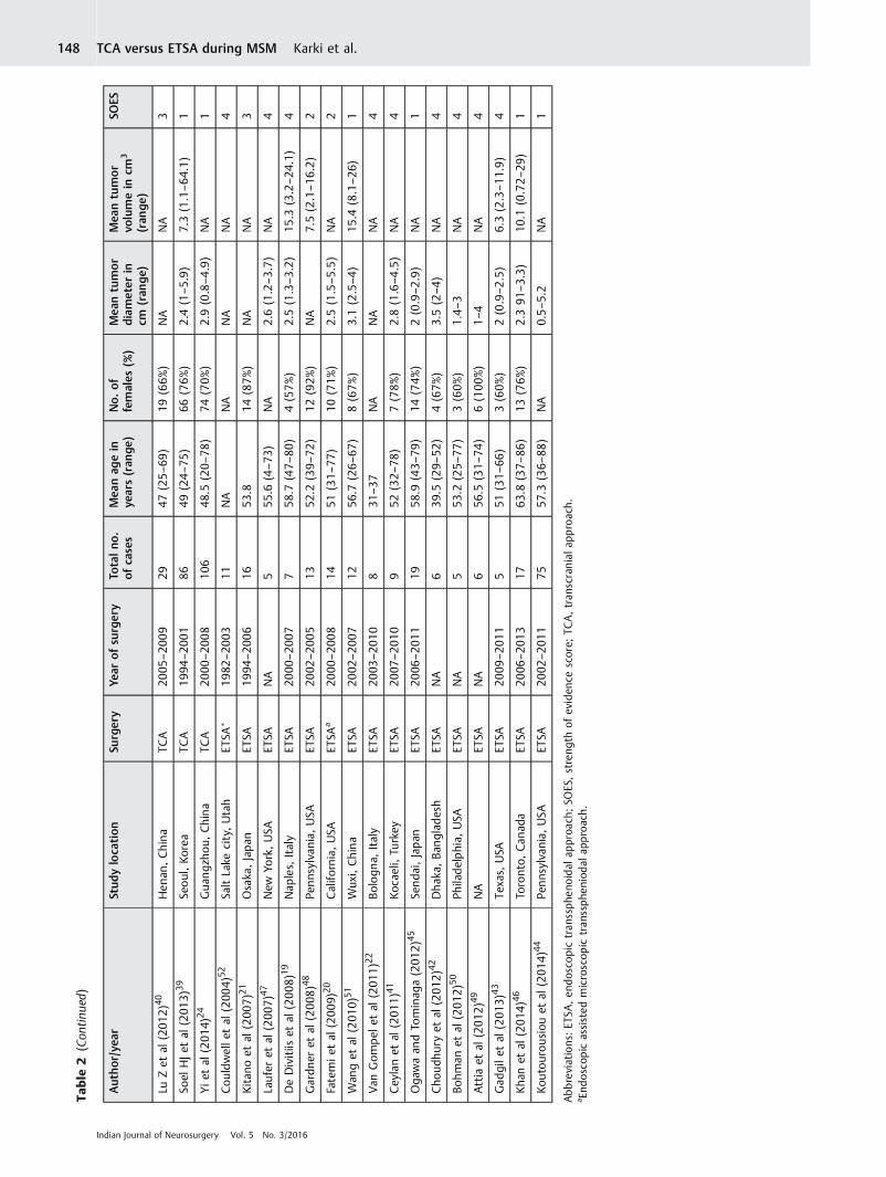
Inclusion and Exclusion CriteriaOnly patients with tumor specifically located in the TS, DS, orPS were included in this study. The studies were reviewedand observations were made regarding study design,methodology, and patient characteristics (►Table 2).Thecases for each study were extracted and the cohortsassessed based on preoperative clinical features (visualdeficits, headache, and endocrinopathy) and postoperativeoutcomes (visual function, GTR, occurrence of CSF leakage,and tumor recurrence). Similarly, morbidity assessment wasbased on frequency of infection (pneumonia, meningitis, andwound/bone flap infection), seizure, hemorrhage, visualworsening, endocrinopathy, and anosmia. Information onmortality, if available, was captured. Age was categorized into� 50 or > 50 years. Tumor size was categorized into � 2.4or > 2.4 cm. Also, duration of symptoms was categorized into� 12 or > 12 months. The p-values � 0.05 were consideredstatistically significant in all analysis. Duplication of patientswas eliminated in case series where multiple papers werepublished by the same authors or the same institution. Insuch situations, only reports with the largest sample size andrelevant data were selected.
Table 1 Strength of evidence scoring (SOES) system demonstratingthe assignment of scores based on visual assessment, demographicvariables (sex, mean age, size, and volume of tumor, and volume oftumor resection) in patients that were operated for MSM
Included variable SOES
10 or more patientsPostoperative vision assessmentNonvisual complicationReported age, sex, size, or volume orextent of resection
1
10 or more patientsPostoperative vision assessment ORNonvisual complicationReported age, sex, size, or volume orextent of resection
2
10 or more patientsPostoperative vision assessment ORNonvisual complication OR2 of 3 (reported age sex, size, orvolume or extent of resection)
3
Either 10 or more patients ORPostoperative vision assessment ORNonvisual complication OR1of 3 (reported age, sex, size, orvolume or extent of resection)
4
Abbreviation: MSM, midline suprasellar meningioma; OR, odds ratio.
Indian Journal of Neurosurgery Vol. 5 No. 3/2016
TCA versus ETSA during MSM Karki et al.146

Table
2Extrac
tedarticles
withtheirde
mog
raph
icvariab
les
Author/yea
rStud
yloca
tion
Surgery
Year
ofsu
rgery
Totalno
.ofcases
Mea
nag
ein
years(ran
ge)
No.of
females
(%)
Mea
ntumor
diameter
incm
(ran
ge)
Mea
ntumor
volumein
cm3
(ran
ge)
SOES
Araiet
al(200
0)26
Tokyo,
Japan
TCA
NA
2149
9(43%
)NA
NA
1
Ciric
andRo
senb
latt
(200
1)6
NA
TCA
NA
2440
18(75%
)NA
NA
2
Ohtaet
al(200
1)32
Kobecity,Japan
TCA
1980
–200
033
46.7
(15–
69)
29(88%
)NA
NA
2
Zevg
aridiset
al(200
1)16
Mun
ich,
German
yTC
A19
90–1
996
3454
.9(38–
80)
27(79%
)2.8(1.5–6
)NA
2
Fahlbusch
andScott(200
2)13
Erlang
en,German
yTC
A19
83–1
998
4754
.9(28–
74)
39(83%
)NA
NA
2
Goe
letal
(200
2)7
Bombay,India
TCA
1991
–200
170
NA
42(60%
)NA
NA
3
Jallo
andBe
njam
in(200
2)14
New
york,USA
TCA
1983
–200
123
57.7
(4–7
3)15
(65%
)3.3(2–5
)23
.4(8–6
0)1
Chi
andMcD
ermott(200
3)5
Califo
rnia,USA
TCA
1992
–200
221
52.3
793
3%)
NA
NA
2
Schick
andHassler
(200
5)15
Duisburg,German
yTC
A19
91–2
002
5353
(27–
78)
13(25%
)1–
5NA
1
Pamiret
al(200
5)29
Istanb
ul,Tu
rkey
TCA
1987
–200
442
24–7
928
(67%
)NA
7.5–
211
Mathiesen
(200
5)33
Stoc
kholm,Sw
eden
TCA
NA
2958
.323
(79%
)2.3
NA
1
Park
etal
(200
6)18
Seou
l,Ko
rea
TCA
NA
3045
24(80%
)2.6(1.6–6
.3)
12.4
(2.3–1
25.6)
2
Bassioun
ietal
(200
6)23
Essen,
German
yTC
A19
90–2
003
6253
(29–
81)
46(73%
)3.4(2–6
)NA
1
Nakam
uraet
al(200
6)25
Han
over,German
yTC
A19
78–2
002
7254
.3(30–
86)
54(75%
)2.8
NA
1
Otani
etal
(200
6)36
Zurich
,Sw
itzerlan
dTC
ANA
3253
.521
(66%
)2.3
NA
2
Kitan
oet
al(200
7)21
Osaka
,Japan
TCA
1994
–200
612
61.4
10(83%
)NA
NA
3
LiXet
al(200
7)35
Jinan
,China
TCA
1989
–200
343
53.8
(24–
68)
31(72%
)1.8–
5.4
NA
1
DeDivitiis
etal
(200
8)19
Nap
les,
Italy
TCA
1983
–200
644
NA
NA
NA
NA
4
Kim
etal
(200
8)27
Gwan
gju,
Korea
TCA
1998
–200
627
53.8
(36–
73)
22(80%
)3
NA
2
Meh
razinan
dMirfalah.
(200
8)38
Tehran
,Iran
TCA
1997
–200
645
45.7
(22–
75)
35(78%
)2.6(1.4–5
.2)
NA
1
Noz
akie
tal
(200
8)31
Kyoto,
Japan
TCA
1999
–200
722
52.9
(27–
73)
18(81%
)2.3(1.5–3
.5)
NA
2
Fatemie
tal
(200
9)20
Califo
rnia,USA
TCA
2000
–200
89
49(37–
57)
6(73%
)3.3(2.2–5
.5)
NA
4
Gan
naet
al(200
9)8
Toronto,
Can
ada
TCA
2000
–200
724
53.8
20(83%
)2.6
NA
1
Sadean
dLee(200
9)30
Ohio,
USA
TCA
1994
–200
631
NA
NA
2.3(1.1–3
.8)
NA
3
Galal
etal
(201
0)9
Cairo,Eg
ypt
TCA
2000
–200
621
43(27–
65)
14(67%
)NA
NA
1
Land
eiro
etal
(201
0)28
Rio
deJane
iroRJ,Brazil
TCA
1997
–200
823
56.2
(38–
77)
15(65%
)NA
NA
1
Palani
etal
(201
2)34
Hyd
erab
ad,India
TCA
2004
–201
141
NA
26(63%
)NA
NA
4
Jang
etal
(201
2)37
Gwan
gju,
Korea
TCA
2005
–201
124
49.5
(25–
70)
19(79%
)2(0.7–3
.3)
NA
1
Nan
daet
al(201
3)11
Louisian
a,USA
TCA
1990
–201
330
NA
NA
NA
NA
4
(Con
tinue
d)
Indian Journal of Neurosurgery Vol. 5 No. 3/2016
TCA versus ETSA during MSM Karki et al. 147
Table
2(Con
tinued)
Author/yea
rStud
yloca
tion
Surgery
Year
ofsu
rgery
Totalno
.ofcases
Mea
nag
ein
years(ran
ge)
No.of
females
(%)
Mea
ntumor
diameter
incm
(ran
ge)
Mea
ntumor
volumein
cm3
(ran
ge)
SOES
LuZet
al(201
2)40
Hen
an,China
TCA
2005
–200
929
47(25–
69)
19(66%
)NA
NA
3
Soel
HJet
al(201
3)39
Seou
l,Ko
rea
TCA
1994
–200
186
49(24–
75)
66(76%
)2.4(1–5
.9)
7.3(1.1–6
4.1)
1
Yie
tal
(201
4)24
Gua
ngzh
ou,China
TCA
2000
–200
810
648
.5(20–
78)
74(70%
)2.9(0.8–4
.9)
NA
1
Cou
ldwelle
tal
(200
4)52
SaltLake
city,Utah
ETSA
�19
82–2
003
11NA
NA
NA
NA
4
Kitan
oet
al(200
7)21
Osaka
,Japan
ETSA
1994
–200
616
53.8
14(87%
)NA
NA
3
Laufer
etal
(200
7)47
New
York,USA
ETSA
NA
555
.6(4–7
3)NA
2.6(1.2–3
.7)
NA
4
DeDivitiis
etal
(200
8)19
Nap
les,
Italy
ETSA
2000
–200
77
58.7
(47–
80)
4(57%
)2.5(1.3–3
.2)
15.3
(3.2–2
4.1)
4
Gardn
eret
al(200
8)48
Penn
sylvan
ia,USA
ETSA
2002
–200
513
52.2
(39–
72)
12(92%
)NA
7.5(2.1–1
6.2)
2
Fatemie
tal
(200
9)20
Califo
rnia,USA
ETSA
a20
00–2
008
1451
(31–
77)
10(71%
)2.5(1.5–5
.5)
NA
2
Wan
get
al(201
0)51
Wux
i,China
ETSA
2002
–200
712
56.7
(26–
67)
8(67%
)3.1(2.5–4
)15
.4(8.1–2
6)1
Van
Gom
pel
etal
(201
1)22
Bologn
a,Italy
ETSA
2003
–201
08
31–3
7NA
NA
NA
4
Cey
lanet
al(201
1)41
Kocaeli,Tu
rkey
ETSA
2007
–201
09
52(32–
78)
7(78%
)2.8(1.6–4
.5)
NA
4
Oga
waan
dTo
minag
a(201
2)45
Send
ai,Japan
ETSA
2006
–201
119
58.9
(43–
79)
14(74%
)2(0.9–2
.9)
NA
1
Cho
udhu
ryet
al(201
2)42
Dha
ka,Ba
ngladesh
ETSA
NA
639
.5(29–
52)
4(67%
)3.5(2–4
)NA
4
Bohm
anet
al(201
2)50
Philadelphia,
USA
ETSA
NA
553
.2(25–
77)
3(60%
)1.4–
3NA
4
Attia
etal
(201
2)49
NA
ETSA
NA
656
.5(31–
74)
6(100
%)
1–4
NA
4
Gad
gile
tal
(201
3)43
Texas,
USA
ETSA
2009
–201
15
51(31–
66)
3(60%
)2(0.9–2
.5)
6.3(2.3–1
1.9)
4
Kha
net
al(201
4)46
Toronto,
Can
ada
ETSA
2006
–201
317
63.8
(37–
86)
13(76%
)2.391
–3.3)
10.1
(0.72–
29)
1
Koutou
rousiouet
al(201
4)44
Penn
sylvan
ia,USA
ETSA
2002
–201
175
57.3
(36–
88)
NA
0.5–
5.2
NA
1
Abbrev
iation
s:ET
SA,en
dosco
pictran
ssphe
noidal
approa
ch;SO
ES,streng
thof
eviden
cescore;
TCA,tran
scraniala
pproach.
a End
osco
pic
assisted
microscop
ictran
ssphe
niod
alap
proach.
Indian Journal of Neurosurgery Vol. 5 No. 3/2016
TCA versus ETSA during MSM Karki et al.148
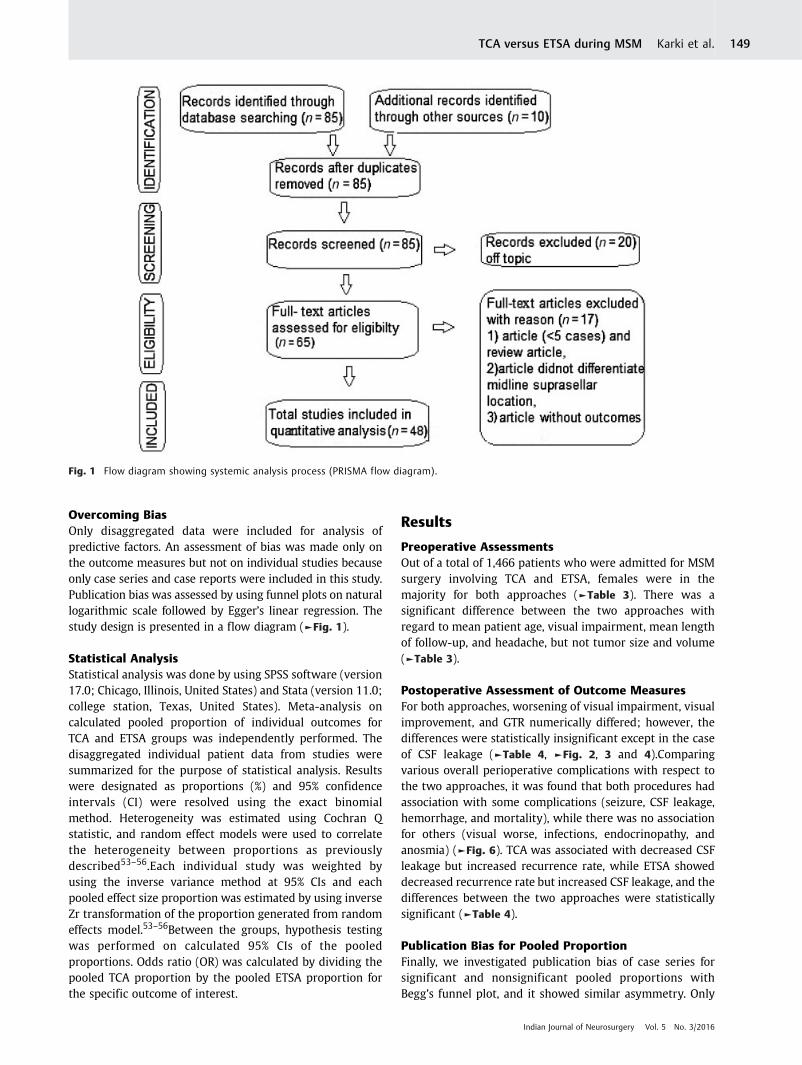
Overcoming BiasOnly disaggregated data were included for analysis ofpredictive factors. An assessment of bias was made only onthe outcome measures but not on individual studies becauseonly case series and case reports were included in this study.Publication bias was assessed by using funnel plots on naturallogarithmic scale followed by Egger’s linear regression. Thestudy design is presented in a flow diagram (►Fig. 1).
Statistical AnalysisStatistical analysis was done by using SPSS software (version17.0; Chicago, Illinois, United States) and Stata (version 11.0;college station, Texas, United States). Meta-analysis oncalculated pooled proportion of individual outcomes forTCA and ETSA groups was independently performed. Thedisaggregated individual patient data from studies weresummarized for the purpose of statistical analysis. Resultswere designated as proportions (%) and 95% confidenceintervals (CI) were resolved using the exact binomialmethod. Heterogeneity was estimated using Cochran Qstatistic, and random effect models were used to correlatethe heterogeneity between proportions as previouslydescribed53–56.Each individual study was weighted byusing the inverse variance method at 95% CIs and eachpooled effect size proportion was estimated by using inverseZr transformation of the proportion generated from randomeffects model.53–56Between the groups, hypothesis testingwas performed on calculated 95% CIs of the pooledproportions. Odds ratio (OR) was calculated by dividing thepooled TCA proportion by the pooled ETSA proportion forthe specific outcome of interest.
Results
Preoperative AssessmentsOut of a total of 1,466 patients who were admitted for MSMsurgery involving TCA and ETSA, females were in themajority for both approaches (►Table 3). There was asignificant difference between the two approaches withregard to mean patient age, visual impairment, mean lengthof follow-up, and headache, but not tumor size and volume(►Table 3).
Postoperative Assessment of Outcome MeasuresFor both approaches, worsening of visual impairment, visualimprovement, and GTR numerically differed; however, thedifferences were statistically insignificant except in the caseof CSF leakage (►Table 4, ►Fig. 2, 3 and 4).Comparingvarious overall perioperative complications with respect tothe two approaches, it was found that both procedures hadassociation with some complications (seizure, CSF leakage,hemorrhage, and mortality), while there was no associationfor others (visual worse, infections, endocrinopathy, andanosmia) (►Fig. 6). TCA was associated with decreased CSFleakage but increased recurrence rate, while ETSA showeddecreased recurrence rate but increased CSF leakage, and thedifferences between the two approaches were statisticallysignificant (►Table 4).
Publication Bias for Pooled ProportionFinally, we investigated publication bias of case series forsignificant and nonsignificant pooled proportions withBegg’s funnel plot, and it showed similar asymmetry. Only
Fig. 1 Flow diagram showing systemic analysis process (PRISMA flow diagram).
Indian Journal of Neurosurgery Vol. 5 No. 3/2016
TCA versus ETSA during MSM Karki et al. 149

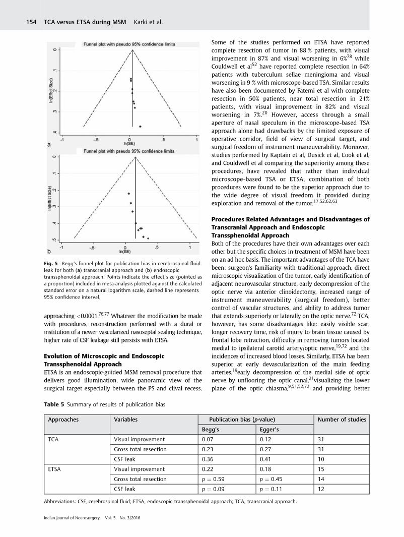
Funnel plot with respect to CSF leakage is shown (►Fig. 5)and it was a nonsignificant plot (p > 0.05). From Egger’s testperformed on the publications used in this study, there wasno bias with specific respect to visual improvement, GTR,and CSF leakage (►Table 5).
Discussion
BackgroundComplete removal of MSM via the TCA, first described by Stirlingand Edin in 1897,57was performed by Cushing and Eisenhardt in
Table 3 Comparison of clinical characteristics of patients who underwent TCA and ETSA for resection of midline suprasellarmeningiomas, from studies in which patient data were reported in disaggregated form
Preoperative variables No. of studies TCA No. of studies ETSA p- Value
Age (y)
<50 y (%) 11 428(44) 1 6(3) 0.001
>50 y (%) 15 524(55) 13 203(97)
Sex
Male (%) 29 336(30) 12 39(28) 0.776
Female (%) 29 782(70) 12 98(71)
Presenting symptoms
Headache (%) 12 244(42) 10 30(20) 0.001
Endocrine abnormalities (%) 7 34(13) 4 10(9) 0.362
Preoperative visual deficits (%) 31 1,075(91) 15 175(81) 0.001
Tumor characteristics
Mean tumor diameter, cm (range) 18 3(0.8–63) 12 2.5(0.5–5.9) 0.135
Mean tumor volume, cm3 (range) 4 14.3(1.1–125.6) 5 11(0.7–29) 0.072
Optic canal involvement (%) 9 149(36) 1 20(26) 0.114
Major vascular encasement (%) 8 135(40) 4 59(47) 0.218
Mean follow-up (mo, range) 27 53(3–192) 12 19(1–98) 0.002
Mean tumor volume, cm3 (range) 4 14.3(1.1–125.6) 5 11(0.7–29) 0.072
Abbreviations: ETSA, endoscopic transsphenoidal approach; TCA, transcranial approach.
Table 4 Comparison of surgical outcomes after resection of midline suprasellar meningioma (MSM) between TCA and ETSAgroups
Variables Proportion OR; 95% CI p-value
GTR
TCA 0.80 1.3; 0.74–7.68 0.43
ETSA 0.83
Visual improved
TCA 0.71 1.2; 0.25–8.92 0.26
ETSA 0.70
CSF leak
TCA 0.08 2.5; 1.78–11.56 0.04
ETSA 0.17
Visual worse
TCA 0.18 1.6;0.43–6.11 0.12
ETSA 0.15
Tumor recurrence
TCA 0.07 1.8; 1.1–6.39 0.02
ETSA 0.05
Abbreviations: CI, confidence interval; CSF, cerebrospinal fluid; ETSA, endoscopic transsphenoidal approach; GTR, gross total resection; OR, oddsratio; TCA, transcranial approach.
Indian Journal of Neurosurgery Vol. 5 No. 3/2016
TCA versus ETSA during MSM Karki et al.150

Fig. 2 It demonstrates comparison between transcranial and endoscopic transsphenoidal approach with respect to postoperative visualimprovement. Forest plot demonstrates the 95% confidence intervals and percentiles weights associated with the individual and combinedstudy groups for (a) transcranial approach group and (b) endoscopic transsphenoidal approach group.
Indian Journal of Neurosurgery Vol. 5 No. 3/2016
TCA versus ETSA during MSM Karki et al. 151

1916.58 Since then, MSMs have been removed by differenttranscranial approaches that include: frontolateral,frontotemporal/pterional, orbitopterional/orbitozygomatic,unilateral subfrontal, bilateral subfrontal, interhemispheric,supraorbital, and pterional.7,13,14,25,30–33,59,60 With time, the
complications, risks, and associated morbidities with TCA gaverise to TSA that was devoid of the previous risks. TSA wasintroduced by Herman Schloffer, who in 1907 resected apituitary tumor via TSA.61 TSA was found to be effective andsafer for suprasellar meningiomas that were predominantly
Fig. 3 It demonstrates comparison between transcranial and endoscopic transsphenoidal approach with respect to gross total resection.Forest plot demonstrates the 95% confidence intervals and percentiles weights associated with the individual and combined study groups for(a) transcranial approach group and (b) endoscopic transsphenoidal approach group.
Indian Journal of Neurosurgery Vol. 5 No. 3/2016
TCA versus ETSA during MSM Karki et al.152

located in the midline17,47,62–66 though its study-basedadvantages and disadvantages need affirmation.13,25,52,67 Eventhough TSA has refined with evolution of microscope-based52,68
or currently by endoscope-based techniques,21,47,69–71 there stillremains the most common problem of CSF leakage. Similarly, tolimit this CSF leakage during ETSA, varieties of closure methodslike: fat and fascial graft,72 fat and synthetic dural substitute,51
mixture (fascial graft, methacrylate, mucoperichondrium, andfibrin glue),73 mixed (fascia lata and Nissel or DuraSeal),47 ormixed (fascial graft and nasoseptal flap)46 for duralreconstruction have been proposed. Among these closure
methods, a study done by de Divitiis19 has reported a higher(29%) rate of CSF leakage that specifically utilized a multilayerclosure involving collagen matrix, dural substitute, areabsorbable plate, and fibrin sealant. Various reconstructiontechniques during the TCA have been suggested to preventpostoperative CSF leakage that occur mainly in the frontalsinus74 and resultant meningitis, including the use of pericranialflap with or without adipose tissue being the most popularone.6,75 Progressively, however, with the evolution ofvascularized nasoseptal flap reconstruction technique, the CSFleakage rate has dramatically decreased to 16% with a p-value
Fig. 4 It demonstrates comparison between transcranial and endoscopic transsphenoidal approach with respect to cerebrospinal fluid leakage.Forest plot demonstrates the 95% confidence intervals and percentiles weights associated with the individual and combined study groups for(a) transcranial approach group and (b) endoscopic transsphenoidal approach group.
Indian Journal of Neurosurgery Vol. 5 No. 3/2016
TCA versus ETSA during MSM Karki et al. 153
approaching <0.0001.76,77 Whatever the modification be madewith procedures, reconstruction performed with a dural orinstitution of a newer vascularized nasoseptal sealing technique,higher rate of CSF leakage still persists with ETSA.
Evolution of Microscopic and EndoscopicTranssphenoidal ApproachETSA is an endoscopic-guided MSM removal procedure thatdelivers good illumination, wide panoramic view of thesurgical target especially between the PS and clival recess.
Some of the studies performed on ETSA have reportedcomplete resection of tumor in 88 % patients, with visualimprovement in 87% and visual worsening in 6%78 whileCouldwell et al52 have reported complete resection in 64%patients with tuberculum sellae meningioma and visualworsening in 9 % with microscope-based TSA. Similar resultshave also been documented by Fatemi et al with completeresection in 50% patients, near total resection in 21%patients, with visual improvement in 82% and visualworsening in 7%.20 However, access through a smallaperture of nasal speculum in the microscope-based TSAapproach alone had drawbacks by the limited exposure ofoperative corridor, field of view of surgical target, andsurgical freedom of instrument maneuverability. Moreover,studies performed by Kaptain et al, Dusick et al, Cook et al,and Couldwell et al comparing the superiority among theseprocedures, have revealed that rather than individualmicroscope-based TSA or ETSA, combination of bothprocedures were found to be the superior approach due tothe wide degree of visual freedom it provided duringexploration and removal of the tumor.17,52,62,63
Procedures Related Advantages and Disadvantages ofTranscranial Approach and EndoscopicTranssphenoidal ApproachBoth of the procedures have their own advantages over eachother but the specific choices in treatment of MSM have beenon an ad hoc basis. The important advantages of the TCA havebeen: surgeon’s familiarity with traditional approach, directmicroscopic visualization of the tumor, early identification ofadjacent neurovascular structure, early decompression of theoptic nerve via anterior clinoidectomy, increased range ofinstrument maneuverability (surgical freedom), bettercontrol of vascular structures, and ability to address tumorthat extends superiorly or laterally on the optic nerve.72 TCA,however, has some disadvantages like: easily visible scar,longer recovery time, risk of injury to brain tissue caused byfrontal lobe retraction, difficulty in removing tumors locatedmedial to ipsilateral carotid artery/optic nerve,19,72 and theincidences of increased blood losses. Similarly, ETSA has beensuperior at early devascularization of the main feedingarteries,19early decompression of the medial side of opticnerve by unflooring the optic canal,21visualizing the lowerplane of the optic chiasma,9,51,52,72 and providing better
Table 5 Summary of results of publication bias
Approaches Variables Publication bias (p-value) Number of studies
Begg’s Egger’s
TCA Visual improvement 0.07 0.12 31
Gross total resection 0.23 0.27 31
CSF leak 0.36 0.41 10
ETSA Visual improvement 0.22 0.18 15
Gross total resection p ¼ 0.59 p ¼ 0.45 14
CSF leak p ¼ 0.09 p ¼ 0.11 12
Abbreviations: CSF, cerebrospinal fluid; ETSA, endoscopic transsphenoidal approach; TCA, transcranial approach.
Fig. 5 Begg’s funnel plot for publication bias in cerebrospinal fluidleak for both (a) transcranial approach and (b) endoscopictranssphenoidal approach. Points indicate the effect size (pointed asa proportion) included in meta-analysis plotted against the calculatedstandard error on a natural logarithm scale, dashed line represents95% confidence interval,
Indian Journal of Neurosurgery Vol. 5 No. 3/2016
TCA versus ETSA during MSM Karki et al.154

cosmetic results.51 This approach also shortens the recoverytime, lessens injury to brain parenchyma (no brainretraction),19 provides clear vision between the internalcarotid artery (ICA) to ICA and easy removal of sellarextension tumors. Moreover, it has also been the choice ofsurgery in elderly patients or patients with majorcomorbidities that cannot stand surgery.21,52 However, ETSAhas been limited in number of cases due to increased risk ofCSF leakage/CSF fistulae19,52 and inadequate removal due tothe nature of tumor (hard/calcified/large/above optic nerve/more laterally located).19
Surgical OutcomesThe reports of the present study relate favorably with those ofother studies.19,20,78,79 For example, Komotar et al observedsignificantly higher rate of GTR in endoscopic group comparedwith transcranial group (92.8 vs 63.2%; p < 0.001).79 Removalof the tumor as much as possible is the ultimate goal ofresection in neurosurgery. Nonetheless, several studies havehighlighted the fact that complete resection at the cost of visualdeterioration or hypothalamic dysfunction should not beattempted. With the advent of modern microsurgery, theamount of GTR rate has leaped from 35 to 76% (macrosurgicalera) to 58 to 100% (of the microsurgical era).25 Resection ofMSM is a complex procedure and depends on various factorslike size of meningioma, arterial encasement, peritumoraledema, and approaches followed.34 Whatever the influentialfactor may be, the extent of resection is paramount inpredicting subsequent recurrence. Studies conducted earlierhave reported the yield of GTR during TCA to be 70 to 100%while that with ETSA to be between 50 to 84%.20,63Contrarily,studies conducted by de Divitiis et al19 have revealed 83.3% GTRduring TCA while 86.4% GTR yield during ETSA. Our findingstoo followed the latter pattern with 80% GTR during TCA and83% GTR during ETSA, with no statistical association (p ¼ 0.43).
One of the undesired results of MSM surgery is tumorrecurrence which is influenced by varieties of factors. Asoutlined by Fahlbusch and Schott,13 these factors can be: 1)extent of tumor resection, 2) histological grading of thetumor, 3) length of follow-up period, and 4) mode and qualityof assessment of tumor recurrence. TSA approach being lessinvasive with a shorter hospital stay, the compliance to long-term follow-up becomes questionable. Most of the previouslyconducted ETSA studies have had 6 to 24 months of follow-ups (mean, 21 � 18 months)51 while the mean follow-upduration for TCA was 51.8 months (0.8–112 months).79
Recurrence rate for MSM ranges from 5% to > 30 % and therate increases with longer follow-ups.1–3,10,12,80 Ourobservations, as shown in ►Table 3, too shared the samefact as seen by lack of 100% follow-up rates with both theprocedures, (TCA, 73.3% vs ETSA, 79%). Moreover, the lengthof mean follow-up period for TCA was longer as comparedwith ETSA (53 vs 19 months) and the p-value approaching0.002. As a result, the recurrence rate with TCA was higher ascompared with ETSA (7 vs 5%; p ¼ 0.02). Meningiomas,although histologically benign, can recur and progress overtime, so longer follow-up is necessary to evaluate the long-term outcome.
As mentioned earlier, good visual outcome is the target ofa successful MSM surgery. In relation to the surgical approachundertaken, recent meta-analysis have demonstrated morevisual improvements with ETSA (75%) as compared with TCA(58.4%), although the observations did not have statisticalsignificance.19 Our studies also demonstrated almost similarrate of visual improvement with no statistically significantdifference (TCA vs ETSA, 71 vs 70%; p ¼ 0.26)), as depictedin ►Table 4. Moreover, some series have identified ETSA todeliver higher visual functional improvement in comparisonto TCA.22,49,69,79,81 Our observations, however, alsodemonstrated higher rate of visual worsening with TCA
Fig. 6 Perioperative complications following transcranial and endoscopic transsphenoidal approach for midline suprasellar meningiomaresection.
Indian Journal of Neurosurgery Vol. 5 No. 3/2016
TCA versus ETSA during MSM Karki et al. 155

than with ETSA and no statistical association (18 vs 15%;p ¼ 0.12). This lower rate of visual worsening encounteredwith the ETSA approach was attributed to minimalmanipulation of compromised ischemic optic apparatus,adequate and early visualization of the subchiasmatic bloodvessels, and precision and safer handling of the opticapparatus.51
Operative ComplicationsOn the other hand, we observed that both TCA and ETSA didnot differ significantly in visual improvement and GTR butpostoperative complications, specifically, seizure andhemorrhage was significantly predominant in TCA ascompared with ETSA.19,78 Some series reported thatsurgical morbidities such as seizure and hemorrhage werelower in patients treated with ETSA as compared with TCA.Symon and Rosenstein also reported that new onset seizurewas a commonly occurring complication of MSM surgerybeing as high as in 10.9% of cases during TCA.82 Our studyalso revealed cases of new onset seizure and the prevalencebeing more (6%) with TCA than with ETSA (1%). Similarly,intracerebral hemorrhage was found to be more with TCAthan ETSA (5 vs 3%). Observation of low cases of seizure andhemorrhage in ETSA in comparison to TCA is expectedbecause in TCA, more or less frontal lobe retraction needs tobe done which may cause rupture of small feeding bloodvessels and injury to brain parenchyma causing seizure. ButETSA has been superior to TCA in the field of earlydevascularization of main feeding vessels and no brainretraction.19 Theoretically, postoperative CSF leakage is apotential complication of approaches and occurs via thefrontal sinus during TCA. But in practice and in numerousresearches namely, Komotar et al79 (21.3 vs 4.3%; p < 0.001),ETSA is found to be associated with higher rates of CSFleakage than with the TCA.19,20,63,83 Our study has alsoshown that postoperative CSF leakage occurs more withETSA than with TCA (17 vs 8%; p ¼ 0.04) (►Table 4).
Finally, we report that TCA and ETSA surgical approaches forMSM removal do not display complete superiority over eachother when assessed on the basis of postoperative outcomemeasures. This was apparent since TCA and ETSA displayedpoor outcomes with regard to recurrence rates and CSF leakagerespectively, coincidentally, two of the commonly usedpostoperation determinants of improvement. Generally, itwas observed that both TCA and ETSA did not improveworsening of visual function, visual improvement, and GTR.However, reduced CSF leakage and increased recurrence ratewas associated with TCA, while reduced recurrent rate andincreased CSF leakage was attributable to ETSA. The poorrecurrence rate associated with TCA may be linked to the longfollow-up period, while the comparatively reduced recurrencerate in ETSA cohorts may be accounted for by the short follow-up period. It was apparent that patients who underwent MSMremoval by both TCA and ETSA have almost similarpreoperative clinical characteristics, an observation whichstrengthens the comparability of the two cohorts. The resultsof this review are not much different from recent studies,which have shown that none of the surgical approaches could
be seen as an end in itself since both have advantages anddisadvantages, thus the choice of TCA or ETSA may bedetermined by tumor characteristics, patient-specific clinicalpresentations, risk/benefits, and prioritization of clinicalobjectives.
Limitations
This study, among other factors, may be limited by caseheterogeneity, methodological heterogeneity which cannotbe cured completely by statistical method, because the studyis retrospective in nature. For instance, length of follow-upand tumor characteristics for both cohorts in some casesdiffered.
Conclusions
Put together, the choice of TCA or ETSA for surgical removalof MSMs may depend on many factors including tumorcharacteristics, patient-specific clinical presentations, risk/benefit assessment, and the clinical objective. Ideally, asurgical approach that can maximize the advantages of bothTCA and ETSA while at the same time minimize the risksassociated with these approaches must be sought.
Conflict of InterestNone.
FundingNone.
AcknowledgmentsWewould like to thank Dr. DejunWu, Dr. Jian Tao, and Dr. LiDekun at the Department of Neurosurgery, Anhui MedicalUniversity for their valuable guidance and revision of ourillustrations.
References1 Al-Mefty O, Smith R. Tuberculum sellae meningiomas. Meningiomas
Raven, New York 1991;395411:395–4112 Al-Mefty O, Holoubi A, Rifai A, Fox JL. Microsurgical removal of
suprasellar meningiomas. Neurosurgery 1985;16(3):364–3723 Brihaye J, Brihaye-van Geertruyden M. Management and surgical
outcome of suprasellar meningiomas. Proceedings of the 8thEuropean Congress of Neurosurgery Barcelona, September 6–11,1987: Springer; 1988:124–129
4 Arifin MZ, Mardjono I, Sidabutar R, Wirjomartani BA, Faried A.Pterional approach versus unilateral frontal approach ontuberculum sellae meningioma: single centre experiences.Asian J Neurosurg 2012;7(1):21–24
5 Chi JH, McDermott MW. Tuberculum sellae meningiomas.Neurosurg Focus 2003;14(6):e6
6 Ciric I, Rosenblatt S. Suprasellar meningiomas. Neurosurgery2001;49(6):1372–1377
7 Goel A, Muzumdar D, Desai KI. Tuberculum sellae meningioma: areport on management on the basis of a surgical experience with70 patients. Neurosurgery 2002;51(6):1358–1363, discussion1363–1364
Indian Journal of Neurosurgery Vol. 5 No. 3/2016
TCA versus ETSA during MSM Karki et al.156
8 Ganna A, Dehdashti AR, Karabatsou K, Gentili F. Fronto-basalinterhemispheric approach for tuberculum sellae meningiomas;long-term visual outcome. Br J Neurosurg 2009;23(4):422–430
9 Galal A, Faisal A, Al-Werdany M, El Shehaby A, Lotfy T, MoharramH. Determinants of postoperative visual recovery in suprasellarmeningiomas. Acta Neurochir (Wien) 2010;152(1):69–77
10 Mirimanoff RO, Dosoretz DE, Linggood RM, Ojemann RG, MartuzaRL. Meningioma: analysis of recurrence and progressionfollowing neurosurgical resection. J Neurosurg 1985;62(1):18–24
11 Nanda A, Ambekar S, Javalkar V, Sharma M. Technical nuances inthe management of tuberculum sellae and diaphragma sellaemeningiomas. Neurosurg Focus 2013;35(6):E7
12 Andrews BT, Wilson CB. Suprasellar meningiomas: the effect oftumor location on postoperative visual outcome. J Neurosurg1988;69(4):523–528
13 Fahlbusch R, Schott W. Pterional surgery of meningiomas of thetuberculum sellae and planum sphenoidale: surgical results withspecial consideration of ophthalmological and endocrinologicaloutcomes. J Neurosurg 2002;96(2):235–243
14 Jallo GI, Benjamin V. Tuberculum sellae meningiomas:microsurgical anatomy and surgical technique. Neurosurgery2002;51(6):1432–1439, discussion 1439–1440
15 Schick U, Hassler W. Surgical management of tuberculum sellaemeningiomas: involvement of the optic canal and visualoutcome. J Neurol Neurosurg Psychiatry 2005;76(7):977–983
16 Zevgaridis D, Medele RJ, Müller A, Hischa AC, Steiger H-J.Meningiomas of the sellar region presenting with visualimpairment: impact of various prognostic factors on surgicaloutcome in 62 patients. Acta Neurochir (Wien) 2001;143(5):471–476
17 Kaptain GJ, Vincent DA, Sheehan JP, Laws ER Jr. Transsphenoidalapproaches for the extracapsular resection of midline suprasellarand anterior cranial base lesions. Neurosurgery 2001;49(1):94–100, discussion 100–101
18 Park C-K, Jung H-W, Yang S-Y, Seol HJ, Paek SH, Kim DG. Surgicallytreated tuberculum sellae and diaphragm sellae meningiomas:the importance of short-term visual outcome. Neurosurgery2006;59(2):238–243, discussion 238–243
19 de Divitiis E, Esposito F, Cappabianca P, Cavallo LM, de Divitiis O.Tuberculum sellae meningiomas: high route or low route? Aseries of 51 consecutive cases. Neurosurgery 2008;62(3):556–563, discussion 556–563
20 Fatemi N, Dusick JR, de Paiva Neto MA, Malkasian D, Kelly DF.Endonasal versus supraorbi tal keyhole removal ofcraniopharyngiomas and tuberculum sellae meningiomas.Neurosurgery 2009;64(5, Suppl 2):269–284, discussion 284–286
21 Kitano M, Taneda M, Nakao Y. Postoperative improvement invisual function in patients with tuberculum sellae meningiomas:results of the extended transsphenoidal and transcranialapproaches. J Neurosurg 2007;107(2):337–346
22 Van Gompel JJ, Frank G, Pasquini E, Zoli M, Hoover J, Lanzino G.Expanded endonasal endoscopic resection of anterior fossameningiomas: report of 13 cases and meta-analysis of theliterature. Neurosurg Focus 2011;30(5):E15
23 Bassiouni H, Asgari S, Stolke D. Tuberculum sellae meningiomas:functional outcome in a consecutive series treated microsurgically.Surg Neurol 2006;66(1):37–44, discussion 44–45
24 Liu Y, Chotai S, Ming C, Jin S, Pan J, Qi S. Characteristics of midlinesuprasellar meningiomas based on their origin and growthpattern. Clin Neurol Neurosurg 2014;125:173–181
25 Nakamura M, Roser F, Struck M, Vorkapic P, Samii M. Tuberculumsellae meningiomas: clinical outcome considering differentsurgical approaches. Neurosurgery 2006;59(5):1019–1028,discussion 1028–1029
26 Arai H, Sato K, Okuda O, et al. Transcranial transsphenoidalapproach for tuberculum sellae meningiomas. Acta Neurochir(Wien) 2000;142(7):751–756, discussion 756–757
27 Kim T-W, Jung S, Jung T-Y, Kim I-Y, Kang S-S, Kim S-H. Prognosticfactors of postoperative visual outcomes in tuberculum sellaemeningioma. Br J Neurosurg 2008;22(2):231–234
28 Landeiro JA, Gonçalves MB, Guimarães RD, et al. Tuberculumsellae meningiomas: surgical considerations. Arq Neuropsiquiatr2010;68(3):424–429
29 Pamir MN, Özduman K, Belirgen M, Kilic T, Özek MM. Outcomedeterminants of pterional surgery for tuberculum sellaemeningiomas. Acta Neurochir (Wien) 2005;147(11):1121–1130,discussion 1130
30 Sade B, Lee JH. High incidence of optic canal involvement intuberculum sellae meningiomas: rationale for aggressive skullbase approach. Surg Neurol 2009;72(2):118–123, discussion123
31 Nozaki K, Kikuta K, Takagi Y, Mineharu Y, Takahashi JA,Hashimoto N. Effect of early optic canal unroofing on theoutcome of visual functions in surgery for meningiomas of thetuberculum sellae and planum sphenoidale. Neurosurgery 2008;62(4):839–844, discussion 844–846
32 Ohta K, Yasuo K, Morikawa M, Nagashima T, Tamaki N. Treatmentof tuberculum sellae meningiomas:a long-term follow-up study.J Clin Neurosci 2001;8(Suppl 1):26–31
33 Mathiesen T, Kihlström L. Visual outcome of tuberculum sellaemeningiomas after extradural optic nerve decompression.Neurosurgery 2006;59(3):570–576, discussion 570–576
34 Palani A, Panigrahi MK, Purohit AK. Tuberculum sellaemening iomas: a ser ies o f 41 cases ; surg ica l andophthalmological outcomes with proposal of a new prognosticscoring system. J Neurosci Rural Pract 2012;3(3):286–293
35 Li X, Liu M, Liu Y, Zhu S. Surgical management of tuberculumsellae meningiomas. J Clin Neurosci 2007;14(12):1150–1154
36 Otani N, Muroi C, Yano H, Khan N, Pangalu A, Yonekawa Y.Surgical management of tuberculum sellae meningioma: role ofselective extradural anterior clinoidectomy. Br J Neurosurg 2006;20(3):129–138
37 Jang W-Y, Jung S, Jung T-Y, Moon K-S, Kim I-Y. The contralateralsubfrontal approach can simplify surgery and provide favorablevisual outcome in tuberculum sellae meningiomas. NeurosurgRev 2012;35(4):601–607, discussion 607–608
38 Mehrazin M, Mirfalah R. Early postoperative visual outcome inmicrosurgically treated suprasellar meningiomas predict long-term visual outcome. Turk Neurosurg 2008;18(4):380–386
39 Seol HJ, Park H-Y, Nam D-H, et al. Clinical outcomes oftuberculum sellae meningiomas focusing on reversibility ofpostoperative visual function. Acta Neurochir (Wien) 2013;155(1):25–31
40 Lu ZF, Cheng XB, Zhao YG, Shi BZ. Twenty-nine cases of resectionof suprasellar meningioma through small bone window: aninterhemispheric approach. Contemp Oncol (Pozn) 2013;17(6):525–529
41 Ceylan S, Koc K, Anık I. Extended endoscopic transsphenoidalapproach for tuberculum sellae meningiomas. Acta Neurochir(Wien) 2011;153(1):1–9
42 Chowdhury FH, Haque MR, Goel AH, Kawsar KA. Endoscopicendonasal extended transsphenoidal removal of tuberculumsellae meningioma (TSM): an experience of six cases. Br JNeurosurg 2012;26(5):692–699
43 Gadgil N, Thomas JG, Takashima M, Yoshor D. Endoscopicresection of tuberculum sellae meningiomas. J Neurol Surg BSkull Base 2013;74(4):201–210
44 Koutourousiou M, Fernandez-Miranda JC, Stefko ST, Wang EW,Snyderman CH, Gardner PA. Endoscopic endonasal surgery forsuprasellar meningiomas: experience with 75 patients.J Neurosurg 2014;120(6):1326–1339
45 Ogawa Y, Tominaga T. Extended transsphenoidal approach fortuberculum sellae meningioma—what are the optimum and criticalindications? Acta Neurochir (Wien) 2012;154(4):621–626
Indian Journal of Neurosurgery Vol. 5 No. 3/2016
TCA versus ETSA during MSM Karki et al. 157

46 Khan OH, Krischek B, Holliman D, et al. Pure endoscopic expandedendonasal approach for olfactory groove and tuberculum sellaemeningiomas. J Clin Neurosci 2014;21(6):927–933
47 Laufer I, Anand VK, Schwartz TH. Endoscopic, endonasalextended transsphenoidal, transplanum transtuberculumapproach for resection of suprasellar lesions. J Neurosurg 2007;106(3):400–406
48 Gardner PA, Kassam AB, Thomas A, et al. Endoscopic endonasalresection of anterior cranial base meningiomas. Neurosurgery2008;63(1):36–52, discussion 52–54
49 Attia M, Kandasamy J, Jakimovski D, et al. The importance andtiming of optic canal exploration and decompression duringendoscopic endonasal resection of tuberculum sella and planumsphenoidale meningiomas. Neurosurgery 2012;71(1, SupplOperative):58–67
50 Bohman L-E, Stein SC, Newman JG, et al. Endoscopic versus openresection of tuberculum sellae meningiomas: a decision analysis.ORL J Otorhinolaryngol Relat Spec 2012;74(5):255–263
51 Wang Q, Lu X-J, Ji W-Y, et al. Visual outcome afterextended endoscopic endonasal transsphenoidal surgery fortuberculum sellae meningiomas. World Neurosurg 2010;73(6):694–700
52 Couldwell WT, Weiss MH, Rabb C, Liu JK, Apfelbaum RI, FukushimaT. Variations on the standard transsphenoidal approach to thesellar region, with emphasis on the extended approaches andparasellar approaches: surgical experience in 105 cases.Neurosurgery 2004;55(3):539–547, discussion 547–550
53 DerSimonian R. Combining evidence from clinical trials. AnesthAnalg 1990;70(5):475–476
54 DerSimonian R, Charette LJ, McPeek B, Mosteller F. Reporting onmethods in clinical trials. N Engl J Med 1982;306(22):1332–1337
55 DerSimonian R, Laird N. Meta-analysis in clinical trials. ControlClin Trials 1986;7(3):177–188
56 DerSimonian R. Meta-analysis in the design and monitoring ofclinical trials. Stat Med 1996;15(12):1237–1248, discussion1249–1252
57 Stirling J, Edin M. Tumor of the meninges in the region of thepituitary body, pressing on the chiasma. Ann Ophthalmol 1897;6:15–16
58 Cushing H, Eisenhardt L. Suprasellar meningiomas. Meningiomas:Their Classification, Regional Behaviour, Life History, and SurgicalEnd Results. UK. Hafner Publishing Co Ltd; 1938:224–49
59 Benjamin V, Russell SM. The microsurgical nuances of resectingtuberculum sellae meningiomas. Neurosurgery 2005;56(2,Suppl):411–417, discussion
60 Noguchi A, Balasingam V, McMenomey SO, Delashaw JB Jr.Supraorbital craniotomy for parasellar lesions. Technical note.J Neurosurg 2005;102(5):951–955
61 Schmidt RF, Choudhry OJ, Takkellapati R, Eloy JA, Couldwell WT,Liu JK. Hermann Schloffer and the origin of transsphenoidalpituitary surgery. Neurosurg Focus 2012;33(2):E5
62 Dusick JR, Esposito F, Kelly DF, et al. The extended directendonasal transsphenoidal approach for nonadenomatoussuprasellar tumors. J Neurosurg 2005;102(5):832–841
63 Cook SW, Smith Z, Kelly DF. Endonasal transsphenoidal removalof tuberculum sellae meningiomas: technical note. Neurosurgery2004;55(1):239–244, discussion 244–246
64 Kassam AB, Thomas A, Carrau RL, et al. Endoscopicreconstruction of the cranial base using a pedicled nasoseptalf lap. Neurosurgery 2008;63(1, Suppl 1):ONS44–ONS52,discussion ONS52–ONS53
65 Kouri JG, Chen MY, Watson JC, Oldfield EH. Resection of suprasellartumors by using a modified transsphenoidal approach. Report offour cases. J Neurosurg 2000;92(6):1028–1035
66 Maira G, Anile C, Albanese A, Cabezas D, Pardi F, Vignati A. Therole of transsphenoidal surgery in the treatment ofcraniopharyngiomas. J Neurosurg 2004;100(3):445–451
67 Honegger J, Fahlbusch R, Buchfelder M, Huk WJ, Thierauf P. Therole of transsphenoidal microsurgery in the management of sellarand parasellar meningioma. Surg Neurol 1993;39(1):18–24
68 Fatemi N, Dusick JR, de Paiva Neto MA, Kelly DF. The endonasalmicroscopic approach for pituitary adenomas and otherparasellar tumors: a 10-year experience. Neurosurgery 2008;63(4, Suppl 2):244–256, discussion 256
69 Cappabianca P, Cavallo L, Esposito F, De Divitiis O, Messina A, DeDivitiis E. Extended endoscopic endonasal approach to themidline skull base: the evolving role of transsphenoidalsurgery. Adv Tech Stand Neurosurg 2008;33:151–199
70 de Divitiis E, Cavallo LM, Esposito F, Stella L, Messina A. Extendedendoscopic transsphenoidal approach for tuberculum sellaemeningiomas. Neurosurgery 2007;61(5, Suppl 2):229–237,discussion 237–238
71 Liu JK, Christiano LD, Patel SK, Tubbs RS, Eloy JA. Surgical nuancesfor removal of tuberculum sellae meningiomas with optic canalinvolvement using the endoscopic endonasal extendedtranssphenoidal transplanum transtuberculum approach.Neurosurg Focus 2011;30(5):E2
72 Bowers CA, Altay T, Couldwell WT. Surgical decision-makingstrategies in tuberculum sellae meningioma resection. NeurosurgFocus 2011;30(5):E1
73 Ceylan S, Koc K, Anik I. Extended endoscopic approaches formidline skull-base lesions. Neurosurg Rev 2009;32(3):309–319,discussion 318–319
74 Chokyu I, Goto T, Ishibashi K, Nagata T, Ohata K. Bilateral subfrontalapproach for tuberculum sellae meningiomas in long-termpostoperative visual outcome. J Neurosurg 2011;115(4):802–810
75 Mahmoud M, Nader R, Al-Mefty O. Optic canal involvement intuberculum sellae meningiomas: influence on approach,recurrence, and visual recovery. Neurosurgery 2010;67(3, SupplOperative ):ons108–ons118, discussion ons118–ons119
76 Hadad G, Bassagasteguy L, Carrau RL, et al. A novel reconstructivetechnique after endoscopic expanded endonasal approaches: vascularpedicle nasoseptal flap. Laryngoscope 2006;116(10):1882–1886
77 Zanation AM, Carrau RL, Snyderman CH, et al. Nasoseptal flapreconstruction of high flow intraoperative cerebral spinal fluidleaks during endoscopic skull base surgery. Am J Rhinol Allergy2009;23(5):518–521
78 Clark AJ, Jahangiri A, Garcia RM, et al. Endoscopic surgery fortuberculum sellae meningiomas: a systematic review and meta-analysis. Neurosurg Rev 2013;36(3):349–359
79 Komotar RJ, Starke RM, Raper DM, Anand VK, Schwartz TH.Endoscopic endonasal versus open transcranial resection ofanterior midline skull base meningiomas. World Neurosurg2012;77(5–6):713–724
80 Rosenstein J, Symon L. Surgical management of suprasellarmeningioma. Part 2: Prognosis for visual function followingcraniotomy. J Neurosurg 1984;61(4):642–648
81 Dehdashti AR, Ganna A, Witterick I, Gentili F. Expandedendoscopic endonasal approach for anterior cranial base andsuprasellar lesions: indications and limitations. Neurosurgery2009;64(4):677–687, discussion 687–689
82 Symon L, Rosenstein J. Surgical management of suprasellarmeningioma. Part 1: The influence of tumor size, duration ofsymptoms, and microsurgery on surgical outcome in 101consecutive cases. J Neurosurg 1984;61(4):633–641
83 Wang Q, Lu XJ, Li B, Ji WY, Chen KL. Extended endoscopic endonasaltranssphenoidal removal of tuberculum sellae meningiomas: apreliminary report. J Clin Neurosci 2009;16(7):889–893
Indian Journal of Neurosurgery Vol. 5 No. 3/2016
TCA versus ETSA during MSM Karki et al.158


![Endoscopic Transsphenoidal Management of Ecchordosis ... · ] } v W Lindemann TL, Kamrava B, Chakraborty B, et al. (2019) Endoscopic Transsphenoidal Management of Ecchordosis Physaliphora](https://img.pdfslide.us/doc/110x75/5e92e904abb71e0cef2efcf2/endoscopic-transsphenoidal-management-of-ecchordosis-v-w-lindemann-tl-kamrava.jpg)
![Transsphenoidal Hypophysectomy - American Journal of ... · The transsphenoidal approach is contraindicated when the sellar lesion involves the brain [7, 8], cavernous sinus, or middle](https://img.pdfslide.us/doc/110x75/5ec9e438caa2204df94a718f/transsphenoidal-hypophysectomy-american-journal-of-the-transsphenoidal-approach.jpg)