Embed Size (px)
Citation preview
© 2016 Surgical Neurology International | Published by Wolters Kluwer - Medknow
Editor:James I. Ausman, MD, PhD University of California, Los Angeles, CA, USA
OPEN ACCESSFor entire Editorial Board visit : http://www.surgicalneurologyint.com
Sir,
Spontaneous chemical meningitis in craniopharyngioma is a rare phenomenon. The cause of this meningitis is due to rupture of craniopharyngioma and release of chemical contents particularly cholesterol crystals. We report an interesting case of chemical meningitis due to a leaking craniopharyngioma rather than a ruptured one. Very few cases of leaking craniopharyngioma have been reported in the literature.[2,5]
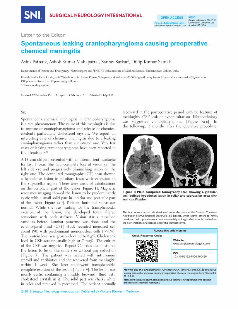
A 13‑year‑old girl presented with an intermittent headache for last 1 year. She had complete loss of vision on the left side eye and progressively diminishing vision on the right one. The computed tomography (CT) scan showed a hypodense lesion in pituitary fossa with extension to the suprasellar region. There were areas of calcifications on the peripheral part of the lesion [Figure 1]. Magnetic resonance imaging showed the lesion to be predominantly cystic with a small solid part in inferior and posterior part of the lesion [Figure 2a‑f]. Patients’ hormonal status was normal. While she was waiting for the transphenoidal excision of the lesion, she developed fever, altered sensorium with neck stiffness. Vision status remained same as before. Lumbar puncture was done, and the cerebrospinal fluid (CSF) study revealed increased cell count (90) with predominant mononuclear cells (>90%). The protein level was grossly elevated to 8 g%. Cholesterol level in CSF was unusually high at 7 mg%. The culture of the CSF was negative. Repeat CT scan demonstrated the lesion to be of the same size without any reduction [Figure 3]. The patient was treated with intravenous steroid and antibiotics and she recovered from meningitis within 1 week. She later underwent transphenoidal complete excision of the lesion [Figure 4]. The lesion was mostly cystic containing a muddy brownish fluid with cholesterol crystals in it. The solid part was chalky white in color and removed in piecemeal. The patient normally
recovered in the postoperative period with no features of meningitis, CSF leak or hypopituitarism. Histopathology was suggestive craniopharyngioma [Figure 5a‑c]. In the follow‑up, 2 months after the operative procedure,
Letter to the Editor
Spontaneous leaking craniopharyngioma causing preoperative chemical meningitisAshis Patnaik, Ashok Kumar Mahapatra1, Saurav Sarkar2, Dillip Kumar Samal2
Departments of Trauma and Emergency, 1Neurosurgery and 2ENT, All India Institute of Medical Science, Bhubaneswar, Odisha, India
E‑mail: *Ashis Patnaik ‑ [email protected]; Ashok Kumar Mahapatra ‑ [email protected]; Saurav Sarkar ‑ [email protected]; Dillip Kumar Samal ‑ [email protected] *Corresponding author
Received: 07 December 15 Accepted: 19 February 16 Published: 14 April 16
Access this article online
Quick Response Code:Website: www.surgicalneurologyint.com
DOI: 10.4103/2152-7806.180466
This is an open access article distributed under the terms of the Creative Commons Attribution-NonCommercial-ShareAlike 3.0 License, which allows others to remix, tweak, and build upon the work non-commercially, as long as the author is credited and the new creations are licensed under the identical terms.
How to cite this article: Patnaik A, Mahapatra AK, Sarkar S, Samal DK. Spontaneous leaking craniopharyngioma causing preoperative chemical meningitis. Surg Neurol Int 2016;7:41.http://surgicalneurologyint.com/Spontaneous-leaking-craniopharyngioma-causing-preoperative-chemical-meningitis/
Figure 1: Plain computed tomography scan showing a globular, well-defined hypodense lesion in sellar and suprasellar area with wall calcification

Surgical Neurology International 2016, 7:41 http://www.surgicalneurologyint.com/content/7/1/41
patients’ vision improved in the right eye but there was no improvement on the left side.
Aseptic or chemical meningitis is a rare complication in craniopharyngiomas particularly cystic ones, due to rupture and spillage of its contents containing cholesterol into the subarachnoid space, secreted by its squamous epilthelial lining. The occurrence of meningitis following rupture of the cyst is directly related to the cholesterol contents as its absence makes the rupture asymptomatic. Takahashi et al.,[8] reported two cases of spontaneous rupture of craniopharyngiomas without any meningitic symptoms. In both of these cases, the cysts did not contain cholesterol crystals. The rupture of the cyst also leads partial decompression leading improvement of the symptoms. In our case, the meningitis followed the leakage of its contents rather than frank rupture as
evidenced by no change in cyst size in the repeat scan. There was no change in the vision as the rupture would have resulted in partly decompression on the visual pathway leading to vision improvement.
Suprasellar cystic tumors such as craniopharyngioma, dermoid, and epidermoid, Rathke’s cleft cyst can rupture spontaneously with remission of symptoms particularly those caused by a compressive effect like vision symptoms. Cerebral infarction due to vasospasm following craniopharyngioma cyst rupture has also been reported in the literature.[7] Few cases of spontaneous rupture of craniopharyngioma have been reported until now.[1,3,4,6‑9] This rupture can result in chemical meningitis depending upon the cholesterol content of the cyst fluid.
Figure 3: Computed tomography images of the lesion after meningitis showing the lesion to be of same size as before
Figure 4: Complete excision of the lesion except a small part of its wall attached to the surrounding vessels
Figure 2: Magnetic resonance imaging pictures of the lesion. (a) T1-weighted sagittal image showing the lesion containing fat containing T1 hyperintense antero-superior and hypointense solid part in postero-inferior part. (b) T2-weighted coronal image of the lesion. (c) Diffusion image showing the solid part predominantly on left side. (d-f) Contrast images show the lesion to be minimally patchy enhancing
a b c
d e f
Surgical Neurology International 2016, 7:41 http://www.surgicalneurologyint.com/content/7/1/41
The exact mechanism of cyst rupture in these tumors is not clearly known, but it may be induced by the progressive enlargement of the cyst causing weakness of the cyst wall and finally rupture. The leaking variety, as in the present case, can occur if the rent in the cyst wall is small enough to prevent the significant escape of cystic fluid into the subarachnoid space. This also explains the nonreduction in the size of the cyst as well as no relief of compressive effect of the cyst on surrounding structures.
Our case was exclusive in that the meningitis episode occurred preoperatively without any reduction in the cyst size raising possibility of a bacterial cause. However, the CSF study established the chemical nature of the lesion. Neurosurgeons should be aware of such spontaneous leakage or rupture of the cyst in preoperative stage
causing chemical meningitis in a cystic tumor. CSF and radiological examination must be done to establish the exact cause. Treatment is directed toward immediate management of meningitis followed by removal of the cause.
Financial support and sponsorshipNil.
Conflicts of interestThere are no conflicts of interest.
REFERENCES
1. Hadden D, Allen I. Chemical meningitis due to rupture of a craniopharyngioma cyst. J R Soc Med 2004;97:585-6.
2. Krueger DW, Larson EB. Recurrent fever of unknown origin, coma, and meningismus due to a leaking craniopharyngioma. Am J Med 1988;84 (3 Pt 1):543-5.
3. Kumar A, Kasliwal MK, Suri A, Sharma BS. Spontaneous asymptomatic rupture of cystic craniopharyngioma in a child: Case report and review of the literature. J Child Neurol 2010;25:1555-8.
4. Okamoto H, Harada K, Uozumi T, Goishi J. Spontaneous rupture of a craniopharyngioma cyst. Surg Neurol 1985;24:507-10.
5. Rajput D, Srivastva A, Kumar R, Mahapatra A. Recurrent chemical meningitis in craniopharyngioma without reduction in size of cyst: Case report of two cases and review of the literature. Turk Neurosurg 2012;22:233-6.
6. Satoh H, Uozumi T, Arita K, Kurisu K, Hotta T, Kiya K, et al. Spontaneous rupture of craniopharyngioma cysts. A report of five cases and review of the literature. Surg Neurol 1993;40:414-9.
7. Shida N, Nakasato N, Mizoi K, Kanaki M, Yoshimoto T. Symptomatic vessel narrowing caused by spontaneous rupture of craniopharyngioma cyst – Case report. Neurol Med Chir (Tokyo) 1998;38:666-8.
8. Takahashi T, Kudo K, Ito S, Suzuki S. Spontaneously ruptured craniopharyngioma cyst without meningitic symptoms – Two case reports. Neurol Med Chir (Tokyo) 2003;43:150-2.
9. Tokiwa K, Nakayama K, Miyasaka Y, Matsumori K, Beppu T, Asao T. Cystic craniopharyngioma with spontaneous shrinkage of the cyst. Case report. Neurol Med Chir (Tokyo) 1984;24:954-7.
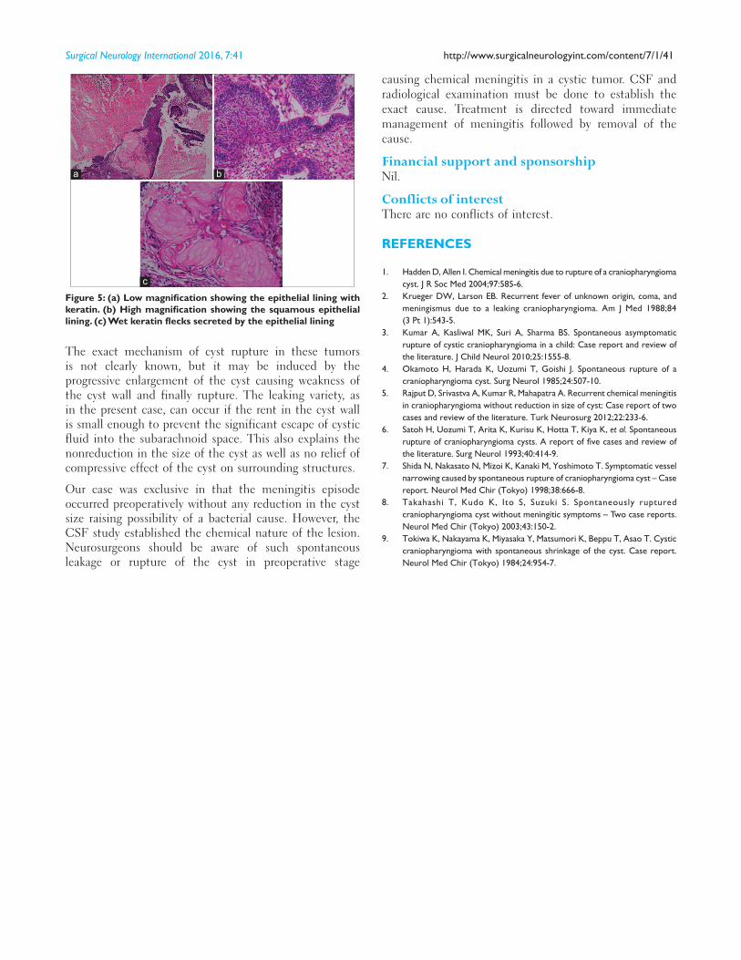
Figure 5: (a) Low magnification showing the epithelial lining with keratin. (b) High magnification showing the squamous epithelial lining. (c) Wet keratin flecks secreted by the epithelial lining
ba
c