Embed Size (px)
Citation preview
Saggu TK et al. Central Odontogenic Fibroma (WHO type).
165
Journal of Advanced Medical and Dental Sciences Research |Vol. 2|Issue 3| July-September 2014
Case Report
Central Odontogenic Fibroma (WHO type) mimicking Fibro-osseous Lesion- A Case Report
Tajinder Kaur Saggu1, Shams Ul Nisa2, Ramandeep Singh Brar3, Saloni Sharma1
Departments of 1Oral & Maxillofacial Pathology, 3Oral & Maxillofacial Surgery Dasmesh Institute of Research & Dental Sciences, Faridkot- 151203, Punjab, India. 2Oral Medicine and Radiology, Bharati Vidyapeeth Deemed University Dental College and Hospital Pune, Maharashtra, India Corresponding Author:
Dr. Tajinder Kaur Saggu
Department of Oral & Maxillofacial
Pathology,
Dasmesh Institute of Research &
Dental Sciences,
Faridkot- 151203, Punjab, India
E-mail: [email protected]
Received: 22-07-2014
Revised: 22-08-2014
Accepted: 26-08-2014
This article may be cited as: Saggu TK, Ul Nisa S, Brar RS, Sharma S. Central Odontogenic Fibroma (WHO type) mimicking Fibro-osseous Lesion- A Case Report. J Adv Med Dent Scie Res 2014;2(3):165-170.
ntroduction: Odontogenic fibromas (OFs) were defined in the latest classification of the World Health
Organization (WHO) as a uncommon neoplasm characterized by an erratic quantity of inactive odontogenic epithelium in a rather-mature fibrous stroma.[1] OF is considered to be an “Elusive and controversial tumor” due to its rarity and the uncertainty.[2] It is a benign neoplasm, representing 1% to 2.5% of oral biopsy specimens.[3] It occurs either as a
central (intra-osseous) lesion (COF) or as peripheral lesion (POF).[3] POF is more common than it counterpart by ratio of 1.4:1.[2] The age group for the occurrence of OF varies from 4 to 80 years, with an average of 40 years. It may affect both genders, but has slight female predilection.[1] It has predilection to occur in the mandible and is generally asymptomatic except for the swelling of jaw.[2] COF radiographically presents as uni- or multilocular radiolucencies
I
Abstract: The odontogenic fibroma is a relatively rare, slow-growing, benign odontogenic neoplasm of the jaws, with the potential to recur after excision. The World Health Organization (WHO) classified odontogenic fibroma into two variants: 1) WHO type (Epithelial-rich) 2) simple type (epithelial-poor type). It commonly occurs during middle age and has predilection for mandible. This article presents a rare case of Central Odontogenic Fibroma (WHO type) in a 24-year-old female demonstrated radiographically as mixed lesion involving right maxillary premolar – molar region. OF can mimick fibro-ossoeus lesions so, we recommend that vigilant comprehensive analysis of all the pertaining clinical, radiographic and histopathological aspects should be carried out with long term follow up after required treatment. Key words: Odontogenic fibroma, Intra-osseous, benign.
Saggu TK et al. Central Odontogenic Fibroma (WHO type).
166
Journal of Advanced Medical and Dental Sciences Research |Vol. 2|Issue 3| July-September 2014
with well-defined borders. In some rare cases, it might present mixed radiolucent and radiopaque features and undefined borders.[4]
Root resorption is common and lesion located between teeth often cause root divergence.[5] Microscopically, it is characterized by proliferation of cellular fibrous or fibro-myxomatous connective tissue that exhibits variable amounts of odontogenic epithelium, mostly designated as epithelium-poor or epithelium-rich subtype. Foci of calcification in the form of dentinoid, cementicles, or bone may also be seen.[3] Here we presents a rare case of Central Odontogenic Fibroma (WHO type) in a 24-year-old female involving right posterior region of maxilla i.e premolar – molar region with emphasis on its differential diagnosis.
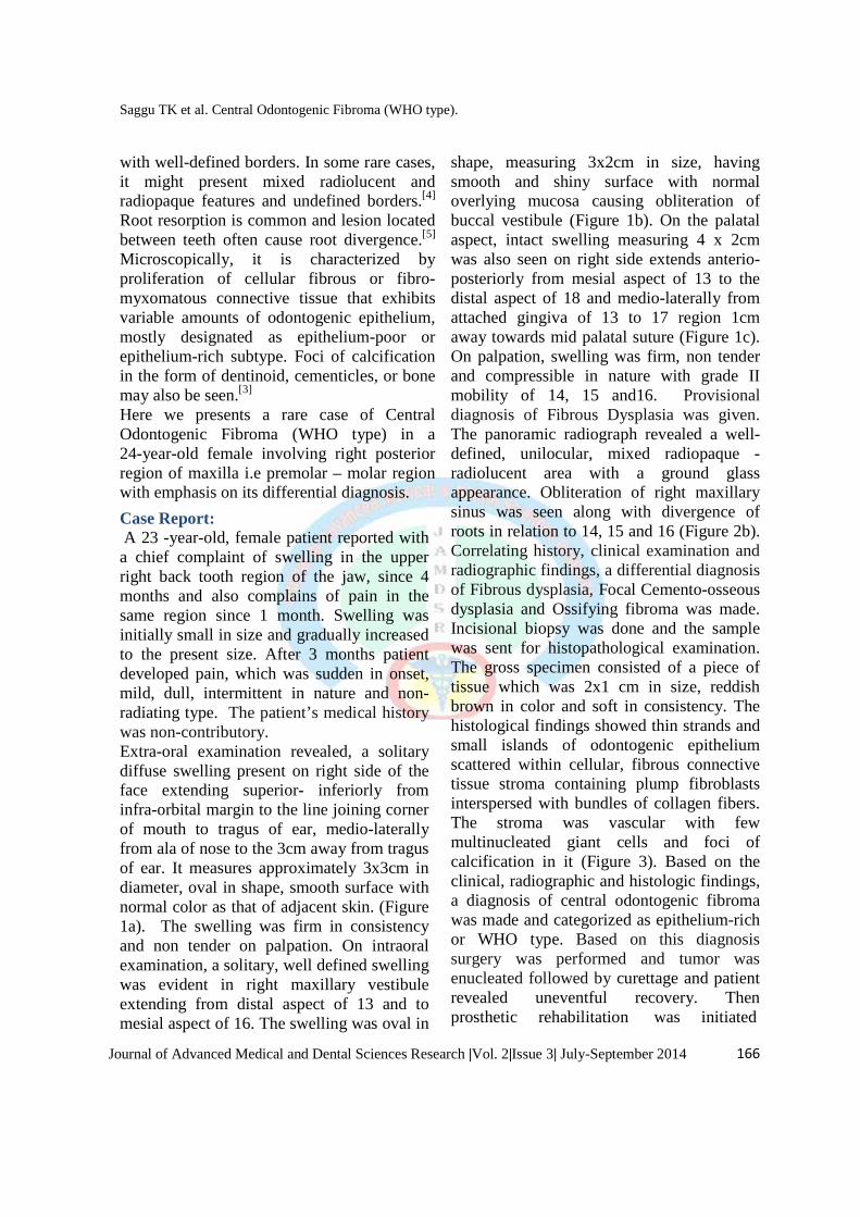
Case Report: A 23 -year-old, female patient reported with a chief complaint of swelling in the upper right back tooth region of the jaw, since 4 months and also complains of pain in the same region since 1 month. Swelling was initially small in size and gradually increased to the present size. After 3 months patient developed pain, which was sudden in onset, mild, dull, intermittent in nature and non- radiating type. The patient’s medical history was non-contributory. Extra-oral examination revealed, a solitary diffuse swelling present on right side of the face extending superior- inferiorly from infra-orbital margin to the line joining corner of mouth to tragus of ear, medio-laterally from ala of nose to the 3cm away from tragus of ear. It measures approximately 3x3cm in diameter, oval in shape, smooth surface with normal color as that of adjacent skin. (Figure 1a). The swelling was firm in consistency and non tender on palpation. On intraoral examination, a solitary, well defined swelling was evident in right maxillary vestibule extending from distal aspect of 13 and to mesial aspect of 16. The swelling was oval in
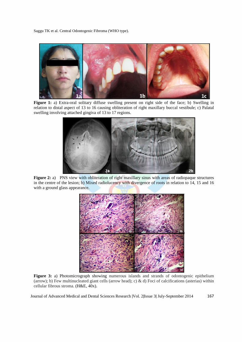
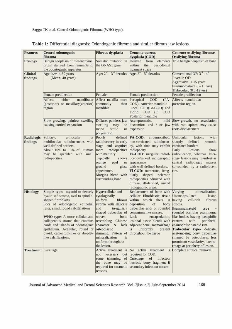
shape, measuring 3x2cm in size, having smooth and shiny surface with normal overlying mucosa causing obliteration of buccal vestibule (Figure 1b). On the palatal aspect, intact swelling measuring 4 x 2cm was also seen on right side extends anterio-posteriorly from mesial aspect of 13 to the distal aspect of 18 and medio-laterally from attached gingiva of 13 to 17 region 1cm away towards mid palatal suture (Figure 1c). On palpation, swelling was firm, non tender and compressible in nature with grade II mobility of 14, 15 and16. Provisional diagnosis of Fibrous Dysplasia was given. The panoramic radiograph revealed a well-defined, unilocular, mixed radiopaque - radiolucent area with a ground glass appearance. Obliteration of right maxillary sinus was seen along with divergence of roots in relation to 14, 15 and 16 (Figure 2b). Correlating history, clinical examination and radiographic findings, a differential diagnosis of Fibrous dysplasia, Focal Cemento-osseous dysplasia and Ossifying fibroma was made. Incisional biopsy was done and the sample was sent for histopathological examination. The gross specimen consisted of a piece of tissue which was 2x1 cm in size, reddish brown in color and soft in consistency. The histological findings showed thin strands and small islands of odontogenic epithelium scattered within cellular, fibrous connective tissue stroma containing plump fibroblasts interspersed with bundles of collagen fibers. The stroma was vascular with few multinucleated giant cells and foci of calcification in it (Figure 3). Based on the clinical, radiographic and histologic findings, a diagnosis of central odontogenic fibroma was made and categorized as epithelium-rich or WHO type. Based on this diagnosis surgery was performed and tumor was enucleated followed by curettage and patient revealed uneventful recovery. Then prosthetic rehabilitation was initiated
Saggu TK et al. Central Odontogenic Fibroma (WHO type).
167
Journal of Advanced Medical and Dental Sciences Research |Vol. 2|Issue 3| July-September 2014
Figure 1: a) Extra-oral solitary diffuse swelling present on right side of the face; b) Swelling in relation to distal aspect of 13 to 16 causing obliteration of right maxillary buccal vestibule; c) Palatal swelling involving attached gingiva of 13 to 17 regions. Figure 2: a) PNS view with obliteration of right maxillary sinus with areas of radiopaque structures in the centre of the lesion; b) Mixed radiolucency with divergence of roots in relation to 14, 15 and 16 with a ground glass appearance.
Figure 3: a) Photomicrograph showing numerous islands and strands of odontogenic epithelium (arrow); b) Few multinucleated giant cells (arrow head); c) & d) Foci of calcifications (asterias) within cellular fibrous stroma. (H&E, 40x).
Saggu TK et al. Central Odontogenic Fibroma (WHO type).
168
Journal of Advanced Medical and Dental Sciences Research |Vol. 2|Issue 3| July-September 2014
Table 1: Differential diagnosis: Odondogenic fibroma and similar fibrous jaw lesions
Features Central odontogenic fibroma
Fibrous dysplasia Cemento-osseous dysplasia (COD)
Cemento-ossifying fibroma/ Ossifying fibroma
Etiology Benign neoplasm of mesenchymal origin derived from remnants of the odontogenic apparatus
Somatic mutation in the GNAS1 gene
Derived from elements within the periodontal ligament space
True benign neoplasm of bone
Clinical findings
Age: b/w 4-80 years (Mean- 40 years)
Age: 2nd - 3rd decades
Age: 3rd - 5th decades
Conventional OF: 3rd - 4th Juvenile OF: Aggressive: < 15 years Psammomatoid: (5- 15 yrs) Trabecular: (8.5-12 yrs)
Female predilection Female Female predilection Female predilection Affects either mandibular (posterior) or maxillary(anterior) region
Affect maxilla more commonly than mandible.
Periapical COD (PA-COD)- Anterior mandible Focal COD(Fo-COD) and Florid COD (Fl COD Posterior mandible
Affects mandibular posterior region.
Slow growing, painless swelling causing cortical expansion
Diffuse, painless jaw swelling may be mono stotic or polyostotic
Asymptomatic, mild discomfort and / or jaw expansion.
Slow-growth, no association with root apices, may cause roots displacement.
Radiologic findings
Solitary, unilocular or multilocular radiolucencies with well-defined borders. About 10% to 15% of tumors may be speckled with small radiopacities.
Poorly defined radiolucency in early stage and acquires more radiopacities with maturity. Typically shows orange peel or ground glass appearance. Margins blend with surrounding bone.
PA-COD: circumscribed, non-corticated radiolucen- cy, with time may exhibit radiopacity Fo-COD: irregular radiol- ucency/mixed radiographic appearance with well-defined borders. Fl-COD: numerous, irreg-ularly shaped, sclerotic radiopacities admixed with diffuse, ill-defined, mixed radiographic areas.
Unilocular lesions with sharply defined smooth, corticated borders Early lesions show radiolucency, whereas later-stage lesions may manifest as central radiopaque masses surrounded by a radiolucent rim.
Histology Simple type: myxoid to densely hyalinised stroma, oval to spindle-shaped fibroblasts. Foci of odontogenic epithelial rests, small, round calcifications WHO type: A more cellular and collagenous stroma that contains cords and islands of odontogenic epithelium. Acellular, round or ovoid, cementum-like or droplet-like calcifications.
Hypercellular and cytologically uniform fibrous stroma with delicate and irregularly shaped trabeculae of woven bone resembling Chinese character & lack osteoblastic rimming. Pattern of mineralization is uniform throughout the lesion.
Replacement of bone with cellular fibroblastic tissue within which there is deposition of bony trabeculae and/ or rounded cementum like masses. Lack encapsulation, lesional tissue blends with adjacent bone Haemorrhage is uniformly present throughout the tissue
Varying mineralization, Unenc-apsulated lesion having cell-rich fibrous stroma. Psammomatoid type -rounded acellular psammoma like bodies having basophilic centres with peripheral eosinophilic osteoid rim. Trabecular type- delicate, anatomosing bony trabeculae rimmed by osteoblasts, less prominent vascularity, haemo- rrhage at periphery of lesion.
Treatment Curettage. Active treatment is not necessary but some trimming of the bone may be required for cosmetic reasons.
No active treatment is required for COD. Curettage of infected/ necrotic bony fragment if secondary infection occurs.
Complete surgical removal.
Saggu TK et al. Central Odontogenic Fibroma (WHO type).
169
Journal of Advanced Medical and Dental Sciences Research |Vol. 2|Issue 3| July-September 2014
to restore function and esthetics. Patient was followed up for 15 months without any report of recurrence.
Discussion: Central odontogenic fibroma (COF) has been described as an unusual, slow-growing tumor of the jaw. It appears as an asymptomatic extension of the cortical plate of the mandible or maxilla.[4] Unlike ameloblastoma, it is not locally invasive.[3] Shafer et al considered odontogenic fibroma a distinct neoplasm with its own histopathologic and clinical features separating it from other odontogenic tumors.
[2] COFs usually occur in adults and shows a female predilection (M:F= 1:2.8). [6] The sex and the age of the patient we described in this paper are consistent with the literature. The most common site for the presentation of COFs is the mandibular posterior (premolar/molar) region, while in the maxilla they are mostly observed in the anterior region[1] unlike in our case where the lesion was present in maxillary posterior region and which was rare according to its site.
Usually, COF exhibits slow growing painless swelling that results in cortical expansion. Clinical symptoms such as pain and paresthesia are uncommon[7] but in our case, patient presented with gradually increasing asymptomatic swelling which eventually became painful. In some cases, it presents as slowly enlarging hard swelling which is sometimes recognized by presence of slow growing diastema due to dislocation of adjacent teeth. The affected patients may have tooth mobility or displacement.[8] In present case, the bony hard swelling was associated with mobility of involved teeth (14, 15,16). Radiological examination showed that the odontogenic fibroma is an eminently unilocular radiolucency with well-defined margins and large size lesions tend to have multilocular appearance with poorly defined or diffused borders in rare case.[7] In few
cases, radiopacity can be seen within a radiolucent area, as in our case, the lesion showed mixed radiopaque- radiolucent areas with glass ground appearance.[9] The great variation and overlap in radiologic appearance of the COFs with other lesions means it should be carefully differentiated from similar fibrous lesions.(Table 1) [2,5,10,11]. Histologically, the CODF is categorized into two types: the epithelium-rich type (WHO-type) and the epithelium poor type (Simple type). The epithelium rich type shows more epithelial islands or strands distributed in fibrous tissue. It contains mineralized material that has been interpreted as osteoid, cementum-like or dysplastic dentin. The epithelium poor type lesions are less cellular and the epithelial tissue is not necessarily presented. It is generally believed that odontogenic epithelium is not always presented in the COF and the WHO type occurs much less commonly than the simple type.[8,12] In our case, thin strands and islands of odontogenic epithelium were seen scattered within cellular fibrous stroma. Foci of calcification and few multinucleated giant cells were seen along with odontogenic epithelium. Thus the diagnosis of central odontogenic fibroma i.e epithelium-rich or WHO type was made. Different treatment recommendations were suggested in the literature for CODF. Recurrences have been reported, but are very uncommon. One piece enucleation of the lesion is possible because it has no attachment to a tooth root and bone but, a few cases, which have grown large, may necessitate tooth extraction to have more access for total removal.[6] In our case, enucleation followed by curettage was done and the lesion was removed. Extraction of 15 and 16 was done followed by prosthetic rehabilitation to restore function and esthetics. The present case showed no sign of recurrence after 15 months of follow-up.
Saggu TK et al. Central Odontogenic Fibroma (WHO type).
170
Journal of Advanced Medical and Dental Sciences Research |Vol. 2|Issue 3| July-September 2014
We retrieved pubmed and medline literature accessed on April 3, 2014 and we obtained 20 cases of central odontogenic fibroma- WHO type.
Conclusion: It is imperative that all fibrous lesions of the jaws should also be taken into account to arrive at final diagnosis. Keeping in mind that OF can mimick fibro-ossoeus lesions so, we recommend that vigilant comprehensive analysis of all the pertaining clinical, radiographic and histopathological aspects should be carried out. We also emphasize on the fact OF have tendency for recurrence so it should be followed up for long term to detect any signs of recurrence.
References: 1. Niklander S, Martinez R, Deichler J,
Esguep A. J Dent Sci 2011; 6: 123-127. 2. Shafer WG, Hine MK, Levy BM. Shafer's
textbook of oral pathology. 6th ed. Elsevier; 2009.
3. Armas JM, Hunter KD, Jenkins MM. Odontogenic fibroma: An unusual presentation. J Oral Maxillofac Pathol 2008; 12:68-71.
4. Daskala I, Kalyvas D, Kolokoudias M, Vlachodimitropoulos D, Alexandridis C. Central odontogenic fibroma of the mandible: a case report: J Oral Scie 2009;51( 3): 457-461.
5. Neville, Damm, Allen, Bouquet, oral and maxillofacial pathology, 3rd ed. Elsevier.
6. Brazão-Silva MT, Fernandes AV, Durighetto-Júnior AF, Cardoso SV, Loyola AM Central odontogenic fibroma: a case report with long-term follow-up. Head & Face Medicine 2010, 6:20.
7. Kyung-Soo Nah. Central odontogenic fibroma, a case report. Imaging Scie Dent 2011; 41: 85-8.
8. Talukder S, Agarwal R, Gupta P, BS Santosh, Misra D. Central Odontogenic fibroma(WHO type): A case report and review of literature. J Indian acad oral med radiol 2011; 23(3): 259-262.
9. Sato FRL, de Moraes M; Central Odontogenic Fibroma: Description of a Case and Review, Int J Oral-Med Sci 2008;7(1):50-53.
10. Hall G. Fibro-osseous lesions of the head and neck. Diagnostic Histopathol 2012; 18(4):149-158.
11. Faizan Alawi. Benign Fibro-osseous Diseases of the Maxillofacial Bones A Review and Differential Diagnosis. Am J Clin Pathol 2002;118(Suppl 1):S50-S70.
12. Chuang HP and Tsai LL. Central Odontogenic Fibroma of Mandible — A Case Report: Taiwan J Oral Maxillofac Surg September 2008;19: 179-185.
13. Hwang E, Lee S. Central odontogenic fibroma of the simple type. Korean J Oral Maxillofac Radiol 2002; 32:227-30.
14. Kinney L, Bradford J, Cohen M, Glickman R. The Aggressive Odontogenic Fibroma: Report of a Case. J Oral Maxillofac Surg 1993; 51:321-4.
Corresponding Author: Dr. Tajinder Kaur Senior Lecturer, Department of Oral & Maxillofacial Pathology, Dasmesh Institute of Research & Dental Sciences, Faridkot- 151203, Punjab, India
Source of support: Nil Conflict of interest: None declared


![Fibroma of the Maxilla Trabecular Variant Juvenile … · contains cementicles [2], and while it is of odontogenic origin, it predominantly occurs in the second and third decades](https://img.pdfslide.us/doc/110x75/5b810d1f7f8b9a2b6f8b7676/fibroma-of-the-maxilla-trabecular-variant-juvenile-contains-cementicles-2.jpg)