Embed Size (px)
Citation preview
940
Introduction Reactive lesions of gingiva are clinically and histologically non-neoplastic nodular swellings that develop in response to chronic and recurring tissue injury which stimulates an exuberant tissue response. These mainly include focal fibrous hyperplasia, pyogenic granuloma, peripheral ossifying fibroma and peripheral giant cell granuloma. Clinically, these lesions mimic various groups of pathologic processes and therefore often present a diagnostic challenge [1]. Peripheral Ossifying Fibroma (POF) is described as any solitary growth on the gingiva thought to arise from the periodontal ligament, most commonly in the region of the interdental papillae. While some consider it as a benign neoplasm, others suggest it to be a non-neoplastic inflammatory response of the connective tissue or superficial periodontal ligament to low grade irritation, such as trauma, plaque, calculus, masticatory forces, ill- fitting dental appliances and poor quality restorations [2,3]. It usually measures <1.5 cm in diameter, has a slight predilection for females and is more commonly seen in the anterior maxilla of young individuals. There is still considerable confusion regarding its nomenclature and etiopathogenesis. Here we present a case of a massive rapidly proliferating POF in the posterior mandible of an elderly male chronic smoker where most of the clinical findings didn’t seem to correlate with the general characteristics of this lesion.
Case Report A 58-year-old Indian male reported to a private clinic in Meerut with a complaint of a progressive, non painful growth in the left lower back region of his mouth for the past 2-3 months resulting in discomfort during speech and mastication. He and his family were extremely worried thinking it as cancer. Patient’s history revealed that he was a smoker, smoking 15-20 bidis a day for the past 26 years and that he had lost 6-7 kg of weight in the past six months. There was no history of any trauma or injury. His family history was non contributory. There was no history of associated symptoms
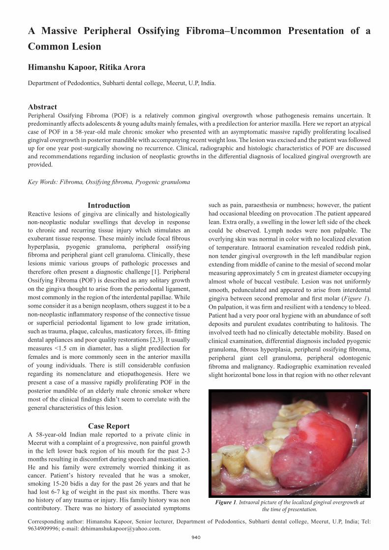
such as pain, paraesthesia or numbness; however, the patient had occasional bleeding on provocation .The patient appeared lean. Extra orally, a swelling in the lower left side of the cheek could be observed. Lymph nodes were non palpable. The overlying skin was normal in color with no localized elevation of temperature. Intraoral examination revealed reddish pink, non tender gingival overgrowth in the left mandibular region extending from middle of canine to the mesial of second molar measuring approximately 5 cm in greatest diameter occupying almost whole of buccal vestibule. Lesion was not uniformly smooth, pedunculated and appeared to arise from interdental gingiva between second premolar and first molar (Figure 1). On palpation, it was firm and resilient with a tendency to bleed. Patient had a very poor oral hygiene with an abundance of soft deposits and purulent exudates contributing to halitosis. The involved teeth had no clinically detectable mobility. Based on clinical examination, differential diagnosis included pyogenic granuloma, fibrous hyperplasia, peripheral ossifying fibroma, peripheral giant cell granuloma, peripheral odontogenic fibroma and malignancy. Radiographic examination revealed slight horizontal bone loss in that region with no other relevant
A Massive Peripheral Ossifying Fibroma–Uncommon Presentation of a Common Lesion
Himanshu Kapoor, Ritika Arora
Department of Pedodontics, Subharti dental college, Meerut, U.P, India.
AbstractPeripheral Ossifying Fibroma (POF) is a relatively common gingival overgrowth whose pathogenesis remains uncertain. It predominantly affects adolescents & young adults mainly females, with a predilection for anterior maxilla. Here we report an atypical case of POF in a 58-year-old male chronic smoker who presented with an asymptomatic massive rapidly proliferating localised gingival overgrowth in posterior mandible with accompanying recent weight loss. The lesion was excised and the patient was followed up for one year post-surgically showing no recurrence. Clinical, radiographic and histologic characteristics of POF are discussed and recommendations regarding inclusion of neoplastic growths in the differential diagnosis of localized gingival overgrowth are provided.
Key Words: Fibroma, Ossifying fibroma, Pyogenic granuloma
Corresponding author: Himanshu Kapoor, Senior lecturer, Department of Pedodontics, Subharti dental college, Meerut, U.P, India; Tel: 9634909996; e-mail: [email protected].
Figure 1. Intraoral picture of the localized gingival overgrowth at the time of presentation.
941
OHDM - Vol. 13 - No. 4 - December, 2014
findings. Complete hemo gram of the patient was within normal limits.
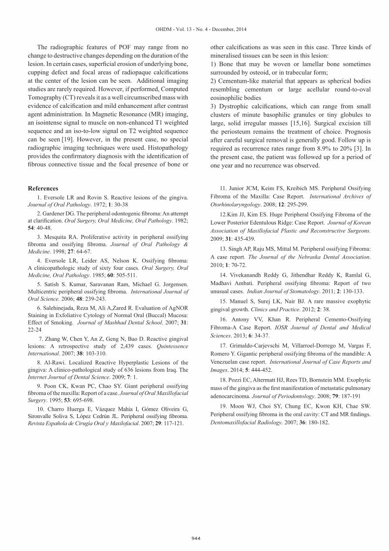
Examination by a Physician and investigations did not reveal any relevant medical background. Patient was motivated to quit the habit of smoking and instructed regarding maintenance of oral hygiene. After an initial visit of supragingival scaling and removal of local deposits, the lesion was completely excised along with some surrounding normal tissue under local anaesthesia and sent for histopathologic examination. The area was carefully curetted, irrigated and covered by a periodontal dressing. The lesion measuring about 5.5×3×2 cm ; on histopathological examination revealed stratified squamous epithelium with multiple foci of surface ulceration. The deeper part showed dense aggregates of spindle-shaped fibroblasts, bundles of collagen fibers along with some dystrophic calcification and focal areas of basophilic small globules of cementum like material.
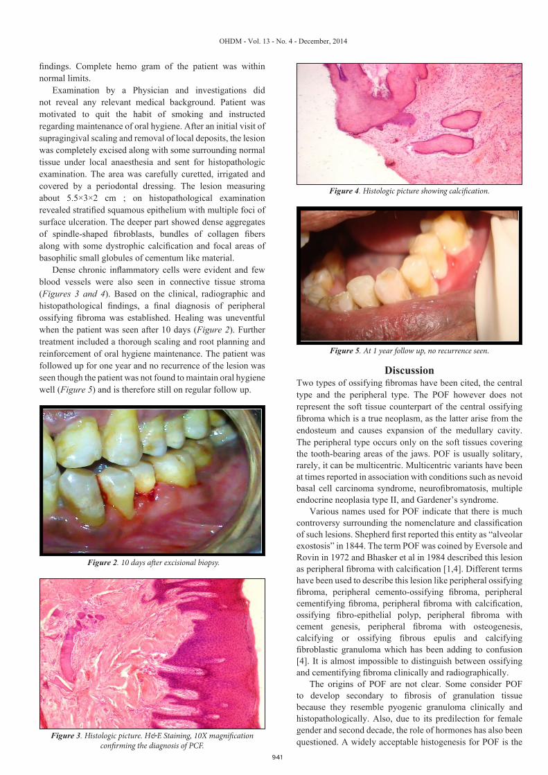
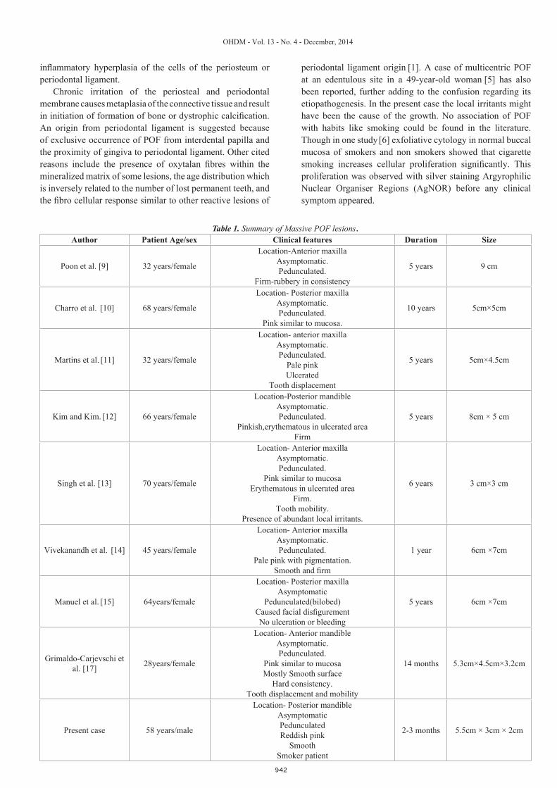
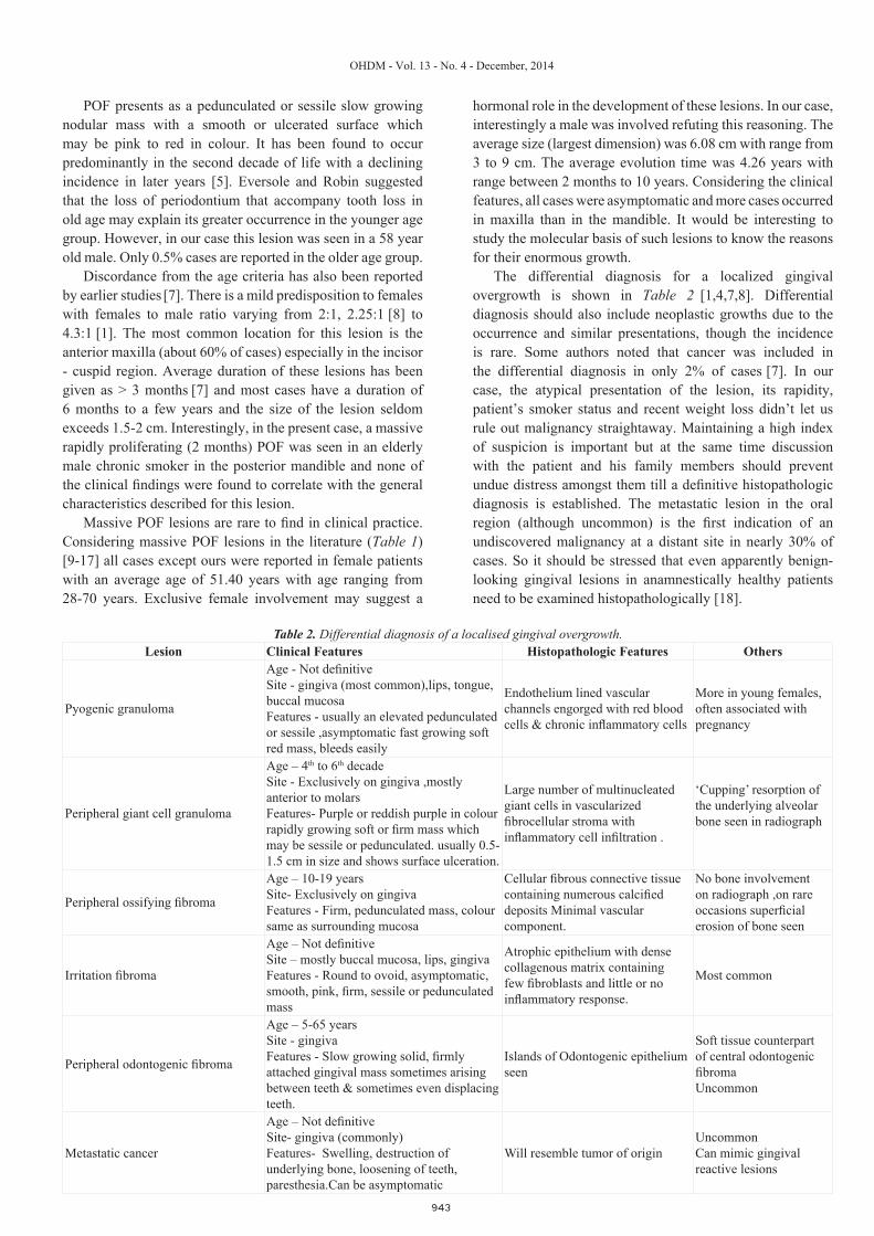
Dense chronic inflammatory cells were evident and few blood vessels were also seen in connective tissue stroma (Figures 3 and 4). Based on the clinical, radiographic and histopathological findings, a final diagnosis of peripheral ossifying fibroma was established. Healing was uneventful when the patient was seen after 10 days (Figure 2). Further treatment included a thorough scaling and root planning and reinforcement of oral hygiene maintenance. The patient was followed up for one year and no recurrence of the lesion was seen though the patient was not found to maintain oral hygiene well (Figure 5) and is therefore still on regular follow up.
Discussion Two types of ossifying fibromas have been cited, the central type and the peripheral type. The POF however does not represent the soft tissue counterpart of the central ossifying fibroma which is a true neoplasm, as the latter arise from the endosteum and causes expansion of the medullary cavity. The peripheral type occurs only on the soft tissues covering the tooth-bearing areas of the jaws. POF is usually solitary, rarely, it can be multicentric. Multicentric variants have been at times reported in association with conditions such as nevoid basal cell carcinoma syndrome, neurofibromatosis, multiple endocrine neoplasia type II, and Gardener’s syndrome.
Various names used for POF indicate that there is much controversy surrounding the nomenclature and classification of such lesions. Shepherd first reported this entity as “alveolar exostosis” in 1844. The term POF was coined by Eversole and Rovin in 1972 and Bhasker et al in 1984 described this lesion as peripheral fibroma with calcification [1,4]. Different terms have been used to describe this lesion like peripheral ossifying fibroma, peripheral cemento-ossifying fibroma, peripheral cementifying fibroma, peripheral fibroma with calcification, ossifying fibro-epithelial polyp, peripheral fibroma with cement genesis, peripheral fibroma with osteogenesis, calcifying or ossifying fibrous epulis and calcifying fibroblastic granuloma which has been adding to confusion
[4]. It is almost impossible to distinguish between ossifying and cementifying fibroma clinically and radiographically.
The origins of POF are not clear. Some consider POF to develop secondary to fibrosis of granulation tissue because they resemble pyogenic granuloma clinically and histopathologically. Also, due to its predilection for female gender and second decade, the role of hormones has also been questioned. A widely acceptable histogenesis for POF is the
Figure 2. 10 days after excisional biopsy.
Figure 4. Histologic picture showing calcification.
Figure 5. At 1 year follow up, no recurrence seen.
Figure 3. Histologic picture. H&E Staining, 10X magnification confirming the diagnosis of PCF.
942
OHDM - Vol. 13 - No. 4 - December, 2014
inflammatory hyperplasia of the cells of the periosteum or periodontal ligament.
Chronic irritation of the periosteal and periodontal membrane causes metaplasia of the connective tissue and result in initiation of formation of bone or dystrophic calcification. An origin from periodontal ligament is suggested because of exclusive occurrence of POF from interdental papilla and the proximity of gingiva to periodontal ligament. Other cited reasons include the presence of oxytalan fibres within the mineralized matrix of some lesions, the age distribution which is inversely related to the number of lost permanent teeth, and the fibro cellular response similar to other reactive lesions of
periodontal ligament origin [1]. A case of multicentric POF at an edentulous site in a 49-year-old woman [5] has also been reported, further adding to the confusion regarding its etiopathogenesis. In the present case the local irritants might have been the cause of the growth. No association of POF with habits like smoking could be found in the literature. Though in one study [6] exfoliative cytology in normal buccal mucosa of smokers and non smokers showed that cigarette smoking increases cellular proliferation significantly. This proliferation was observed with silver staining Argyrophilic Nuclear Organiser Regions (AgNOR) before any clinical symptom appeared.
Author Patient Age/sex Clinical features Duration Size
Poon et al. [9] 32 years/female
Location-Anterior maxillaAsymptomatic.Pedunculated.
Firm-rubbery in consistency
5 years 9 cm
Charro et al. [10] 68 years/female
Location- Posterior maxillaAsymptomatic.Pedunculated.
Pink similar to mucosa.
10 years 5cm×5cm
Martins et al. [11] 32 years/female
Location- anterior maxillaAsymptomatic.Pedunculated.
Pale pinkUlcerated
Tooth displacement
5 years 5cm×4.5cm
Kim and Kim. [12] 66 years/female
Location-Posterior mandibleAsymptomatic.Pedunculated.
Pinkish,erythematous in ulcerated areaFirm
5 years 8cm × 5 cm
Singh et al. [13] 70 years/female
Location- Anterior maxillaAsymptomatic.Pedunculated.
Pink similar to mucosaErythematous in ulcerated area
Firm.Tooth mobility.
Presence of abundant local irritants.
6 years 3 cm×3 cm
Vivekanandh et al. [14] 45 years/female
Location- Anterior maxillaAsymptomatic.Pedunculated.
Pale pink with pigmentation.Smooth and firm
1 year 6cm ×7cm
Manuel et al. [15] 64years/female
Location- Posterior maxillaAsymptomatic
Pedunculated(bilobed)Caused facial disfigurementNo ulceration or bleeding
5 years 6cm ×7cm
Grimaldo-Carjevschi et al. [17] 28years/female
Location- Anterior mandibleAsymptomatic.Pedunculated.
Pink similar to mucosaMostly Smooth surface
Hard consistency.Tooth displacement and mobility
14 months 5.3cm×4.5cm×3.2cm
Present case 58 years/male
Location- Posterior mandibleAsymptomaticPedunculatedReddish pink
SmoothSmoker patient
2-3 months 5.5cm × 3cm × 2cm
Table 1. Summary of Massive POF lesions.
943
OHDM - Vol. 13 - No. 4 - December, 2014
POF presents as a pedunculated or sessile slow growing nodular mass with a smooth or ulcerated surface which may be pink to red in colour. It has been found to occur predominantly in the second decade of life with a declining incidence in later years [5]. Eversole and Robin suggested that the loss of periodontium that accompany tooth loss in old age may explain its greater occurrence in the younger age group. However, in our case this lesion was seen in a 58 year old male. Only 0.5% cases are reported in the older age group.
Discordance from the age criteria has also been reported by earlier studies [7]. There is a mild predisposition to females with females to male ratio varying from 2:1, 2.25:1 [8] to 4.3:1 [1]. The most common location for this lesion is the anterior maxilla (about 60% of cases) especially in the incisor - cuspid region. Average duration of these lesions has been given as > 3 months [7] and most cases have a duration of 6 months to a few years and the size of the lesion seldom exceeds 1.5-2 cm. Interestingly, in the present case, a massive rapidly proliferating (2 months) POF was seen in an elderly male chronic smoker in the posterior mandible and none of the clinical findings were found to correlate with the general characteristics described for this lesion.
Massive POF lesions are rare to find in clinical practice. Considering massive POF lesions in the literature (Table 1)[9-17] all cases except ours were reported in female patients with an average age of 51.40 years with age ranging from 28-70 years. Exclusive female involvement may suggest a
hormonal role in the development of these lesions. In our case, interestingly a male was involved refuting this reasoning. The average size (largest dimension) was 6.08 cm with range from 3 to 9 cm. The average evolution time was 4.26 years with range between 2 months to 10 years. Considering the clinical features, all cases were asymptomatic and more cases occurred in maxilla than in the mandible. It would be interesting to study the molecular basis of such lesions to know the reasons for their enormous growth.
The differential diagnosis for a localized gingival overgrowth is shown in Table 2 [1,4,7,8]. Differential diagnosis should also include neoplastic growths due to the occurrence and similar presentations, though the incidence is rare. Some authors noted that cancer was included in the differential diagnosis in only 2% of cases [7]. In our case, the atypical presentation of the lesion, its rapidity, patient’s smoker status and recent weight loss didn’t let us rule out malignancy straightaway. Maintaining a high index of suspicion is important but at the same time discussion with the patient and his family members should prevent undue distress amongst them till a definitive histopathologic diagnosis is established. The metastatic lesion in the oral region (although uncommon) is the first indication of an undiscovered malignancy at a distant site in nearly 30% of cases. So it should be stressed that even apparently benign-looking gingival lesions in anamnestically healthy patients need to be examined histopathologically [18].
Lesion Clinical Features Histopathologic Features Others
Pyogenic granuloma
Age - Not definitiveSite - gingiva (most common),lips, tongue, buccal mucosaFeatures - usually an elevated pedunculated or sessile ,asymptomatic fast growing soft red mass, bleeds easily
Endothelium lined vascular channels engorged with red blood cells & chronic inflammatory cells
More in young females, often associated with pregnancy
Peripheral giant cell granuloma
Age – 4th to 6th decadeSite - Exclusively on gingiva ,mostly anterior to molarsFeatures- Purple or reddish purple in colour rapidly growing soft or firm mass which may be sessile or pedunculated. usually 0.5-1.5 cm in size and shows surface ulceration.
Large number of multinucleated giant cells in vascularized fibrocellular stroma with inflammatory cell infiltration .
‘Cupping’ resorption of the underlying alveolar bone seen in radiograph
Peripheral ossifying fibroma
Age – 10-19 years Site- Exclusively on gingivaFeatures - Firm, pedunculated mass, colour same as surrounding mucosa
Cellular fibrous connective tissue containing numerous calcified deposits Minimal vascular component.
No bone involvement on radiograph ,on rare occasions superficial erosion of bone seen
Irritation fibroma
Age – Not definitiveSite – mostly buccal mucosa, lips, gingivaFeatures - Round to ovoid, asymptomatic, smooth, pink, firm, sessile or pedunculated mass
Atrophic epithelium with dense collagenous matrix containing few fibroblasts and little or no inflammatory response.
Most common
Peripheral odontogenic fibroma
Age – 5-65 yearsSite - gingivaFeatures - Slow growing solid, firmly attached gingival mass sometimes arising between teeth & sometimes even displacing teeth.
Islands of Odontogenic epithelium seen
Soft tissue counterpart of central odontogenic fibromaUncommon
Metastatic cancer
Age – Not definitiveSite- gingiva (commonly)Features- Swelling, destruction of underlying bone, loosening of teeth, paresthesia.Can be asymptomatic
Will resemble tumor of originUncommonCan mimic gingival reactive lesions
Table 2. Differential diagnosis of a localised gingival overgrowth.
944
OHDM - Vol. 13 - No. 4 - December, 2014
The radiographic features of POF may range from no change to destructive changes depending on the duration of the lesion. In certain cases, superficial erosion of underlying bone, cupping defect and focal areas of radiopaque calcifications at the center of the lesion can be seen. Additional imaging studies are rarely required. However, if performed, Computed Tomography (CT) reveals it as a well circumscribed mass with evidence of calcification and mild enhancement after contrast agent administration. In Magnetic Resonance (MR) imaging, an isointense signal to muscle on non-enhanced T1 weighted sequence and an iso-to-low signal on T2 weighted sequence can be seen [19]. However, in the present case, no special radiographic imaging techniques were used. Histopathology provides the confirmatory diagnosis with the identification of fibrous connective tissue and the focal presence of bone or
other calcifications as was seen in this case. Three kinds of mineralised tissues can be seen in this lesion: 1) Bone that may be woven or lamellar bone sometimessurrounded by osteoid, or in trabecular form; 2) Cementum-like material that appears as spherical bodiesresembling cementum or large acellular round-to-oval eosinophilic bodies 3) Dystrophic calcifications, which can range from smallclusters of minute basophilic granules or tiny globules to large, solid irregular masses [15,16]. Surgical excision till the periosteum remains the treatment of choice. Prognosis after careful surgical removal is generally good. Follow up is required as recurrence rates range from 8.9% to 20% [3]. In the present case, the patient was followed up for a period of one year and no recurrence was observed.
References1. Eversole LR and Rovin S. Reactive lesions of the gingiva.
Journal of Oral Pathology. 1972; 1: 30-382. Gardener DG. The peripheral odontogenic fibroma: An attempt
at clarification. Oral Surgery, Oral Medicine, Oral Pathology. 1982; 54: 40-48.
3. Mesquita RA. Proliferative activity in peripheral ossifyingfibroma and ossifying fibroma. Journal of Oral Pathology & Medicine. 1998; 27: 64-67.
4. Eversole LR, Leider AS, Nelson K. Ossifying fibroma:A clinicopathologic study of sixty four cases. Oral Surgery, Oral Medicine, Oral Pathology. 1985; 60: 505-511.
5. Satish S. Kumar, Saravanan Ram, Michael G. Jorgensen.Multicentric peripheral ossifying fibroma. International Journal of Oral Science. 2006; 48: 239-243.
6. Salehinejada, Reza M, Ali A,Zared R. Evaluation of AgNORStaining in Exfoliative Cytology of Normal Oral (Buccal) Mucosa: Effect of Smoking. Journal of Mashhad Dental School. 2007; 31: 22-24
7. Zhang W, Chen Y, An Z, Geng N, Bao D. Reactive gingivallesions: A retrospective study of 2,439 cases. Quintessence International. 2007; 38: 103-310.
8. Al-Rawi. Localized Reactive Hyperplastic Lesions of thegingiva: A clinico-pathological study of 636 lesions from Iraq. The Internet Journal of Dental Science. 2009; 7: 1.
9. Poon CK, Kwan PC, Chao SY. Giant peripheral ossifyingfibroma of the maxilla: Report of a case. Journal of Oral Maxillofacial Surgery. 1995; 53: 695-698.
10. Charro Huerga E, Vázquez Mahía I, Gómez Oliveira G,Sironvalle Soliva S, López Cedrún JL. Peripheral ossifying fibroma.Revista Española de Cirugía Oral y Maxilofacial. 2007; 29: 117-121.
11. Junior JCM, Keim FS, Kreibich MS. Peripheral OssifyingFibroma of the Maxilla: Case Report. International Archives of Otorhinolaryngology. 2008; 12: 295-299.
12.Kim JJ, Kim ES. Huge Peripheral Ossifying Fibroma of theLower Posterior Edentulous Ridge: Case Report. Journal of Korean Association of Maxillofacial Plastic and Reconstructive Surgeons. 2009; 31: 435-439.
13. Singh AP, Raju MS, Mittal M. Peripheral ossifying Fibroma:A case report. The Journal of the Nebraska Dental Association. 2010; 1: 70-72.
14. Vivekanandh Reddy G, Jithendhar Reddy K, Ramlal G,Madhavi Ambati. Peripheral ossifying fibroma: Report of two unusual cases. Indian Journal of Stomatology. 2011; 2: 130-133.
15. Manuel S, Surej LK, Nair BJ. A rare massive exophyticgingival growth. Clinics and Practice. 2012; 2: 38.
16. Antony VV, Khan R. Peripheral Cemento-OssifyingFibroma-A Case Report. IOSR Journal of Dental and Medical Sciences. 2013; 6: 34-37.
17. Grimaldo-Carjevschi M, Villarroel-Dorrego M, Vargas F,Romero Y. Gigantic peripheral ossifying fibroma of the mandible: A Venezuelan case report. International Journal of Case Reports and Images. 2014; 5: 444-452.
18. Pozzi EC, Altermatt HJ, Rees TD, Bornstein MM. Exophyticmass of the gingiva as the first manifestation of metastatic pulmonary adenocarcinoma. Journal of Periodontology. 2008; 79: 187-191
19. Moon WJ, Choi SY, Chung EC, Kwon KH, Chae SW.Peripheral ossifying fibroma in the oral cavity: CT and MR findings. Dentomaxillofacial Radiology. 2007; 36: 180-182.