Embed Size (px)
Citation preview
Copyright © 2006 Pearson Education, Inc., publishing as Benjamin Cummings
C. Bones of the Pelvic Girdle1. 2 coxal bones (a.k.a hip bones):
-bony pelvis is made up of hip bones, sacrum, & coccyx
-pelvic bones are large & heavy & attach to
the axial skeleton via sacrum/coccyx
-The total weight of the upper body rests on the pelvis
-Protects reproductive organs, urinary bladder, &
part of large intestine
Copyright © 2006 Pearson Education, Inc., publishing as Benjamin Cummings
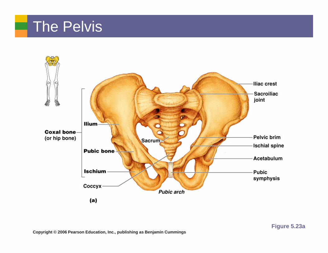
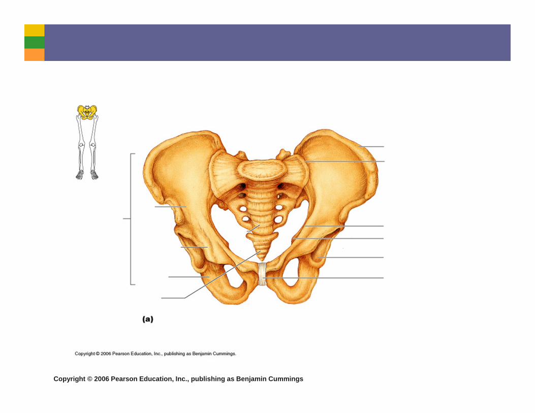
The Pelvis
Figure 5.23a
Copyright © 2006 Pearson Education, Inc., publishing as Benjamin Cummings
Copyright © 2006 Pearson Education, Inc., publishing as Benjamin Cummings
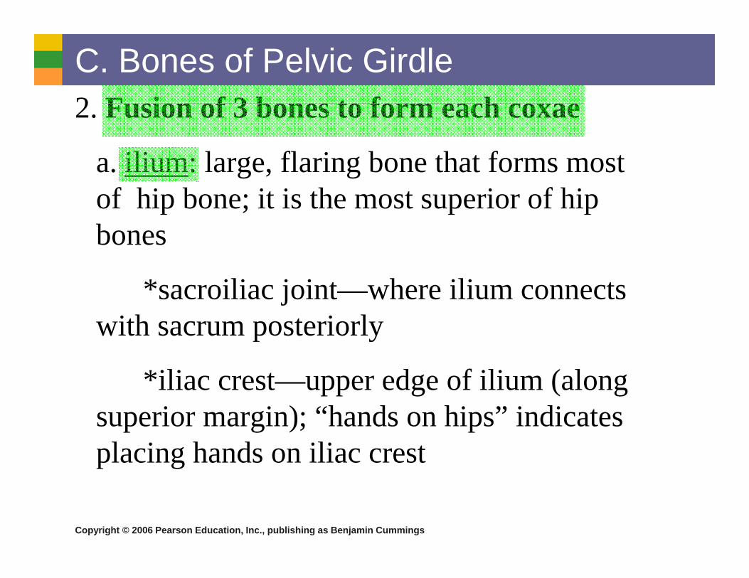
C. Bones of Pelvic Girdle2. Fusion of 3 bones to form each coxae
a. ilium: large, flaring bone that forms most of hip bone; it is the most superior of hip bones
*sacroiliac joint—where ilium connects with sacrum posteriorly
*iliac crest—upper edge of ilium (along superior margin); “hands on hips” indicates placing hands on iliac crest

Copyright © 2006 Pearson Education, Inc., publishing as Benjamin Cummings
C. Bones of Pelvic Girdleb. ischium: forms the most inferior part of the coxal
bone; known as the “sit-down bone”
c. pubis (pubic bone): the most anterior part of a coxal bone
*obturator foramen—an opening that allows blood vessels & nerves to pass into anterior part of thigh
*pubic symphysis—pubic bones of each hip bone fuse anteriorly to form this cartilaginous joint
*acetabulum—receives the head of the thigh bone; location where ilium, ischium, & pubis fuse at deep socket

Copyright © 2006 Pearson Education, Inc., publishing as Benjamin Cummings
The Pelvis: Right Coxal Bone
Figure 5.23b

Copyright © 2006 Pearson Education, Inc., publishing as Benjamin Cummings
C. Bones of Pelvic Girdle3. False & True pelvis: the
true pelvis is surrounded by bone, lies inferior to flaring parts of ilia and pelvic brim
*Woman’s true pelvis must be large enough to allow infant’s head (largest part of infant) to pass during childbirth
MaleMale
Female

Copyright © 2006 Pearson Education, Inc., publishing as Benjamin Cummings
C. Bones of Pelvic Girdle4. 6 differences between male & female pelvis
1. female inlet is LARGER & more circular
2. female pelvis as whole is shallower, and bones are lighter and thinner
3. female ilia flare more laterally
4. female sacrum is shorter & less curved
5. female ischial spines are shorter & farther apart; thus outlet is LARGER
6. female pubic arch is more rounded b/c angle of pubic arch is greater
Copyright © 2006 Pearson Education, Inc., publishing as Benjamin Cummings
D. Bones of the Lower Limbs Lower limbs carry our total body weight when
we are erect
Lower limb bones therefore are much thicker & stronger than upper limb bones
Lower limbs have 3 segments—thigh, leg, foot
Figure 5.24a–b

Copyright © 2006 Pearson Education, Inc., publishing as Benjamin Cummings
D. Bones of Lower Limbs1. Thigh – has only one bone
a. femur: the thigh bone is the heaviest, strongest bone in body
-on proximal end, has a ball-like head, a neck, and greater/lesser trochanters
-head of femur fits into acetabulum of hip bone to form hip joint

Copyright © 2006 Pearson Education, Inc., publishing as Benjamin Cummings
Copyright © 2006 Pearson Education, Inc., publishing as Benjamin Cummings
D. Bones of Lower Limbs(a. femur continued)
*lateral/medial condyles—projections that articulate with tibia bone below; located on distal end of femur
Copyright © 2006 Pearson Education, Inc., publishing as Benjamin Cummings
D. Bones of Lower Limbs2. Leg: 2 bones form the skeleton of the leg
a. tibia: shinbone
-larger & more medial
*lateral/medial condyles—located at proximal end of tibia; articulate with distal end of femur to form knee joint
*medial malleolus—located at distal end of tibia, forms the inner bulge of ankle
*anterior border—sharp ridge on anterior surface of tibia, unprotected by muscles so is felt easily beneath the skin
Copyright © 2006 Pearson Education, Inc., publishing as Benjamin Cummings
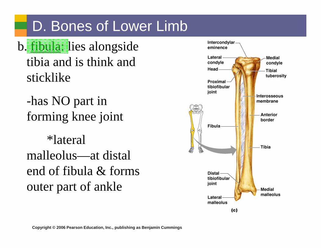
D. Bones of Lower Limbb. fibula: lies alongside
tibia and is think and sticklike
-has NO part in forming knee joint
*lateral malleolus—at distal end of fibula & forms outer part of ankle

Copyright © 2006 Pearson Education, Inc., publishing as Benjamin Cummings

Copyright © 2006 Pearson Education, Inc., publishing as Benjamin Cummings
D. Bones of Lower Limb3. Foot—composed of tarsals,
metatarsals, & phalanges.
-2 important functions: supports body weight & serves as lever that allows us to propel our bodies forward when we walk/run
a. tarsals: 7 ankle bones (see fig. 5.25)
Copyright © 2006 Pearson Education, Inc., publishing as Benjamin Cummings
Copyright © 2006 Pearson Education, Inc., publishing as Benjamin Cummings
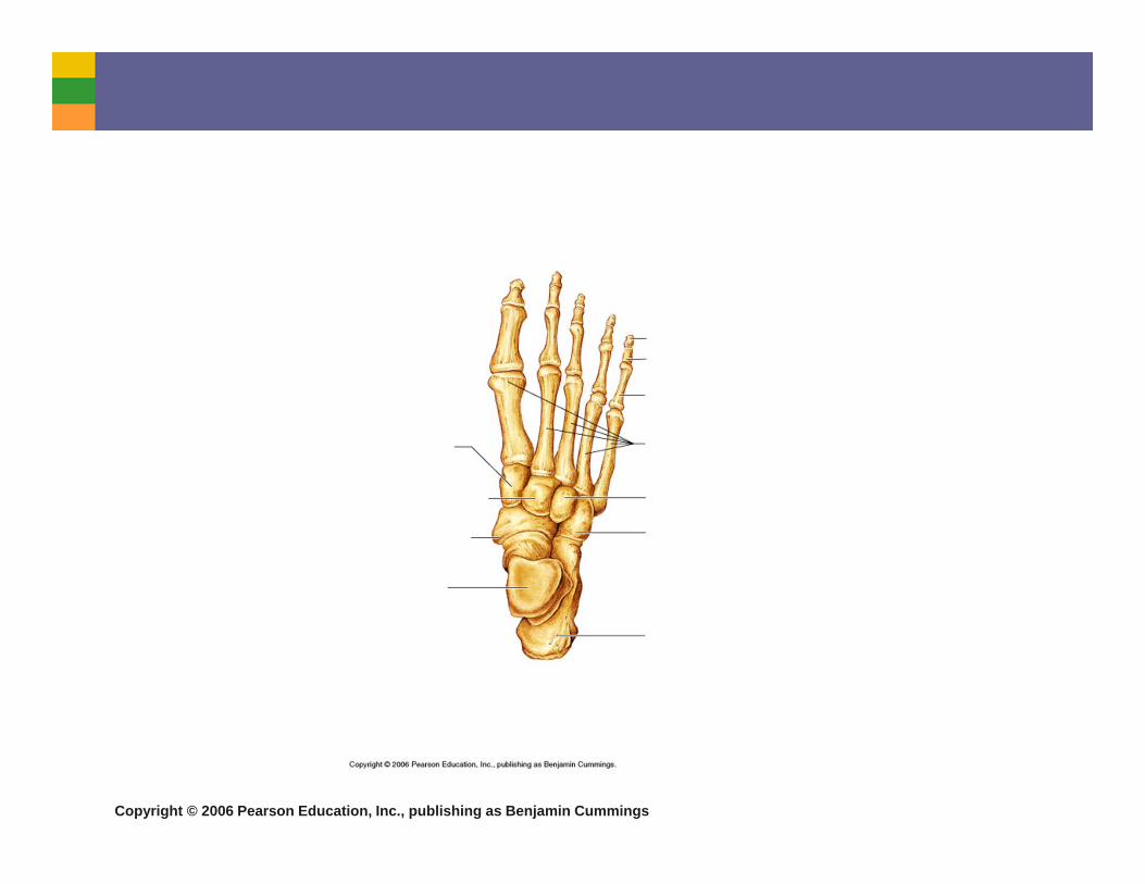
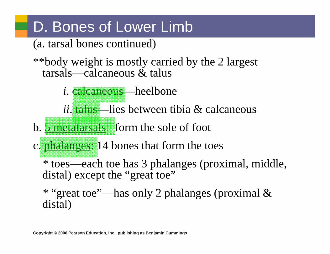
D. Bones of Lower Limb(a. tarsal bones continued)**body weight is mostly carried by the 2 largest
tarsals—calcaneous & talusi. calcaneous—heelboneii. talus—lies between tibia & calcaneous
b. 5 metatarsals: form the sole of footc. phalanges: 14 bones that form the toes
* toes—each toe has 3 phalanges (proximal, middle, distal) except the “great toe”* “great toe”—has only 2 phalanges (proximal & distal)
Copyright © 2006 Pearson Education, Inc., publishing as Benjamin Cummings
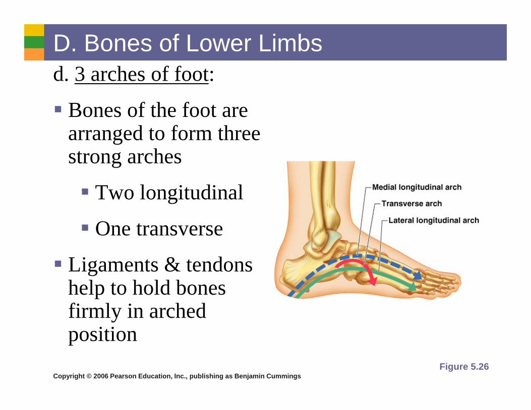
D. Bones of Lower Limbs d. 3 arches of foot:
Bones of the foot are arranged to form three strong arches
Two longitudinal
One transverse
Ligaments & tendons help to hold bones firmly in arched position
Figure 5.26
![muscles [modalità compatibilità] · MUSCLES OF DEEP BACK AND GLUTEAL REGION. 20 • Origin: Upper portion of ilium, the sacrum and coccyx • Insertion: Gluteal tuberosity and iliotibial](https://img.pdfslide.us/doc/110x75/6043690df5743956287e7a5d/muscles-modalit-compatibilit-muscles-of-deep-back-and-gluteal-region-20-a.jpg)