Embed Size (px)
Citation preview
Chapter 9/19
Sacrum/Coccyx
Sacrum
• 5 fused vertebrae
• 4 sets of ________________– Pelvic (Anterior) & Posterior
Sacrum
• _________ – Wings of sacrum
• Superior articulating process– ______________formed with 5th l-spine
vertebra inferior articulating process
Sacrum
• _____________ – Anterior protrusion
• _____________– Continuation of vertebral Foramen
Sacrum
• ________________– Fused spinous processes
• _______________– Joint surface of SI joint
• ______________– Inferior articulating process
Coccyx
• Tailbone
• ___________coccyx segments
• Most distal aspect of spinal column
Coccyx
• Transverse process
• _________
• _________
• Larger at the base and cones toward apex
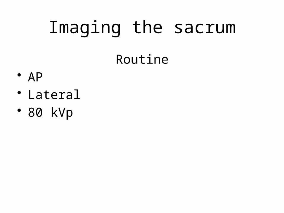
Imaging the sacrum
Routine• AP• Lateral• 80 kVp
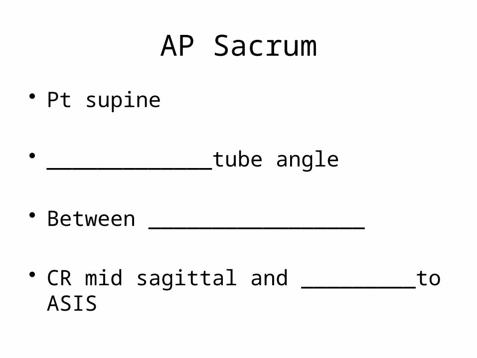
AP Sacrum
• Pt supine
• _____________tube angle
• Between _________________
• CR mid sagittal and _________to ASIS
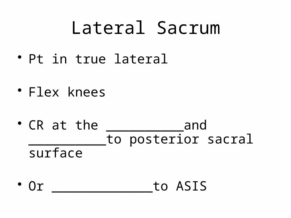
Lateral Sacrum
• Pt in true lateral
• Flex knees
• CR at the __________and __________to posterior sacral surface
• Or _____________to ASIS
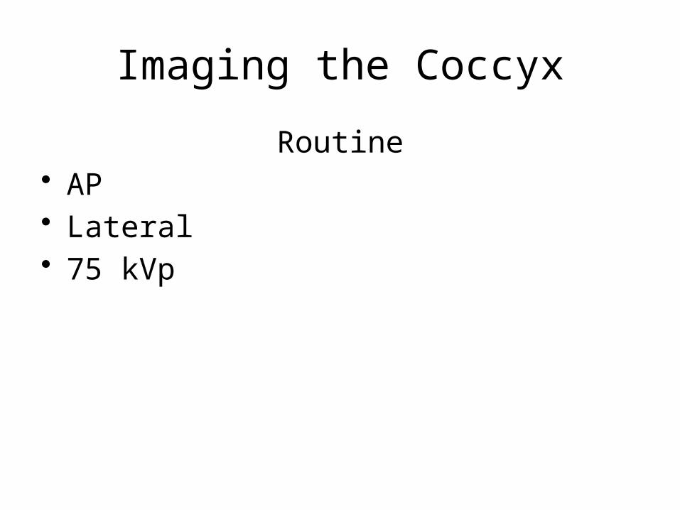
Imaging the Coccyx
Routine• AP• Lateral• 75 kVp
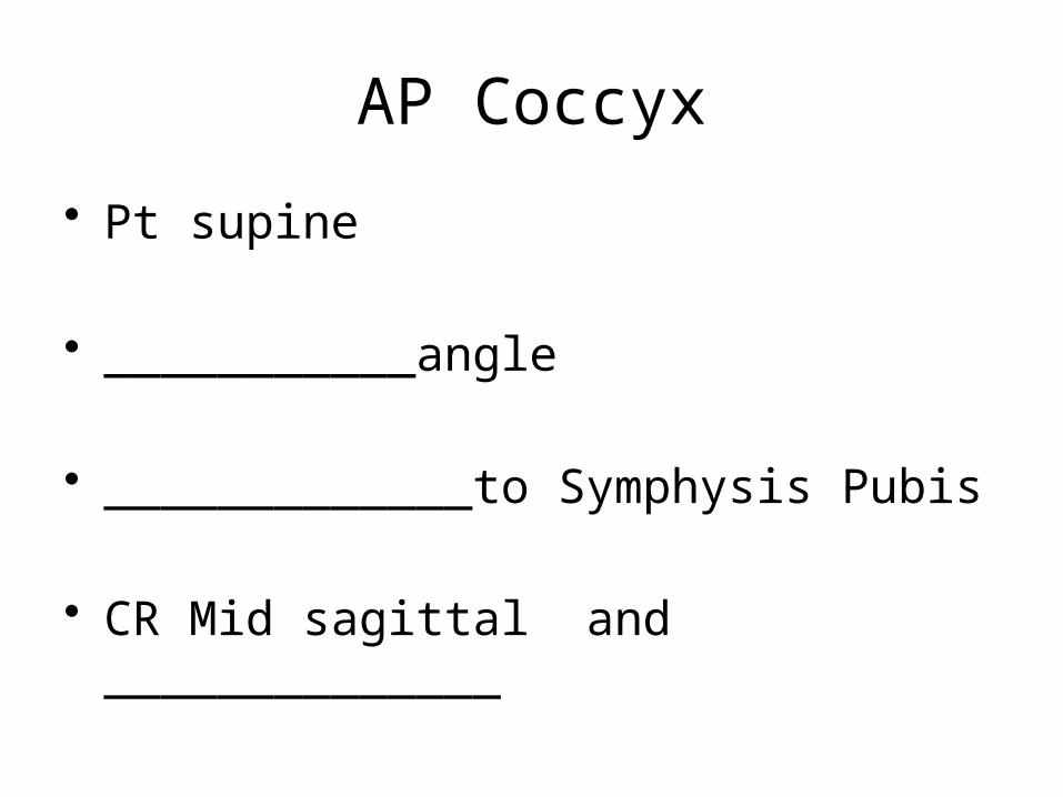
AP Coccyx
• Pt supine
• ___________angle
• _____________to Symphysis Pubis
• CR Mid sagittal and ______________
Lateral Coccyx
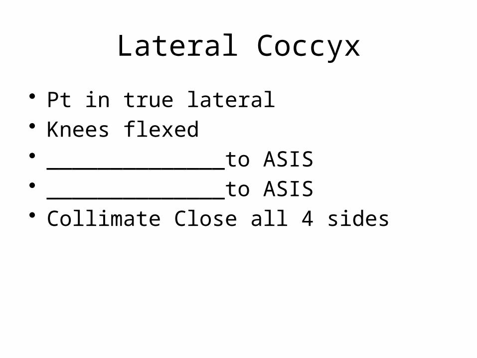
• Pt in true lateral• Knees flexed• ______________to ASIS• ______________to ASIS• Collimate Close all 4 sides
Chapter 22
Myelogram
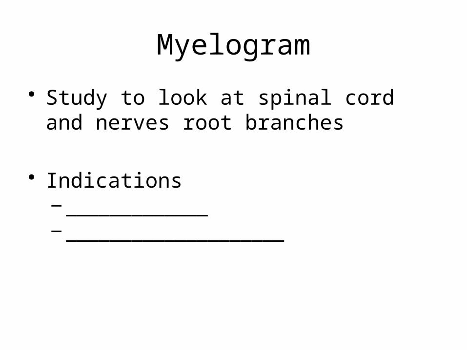
Myelogram
• Study to look at spinal cord and nerves root branches
• Indications – _____________– ____________________
Myelogram
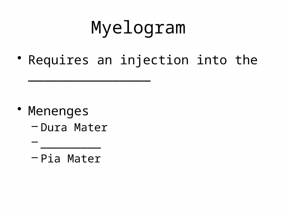
• Requires an injection into the ________________
• Menenges– Dura Mater– _________– Pia Mater
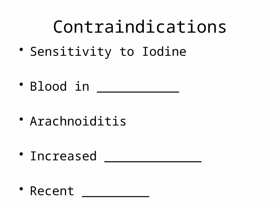
Contraindications• Sensitivity to Iodine
• Blood in ___________
• Arachnoiditis
• Increased _____________
• Recent _________
Equipment
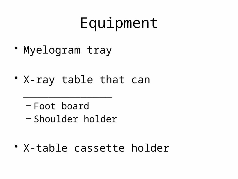
• Myelogram tray
• X-ray table that can ______________– Foot board– Shoulder holder
• X-table cassette holder
Equipment
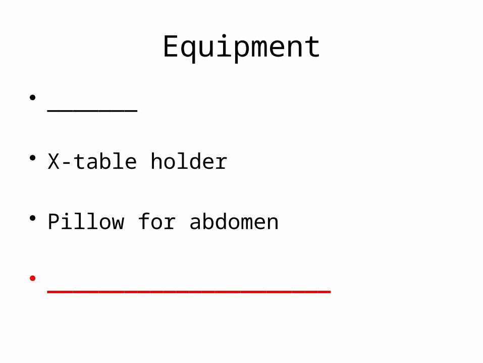
• _______
• X-table holder
• Pillow for abdomen
• ______________________
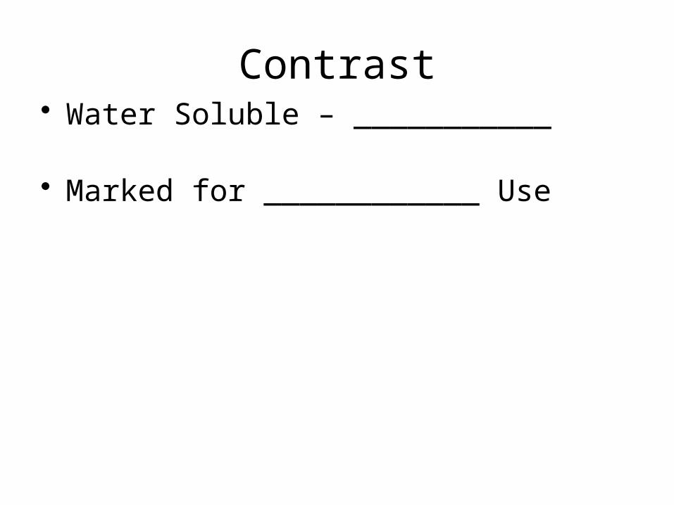
Contrast• Water Soluble – ___________
• Marked for ____________ Use
Injection
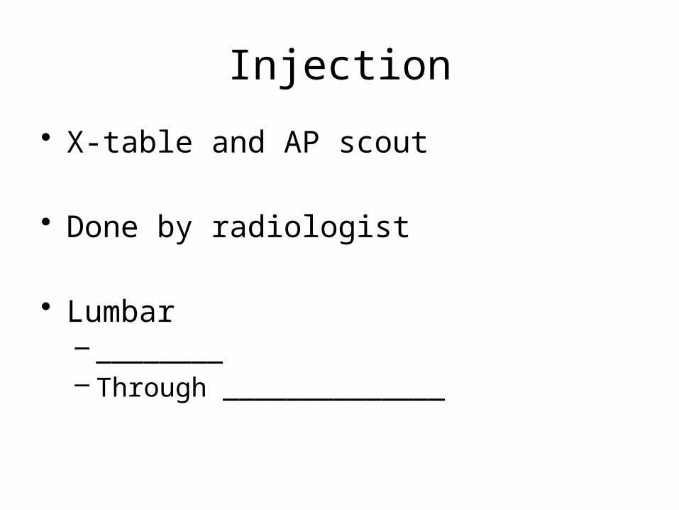
• X-table and AP scout
• Done by radiologist
• Lumbar – ________– Through ______________
Injection
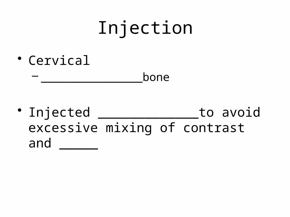
• Cervical– _______________bone
• Injected _____________to avoid excessive mixing of contrast and _____
Lumbar injection
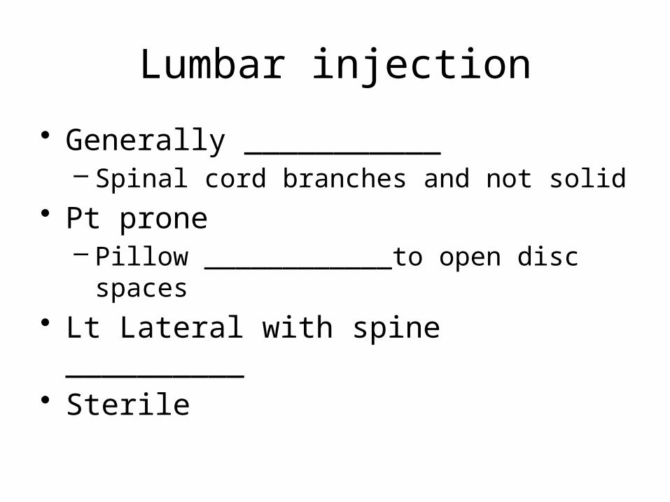
• Generally ___________– Spinal cord branches and not solid
• Pt prone– Pillow ____________to open disc spaces
• Lt Lateral with spine __________• Sterile
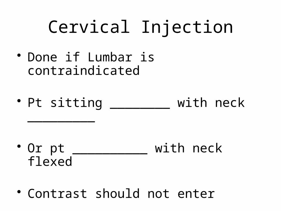
Cervical Injection
• Done if Lumbar is contraindicated
• Pt sitting ________ with neck _________
• Or pt __________ with neck flexed
• Contrast should not enter _________– ___________the neck
What happens
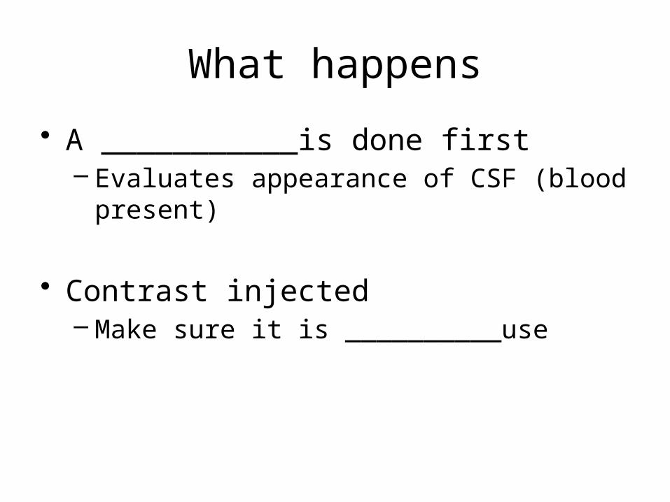
• A ___________is done first– Evaluates appearance of CSF (blood present)
• Contrast injected– Make sure it is __________use
What Happens
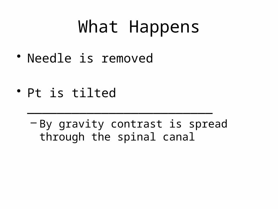
• Needle is removed
• Pt is tilted _________________________– By gravity contrast is spread through the
spinal canal
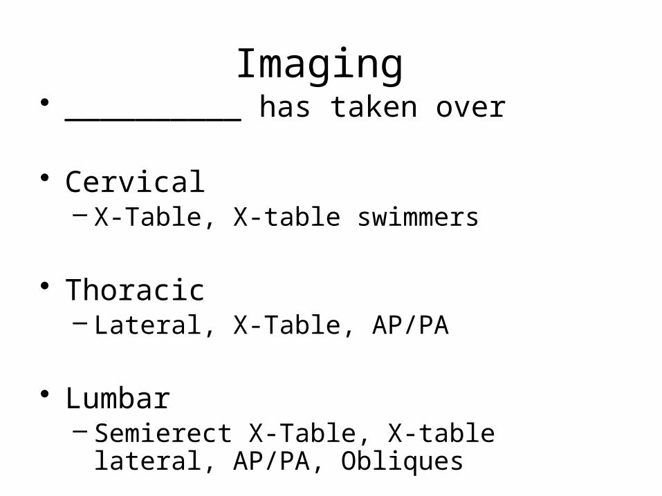
Imaging • __________ has taken over
• Cervical – X-Table, X-table swimmers
• Thoracic– Lateral, X-Table, AP/PA
• Lumbar– Semierect X-Table, X-table lateral, AP/PA,
Obliques
Post Exam Care
• Bandage the injection site
• Place pt semi erect _______________.
• Restricted to the bed.
Complications
• Air into the ____________ of the brain• Spinal needle irritating nerves• Excessive ______________ bleeding• Contrast into ventricular areas
– Can cause _______________• Reaction to contrast


![muscles [modalità compatibilità] · MUSCLES OF DEEP BACK AND GLUTEAL REGION. 20 • Origin: Upper portion of ilium, the sacrum and coccyx • Insertion: Gluteal tuberosity and iliotibial](https://img.pdfslide.us/doc/110x75/6043690df5743956287e7a5d/muscles-modalit-compatibilit-muscles-of-deep-back-and-gluteal-region-20-a.jpg)