Embed Size (px)
Citation preview
ROGANIDANA
By: Dr.Mahamad Yunus S.Nabooji Dept of Roganidana & V.V ([email protected]) Page 1
BASIC PATHOLOGY
SHORT NOTES
(EXAM POINTS)
(For 2nd year BAMS)
By
Dr.Mahamad Yunus S.Nabooji
Associate Professor Dept of Roganidana & V.V
Shri.J.G.C.H.S.Ayurvedic Medical College Ghataprabha
Karnataka
Shri J.G.C.H.S
AYURVEIC MEDICAL
COLLEGE GHATAPRABHA,
KARNATAKA
ROGANIDANA
By: Dr.Mahamad Yunus S.Nabooji Dept of Roganidana & V.V ([email protected]) Page 2
INTRODUCTION TO PATHOLOGY AND ITS SUB-DIVISIONS
DEF: Pathos: suffering, logos: study. Thus the pathology is scientific study of changes
(suffering) in the structure and function of the body in disease (impaired health) and it
answers the disease in terms of its aetiology, pathogenesis, prognosis and treatment plan.
Lesion: Characteristics changes occurred in cell or tissue as the result of disease.
Aetiology: Causative factor
Pathogenesis: Mechanism by which lesion or disease produced
Prognosis: what is going to happen, curability or non-curability of the disease?
Diagnosis: Naming the disease or answer to the pathogenesis
Treatment: What can be done to disease?
Prevention: how to avoid the complications and spread of the disease
BRACHES: Mainly two General pathology (dealing with general principles of disease)
and Systemic pathology (study of disease pertaining to specific organ or tissue.
• Morphological Braches:
1) Histopathology: Also known as tissue pathology or anatomic pathology. It
includes surgical pathology (study of removed cell or tissue by biopsy),
Experimental pathology ( study of disease in experimental animal) and forensic
pathology (study of organ removed from post-mortem)
2) Cytopathology: Study of cells shed off from lesion (Exfoliative cytology) and fine
needle aspiration cytology (FNAC)
Haematology: Deals with disease of blood it includes laboratory haematology and clinical
haematology
ROGANIDANA
By: Dr.Mahamad Yunus S.Nabooji Dept of Roganidana & V.V ([email protected]) Page 3
CELL INJURY
Cell Injury: As the effect of various stresses due to etiological agents a cell results in change
in internal and external environment. It is reversible when stress is mild to moderate and
irreversible when it is severe
Causes: Hypoxia (by blood loss), Physical agents (trauma, radiation etc), chemical agents
and drugs, infectious agents, immunological reactions, Genetic, Nutritional imbalance.
Types: Reversible (Recovery of cell damage once stress removed) and Irreversible (No
recovery / Cell death)
• Reversible: Occurred mainly due to Alteration in plasma membrane (i.e. Bleb
formation, loosening of intracellular attachment and steatosis: means fat
accumulation within cell), Change in mitochondria (Swelling or hydropic,
rarefaction, phospholipids amorphous densities), Nuclear change (Disaggregation of
granular and fibrillar element) and ER changes (Dilatation, detachment and
disaggregation)
• Irreversible: Mainly occurred as the result of Swelling of ER, Lysosomes rupture,
nuclear condensation, nuclear lysis, Membrane blebs, and swollen mitochondria
with amorphous densities.
The irreversible is mainly of two types Necrosis and Apoptosis
NECROSIS: It is death of localized area of tissue followed latter by degradation of
tissue by lysosomal enzyme mainly occur in inflammation and in hypoxia.
APOPTOSIS: also known as coordinated and programmed death of cell occurred
mainly in pathological condition but not in inflammation
Changes after cell death:
• Gangrene: Putrefaction of necrosis, two types dry gangrene e.g. Buerger’s
disease, Raynaud’s disease and wet gangrene e.g. Bed sore, diabetic foot
• Pathological Calcification: Deposition of calcium salt in tissues other than
enamel of tooth occur both in dead tissue and degenerated tissue
ROGANIDANA
By: Dr.Mahamad Yunus S.Nabooji Dept of Roganidana & V.V ([email protected]) Page 4
CELLUALR ADAPTATION
• Form of Reversible cell injury
• Capability of adjusting their structure and functions in response to various
physiological and pathological stimuli (Mild to Moderate) is known as cell or cellular
adaptation
TYPES:
1) Physiological: Occurs in response to an stimulus and ceases once the need of
adaptation has ceased e.g. change in breast and uterus during pregnancy due to
influence of harmones
2) Pathological: Occurs in response to injury or pathogens by producing cell stress
proteins, which protect from damage and help in recovery
• Atrophy: Reduction of cell size which result in shrinkage of cell or organ.
Due to degradation of cell protein by lysosomal enzymes e.g. ischemic
atrophy, disuse atrophy of muscle
• Hypertrophy: Increase in the size of cells which result in enlargement of
cell or organ. Due to increased production of cellular protein,
intracellular factor and growth factor e.g. Left ventricular hypertrophy
• Hyperplasia: Increased number of cells in an organ or tissue. Due to
synthesis of DNA and Proliferation of cell by local production of growth
factor e.g. Hyperplasia of endometrium and hyperplasia of prostate
• Metaplasia: Replacement of one adult cell type with another. Due to long
time persist of cause leads to Metaplasia by the influence of different
factors like growth, cytokines etc. e.g. Squamous metaplasia, columnar
metaplasia
(Note: Both Hyperplasia and Metaplasia are fertile soil for the
development of malignancy in future if cause or stimuli is not removed)
ROGANIDANA
By: Dr.Mahamad Yunus S.Nabooji Dept of Roganidana & V.V ([email protected]) Page 5
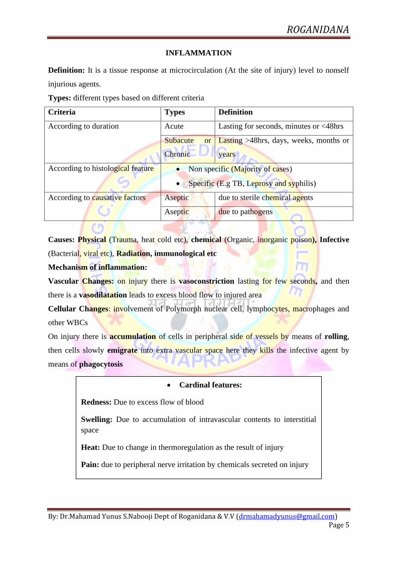
INFLAMMATION
Definition: It is a tissue response at microcirculation (At the site of injury) level to nonself
injurious agents.
Types: different types based on different criteria
Causes: Physical (Trauma, heat cold etc), chemical (Organic, inorganic poison), Infective
(Bacterial, viral etc), Radiation, immunological etc
Mechanism of inflammation:
Vascular Changes: on injury there is vasoconstriction lasting for few seconds, and then
there is a vasodilatation leads to excess blood flow to injured area
Cellular Changes: involvement of Polymorph nuclear cell, lymphocytes, macrophages and
other WBCs
On injury there is accumulation of cells in peripheral side of vessels by means of rolling,
then cells slowly emigrate into extra vascular space here they kills the infective agent by
means of phagocytosis
Criteria Types Definition
According to duration Acute Lasting for seconds, minutes or <48hrs
Subacute or
Chronic
Lasting >48hrs, days, weeks, months or
years
According to histological feature • Non specific (Majority of cases)
• Specific (E.g TB, Leprosy and syphilis)
According to causative factors Aseptic due to sterile chemical agents
Aseptic due to pathogens
• Cardinal features:
Redness: Due to excess flow of blood
Swelling: Due to accumulation of intravascular contents to interstitial
space
Heat: Due to change in thermoregulation as the result of injury
Pain: due to peripheral nerve irritation by chemicals secreted on injury
ROGANIDANA
By: Dr.Mahamad Yunus S.Nabooji Dept of Roganidana & V.V ([email protected]) Page 6
TISSUE HEALING AND REPAIR
Healing is the process by which the cells in the body regenerate (replacement of dead
cell by new cell) and repair (replacement of injured cell by scar tissue) to reduce the size of a
damaged or necrotic area. Most of the injuries will heal using a mixture of both mechanisms.
Mechanism: The process of healing has following steps
• Initial haemorrhage (Clotting phase): As the result of injury the wound space filled
with blood which then clots to stop bleeding and infection
• Inflammatory phase: occurs with 24hrs with appearance of polymorphs from the
margins of incision by 3rd day these are replaced by macrophages
• Proliferative phase (epithelial change): Formation of epithelial spur over the area of
incision by means of proliferating and migration of granulation tissue into type III
collagen
• Maturation phase: unnecessary vessels formed in granulation tissue are removed
by apoptosis, and type III collagen is largely replaced by type I. This phase can last a
year or longer. Ultimately a scar made of collagen, containing a small number of
fibroblasts is left.
EDEMA
Def: Abnormal and excess accumulation of free fluid in the interstitial space
Types:
a) Localised (Confined to organ or limb, e.g. allergic edema, pulmonary edema) and
Generalised ( when it is systemic in distribution also known as anasarca or dropsy
e.g. renal edema, cardiac edema nutritional edema)
b) Transudate (fluid accumulated without change in vascular permeability protein
content low e.g. cardiac and renal edema), Exudates edema (fluid accumulated due
to change in vascular permeability protein content high e.g. inflammatory edema)
c) Pitting ( formation of pit on pressure), Non-Pitting ( absence of pit on press)
Causes or mechanism:
Decreased plasma oncotic pressure, increased capillary hydrostatic pressure, Lymphatic
obstruction, increased oncotic pressure of interstitial space, decreased hydrostatic pressure of
interstitial space, increased capillary permeability, and Sodium water retention.
(Note: Subcutaneous edema: cardiac and renal failure, lower eyelid edema: renal disease,
elephantiasis: filariasis)
ROGANIDANA
By: Dr.Mahamad Yunus S.Nabooji Dept of Roganidana & V.V ([email protected]) Page 7
SHOCK
Def: Also known as cardiovascular collapse, as the result of reduced circulating blood
volume and or inadequate perfusion of cells and tissue.
Pathogenesis: All forms of shock involve: Reduced effective circulatory volume, impaired
tissue oxygenation, Release of inflammatory mediators.
Types:
Hypovolaemic shock: due to acute haemorrhage, Burns, Excessive use diuretics etc
Cardiogenic shock: due to MI, Cardiac arrhythmias, pulmonary embolism etc
Septic Shock: Due to Gram negative and positive bacterial infection
Other shock: includes traumatic shock due to sever injury, neurogenic shock due to head
injury
Clinical features: Low blood pressure, low body temperature, feeble pulse, pale face,
shallow respiration and cold clammy skin
Stages: Reversible and irreversible stage of shock
HAEMORRHAGE
Def: Escape of blood from the rupture or non ruptured blood vessel is known as
haemorrhage
Types:
• Internal (bleeding within body) and external (bleeding out of body) haemorrhage,
• Acute (sudden and massive bleeding) and chronic (small amount over a period)
haemorrhage
• Class I Haemorrhage involves up to 15% of blood volume. There is typically no
change in vital signs. Class II Haemorrhage involves 15-30% of total blood volume
tachycardia, difference between the systolic and diastolic blood pressures. peripheral
vasoconstriction. pale and be cool skin.. Blood transfusion is not usually required.
Class III Haemorrhage involves loss of 30-40% of circulating blood volume, blood
pressure drops, the heart rate increases, shock, diminished capillary refill, blood
transfusion are usually necessary. Class IV Haemorrhage involves loss of >40% of
circulating blood volume, aggressive resuscitation is required to prevent death.
Causes: Trauma to the vessel, inflammation of vessel wall, vascular diseases like
atherosclerosis, aneurysm etc, elevated pressure within vessel, low platelet count,
haemophilia etc,
Effect: the effect of blood loss depends on the amount, speed and site of blood loss. Up to
33% of sudden blood is fatal may cause death
ROGANIDANA
By: Dr.Mahamad Yunus S.Nabooji Dept of Roganidana & V.V ([email protected]) Page 8
THROMBOSIS (Clot)
Def: Process of formation of clot mass in circulation from the constituents of blood, the mass
is called as clot or thrombus
Pathophysiology: Epithelial injury, plate abnormality, alteration of blood flow, abnormality
of coagulation system
Types: Venous thrombosis (Deep or superficial vein) and Arterial thrombosis
The effect of thrombosis will depend upon their location, size and nature of thrombi
EMBOLISM
Def: It is the process of partial or complete obstruction some part of cardiovascular system by
mass (embolus) carried in circulation. This embolus may be a blood originated (thrombus)
or fat (fat embolus) or air (air embolus) etc,
Types:
• Venous, arterial, paradoxical (venous to artery)
• Solid, liquid and Gaseous embolism
• Bland (when embolus is sterile) and Septic ( infected embolus)
ISCHAEMIA
Def: It is deficient blood supply to a part of tissue relative to its metabolic needs
Types: Complete ischemia and Partial Ischemia
Pathophysiology: Ischemia either due to Hypoxia (low oxygen, low haemoglobin, or low
blood supply), Mall nourishment of cell and inadequate clearance of metabolites
Causes: Occlusion (due to thrombus or embolus), Trauma, Others (Atherosclerosis,
hypoglycaemia etc)
INFARCTION
Def: It is the process of tissue death (necrosis) as the result of ischemia
Causes: Occlusion (due to thrombus or embolus), Trauma, Others (Atherosclerosis,
hypoglycaemia etc)
Pathogenesis: As the result of injury or ischemia there will be slowly death of the cell or
tissue as the result of changes in vascular and cellular level like irreversible form of cell
injury
Types:
• Anaemic and haemorrhagic infarction
• Recent and old infarction
ROGANIDANA
By: Dr.Mahamad Yunus S.Nabooji Dept of Roganidana & V.V ([email protected]) Page 9
IMMUNITY
Def: The term immunity defined as resistance exhibited by the host against any foreign
antigen including microorganisms.
Types: Innate and acquired immunity
Innate immunity: It is a resistance which individual possess by birth
• Species immunity: resistance to a pathogen shown by all members of particular
species. E.g B. Anthracis infects human beings but not chickens.
• Racial immunity: within one species different races May exhibits differences in
resistance E.g American negroes are more susceptible than white to tuberculosis
• Individual Immunity: Resistance to infection varies with different individual of
same race and same species. E,g,Homogenous twins exhibit similar degree of
resistance to Tuberculosis
Acquired immunity: The resistance acquired by an individual during life. Two types
Active and Passive
• Active: resistance developed as a result of contact with an antigen; this contact
may be in the form of natural infection or by vaccination
a) Natural: Through clinical or subclinical infection e.g: Post expose of small pox
infection
b) Artificial: Induced by the vaccination e.g: By vaccine
• Passive immunity: It’s induced in an individual by performed antibodies against
infective agent or toxin
a)Natural: through transplacental maternal IgG antibodies e.g: Its transferred from
mother to foetus
b)Artificial: Through antiserum injection e.g: Human ATS
• Mechanism: By the Involment of Humeral and cell mediated immunity
ROGANIDANA
By: Dr.Mahamad Yunus S.Nabooji Dept of Roganidana & V.V ([email protected]) Page 10
IMMUNE RESPONSE
Def: specific reactivity induced in a host following an antigen stimulus is known as immune
response.
Types: Humoral or antibody mediated immune response and Cell mediated immune response
Humoral immune response (HMI/AMI):
• Mediated by macromolecules found in extracellular fluids such as antibodies, c-
proteins and antimicrobial component
• Provides primary defense against most extracellular bacteria and viruses of
Respiratory or GIT
• Participate in immediate hypersensitivity and certain autoimmune diseases
• In response to antigen (foreign body) first the B-cell converted into Plasma cell
(Matured B-cell), these cells will produce different chemicals like antibody etc with
the help of T-helper cells. These secreted chemicals will take part in immune
response
Cell mediated immune response (CMI):
• Protects against fungi, viruses and intracellular bacteria
• Participate in Delay hypersensitivity and in certain autoimmune diseases
• Provide immunity against cancer.
• It works by the activation of phagocytes, antigen specific cytotoxic T-lymphocytes
in response to antigen take part in immune response, here the antibodies are absent
BASIC KNOWLEDGE OF AUTOIMMUNE DISEASES
• Normally immune system recognized its own tissue and tissue antigen as ‘SELF’ and
not produces antibodies against them
• Autoimmunity is a condition when body produces antibodies and immunologically
component T-cells against self antigen leads to structural and functional damage of
tissue and leads to autoimmune diseases
Autoimmune diseases: Classified into
• Haemocytolytic diseases: These conditions involve various cells of blood circulation.
E.g. Autoimmune hemolytic anemia, leucopenia and thrombocytopenia
• Localised or organ specific diseases: Specific organs are target for autoimmune
reactions. E.g. Grave’s disease, Addison’s disease, pernicious anemia etc
ROGANIDANA
By: Dr.Mahamad Yunus S.Nabooji Dept of Roganidana & V.V ([email protected]) Page 11
• Systemic or non- organ specific diseases: Immune system response against a variety
of self antigen and involves damage to several organs and tissue system. E.g.
Rheumatoid arthritis, SLE (systemic lupus erythemtosus)
Mechanism:
May occur either by Humoral or Cellular immune response against self antigen
usually causes tissue damage or disease by TYPE-II and Type-III hypersensitivity.
Sometimes there is Type-IV.
• The immune response can be arrested by immunosuppressive therapy
IMMUNE DEFICIENCY DISEASE
• Diseases produced when the defense mechanism of the host is impaired
• Two types mainly primary immune deficiency diseases and secondary immune
deficiency diseases
• Primary immune deficiency diseases produce when abnormalities in the development
of immune mechanism
• Secondary immune deficiency diseases produce due to consequence of some other
diseases, malnutrition, drugs etc.
• These immune deficiency diseases involve specific abnormal (depression) immune
functions- Humoral immunity, cell mediated immunity or both – or nonspecific
mechanisms such as phagocytosis and complement system
• Primary immune deficiency diseases includes Humoral immune deficiencies (e.g.
X-linked agammaglobulinaemia etc.), cellular immune deficiencies (e.g. Thymic
hypoplasia etc.), combined immune deficiencies (e.g. ataxia telangiectasia etc.),
Disorders of complement (e.g. complement component and inhibitor deficiencies)
and Disorders of phagocytosis (e.g. chronic granulomatous diseases etc.)
• Secondary immune deficiency diseases includes lymphoid malignancy, lymphatic
leukemia AIDS, Hodgkin’s lymphoma etc.
ROGANIDANA
By: Dr.Mahamad Yunus S.Nabooji Dept of Roganidana & V.V ([email protected]) Page 12
HYPERSENSITIVITY
Definition: Hypersensitivity refers to a condition in which immune response result in
excessive reaction leading to tissue damage, disease or even death in the sensitized host.
Hypersensitivity occurs in individual who have had previous contact with the antigen or
foreign substance
Priming or sensitizing dose: The initial contact sensitize the immune system by priming
appropriate B or T lymphocytes it is known as primi or sensitizing dose
Shocking dose: Subsequent contact with the same antigen causes hypersensitivity is known
as shocking dose
Allergy is most commonly used as a synonym for hypersensitivity. The term allergy
means an altered state of reactivity to an antigen; it may include both protective as well as
injurious immune response.
Classification: Hypersensitivity reactions are classified into two types, immediate and
delayed types based on the time required by sensitized host to develop clinical reactions upon
exposure to the shocking dose of the antigen
Comb and Gel classified hyper sensitivity reactions into four types type-I to IV
• Type I (Anaphylactic)
• Type II (Cytotoxic)
• Type III (Immune complex)
• Type IV (Cell mediated)
Type I, II, and III depend on the interaction of antigen with humoral antibodies and are
known as immediate type of reaction and Type IV is mediated by T or B lymphocyte and it is
delayed type of reaction
Difference between Immediate and Delayed Hypersensitivity
Feature Immediate Delayed
Onset and duration Appears and recedes rapidly Appears slowly in 24-72hrs and
last longer
Immune response Antibody and antigen
mediated
Cell mediated (T-lymphocytes)
Passive transfer Possible with serum Possible with lymphocyte or
transfer factor
Desensitization Easy but short lived Difficulty but long lasting
Induction Antigens or haptens, by any
route
By antigen injected
intradermally or by
ROGANIDANA
By: Dr.Mahamad Yunus S.Nabooji Dept of Roganidana & V.V ([email protected]) Page 13
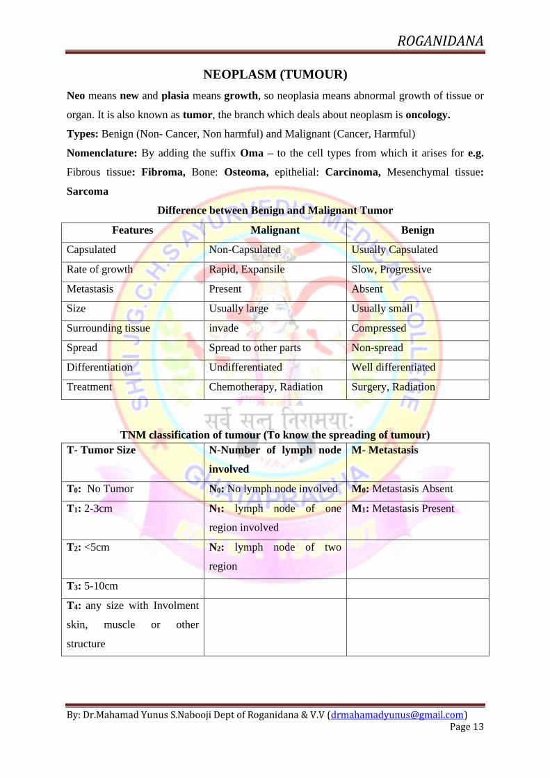
NEOPLASM (TUMOUR)
Neo means new and plasia means growth, so neoplasia means abnormal growth of tissue or
organ. It is also known as tumor, the branch which deals about neoplasm is oncology.
Types: Benign (Non- Cancer, Non harmful) and Malignant (Cancer, Harmful)
Nomenclature: By adding the suffix Oma – to the cell types from which it arises for e.g.
Fibrous tissue: Fibroma, Bone: Osteoma, epithelial: Carcinoma, Mesenchymal tissue:
Sarcoma
Difference between Benign and Malignant Tumor
Features Malignant Benign
Capsulated Non-Capsulated Usually Capsulated
Rate of growth Rapid, Expansile Slow, Progressive
Metastasis Present Absent
Size Usually large Usually small
Surrounding tissue invade Compressed
Spread Spread to other parts Non-spread
Differentiation Undifferentiated Well differentiated
Treatment Chemotherapy, Radiation Surgery, Radiation
TNM classification of tumour (To know the spreading of tumour)
T- Tumor Size N-Number of lymph node
involved
M- Metastasis
T0: No Tumor N0: No lymph node involved M0: Metastasis Absent
T1: 2-3cm N1: lymph node of one
region involved
M1: Metastasis Present
T2: <5cm N2: lymph node of two
region
T3: 5-10cm
T4: any size with Involment
skin, muscle or other
structure
ROGANIDANA
By: Dr.Mahamad Yunus S.Nabooji Dept of Roganidana & V.V ([email protected]) Page 14
NUTRITIONAL DISORDERS
• Macro nutrients: Required in large quantity e.g. Carbohydrate, Protein, fat etc
• Micro nutrients: Required in less quantity e.g. Vitamins and minerals
• An adequate amount of nutrients are required for the body to provide energy, if there
is a over nutrition will cause over weight or obesity will responsible for many diseases
like diabetes, hypertension, stone formation, degenerative diseases like OA, and
increase the incidence of cancer.
• If there is Malnutrition then there will be growth retardation, mental illness,
immunodeficiency disorders. It is occur in two form primary (Malnutrition due
missing of one or all component) and secondary (due to mal absorption of
component).
Disorders of Micro Nutrients:
• Vitamins: Vitamin A (Night blindness, reduced hair growth), Vitamin B1
(wet and dry beriberi), Vitamin B2 (Intestinal keratitis ), Vitamin B3 (
Pellagra), Vitamin B6 (Anaemia, convulsions), Vitamin B12 ( Pernicious
anaemia, nerve damage), Vitamin C (Scurvy), Vitamin D (Rickets,
osteoporosis), Vitamin E (Nerve abnormality), Vitamin K (Defective blood
coagulation).
• Minerals: Calcium (OA, RA etc), Iron (Anaemia, sore mouth), Iodine
(Goitre, Hypothyroidism, hair loss), Potassium (Hypokalemia, muscular
weakness), Sodium (Hyponitremia, digestive disorders)
Disorders of Macro nutrients:
• Carbohydrate: Overweight, obesity, diabetes and cardiovascular diseases etc
and due deficiency leads to mood swing, ketosis, reduced staming
• Fat: deficiency increased the risk of atherosclerosis, behavioural problem,
depression, cognitive decline etc
Proteins: deficiency leads to Kwashiorkor and marasmus
ROGANIDANA
By: Dr.Mahamad Yunus S.Nabooji Dept of Roganidana & V.V ([email protected]) Page 15
INFECTION
Infection is the lodgment and multiplication of a parasite in the body. All infection do not
invariably result in disease
TYPES: Primary (Initial infection), Secondary (When body resistance is lowered by a
preexisting infection, a new parasite set up an infection), Cross infection (When a patient
already suffering from a disease acquires a new infection from another host), Nosocomial
infection (Cross infection acquired in hospital), Iatrogenic infection (Physician or nurse
induced infection resulting from drug therapy or investigating procedures), Subclinical
infection (When clinical features are not apparent), Latent infection (Following infection,
some parasites remain in the host in hidden form and produce disease when host immune is
reduced), Atypical infection (Characteristic features are not present) and Typical infection
(Characteristic features are present)
MODE OF TRANSMISSION: Infections spread from one host to another by a variety of
mechanism. Contact (e.g. syphilis AIDA), Inhalation (e.g. common cold, whooping cough),
Ingestion (Cholera, dysentery etc), Inoculation (e.g. Rabies virus inoculated directly by the
bite of rabid animal), Vectors (e.g. flies, fleas, ticks, mites), Transplacental (through
placental barrier and infect the fetus in utero known as teratogenic infections) and
Iatrogenic and laboratory infections (Through injections, blood transfusions etc)
TYPES OF INFECTIOUS DISEASES: Localised and Generalized. E.g. Bacteraemia
(Circulation of the bacteria in the blood), Septicaemia (Multiplication of the bacteria),
Pyaemia (pyogenic abscess as the result of septicemia), Endemic (The disease constantly
present in a particular area e.g. enteric fever), Epidemic (The disease that spreads rapidly,
involving many persons in a particular area at the same time e.g. Meningococcal meningitis),
Pandemic (It’s an epidemic that spreads through many areas of the world involving very
large number of persons within a short period e.g. cholera, influenza etc)
ROGANIDANA
By: Dr.Mahamad Yunus S.Nabooji Dept of Roganidana & V.V ([email protected]) Page 16
CLASSIFICATION OF BACTERIA
Bacteria of medical importance measures 2-5 um (L) * 0.2-1.5 um (W)
A) Depending on their shape:
1) Cocci oval or spherical shape Diplococci: Arranged in pairs, Streptococci: Arranged
in chains, Staphylococci: Arranged in cluster or group, Tetrads: Group of four cocci
Sarcina: Group of eight cocci
2) Bacilli: rod shaped, Coccobacilli: Length of bacteria is approximately same as width
e.g. Brucella, Streptobacilli: These bacilli are arranged in chains e.g. Streptobacillus,
Chinese latter or cuneiform pattern: Arranged at angels to each other e.g.
Corynebacterium, Comma shaped: Curve appearance e.g. Vibrio, Spirilla: Rigid
spiral form e.g. Spirillum
3) Spirochaetes: (spiera: coil; chaite; hair): These are slender, flexuous spiral forms
e.g. Treponema.
4) Actionomycetes: (Actis: ray. Mykes: fungus): These are branching filamentous
bacteria resembling fungi. They have a rigid cell wall.
5) Mycoplasmas: Cell wall deficient bacteria hence don’t possess a stable shape. They
are very small in size (50-300nm in diameter)
6) Rickettsiae and Chlamydiae: These are very small and obligate parasites. Due to
their inability to grow outside living cells,
B) Based on Gram stain: Gram positive Bacteria: e.g. Streptococcus, Staphylococcus,
and Pneumococcus. Gram negative Bacteria: e.g. Salmonella typhi, Vibrio cholera
C) Based on Acid fast stain: Acid fast stain Bacteria: e.g. M.Tuberculi, M. Leprae.
Non acid fast stain Bacteria: e.g. C. Diphtheria, Bacillus
D) Based on Spore: Sporing Bacteria: e.g. Bacillus, Clostridium , Non sporing
Bacteria: e.g. Streptococcus, staphylococcus etc
E) Based on motility: Motile Bacteria: e.g. Salmonella typhi, Non motile Bacteria:
e.g. Strepto and Staphylococcus
ROGANIDANA
By: Dr.Mahamad Yunus S.Nabooji Dept of Roganidana & V.V ([email protected]) Page 17
CLASSIFICATION OF FUNGUS
Study of fungi is known as Mycology, All fungi are eukaryotic, their cell wall contain
chitin, mannan and other polysaccharides, They divide asexually, sexually or by both
processes
Classification of fungi: Fungi are kept under phylum Thallophyata
Taxonomical Classification: Zygomycetes, Ascomycetes, Basidiomycetes, Deuteromycetes
Morphological classification: Yeast, Yeast like fungi, Moulds, Dimorphic fungi
a) YEAST: Round to oval unicellular fungi, Reproduced by budding
b) YEAST LIKE FUNGI: These are partially grows as yeast and partially as chains of
elongated budding cells joined end to end forming pseudophytes. Example is Candida
albicans
c) MOULDS: They grow as branching filaments called hyphae usually 2-10um in
width. Hyphae may be septate or non-septate, They reproduced from both sexual and
asexual spores, Dermatophytes, aspergillus, penicillium are examples for moulds
d) DIMORPHIC FUNGI: They exist as yeast in the host and in the culture at 370c and
as moulds forms in the soil and in the cultures at 22-250c, Blastomyces, dermatitidis,
etc are the examples
• Diseases of fungus are known as MYCOSES mainly classified into, Superficial
mycoses, Subcutaneous mycoses, Systemic mycoses,
CLASSIFICATION OF VIRUSES
The viruses are classified into different types
B/o Nucleus: DNA and RNA virus
B/o Symmetry: Helical and complex
B/o Envelop: Capsid and non capsid virus
SOURCE OF REFERENCE:
• Text book of Pathology – By Harsh Mohan, published by Jaypee Brothers Medical Publishers
• Text book of Microbiology – By C P Baveja, Published by Arya publications
![Basic Plant Pathology.ppt [Repaired] - University of Georgia...Basic Plant Pathology & Troubleshooting Plant Problems Department of Plant Pathology University of Georgia Paul Pugliese,](https://img.pdfslide.us/doc/110x75/5eb3a4ce0756884351764dbb/basic-plant-repaired-university-of-georgia-basic-plant-pathology-troubleshooting.jpg)