Embed Size (px)
Citation preview

Methods in pathologyBasic histological techniques
Dentistry
Department of pathology
Medical faculty Comenius University

PATHOLOGY (pathos + logos)
• scientific study of structural and functionalchanges of diseased tissues
• studies changes in normal anatomy, histology and physiology (pathophysiology)

PATHOLOGY (pathos + logos)
• studies morphologic manifestation of a disease
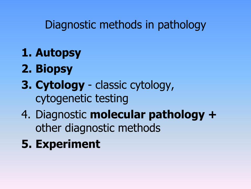
Diagnostic methods in pathology
1. Autopsy
2. Biopsy
3. Cytology - classic cytology, cytogenetic testing
4. Diagnostic molecular pathology + other diagnostic methods
5. Experiment

Autopsy – aims?
• determination of the cause of death
• detection of other / unverified diagnoses
• verification of adequacy of diagnostic and therapeutic procedures
• education – doctors (frequently overlooked diagnoses, detection of new diseases, study of disease epidemiology…) and students
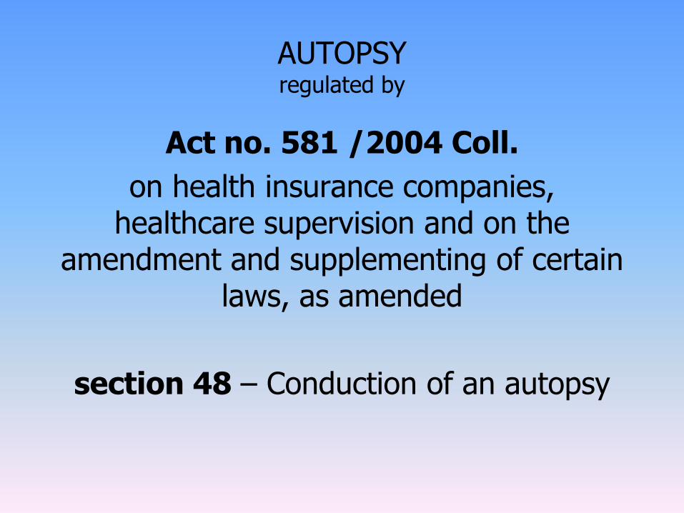
AUTOPSYregulated by
Act no. 581 /2004 Coll.
on health insurance companies, healthcare supervision and on the
amendment and supplementing of certain laws, as amended
section 48 – Conduction of an autopsy

AUTOPSY – why?
• if the cause of death is unknown (confirmation of a disease / diagnostic / therapeutic procedure)
• the diagnosis is not certain (unverified malignant tumors, verified malignant tumors of unknown origin – tumor e loco ignoto)
• if the death is related to a surgery / general anesthesia (mors in tabula)
• if iatrogenic damage is suspected
• if a severe infectious disease is suspected
• sudden death
• organs collected for transplantation
• suspition of inadequate health care
• …

AUTOPSY - process• external examination internal examination
• organs in complexes
• determination of cause of death autopsy protocol

AUTOPSY - process
• during autopsy: samples of tissue for histologic evaluation
• histologic diagnosis
• adjustment of diagnoses, conclusion

Biopsy – aims?
• tissue examination during a patient’s lifetime

Biopsy
• aim: determination of diagnosis – needed for application ofadequate therapy
• any tissues taken from the body for diagnosis of a disease must beprocessed in the histological laboratory to produce microscopicslides that are analyzed in the microscope

Biopsy
• standard way of processing the tissue (order of certainsteps):
1. labeling
2. fixation
3. macroscopic description
4. dehydration, cleaning, impregnation
5. embedding
6. sectioning
7. staining

Biological material
- the material should by adequately labeled - name- age - insurance information- clinical diagnosis- anamnesis- symptoms of disease
Tissue processing in the laboratory0. LABELING

Biological material
- to kill the bacteria and to stop the enzymatic processes in the cells - stop the autolyticchanges- the purpose of fixation is to preserve tissues permanently in as life-like state as possible - should be carried out as soon as possibleafter removal of the tissues
• autolysis (enzymatic decomposition oftissues -> cellular destruction -> greatchanges in the structure)
• microbiological spoilage – bypenetration of bacteria and microbiologicaldecomposition (starts with autolysis, theseprocesses are parallel)
1. FIXATION

Fixation - types of fixatives
1. chemical modification (formalin)• variety of fixatives are available for use (depending on the
type of tissue and features to be demonstrated)
2. physical modification (freezing)

• major groups of fixatives, classified according to mechanism of action:
Aldehydes (formaldehyde, glutaraldehyde) - by formation of cross-linkages in the proteins (between lysine
residues).- does not harm the structure of proteins greatly -> antigenicity is not
lost, formaldehyde is good for immunoperoxidase techniques.
Alcohols (methyl alcohol, ethyl alcohol) - protein denaturation- very good for cytologic smears because they act quickly and give good
nuclear detail
Oxidizing agents (permanganate fixatives, osmium tetroxide). - cross-linkages, but cause extensive denaturation. - used in specialized applications – electron microscopy
Mercurials, picrates fix tissue by an unknown mechanism
Fixation – chemical types of fixatives

Fixation – physical types of fixativesFrozen Samples
• tissues can be preserved by freezing them directly (snap freezing) at -80°C in a cold environment / by immersing in liquidnitrogen.
• freezing makes tissue solid enough to section with microtome(in a cryostat)
• tissue sections are put on a glass slide and are then ready forstaining
• advantages:1. biological and enzymatic activities of proteins do not change
during this process -> suitable for demonstration of enzymes or substances normally washed out (detected by histochemical met.), in techiques for recovery of DNA, mRNA, and proteins
2. takes only several minutes -> intraoperative diagnosticprocedures to guide the surgeon (diagnosis is made quickly)

Gross Examination
- describing the specimen and
- placing all of it / parts of it into a small plastic cassettewhich holds the tissue while it is being processed to a paraffin bloc
- when a malignancy is suspected - the specimen is often covered with ink with the aim to mark the margins of the specimen
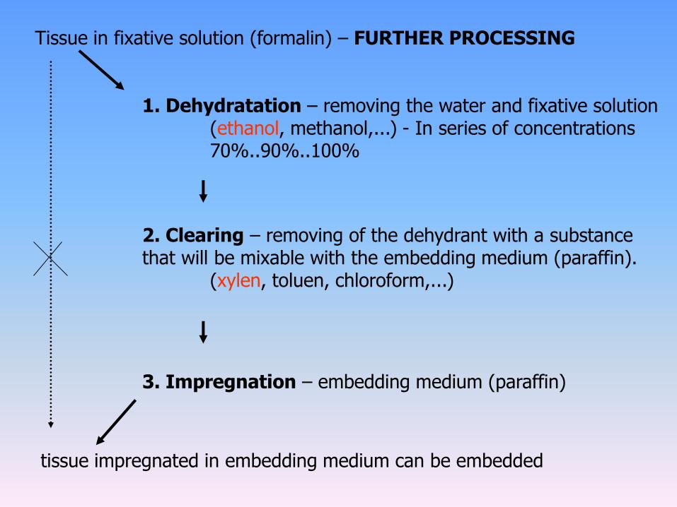
Tissue in fixative solution (formalin) – FURTHER PROCESSING
1. Dehydratation – removing the water and fixative solution(ethanol, methanol,...) - In series of concentrations70%..90%..100%
2. Clearing – removing of the dehydrant with a substance that will be mixable with the embedding medium (paraffin).
(xylen, toluen, chloroform,...)
3. Impregnation – embedding medium (paraffin)
tissue impregnated in embedding medium can be embedded

Dehyderatation and clearing 10% formaline
70% ethanol
95% ethanol
100% ethanol
100% ethanol
100% ethanol
100% ethanol
+Xylen
Xylen
Xylen
Paraffin
Paraffin

Embedding
Aim: processing into a form from which the thin microscopicsections can be prepared.
• tissue can’t be cut right away -> it has to be embedded in a suitable medium
• the medium should be solid, but also cut-able• embedding media must fill all spaces within the tissue
to support cellular components adequately duringmicrotomy
• must be elastic enough to recover sectioning deformation
…advantages of PARAFFIN (similar in density to tissue, adequate viscosity and melting point, can be sectioned at anywhere 3-10 um)

Embedding
• impregnated tissue is put in a metal form -> embedded in liquid warm paraffin
• cools down -> paraffin block with embedded tissue

Sectioning
- samples are cut into sections that can be placed on a slide - microtome and ultramicrotomes

Drying
Storage
Prepared for staining
Deparaffinisation
(paraffin is only used as a medium needed for sectioning, has to be removed before staining)
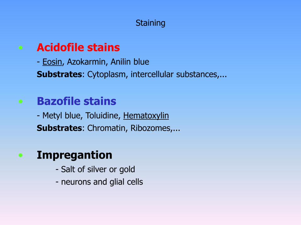
Staining
• Acidofile stains
- Eosin, Azokarmin, Anilin blue
Substrates: Cytoplasm, intercellular substances,...
• Bazofile stains
- Metyl blue, Toluidine, Hematoxylin
Substrates: Chromatin, Ribozomes,...
• Impregantion
- Salt of silver or gold
- neurons and glial cells
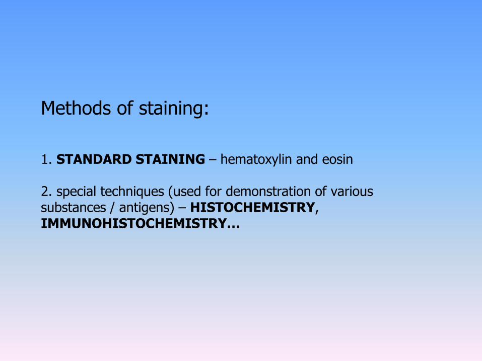
Methods of staining:
1. STANDARD STAINING – hematoxylin and eosin
2. special techniques (used for demonstration of various substances / antigens) – HISTOCHEMISTRY, IMMUNOHISTOCHEMISTRY…

STANDARD STAINING: hematoxylin a eosin
- Nucleus: blue
- Cytoplasm: light red
- Kolagen: red

Special methods - HISTOCHEMISTRY
- if we want to demonstrate certain specific substances / components of cells -> various subst. stain in different colour (depends on characteristics of stained subst. and stain itself)
- amyloid, sacharides, lipids, proteins, NA, connective tissue, nervous tissue...
- congo red, PAS, van Gieson, PWH, Gram, Z-N…

Green trichrome
- Nucleus: blue
- Cytoplasm: lightred
- Collagen: green

Special methods -IMMUNOHISTOCHEMISTRY
- used for demonstration of presence and location of a certain antigen in / on a cell (membrane, cytoplasm, nucleus)
- with the used of specific antibodies and chromogens
- colour is always the same (brown)

IMMUNOHISTOCHEMISTRY – use
• tumors of unknown primary site
• prognostic markers of tumors(expression of HER-2/neu in breast Ca, Ki67) – modification of therapy!!!
• prediction of response to therapy (estrogen receptors in breast Ca)
• infectious diseases – viruses (Ab againstRNA / DNA, HPV, herpesviruses), bacteria, parasites

CYTOLOGY - material
• EXFOLIATIVE
• material: spontaneous detachment of cells from epithelial surfaces, scraping, brushing, lavage of mucosal surfaces
• INTERVENTION
• material: obtained by aspiration, curettage,… / during surgery (FNAC – fine needle aspiration cyt.)
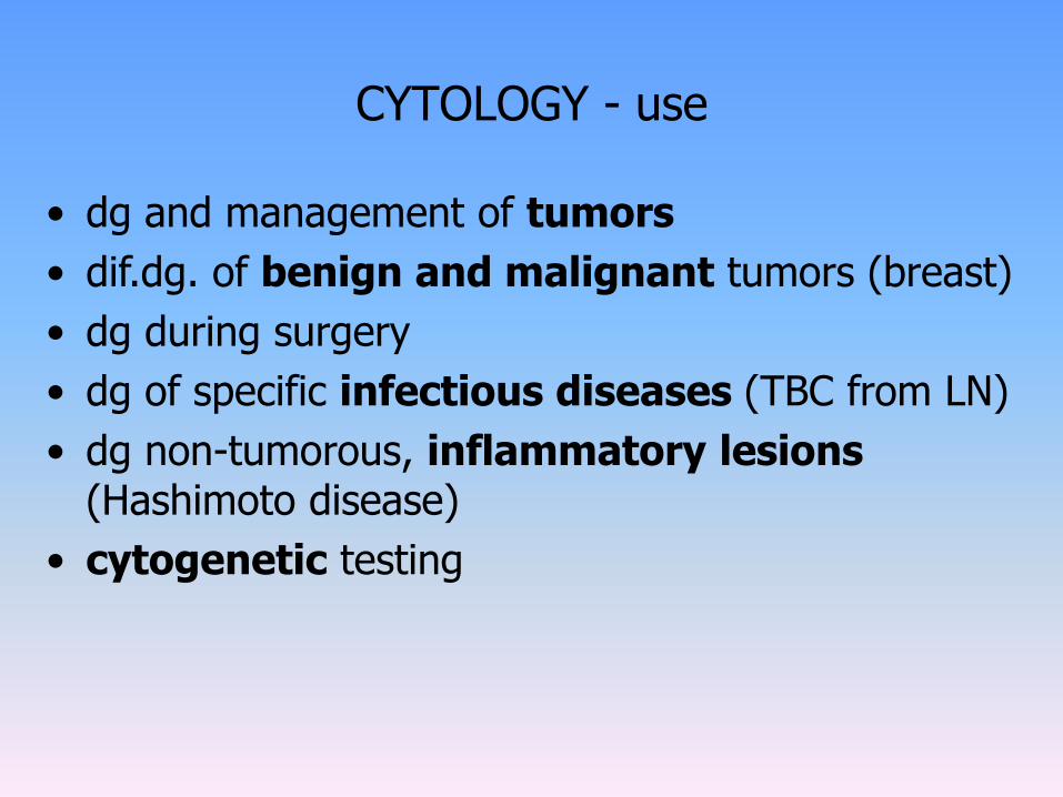
CYTOLOGY - use
• dg and management of tumors
• dif.dg. of benign and malignant tumors (breast)
• dg during surgery
• dg of specific infectious diseases (TBC from LN)
• dg non-tumorous, inflammatory lesions (Hashimoto disease)
• cytogenetic testing

FNAC – fine needle aspiration cytology
• use: palpable lesions (breast, LN, thyroid, soft tissue, salivary glands, intraabdominal lesions, testicles)
• advantages: no need for hospitalisation and anesthesia, method is quick, safe, repeatable, painless and cheap