Embed Size (px)
Citation preview

ANATOMY OF
LARYNX
Dr. Diptiman Baliarsingh
1st Year PG, Dept. of ENT,
Hi-Tech Medical College & Hospital, Bhubaneswar
EMBRYOLOGY
During 4th wk, the tracheo-bronchial diverticulum appears in the ventral wall of primitive pharynx, just below hypobrachial eminence.
The edges of this groove form oesophago-tracheal septum, which fuses caudally leaving a slit like aperture cranially.
The cranial end of tube forms larynx & trachea
The caudal end of tube forms bronchi & lungs The tube is lined by endoderm which forms
the epithelial lining of entire respiratory system.
Arytenoid swellings appear on both sides of tracheo-bronchial diverticulum. The Aryteniod swellings grow upwards and deepen to produce Ary-epiglottic folds
Hypobrachial eminence forms Epiglottis Glottis forms just above the primitive aperture Thyroid cartilage develops from the ventral
ends of the cartilages of the 4th pharyngeal arch
Cricoid cartilage and cartilages of trachea develop from 6th arch
Superior & recurrent laryngeal branches of vagus nerve are derived from 4th & 6th arches which supply the larynx.

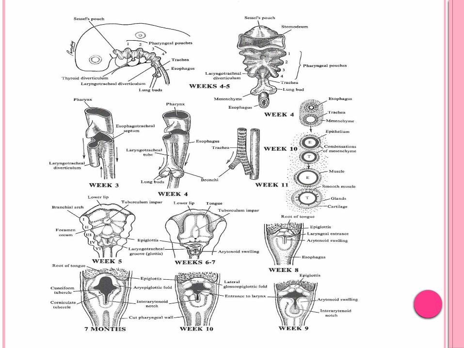
EMBRYOLOGICAL DEVELOPMENT OF LARYNX
Structure Source
Laryngeal mucosa Endoderm of cephalic part of foregut
Laryngeal cartilages Mesenchyme
Epiglottis Hypobranchial eminence
Upper part of thyroid cartilage 4th branchial arch
lower part of thyroid cartilage, cricoid, corniculate, and cunei- form cartilages
6th branchial arch
Intrinsic muscles of larynx 6th branchial arch
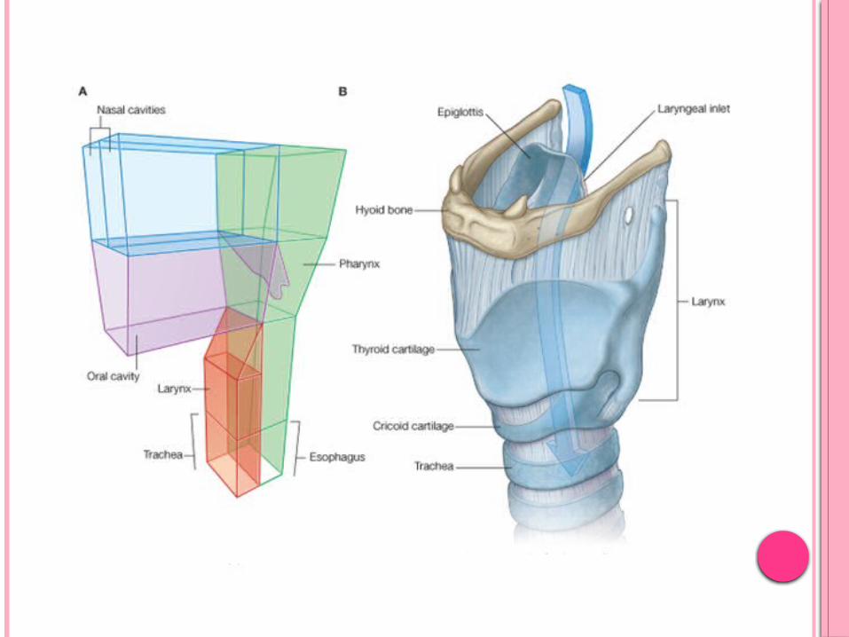
GENERAL ANATOMY
Extent From laryngeal inlet to inferior border of cricoid
cartilage 3rd to 6th cervical vertebrae Little higher in women VC lie at C5 level in adults, C3/C4 in infants
Infantile larynx is smaller & funnel shaped It is narrowest at the junction of sub-glottic
larynx with trachea.* Slight swelling may result in marked airway
obstruction
Laryngeal cartilages are much softer in infants and collapse more easily on forced inspiration
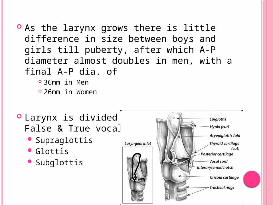
As the larynx grows there is little difference in size between boys and girls till puberty, after which A-P diameter almost doubles in men, with a final A-P dia. of
36mm in Men 26mm in Women
Larynx is divided into 3 parts by False & True vocal cords Supraglottis Glottis Subglottis
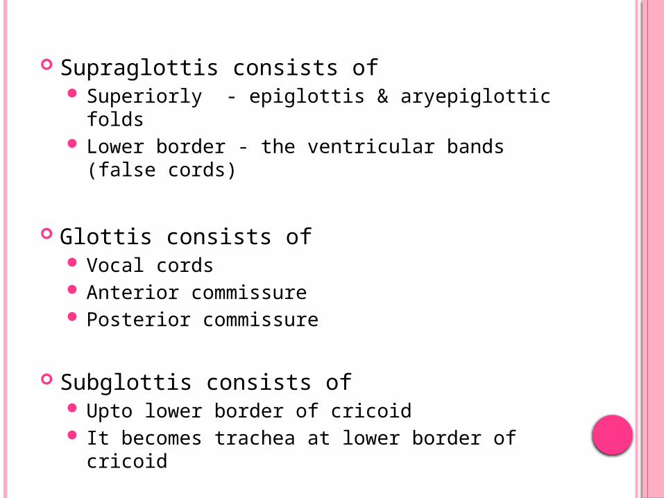
Supraglottis consists of Superiorly - epiglottis & aryepiglottic folds Lower border - the ventricular bands (false cords)
Glottis consists of Vocal cords Anterior commissure Posterior commissure
Subglottis consists of Upto lower border of cricoid It becomes trachea at lower border of cricoid
INFANT LARYNX
Position: Infant larynx is situated higher in the neck. Vocalcords lie at C3/C4 level and during swallowing go up to C1/ C2 level. In adults vocal cords lie at C5 level.
Cartilages: Laryngeal cartilages in infants are soft and collapse easily. Epiglottis: It is omega shaped. Arytenoids: They are relatively large and cover
significant posterior part of glottis. Thyroid: It is flat. Cricoid: The diameter of cricoid is smaller than
glottis.
Cricothyroid and thyrohyoid spaces: They are very narrow. Hyoid bone overlaps thyroid and thyroid overlaps cricoid.
Size: The larynx of an infant is smaller and has a narrower lumen
Shape: It is conical and funnel-shaped
Submucosal tissue: It is thick and loose and becomes easily edematous in response to trauma or inflammation

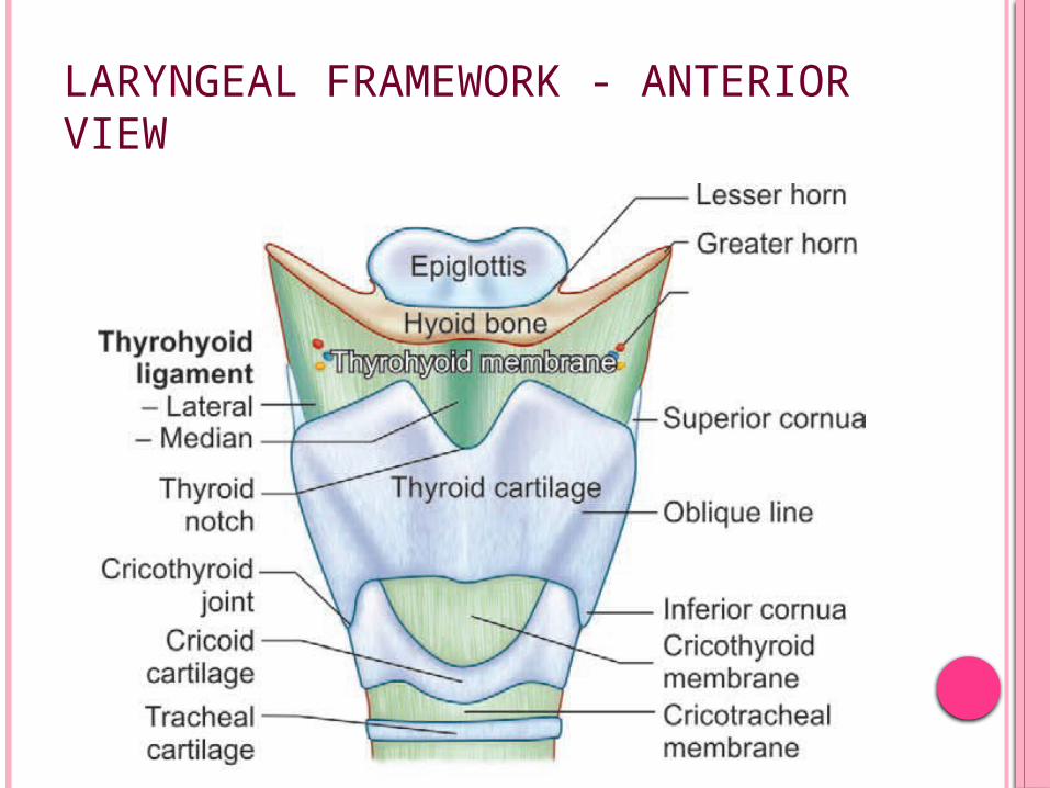
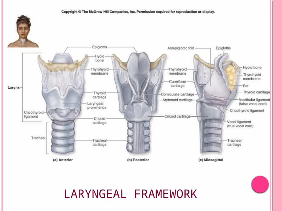
LARYNGEAL FRAMEWORK - ANTERIOR VIEW
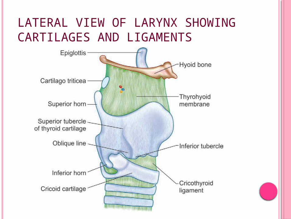
LATERAL VIEW OF LARYNX SHOWING CARTILAGES AND LIGAMENTS

POSTERIOR VIEW OF LARYNX SHOWINGCARTILAGES AND LIGAMENTS
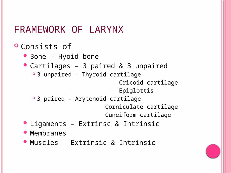
FRAMEWORK OF LARYNX
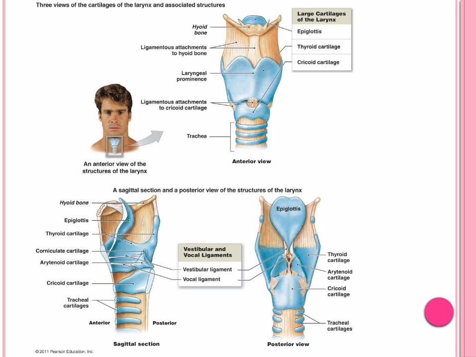
Consists of Bone – Hyoid bone Cartilages – 3 paired & 3 unpaired
3 unpaired – Thyroid cartilage Cricoid cartilage Epiglottis 3 paired – Arytenoid cartilage Corniculate cartilage Cuneiform cartilage
Ligaments – Extrinsc & Intrinsic Membranes Muscles – Extrinsic & Intrinsic
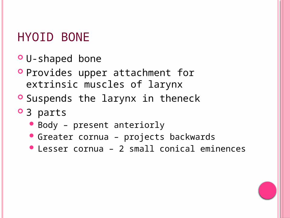
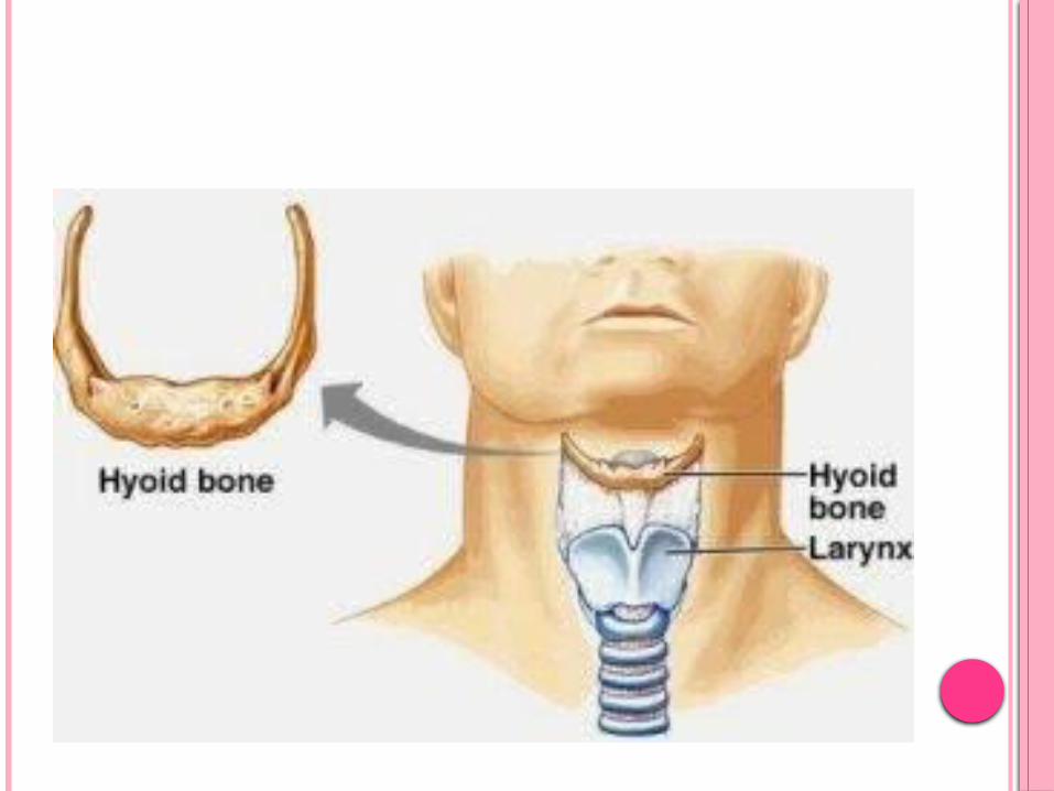
HYOID BONE
U-shaped bone Provides upper attachment for extrinsic
muscles of larynx Suspends the larynx in theneck 3 parts
Body – present anteriorly Greater cornua – projects backwards Lesser cornua – 2 small conical eminences
LARYNGEAL FRAMEWORK - ANTERIOR VIEW
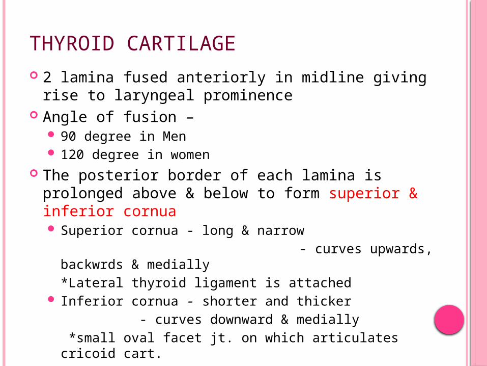
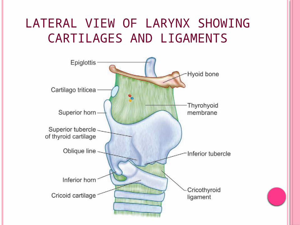
THYROID CARTILAGE 2 lamina fused anteriorly in midline giving rise to
laryngeal prominence Angle of fusion –
90 degree in Men 120 degree in women
The posterior border of each lamina is prolonged above & below to form superior & inferior cornua Superior cornua - long & narrow - curves upwards, backwrds &
medially*Lateral thyroid ligament is attached
Inferior cornua - shorter and thicker - curves downward & medially
*small oval facet jt. on which articulates cricoid cart.
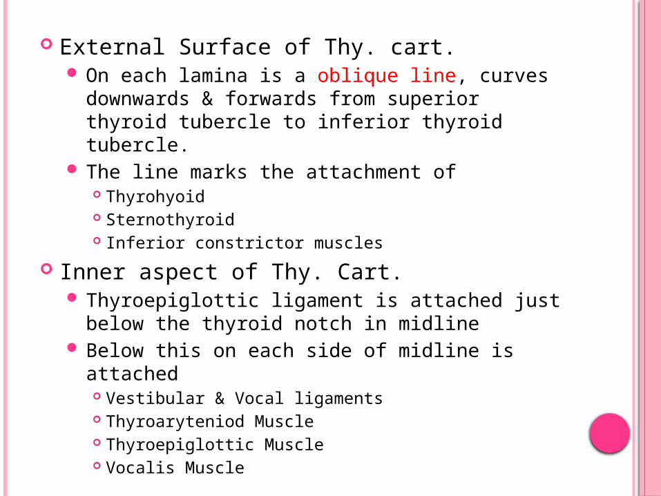
External Surface of Thy. cart. On each lamina is a oblique line, curves
downwards & forwards from superior thyroid tubercle to inferior thyroid tubercle.
The line marks the attachment of Thyrohyoid Sternothyroid Inferior constrictor muscles
Inner aspect of Thy. Cart. Thyroepiglottic ligament is attached just below
the thyroid notch in midline Below this on each side of midline is attached
Vestibular & Vocal ligaments Thyroaryteniod Muscle Thyroepiglottic Muscle Vocalis Muscle

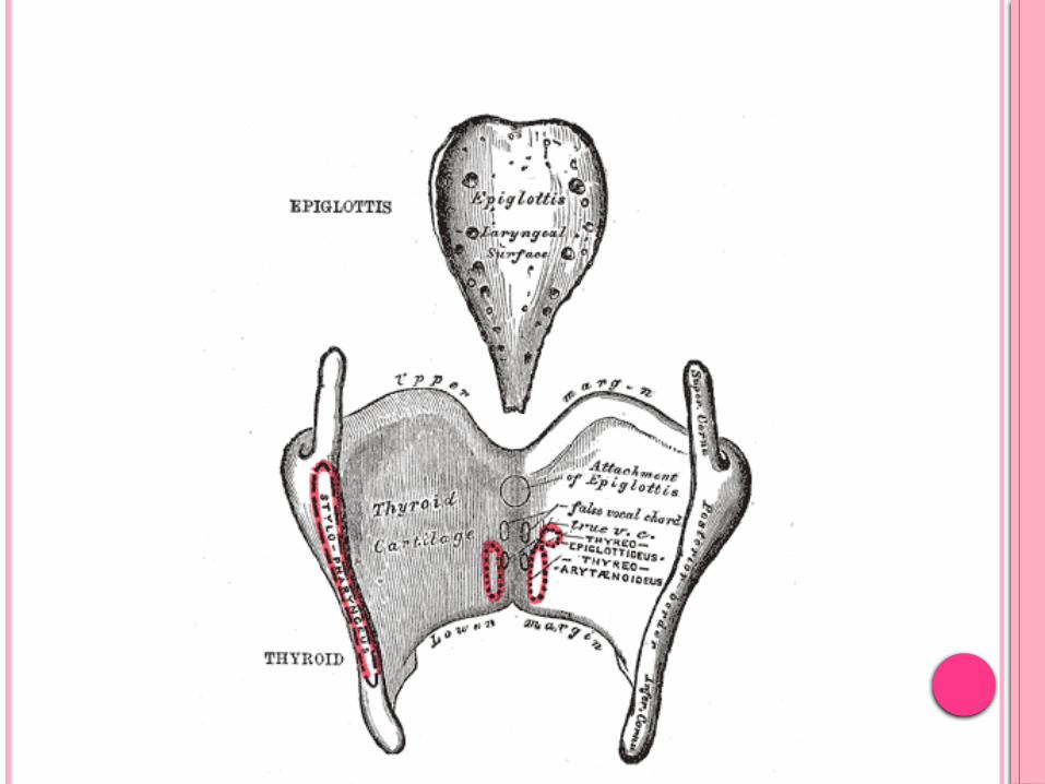
The fusion of the anterior ends of the two vocal ligaments produce Anterior commissure
The superior border of each lamina gives attachment to thyrohyoid ligament
The inferior border gives attachment to cricothyroid ligament
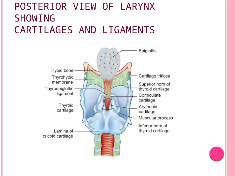
POSTERIOR VIEW OF LARYNX SHOWINGCARTILAGES AND LIGAMENTS
LARYNGEAL FRAMEWORK

CRICOID CARTILAGE
It is the only complete cartilagenous ring in the airway
It forms the inferior part of the larynx It has a deep broad lamina posteriorly &
narrow arch anteriorly with a facet for articulation with the inferior cornu of the thyroid cartilage
The lamina has sloping shoulders on which the articular facets for the arytenoids are found
A vertical ridge in midline of lamina give attachment to longitudinal muscle of oesophaus
Shallow concavity on each side gives origin to posterior cricoaryteniod
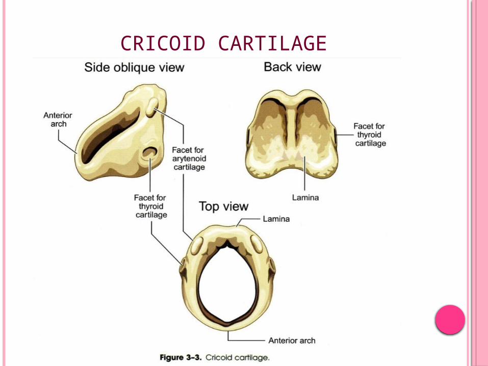
CRICOID CARTILAGE
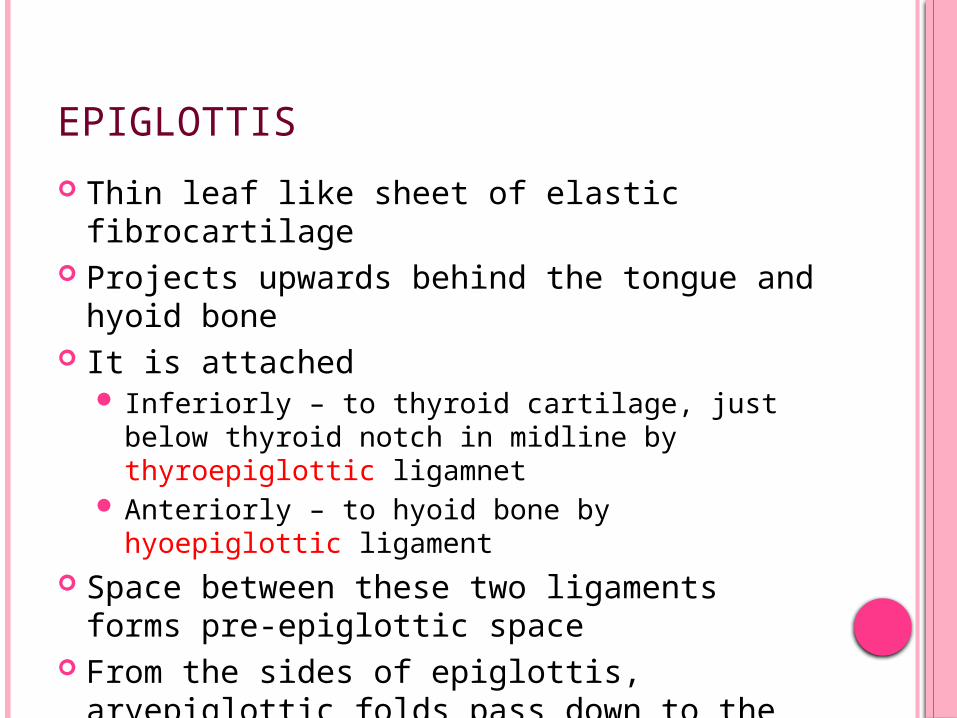
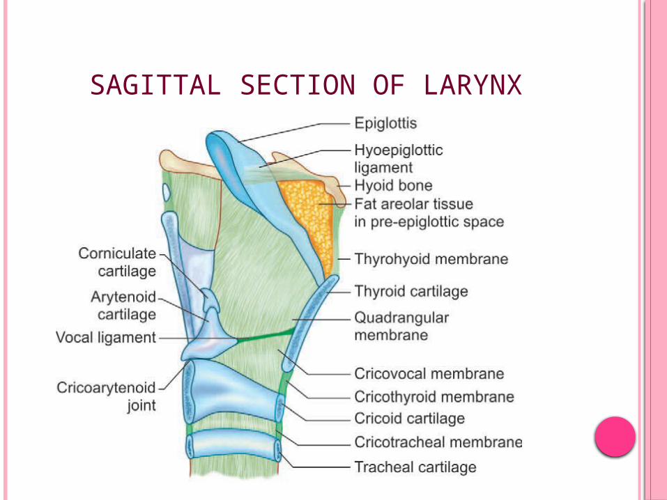
EPIGLOTTIS
Thin leaf like sheet of elastic fibrocartilage Projects upwards behind the tongue and
hyoid bone It is attached
Inferiorly – to thyroid cartilage, just below thyroid notch in midline by thyroepiglottic ligamnet
Anteriorly – to hyoid bone by hyoepiglottic ligament
Space between these two ligaments forms pre-epiglottic space
From the sides of epiglottis, aryepiglottic folds pass down to the apex of aryteniods
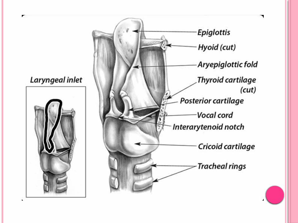
SAGITTAL SECTION OF LARYNX
The posterior surface of Epi. is indented by numerous small pits into which mucus glands project
The anterior surface is coverd by mucous membrane superiorly & forms the posterior wall of vallecula
The mucous membrane overlying epiglottis is reflected on base of tongue forming Glossoepiglottic fold – midline Lateral glossoepiglottic fold – laterally

EPIGLOTTIS

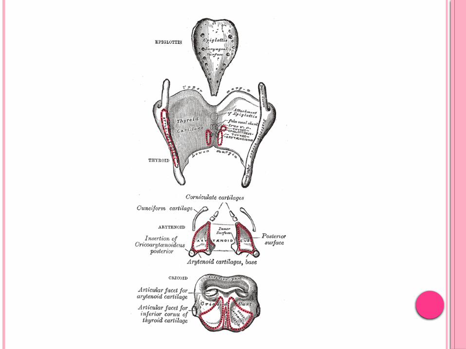
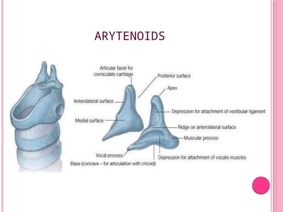
ARYTENOID CARTILAGES
Irregular, three sided pyramid with a
Forward projection – Vocal Process – attached Vocal folds
Lateral projection – Muscular process – attached posterior cricoarytenoid & lateral cricothyroid muscles
Anterolateral surface – is divided into two fossa by a crest from apex into Upper triangular fossa – attachment to Vestibular
ligament Lower triangular fossa – attch. to Vocalis &
Lateral cricoaryteniod muscle
ARYTENOIDS
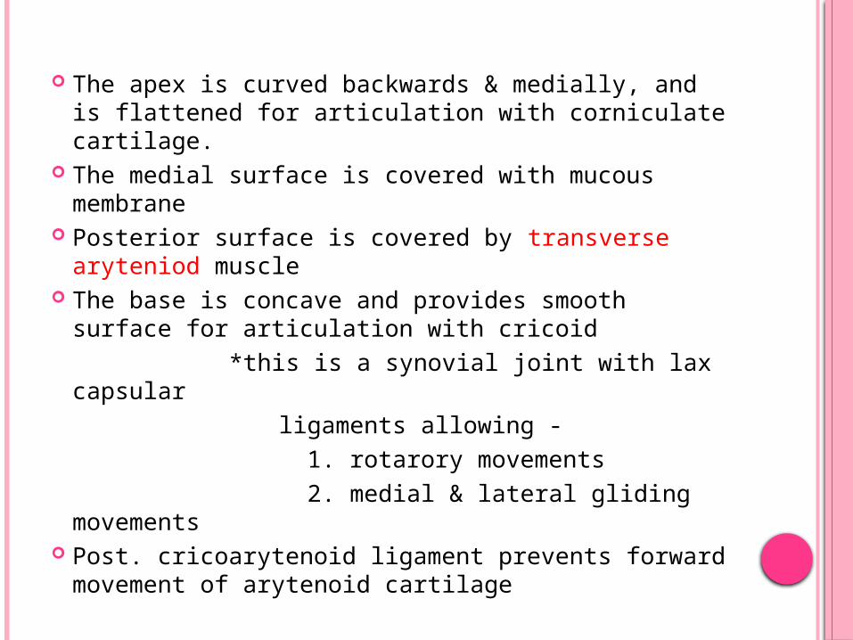
The apex is curved backwards & medially, and is flattened for articulation with corniculate cartilage.
The medial surface is covered with mucous membrane
Posterior surface is covered by transverse aryteniod muscle
The base is concave and provides smooth surface for articulation with cricoid *this is a synovial joint with lax capsular
ligaments allowing - 1. rotarory movements 2. medial & lateral gliding movements Post. cricoarytenoid ligament prevents forward
movement of arytenoid cartilage
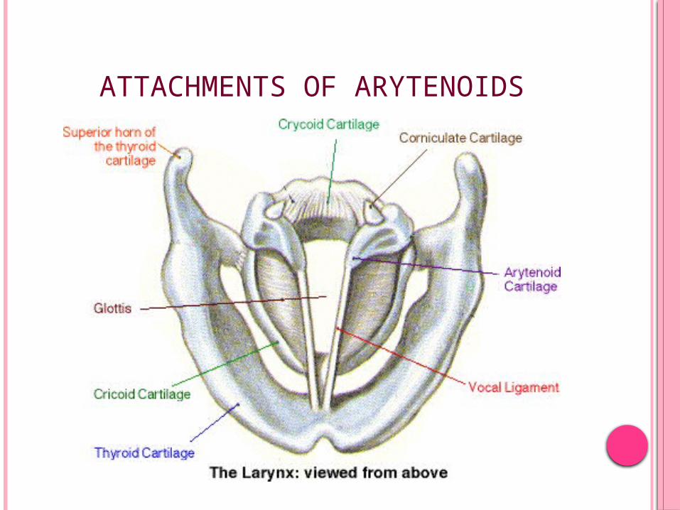
ATTACHMENTS OF ARYTENOIDS
CORNICULATE & CUNEIFORM CARTILAGES
Corniculate Cartilages (of Santorini) 2 small conical nodules of elastic fibrocatilage Articulate by a synovial joint with the apices of
aryteniod cartilages Situated in the posterior part of aryepiglottic fold
Cuneiform Cartilages (of Wrisberg) 2 small elongated flakes of fibroelastic cartilage
(rod shaped cart.) One in each margin of aryepiglottic folds
JOINTS
Cricoarytenoid joint: This synovial joint is formed between the base of
arytenoid and a facet on the upper border of cricoid lamina.
Two types of movements are possible at this joint; rotatory and gliding.
The rotatory movement occurs at a vertical axis and abducts or adducts the vocal cord.
Arytenoids glide laterally and medially and help in closing or opening the posterior part of glottis.
Cricothyroid joint: This synovial joint is formed between the inferior
cornua of thyroid cartilage and a facet on the cricoid cartilage.
LIGAMNETS & MEMBRANES
EXTRINSIC LIGAMENTS They connect laryngeal cartilages to hyoid bone
above & trachea below Superiorly – Thyrohyoid membrane stretches
between upper border of thyroid cartilage & posterior surface of the body & greater cornua of hyoid
The membrane is a fibroelastic tissue & is re-enforced by fibrous tissue in midline as median thyrohyoid ligament & posteriorly as lateral thyrohyoid ligament ( ligament often
contains a small nodule of cartilage – Cartilago Triticea) The membrane is pierced by Internal branch of Sup.
Laryngeal Nerve & Sup. Laryngeal Vessels Cricotracheal ligament unites lower border of cricoid
with first tracheal ring
INTRINSIC LIGAMENTS They
Connect the laryngeal cartilages together Strengthen the capsule of intercartilagenous joints Form a broad sheet of fibroelastic tissue – fibroelastic
membrane Fibroelastic Membrane is divided into upper &
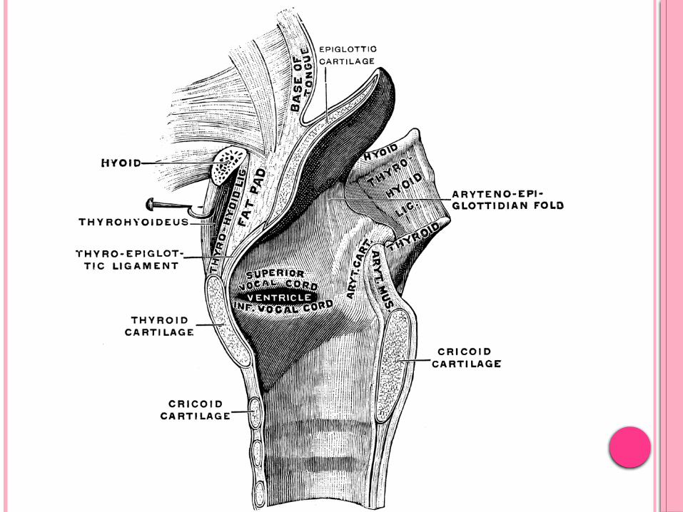
lower part by laryngeal ventricle Upper Quadilateral Membrane – extends
between lateral border of epiglottis & arytenoid cartilages Upper margin forms aryepiglottic fold Lower margin forms vestibular ligament underlying the
vestibular fold (false cords)

SAGITTAL SECTION OF LARYNX
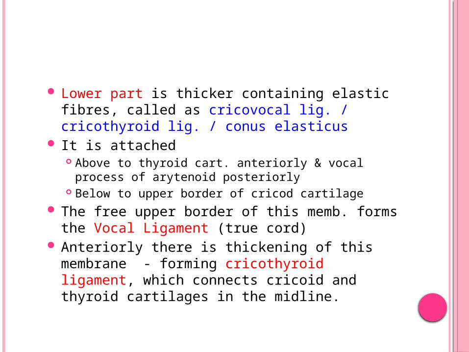
Lower part is thicker containing elastic fibres, called as cricovocal lig. / cricothyroid lig. / conus elasticus
It is attached Above to thyroid cart. anteriorly & vocal process of
arytenoid posteriorly Below to upper border of cricod cartilage
The free upper border of this memb. forms the Vocal Ligament (true cord)
Anteriorly there is thickening of this membrane - forming cricothyroid ligament, which connects cricoid and thyroid cartilages in the midline.
LATERAL VIEW OF LARYNX SHOWING CARTILAGES AND LIGAMENTS
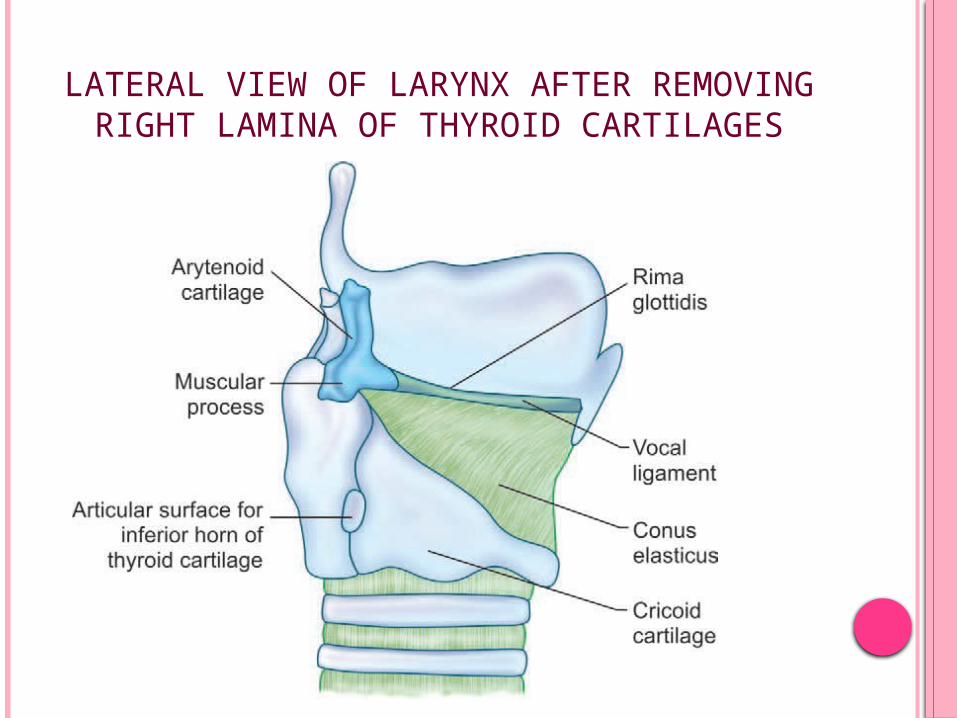
LATERAL VIEW OF LARYNX AFTER REMOVING RIGHT LAMINA OF THYROID CARTILAGES
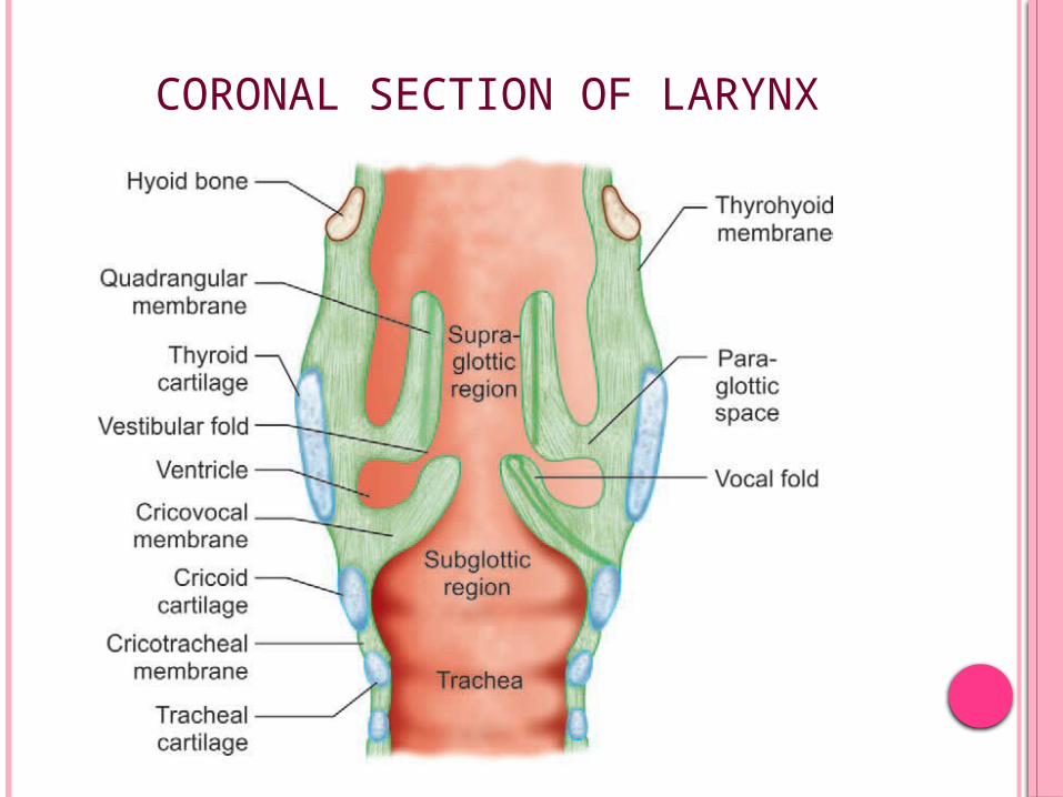
CORONAL SECTION OF LARYNX
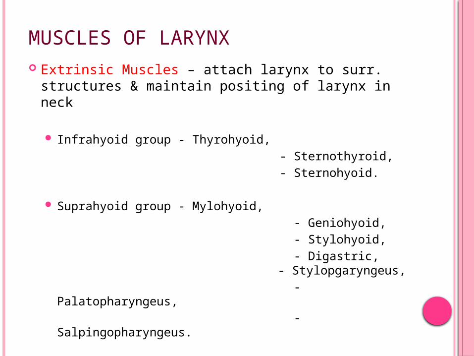
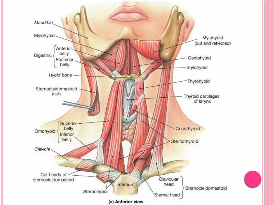
MUSCLES OF LARYNX Extrinsic Muscles – attach larynx to surr.
structures & maintain positing of larynx in neck
Infrahyoid group - Thyrohyoid, - Sternothyroid, - Sternohyoid.
Suprahyoid group - Mylohyoid, - Geniohyoid, - Stylohyoid, - Digastric,
- Stylopgaryngeus, - Palatopharyngeus, - Salpingopharyngeus.
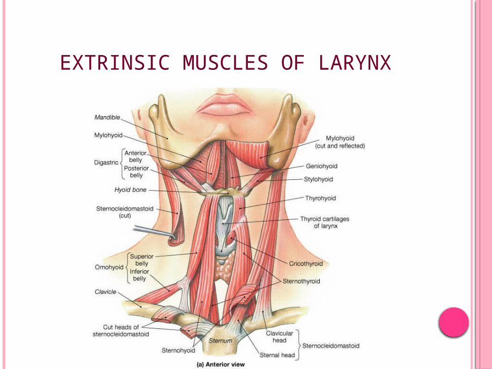
EXTRINSIC MUSCLES OF LARYNX
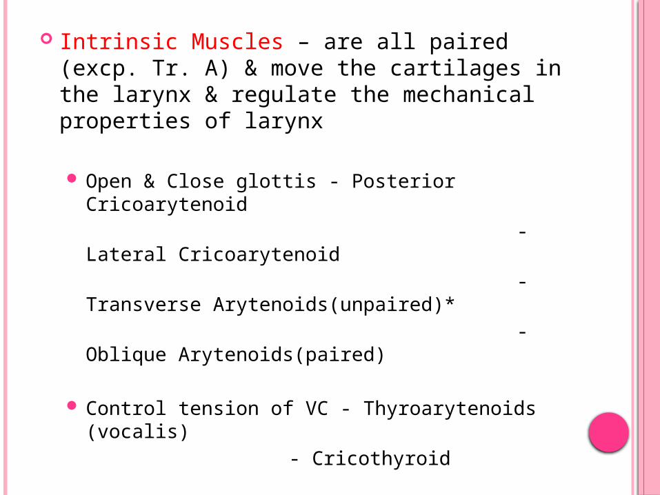
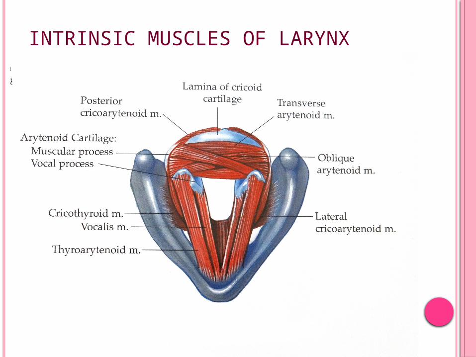
Intrinsic Muscles – are all paired (excp. Tr. A) & move the cartilages in the larynx & regulate the mechanical properties of larynx
Open & Close glottis - Posterior Cricoarytenoid - Lateral Cricoarytenoid - Transverse
Arytenoids(unpaired)* - Oblique Arytenoids(paired)
Control tension of VC - Thyroarytenoids (vocalis) - Cricothyroid
Alter the shape of laryngeal inlet - Aryepiglotticus
- Thyroepiglotticus
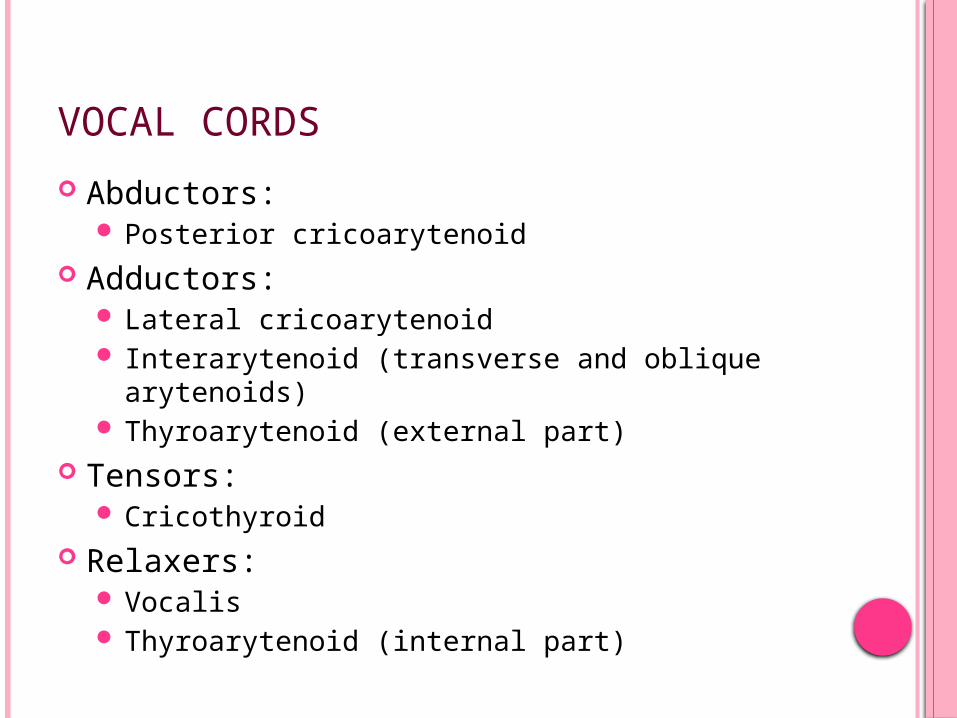
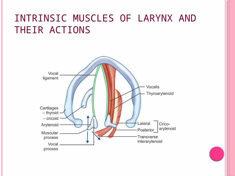
VOCAL CORDS
Abductors: Posterior cricoarytenoid
Adductors: Lateral cricoarytenoid Interarytenoid (transverse and oblique
arytenoids) Thyroarytenoid (external part)
Tensors: Cricothyroid
Relaxers: Vocalis Thyroarytenoid (internal part)
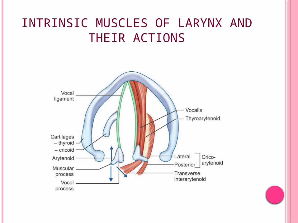
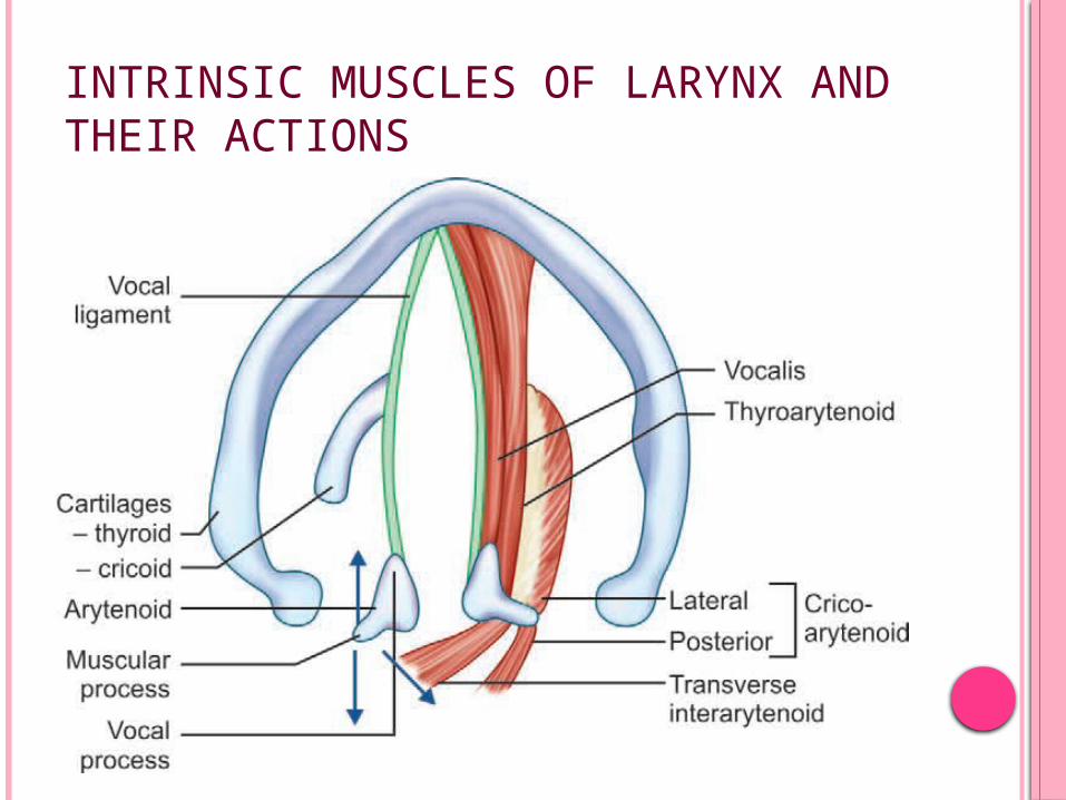
INTRINSIC MUSCLES OF LARYNX AND THEIR ACTIONS
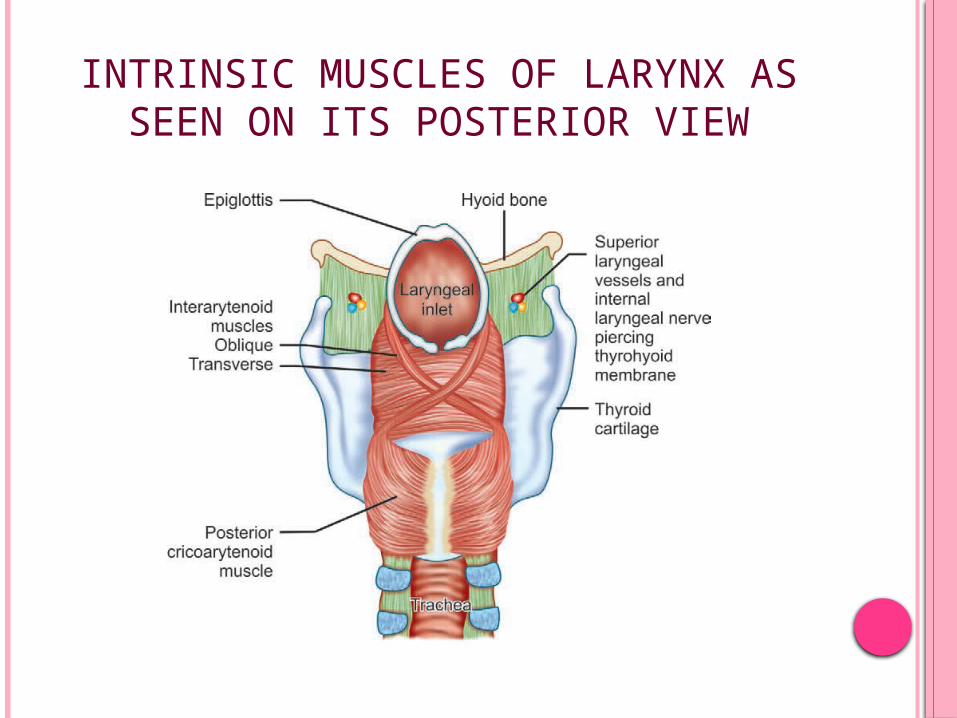
INTRINSIC MUSCLES OF LARYNX AS SEEN ON ITS POSTERIOR VIEW
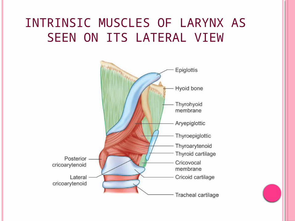
INTRINSIC MUSCLES OF LARYNX AS SEEN ON ITS LATERAL VIEW
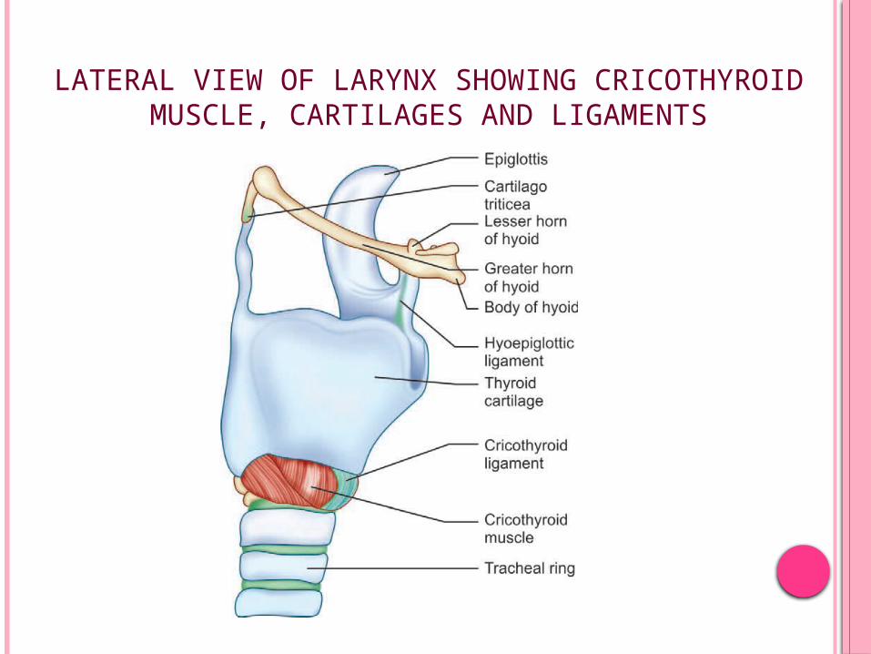
LATERAL VIEW OF LARYNX SHOWING CRICOTHYROID MUSCLE, CARTILAGES AND
LIGAMENTS
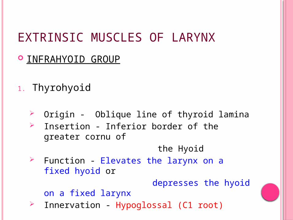
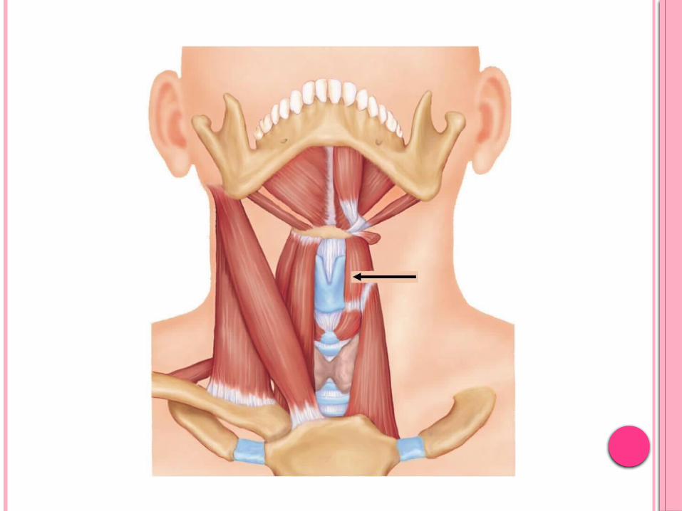
EXTRINSIC MUSCLES OF LARYNX
INFRAHYOID GROUP
1. Thyrohyoid
Origin - Oblique line of thyroid lamina Insertion - Inferior border of the greater cornu
of the Hyoid Function - Elevates the larynx on a fixed hyoid
or depresses the hyoid on a fixed
larynx Innervation - Hypoglossal (C1 root)
2. Sternothyroid
Origin - Posterior surface of manubrium and edge of the first costal cartilage Insertion - Oblique line of the thyroid lamina Function - Depresses the larynx Innervation - Ansa cervicalis (C2, 3 roots)
3. Sternohyoid
Origin - Clavicle and posterior surface of the manubrium
Insertion - Lower edge of the body of the hyoid Function - Depresses the larynx by lowering the
hyoid Innervation - Ansa cervicalis (C1, 2, 3 roots)

SUPRAHYOID GROUP
1. Mylohyoid
Origin – Mylohyoid line in inner aspect of mandible
Insertion – Midline raphe & body of hyoid Function – raises & pulls hyoid anteriorly Innervation – Nr. to Mylohyoid
2. Geniohyoid
Origin – Genial tubercle on mandible Insertion – upper border of the body of hyoid Function – raises & pulls the hyoid forwards Innervation – Hypoglossal (C1 root)

3. Stylohyoid Origin – back of the styloid process Insertion – base of greater cornu of the hyoid Function – retractor & elevator of hyoid for swallowing Innervation – facial nerve
4. Digastric Origin – Digastric notch on the medial surface of the mastoid process Insertion – Lower border of the mandible (a fibrous
sling holds the tendon to the lesser cornu of
hyoid) Function – Anterior belly – pulls the hyoid anteriorly & up Posterior belly – pulls the hyoid posteriorly & up Innervation - Ant. belly – Nr to mylohyoid Post. belly – Facial Nr
5. Stylopharyngeus Origin – Medial aspect of styloid process Insertion – Post. border of lamina of thyroid cart. Function – Elevates the larynx Innervation – Glossopharyngeal Nr
6. Palatopharyngeus Origin – Palatine aponeurosis & post margin of hard
palate Insertion – Post. border of thyroid alar & cornua Function – helps tilts the larynx forwards Innervation – Accessory Nr (pharyngeal plexus)
7. Salpingopharyngeus Origin – Eustachian Tube Insertion – Post. border of thyroid cartilage Function – Elevates the larynx Innervation – Pharyngeal plexus
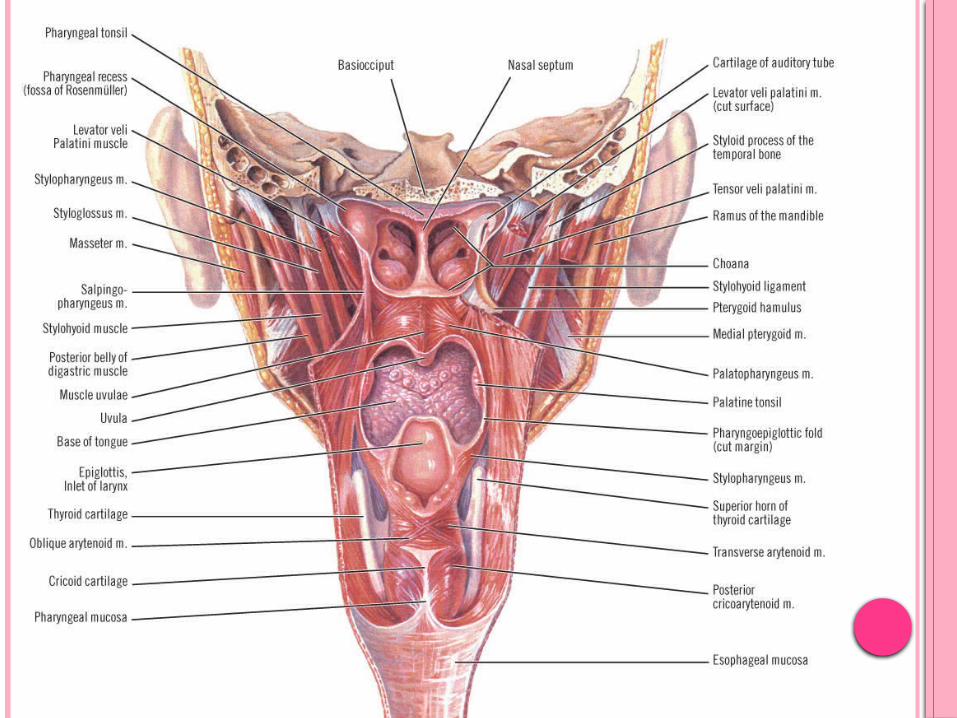

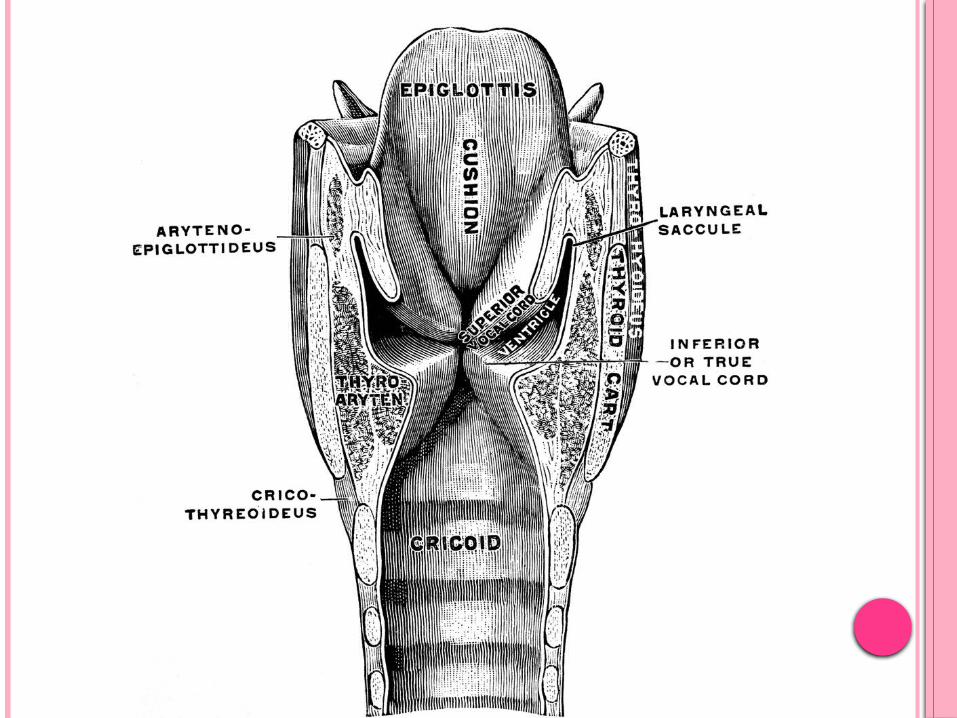
SUPERIOR VIEW OF THE INSIDE OF LARYNX AS SEEN DURING LARYNGOSCOPIC EXAMINATION
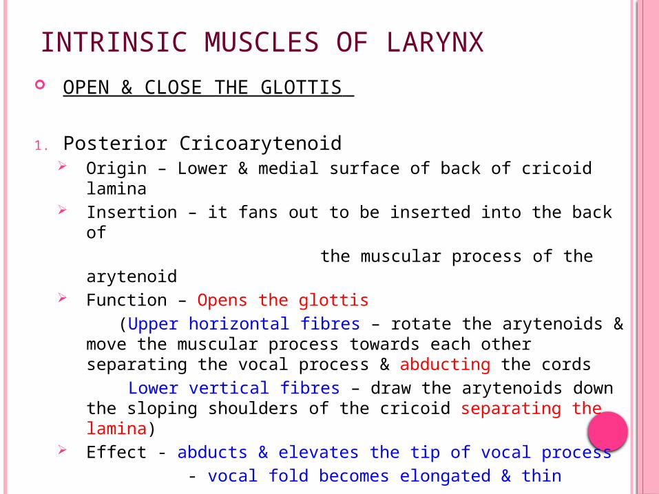
INTRINSIC MUSCLES OF LARYNX OPEN & CLOSE THE GLOTTIS
1. Posterior Cricoarytenoid Origin – Lower & medial surface of back of cricoid
lamina Insertion – it fans out to be inserted into the back of the muscular process of the arytenoid Function – Opens the glottis (Upper horizontal fibres – rotate the arytenoids &
move the muscular process towards each other separating the vocal process & abducting the cords
Lower vertical fibres – draw the arytenoids down the sloping shoulders of the cricoid separating the lamina)
Effect - abducts & elevates the tip of vocal process - vocal fold becomes elongated & thin

INTRINSIC MUSCLES OF LARYNX

INTRINSIC MUSCLES OF LARYNX AND THEIR ACTIONS
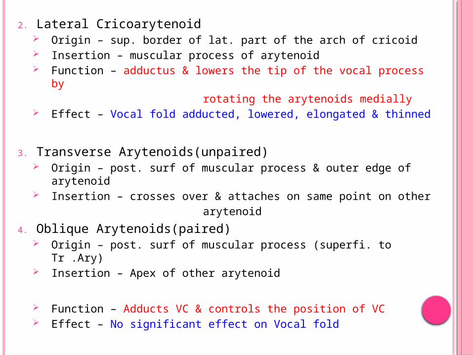
2. Lateral Cricoarytenoid Origin – sup. border of lat. part of the arch of cricoid Insertion – muscular process of arytenoid Function – adductus & lowers the tip of the vocal process
by rotating the arytenoids medially Effect – Vocal fold adducted, lowered, elongated & thinned
3. Transverse Arytenoids(unpaired) Origin – post. surf of muscular process & outer edge of
arytenoid Insertion – crosses over & attaches on same point on other arytenoid
4. Oblique Arytenoids(paired) Origin – post. surf of muscular process (superfi. to Tr .Ary) Insertion – Apex of other arytenoid
Function – Adducts VC & controls the position of VC Effect – No significant effect on Vocal fold

INTRINSIC MUSCLES OF LARYNX
INTRINSIC MUSCLES OF LARYNX AND THEIR ACTIONS
CONTROL THE TENSION OF VOCAL FOLDS
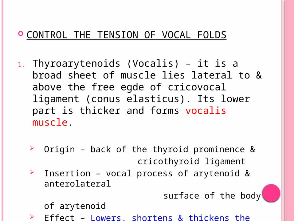
1. Thyroarytenoids (Vocalis) – it is a broad sheet of muscle lies lateral to & above the free egde of cricovocal ligament (conus elasticus). Its lower part is thicker and forms vocalis muscle.
Origin – back of the thyroid prominence & cricothyroid ligament Insertion – vocal process of arytenoid &
anterolateral surface of the body of arytenoid Effect – Lowers, shortens & thickens the vocal folds
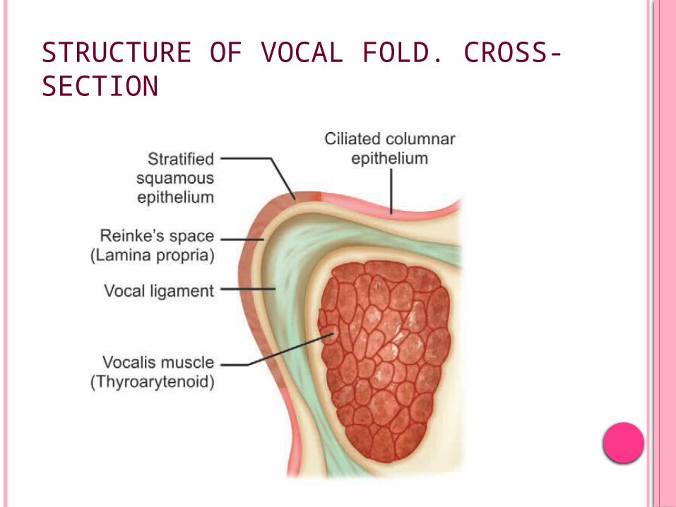
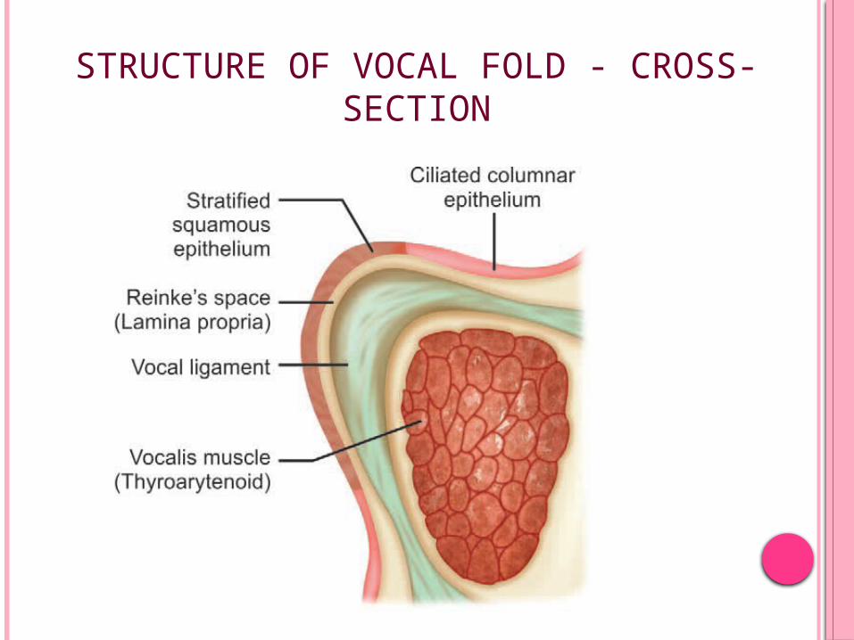
STRUCTURE OF VOCAL FOLD. CROSS-SECTION
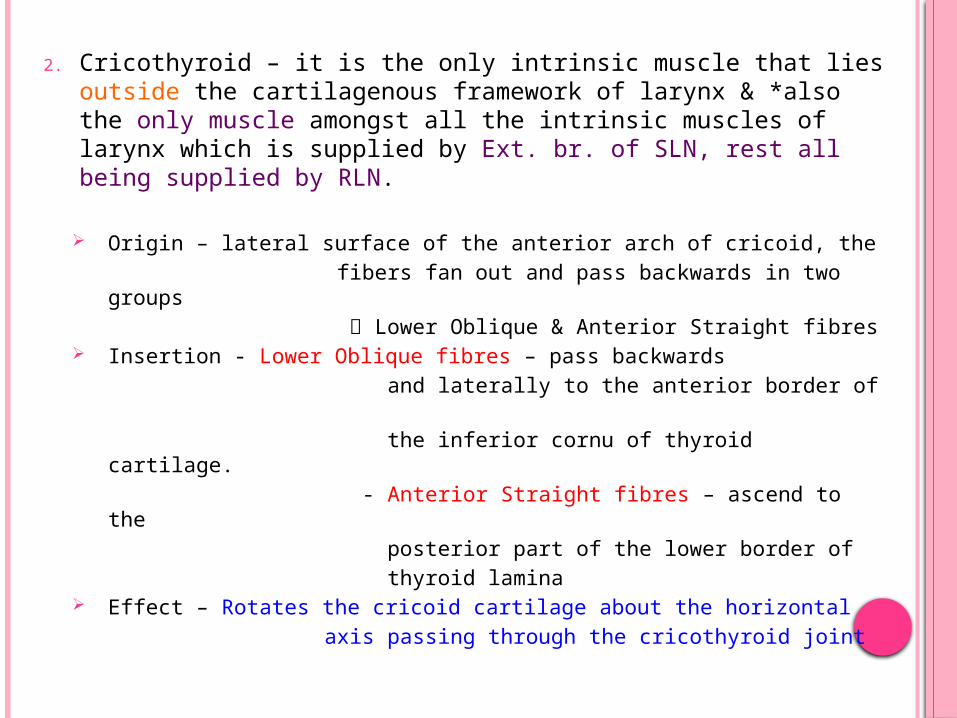
2. Cricothyroid – it is the only intrinsic muscle that lies outside the cartilagenous framework of larynx & *also the only muscle amongst all the intrinsic muscles of larynx which is supplied by Ext. br. of SLN, rest all being supplied by RLN.
Origin – lateral surface of the anterior arch of cricoid, the fibers fan out and pass backwards in two
groups Lower Oblique & Anterior Straight fibres Insertion - Lower Oblique fibres – pass backwards and laterally to the anterior border of the inferior cornu of thyroid cartilage. - Anterior Straight fibres – ascend to the posterior part of the lower border of thyroid lamina Effect – Rotates the cricoid cartilage about the horizontal axis passing through the cricothyroid joint
INTRINSIC MUSCLES OF LARYNX
INTRINSIC MUSCLES OF LARYNX AND THEIR ACTIONS
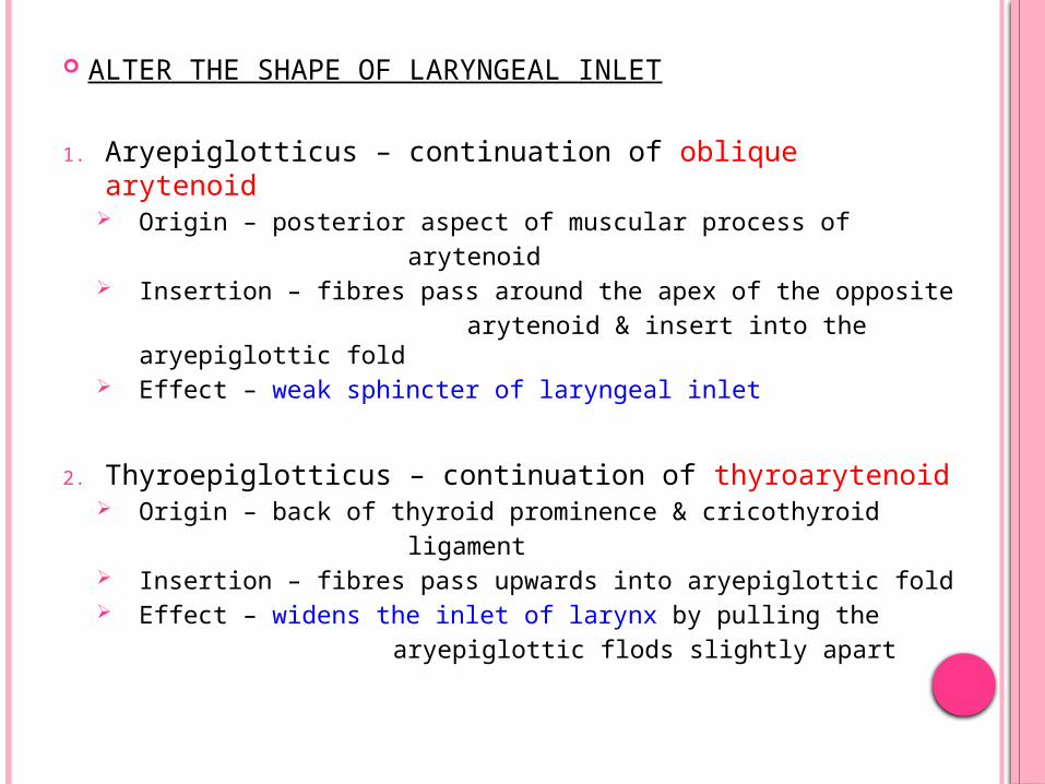
ALTER THE SHAPE OF LARYNGEAL INLET
1. Aryepiglotticus – continuation of oblique arytenoid
Origin – posterior aspect of muscular process of arytenoid Insertion – fibres pass around the apex of the opposite arytenoid & insert into the aryepiglottic
fold Effect – weak sphincter of laryngeal inlet
2. Thyroepiglotticus – continuation of thyroarytenoid Origin – back of thyroid prominence & cricothyroid ligament Insertion – fibres pass upwards into aryepiglottic fold Effect – widens the inlet of larynx by pulling the aryepiglottic flods slightly apart
THE GLOTTIS
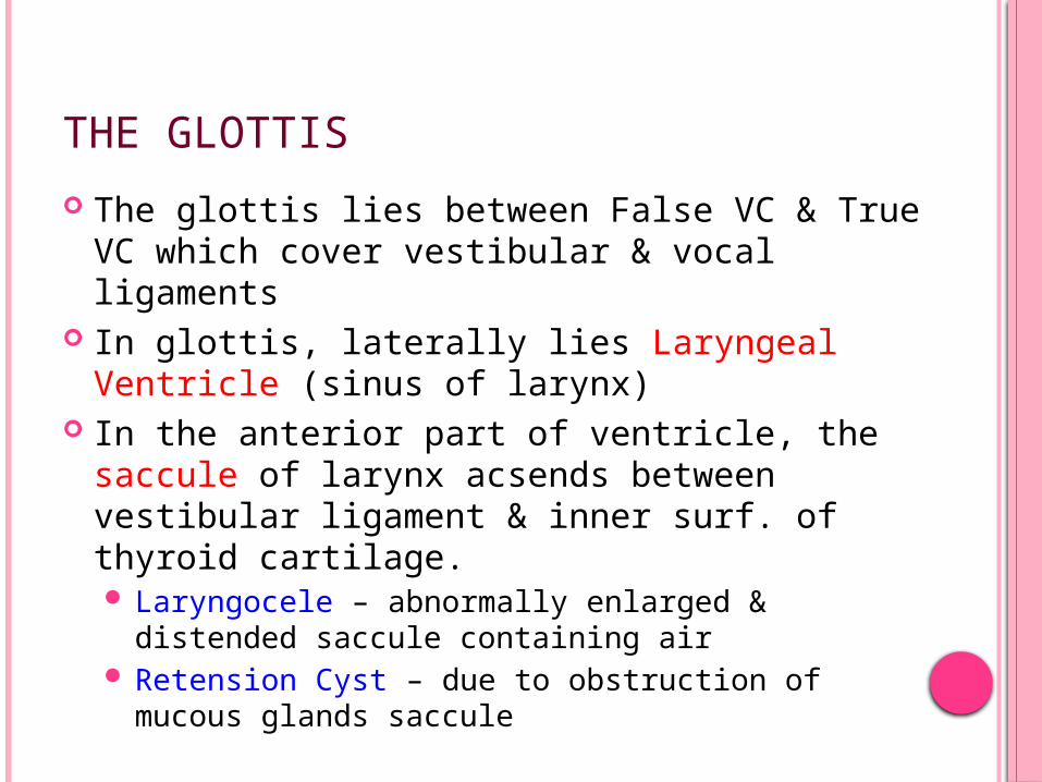
The glottis lies between False VC & True VC which cover vestibular & vocal ligaments
In glottis, laterally lies Laryngeal Ventricle (sinus of larynx)
In the anterior part of ventricle, the saccule of larynx acsends between vestibular ligament & inner surf. of thyroid cartilage. Laryngocele – abnormally enlarged & distended
saccule containing air Retension Cyst – due to obstruction of mucous
glands saccule
The glottis (RIMA GLOTTIDIS) is the narrowest part of adult laryngeal cavity & lies between VC & arytenoids on two sides
The size & shape of glottis varies with the activites of VCs
Vestibular Folds – 2 thick folds of mucous membrane enclosing
vestibular ligament VL is the lower border of upper quadilateral
membrane It is fixed – in Front – at angle of thyroid cartilage just below attch. of epiglottic cart. Behind – anterolateral surface of
arytenoids
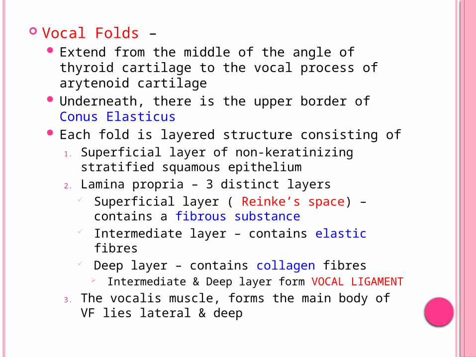
Vocal Folds – Extend from the middle of the angle of thyroid
cartilage to the vocal process of arytenoid cartilage
Underneath, there is the upper border of Conus Elasticus
Each fold is layered structure consisting of 1. Superficial layer of non-keratinizing stratified
squamous epithelium2. Lamina propria – 3 distinct layers
Superficial layer ( Reinke’s space) – contains a fibrous substance
Intermediate layer – contains elastic fibres Deep layer – contains collagen fibres
Intermediate & Deep layer form VOCAL LIGAMENT
3. The vocalis muscle, forms the main body of VF lies lateral & deep
STRUCTURE OF VOCAL FOLD - CROSS-SECTION
The layered structure of vocal fold is not uniform in its entire length.
Anterior end of VF lies a mass of collagen fibres which are connected to inner perichondrium of thyroid cartilage & to deep layer of lamina propria posteriorly
Adjacent to this mass of collagen fibres, posteriorly, lies a mass of elastic fibres continuous with intermediate layer of LP, called Anterior Macula Flava. A similar structure at posterior end of membranous part of VF
These serve as cushions to protect the ends of vocal folds from mechanical damage caused by vocal fold vibration.
Anterior 3/5th of VC is between vocal folds – called Intermembranous part of cord
Remaining 2/5th posteriorly are between vocal process of arytenoid – called Intercartilagenous part of cord
MUCOUS MEMBRANES OF LARYNX
The m.m. lining is closely attached over the posterior surface of epiglottis, corniculate & cuniform cartilages and all over the vocal ligament.
Elsewhere it is loosely attached & prone to edema
Most of larynx* is lined by pseudo-stratified cilliated columnar ‘respiratory-type’ epithelium
* The upper half of posterior surface of epiglottis, the upper part of aryepiglottic fold, posterior glottis & vocal folds are covered with non-keratinizing stratified squamous epithelium.
Mucous glands are freely distributed throughout the mucous membrane & at particularly numerous on the posterior half of epiglottis where they form indentation into the cartilage & in the margins of the lower part of the aryepiglottic folds and saccules.
The vocal folds do not posses any glands & the mucous membrane is lubricated by mucus from the glands of the saccules.
* if these glands cease to function, i.e. after radiation the the sq. epithelium of vocal cords tend to dessicate.
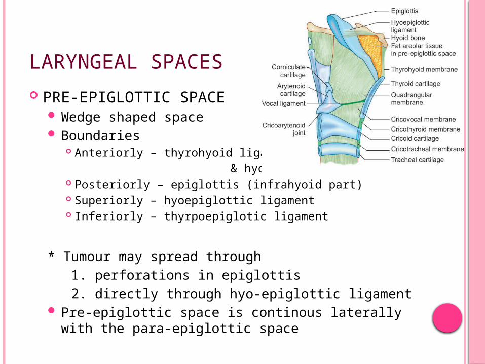
LARYNGEAL SPACES
PRE-EPIGLOTTIC SPACE Wedge shaped space Boundaries
Anteriorly – thyrohyoid ligament & hyoid bone Posteriorly – epiglottis (infrahyoid part) Superiorly – hyoepiglottic ligament Inferiorly – thyrpoepiglotic ligament
* Tumour may spread through 1. perforations in epiglottis 2. directly through hyo-epiglottic ligament Pre-epiglottic space is continous laterally with
the para-epiglottic space
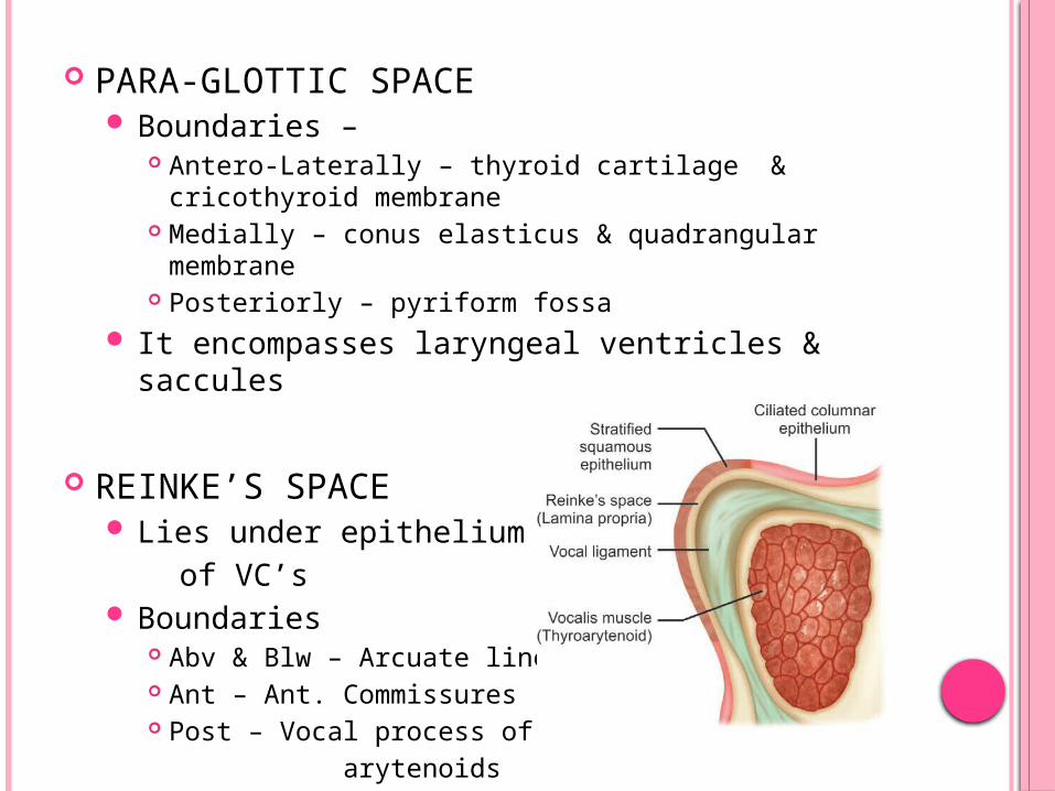
PARA-GLOTTIC SPACE Boundaries –
Antero-Laterally – thyroid cartilage & cricothyroid membrane
Medially – conus elasticus & quadrangular membrane Posteriorly – pyriform fossa
It encompasses laryngeal ventricles & saccules
REINKE’S SPACE Lies under epithelium of VC’s Boundaries
Abv & Blw – Arcuate lines Ant – Ant. Commissures Post – Vocal process of
arytenoids
NERVE SUPPLY OF LARYNX
The motor and sensory supply of larynx is from VAGUS – by superior & recurrent laryngeal nr.s
SUPERIOR LARYNGEAL NERVE Arises from inferior ganglion of vagus & also
receives a branch from superior cervical sympathetic ganglion.
It decends lateral to pharynx behind ICA & at the level of greater horn of hyoid it divides into small external branch & larger internal branch
EXTERNAL BRANCH – motor supply to cricothyroid muscle
INTERNAL BRANCH – pierces thyrohyoid memb. & divides into two main sensory & secretomotor br., & also carries afferent fibres from neuromuscular spindles & other stretch receptors of larynxUpper branch – supplies mucous memb. of lower
part of pharynx, epiglottis, vallecula & vestibule of larynx
Lower branch – supplies aryepiglottic fold & mucous membrane of larynx till level of vocal cords
In its course beneath m.m. of medial wall of pyriform fossa, it is accessible for inj. of LA for providing anaesthesia for most of pyriform fossa.
SLN ends by piercing inf. constricter muscle of pharynx & unites with the ascending br of recurrent laryngeal nerve. – called as Galen’s anastomosis
RECURRENT LARYNGEAL NERVE RIGHT RLN – leaves vagus as it crosses Right sub-
clavian artery & loops under the artery ascending in the TE groove to reach larynx
LEFT RLN – the nerve originates from vagus as it crosses aortic arch, it passes under the arch & ligamentum arteriosum to reach TE groove
In the NECK – both follow same course and pass upwards accompanied by laryngeal branch of inferior thyroid artery
They pass deep to the lower border of inf. constricter muscle & enter the larynx behind cricothyroid joint.
Then divides into motor & sensory branches MOTOR BR – all intrinsic muscles of larynx, except
cricothyroid SENSORY BR – supplies laryngeal mucosa below the
level of vocal cords + aff. fibers from stretch receptors of larynx

NERVES SUPPLYING THE LARYNX AND THEIR RELATIONSESPECIALLY WITH ARTERIES
The relationship between RLN & inferior thyroid art. is variable
It may cross in front of, or behind the artery or may pass between the terminal branches of artery
On the Rt side there is equal chance of the nerve lying in any of three locations but on the Lt side it usually lies posteriorly to artery.
LARYNGEAL VASCULATURE
ARTERIAL SUPPLY Laryngeeal branches of superior & inferior thyroid
arteries Cricothyroid br of superior thyroid artery
The superior laryngeal artery arises from superior thyroid artery – passes deep to thyrohyiod muscle. Together with the int. br of SLN it pierces thyrohyiod memb. to supply larynx
The inferior laryngeal artery arises from inferior thyroid artery at lower border of thyroid gl. And ascends on the traches with RLN. It enters the larynx beneath the lower border of inf constricter to supply it.
The cricothyroid artery passes upper part of cricothyroid ligament to supply larynx.
VENOUS DRAINAGE Accompany arteries
Superior laryngeal vein superior thyroid vein / facial vein IJV
Inferior laryngeal veins inferior thyroid veins bracheocephalic vein
* some veins middle thyroid vein IJV
LYMPHATICS Divided into two groups by vocal folds into upper
& lower drainage LARYNX ABOVE VOCAL FOLDS – drain by vessels
accompanying SL vein Upper deep cervical LNs LARYNX BELOW VOCAL FOLDS prelaryngeal &
pretracheal nodes Lower deep cervical nodes The vocal folds have no lymphatics as they are
firmly bound down to underlying vocal ligament

NERVES SUPPLYING THE LARYNX AND THEIR RELATIONSESPECIALLY WITH ARTERIES
FUNCTIONS OF LARYNX
4 main funtions of larynx -
1. Protection of lower airways Sphincteric closure of laryngeal inlet Cessation of respiration Cough reflex
2. Phonation & speech3. Respiration4. Fixation of chest
LASTLY...
Its that part of our body which helps us to communicate verbally with the whole world...!
From the first cry of the baby to the sweet tunes of a melodius song...!!
Its all about LARYNX...!!!
THANK YOU...
REFERENCES GRAY’s Anatomy - 39th Ed. Scott Brown’s Otorhinolaryngology & Head and Neck Surgery – 7th
Ed. Cumming’s Otolaryngology & Head and Neck Surgery - 5th Ed. Mohan Bansal – 2nd Ed. BD Chaurasia’s – Human Anatomy 3rd Ed.