Embed Size (px)
Citation preview
ArticulationsArticulationsArticulations
Dr. Carmen E. RexachAnatomy 35
Mt. San Antonio College
Classification of joints
• What is an articulation?• Classification Criteria
– How bones are joined together– Degree of mobility
• Minimum components– 2 articulating bones– Intervening tissue
• Fibrous CT or cartilage
Classifications based on structure• Fibrous
– No joint cavity– Ends of bones joined by dense regular CT– Ex: tooth to alveoli of mandible
• Cartilaginous– No joint cavity– Cartilaginous pad between ends of bones– Ex: pubic symphysis
• Synovial– Joint cavity – Articular cartilage caps ends of bones– Synovial fluid-filled joint capsule lined by
synovial membrane– Ex: glenohumeral joint

Categories based on degree of movement
• Synarthrosis– Immovable joint
• Amphiarthrosis– Slightly movable
• Diarthrosis– Freely movable
Least amount of movement
Greatest amount of movement
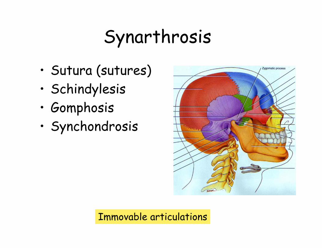
Synarthrosis
• Sutura (sutures) • Schindylesis• Gomphosis• Synchondrosis
Immovable articulations
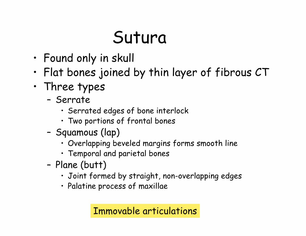
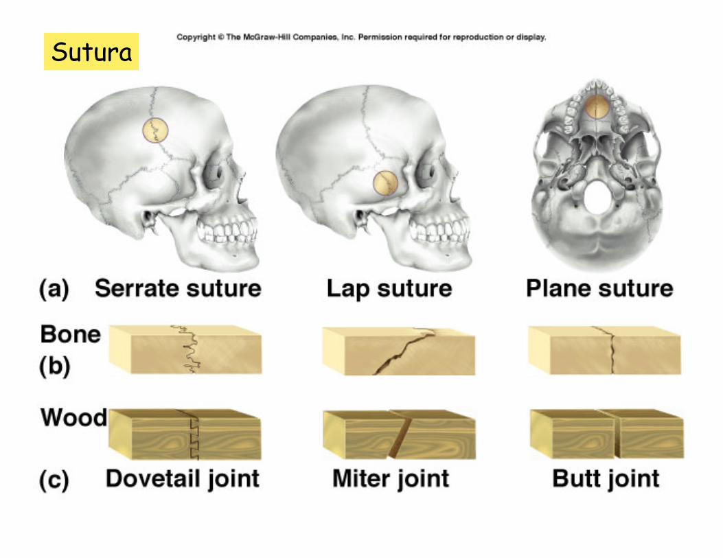
Sutura• Found only in skull• Flat bones joined by thin layer of fibrous CT• Three types
– Serrate• Serrated edges of bone interlock• Two portions of frontal bones
– Squamous (lap)• Overlapping beveled margins forms smooth line• Temporal and parietal bones
– Plane (butt)• Joint formed by straight, non-overlapping edges • Palatine process of maxillae
Immovable articulations
Sutura

Schindylesis• “Web and groove joint”• Thin plate of bone into cleft
or fissure in a separation of the laminae in another bone
• Ex) articulation of sphenoid bone and perpendicular plate of ethmoid bone with vomer
Immovable articulations
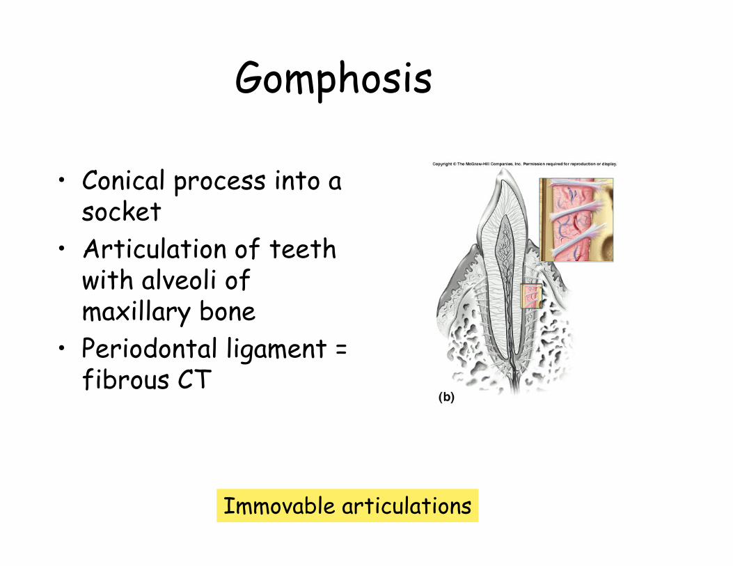
Gomphosis
• Conical process into a socket
• Articulation of teeth with alveoli of maxillary bone
• Periodontal ligament = fibrous CT
Immovable articulations
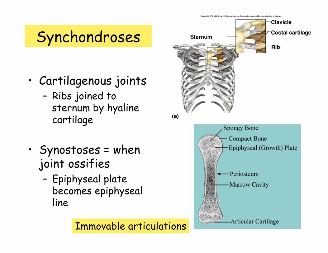
Synchondroses
• Cartilagenous joints– Ribs joined to
sternum by hyaline cartilage
• Synostoses = when joint ossifies– Epiphyseal plate
becomes epiphysealline
Immovable articulations

Amphiarthrosis
• Articulating bones connected in one of two ways:– By broad flattened fibro-cartilage discs
• Hyaline cartilage + collagen fibers• Symphysis
– By interosseus ligaments• Syndesmosis
Slightly moveable articulations
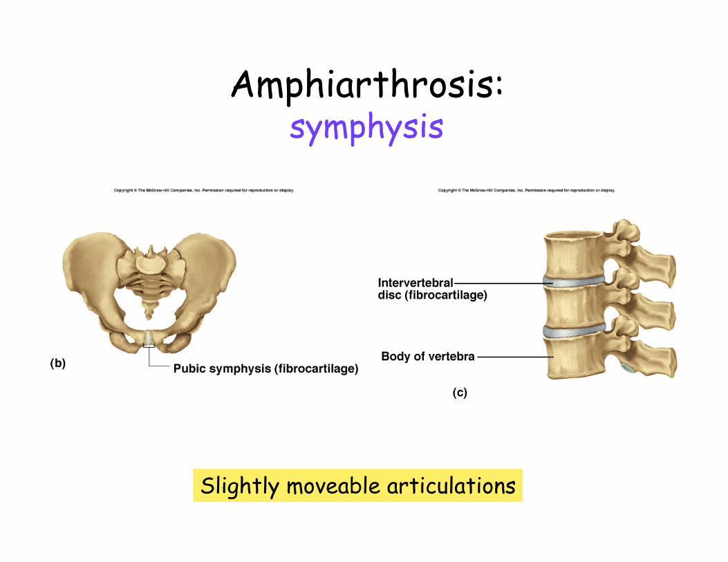
Amphiarthrosis:symphysis
Slightly moveable articulations
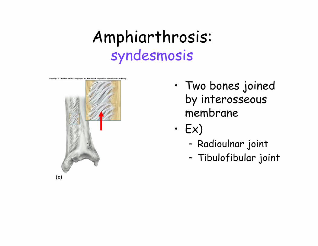
Amphiarthrosis:syndesmosis
• Two bones joined by interosseousmembrane
• Ex)– Radioulnar joint– Tibulofibular joint

Diarthrosis
• Synovial joints– Freely movable– Types
• Hinge • Gliding • Pivot • Saddle • Condyloid• Ball and socket
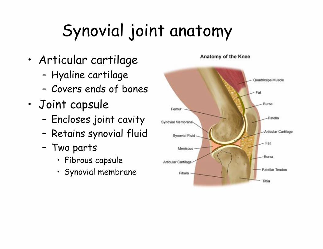
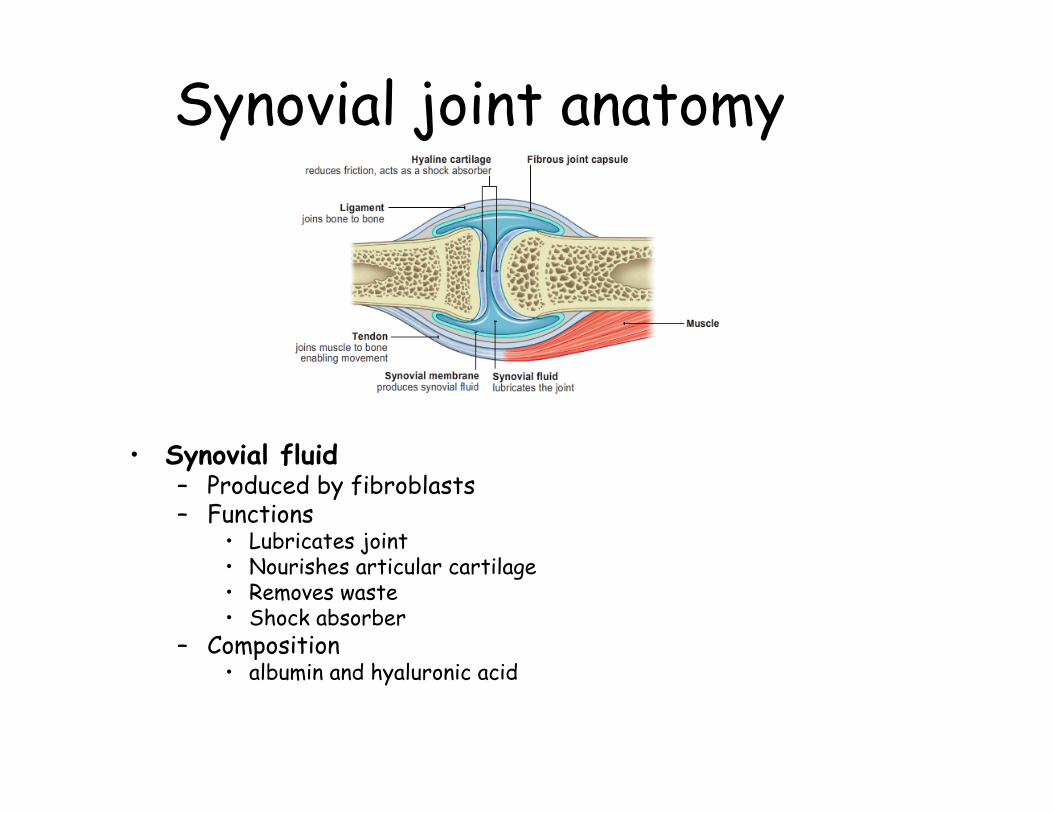
Synovial joint anatomy• Articular cartilage
– Hyaline cartilage – Covers ends of bones
• Joint capsule– Encloses joint cavity– Retains synovial fluid– Two parts
• Fibrous capsule• Synovial membrane
Synovial joint anatomy
• Synovial fluid– Produced by fibroblasts – Functions
• Lubricates joint• Nourishes articular cartilage• Removes waste• Shock absorber
– Composition• albumin and hyaluronic acid
Synovial joint anatomy
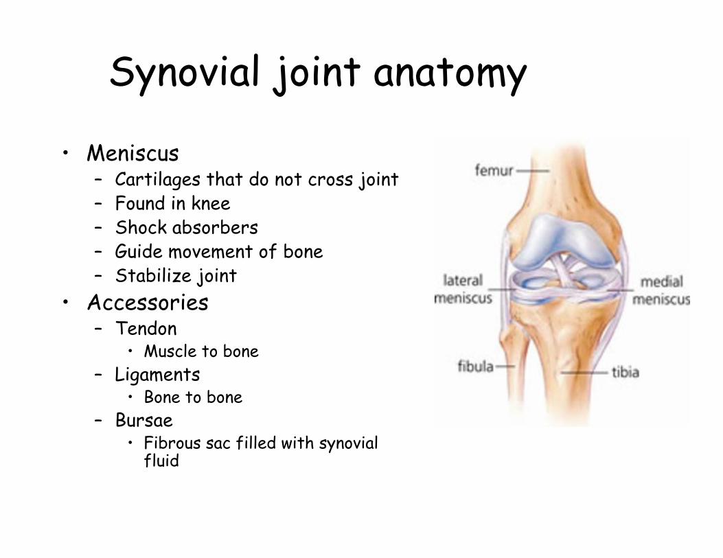
• Meniscus– Cartilages that do not cross joint– Found in knee– Shock absorbers– Guide movement of bone– Stabilize joint
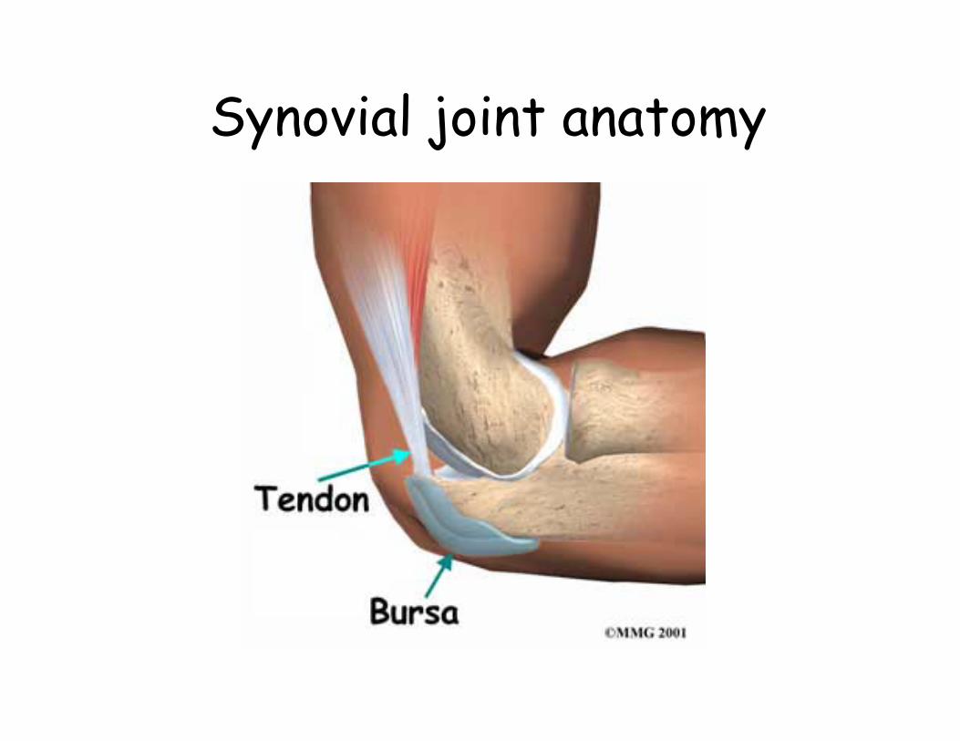
• Accessories– Tendon
• Muscle to bone– Ligaments
• Bone to bone– Bursae
• Fibrous sac filled with synovial fluid
Synovial joint anatomy
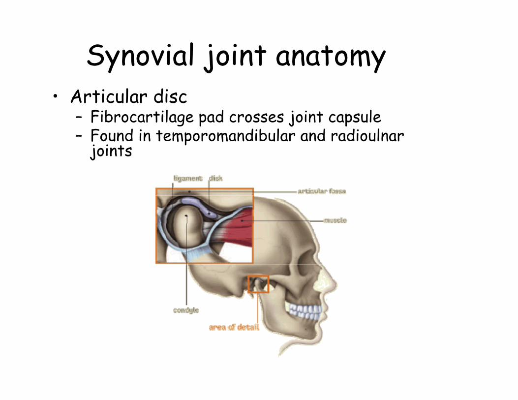
Synovial joint anatomy• Articular disc
– Fibrocartilage pad crosses joint capsule– Found in temporomandibular and radioulnar
joints

Diarthroses:Hinge joints
• Articulation between concave and convex surfaces
• monaxial– movement in only 1
plane• knee, elbow,
between phalanges
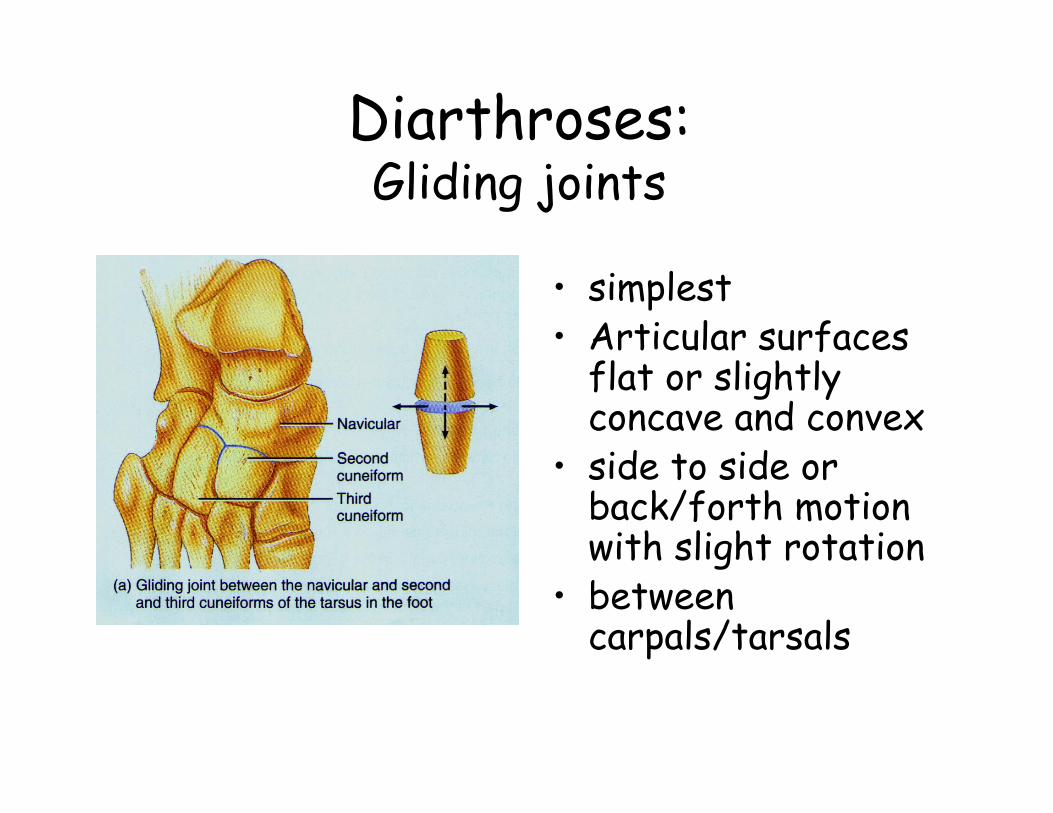
Diarthroses:Gliding joints
• simplest• Articular surfaces
flat or slightly concave and convex
• side to side or back/forth motion with slight rotation
• between carpals/tarsals
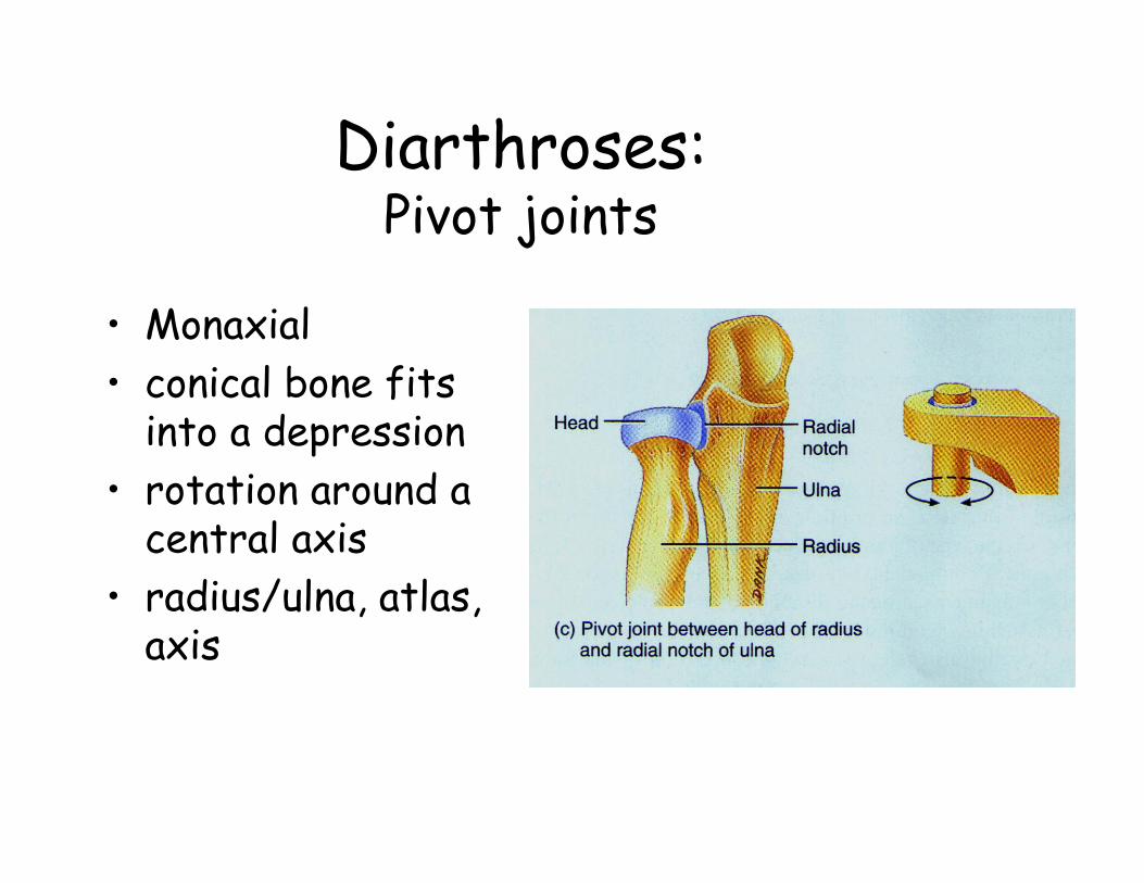
Diarthroses:Pivot joints
• Monaxial• conical bone fits
into a depression• rotation around a
central axis• radius/ulna, atlas,
axis
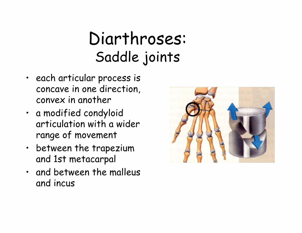
Diarthroses:Saddle joints
• each articular process is concave in one direction, convex in another
• a modified condyloidarticulation with a wider range of movement
• between the trapezium and 1st metacarpal
• and between the malleusand incus
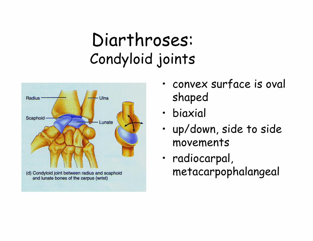
Diarthroses:Condyloid joints
• convex surface is oval shaped
• biaxial• up/down, side to side
movements• radiocarpal,
metacarpophalangeal
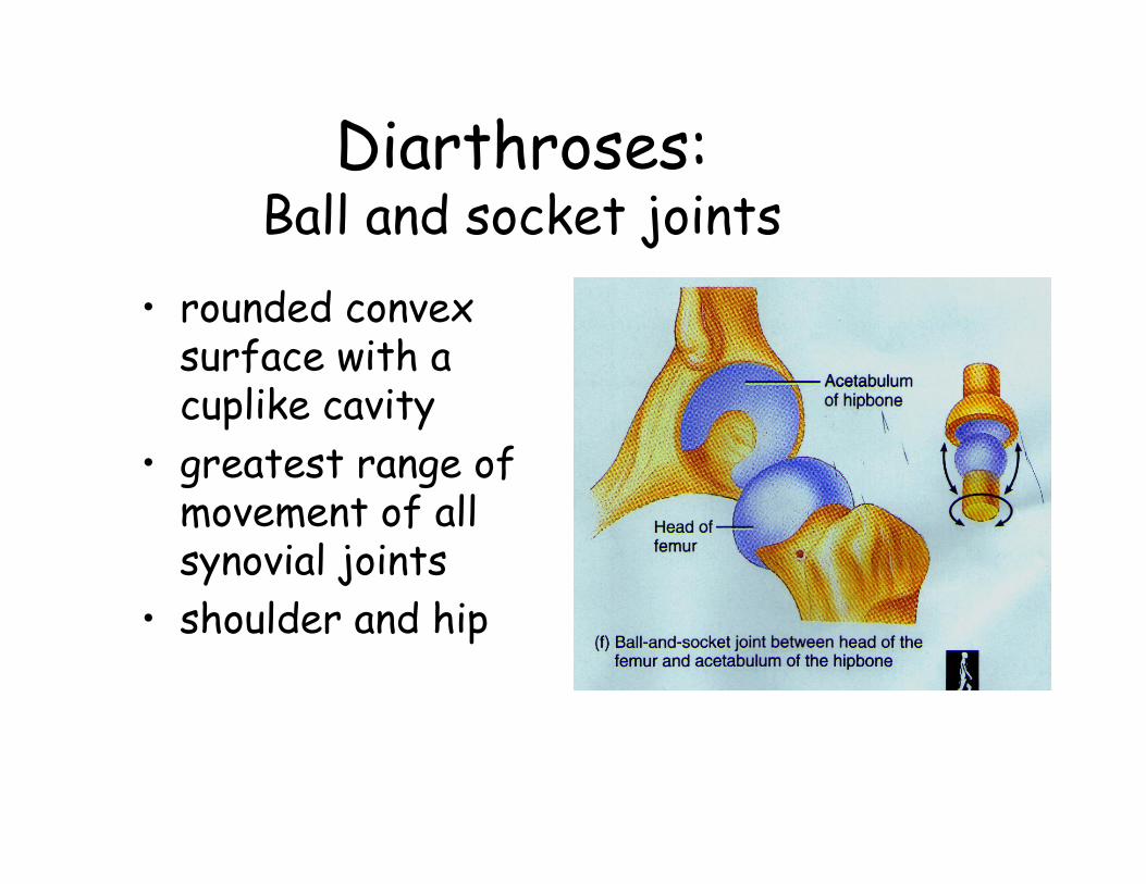
Diarthroses:Ball and socket joints
• rounded convex surface with a cuplike cavity
• greatest range of movement of all synovial joints
• shoulder and hip
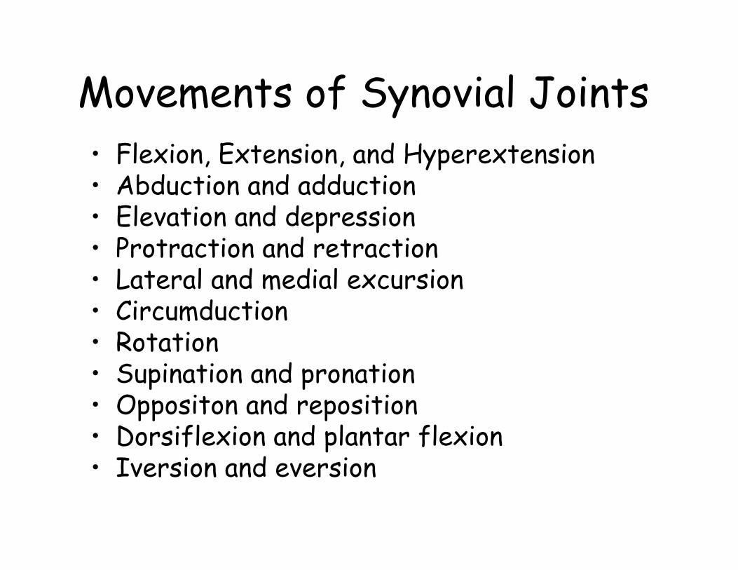
Movements of Synovial Joints• Flexion, Extension, and Hyperextension• Abduction and adduction• Elevation and depression• Protraction and retraction• Lateral and medial excursion• Circumduction• Rotation• Supination and pronation• Oppositon and reposition• Dorsiflexion and plantar flexion• Iversion and eversion
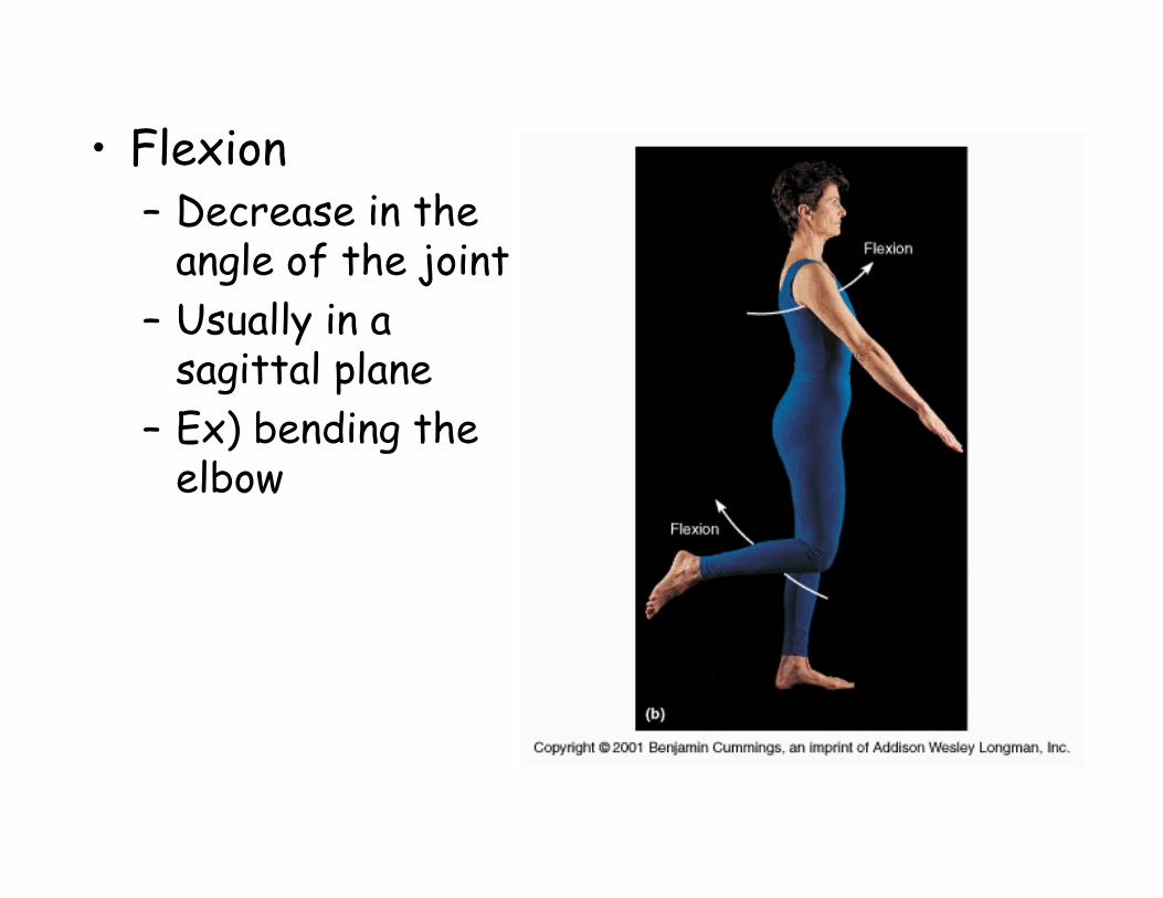
• Flexion– Decrease in the
angle of the joint– Usually in a
sagittal plane– Ex) bending the
elbow
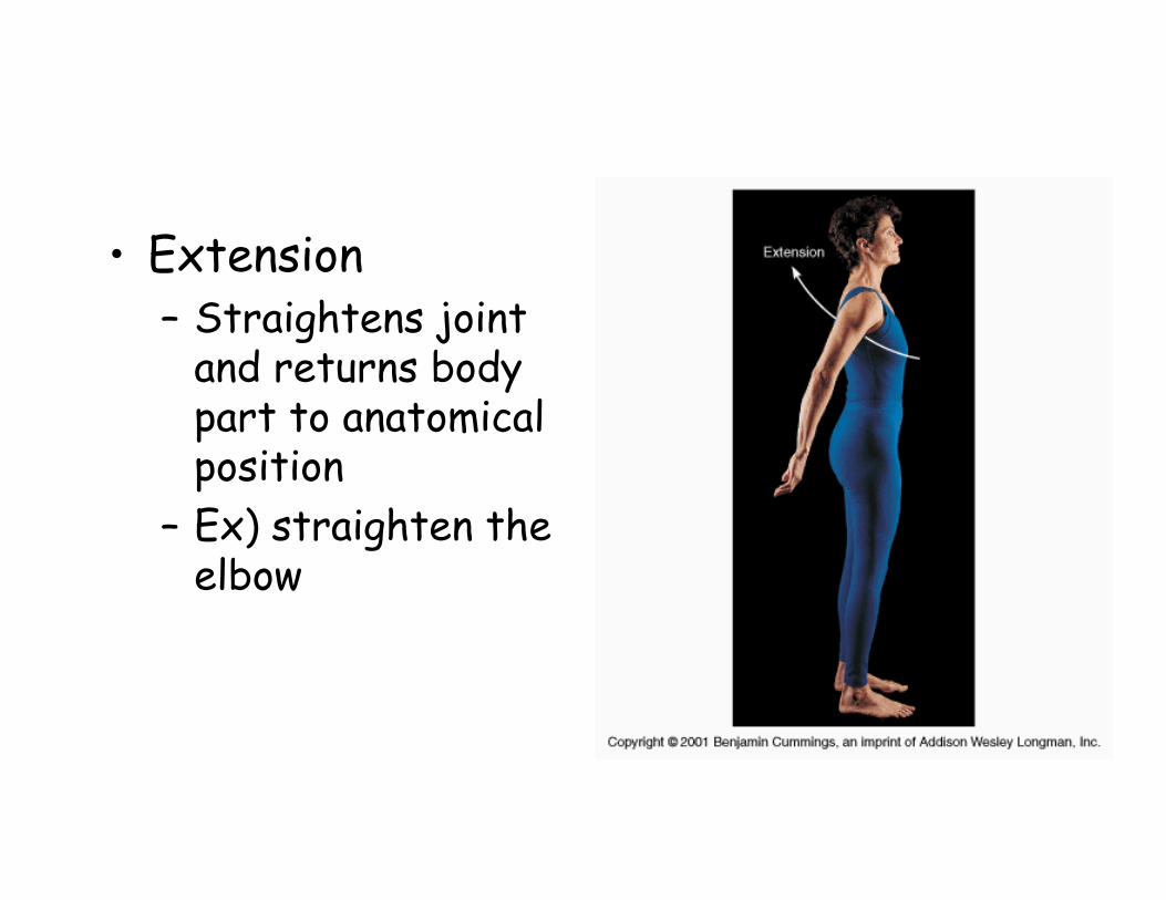
• Extension– Straightens joint
and returns body part to anatomical position
– Ex) straighten the elbow

• Hyperextension– Extension of joint beyond 180o
– Ex) looking upward at the ceiling
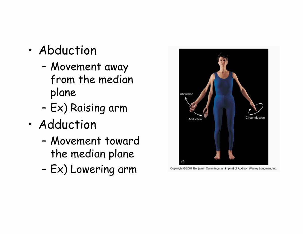
• Abduction– Movement away
from the median plane
– Ex) Raising arm• Adduction
– Movement toward the median plane
– Ex) Lowering arm

• Elevation– Movement that raises bone vertically– Ex) shrugging shoulders
• Depression– Movement that lowers bone vertically– Ex) lowering mandible to open mouth
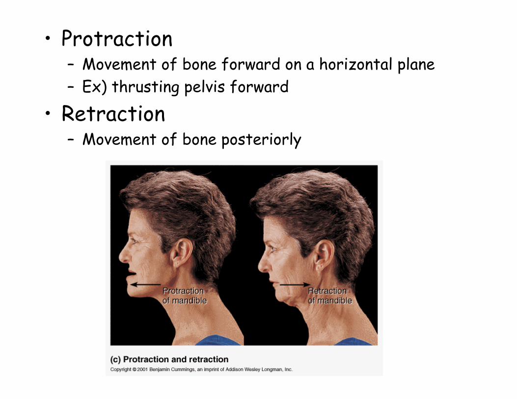
• Protraction– Movement of bone forward on a horizontal plane– Ex) thrusting pelvis forward
• Retraction– Movement of bone posteriorly
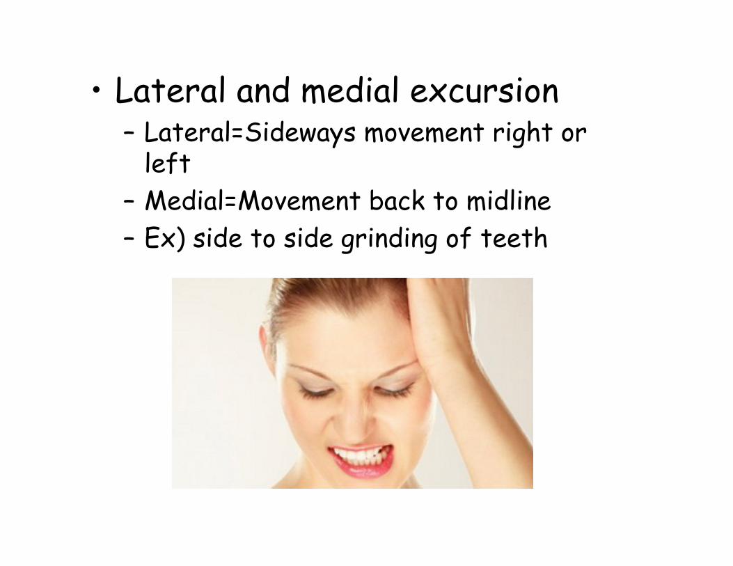
• Lateral and medial excursion– Lateral=Sideways movement right or
left– Medial=Movement back to midline– Ex) side to side grinding of teeth
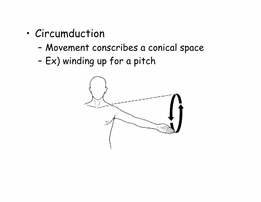
• Circumduction– Movement conscribes a conical space– Ex) winding up for a pitch
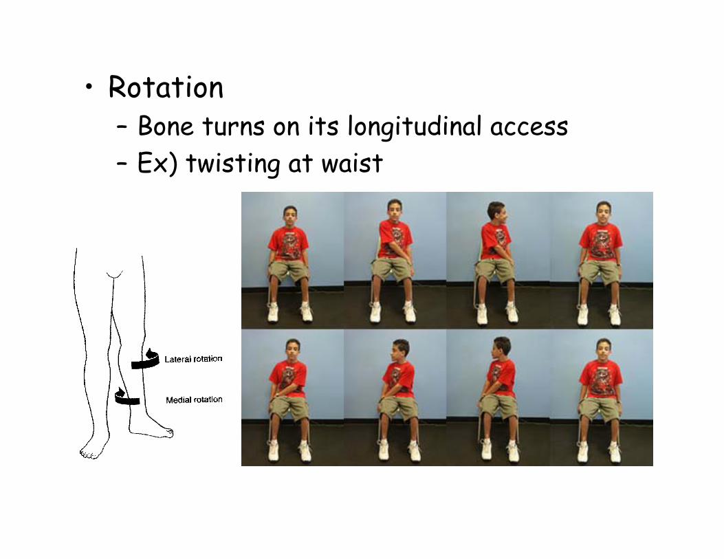
• Rotation– Bone turns on its longitudinal access– Ex) twisting at waist
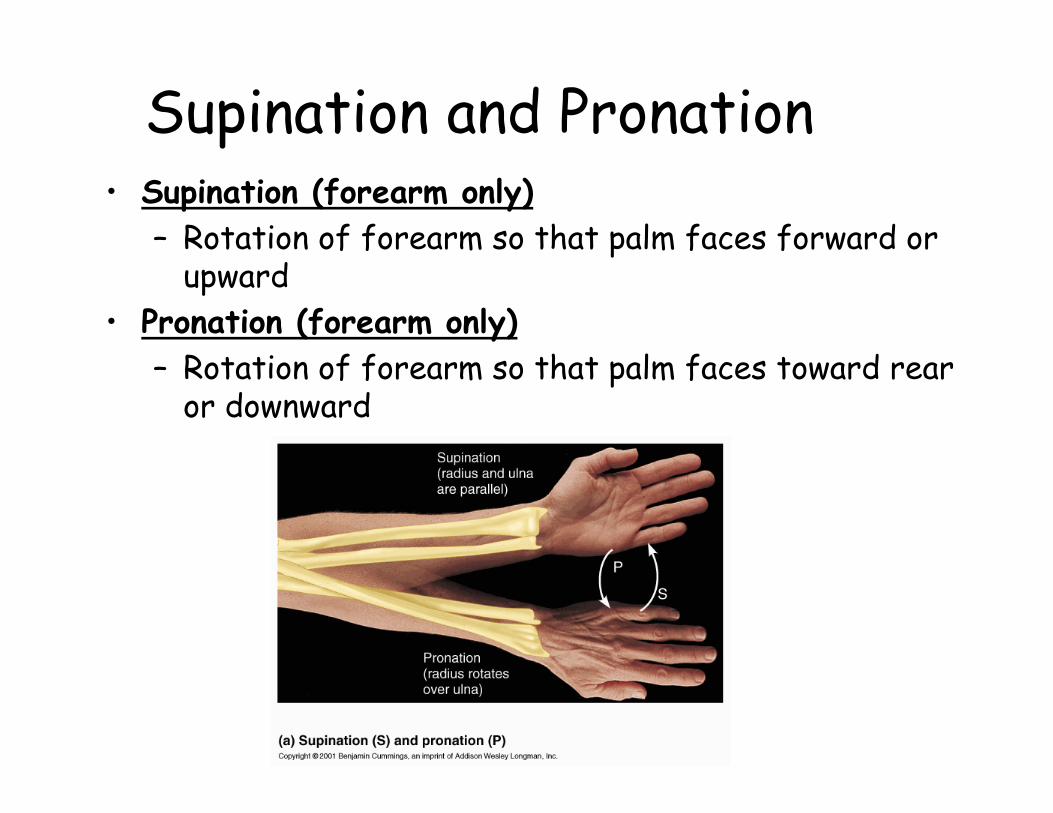
Supination and Pronation• Supination (forearm only)
– Rotation of forearm so that palm faces forward or upward
• Pronation (forearm only)– Rotation of forearm so that palm faces toward rear
or downward
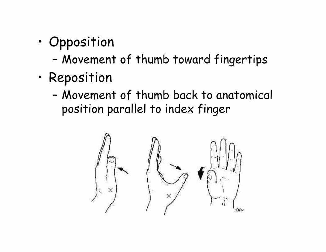
• Opposition– Movement of thumb toward fingertips
• Reposition– Movement of thumb back to anatomical
position parallel to index finger
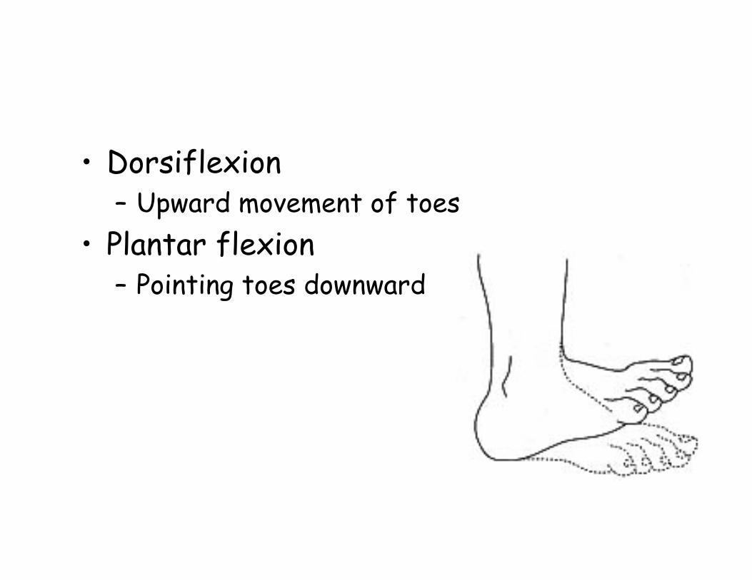
• Dorsiflexion– Upward movement of toes
• Plantar flexion– Pointing toes downward
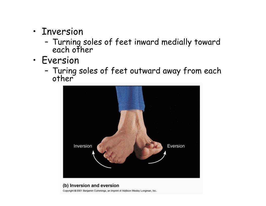
• Inversion– Turning soles of feet inward medially toward
each other• Eversion
– Turing soles of feet outward away from each other
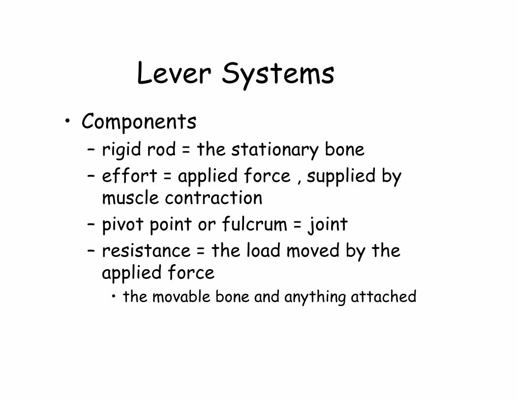
Lever Systems• Components
– rigid rod = the stationary bone– effort = applied force , supplied by
muscle contraction– pivot point or fulcrum = joint– resistance = the load moved by the
applied force • the movable bone and anything attached
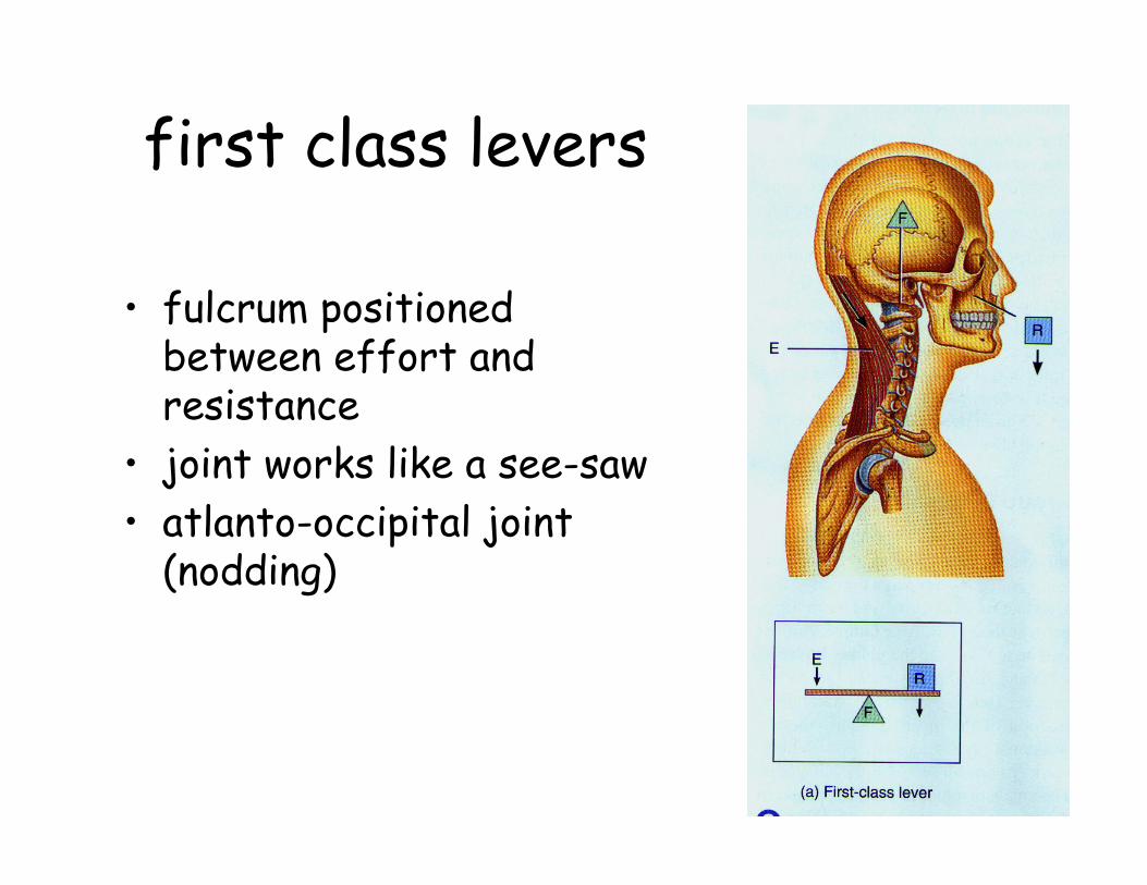
first class levers
• fulcrum positioned between effort and resistance
• joint works like a see-saw• atlanto-occipital joint
(nodding)
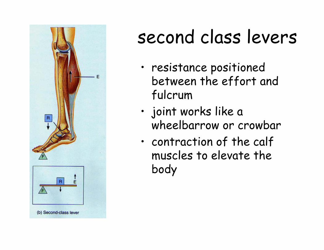
second class levers• resistance positioned
between the effort and fulcrum
• joint works like a wheelbarrow or crowbar
• contraction of the calf muscles to elevate the body
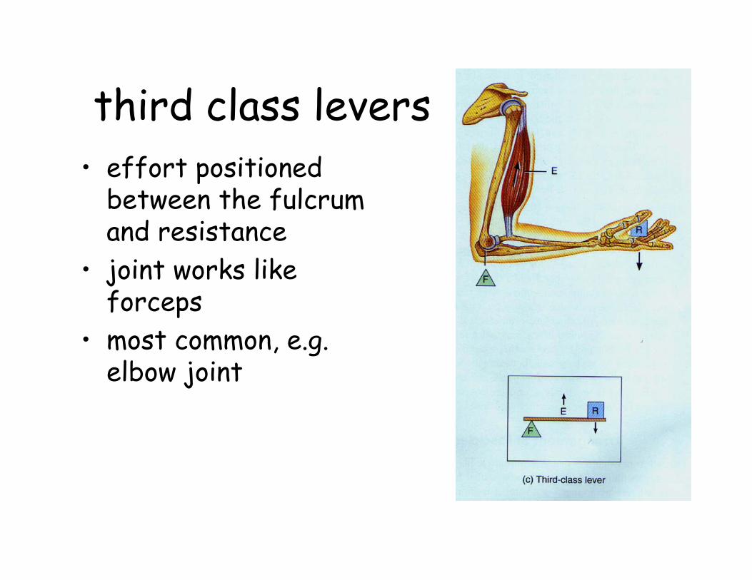
third class levers• effort positioned
between the fulcrum and resistance
• joint works like forceps
• most common, e.g. elbow joint

Herniated disk