Embed Size (px)
Citation preview
Joints and Articulations
Dr. Gary Mumaugh
Joints (Articulations)
• joint (articulation) – any point where two
bones meet, whether or not the bones are
movable at that interface
• arthrology – science of joint structure,
function, and dysfunction
• kinesiology – the study of musculoskeletal
movement
Joints and Their Classification
• joint name – typically derived from the names of the bones involved
• atlanto-occipital joint
• glenohumeral joint
• radioulnar joint
• joints classified according to the manner in which the adjacent bones are bound to each other, with differences in how freely the bones can move
• four major joint categories:
• bony joints
• fibrous joints
• cartilaginous joints
• synovial joints
Bony Joint (Synostosis)
• bony joint, or synostosis – an immovable joint
formed when the gap between two bones ossify,
and they become in effect, a single bone
• frontal and mandibular bones in infants
• cranial sutures in elderly
• attachment of first rib and sternum with old age
• can occur in either fibrous or cartilaginous joint

Fibrous Joints (Synarthrosis)
• fibrous joint, synarthrosis, or synarthrodial joint –
a point at which adjacent bones are bound by
collagen fibers that emerge from one bone, cross
the space between them, and penetrate into the
other
• three kinds of fibrous joints
• sutures
• gomphoses
• syndesmoses
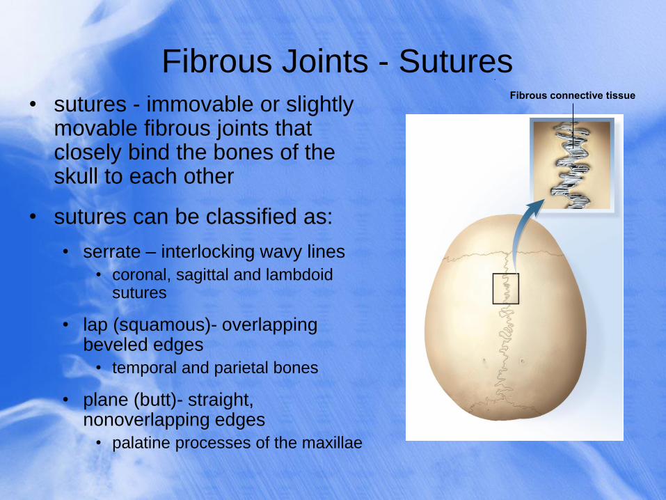
Fibrous Joints - Sutures
• sutures - immovable or slightly movable fibrous joints that closely bind the bones of the skull to each other
• sutures can be classified as:
• serrate – interlocking wavy lines
• coronal, sagittal and lambdoid sutures
• lap (squamous)- overlapping beveled edges
• temporal and parietal bones
• plane (butt)- straight, nonoverlapping edges
• palatine processes of the maxillae
.
Fibrous connective tissue
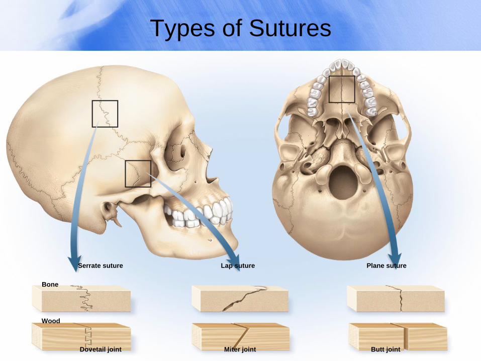
Types of Sutures
Wood
Dovetail joint Miter joint Butt joint
Bone
Serrate suture Lap suture Plane suture
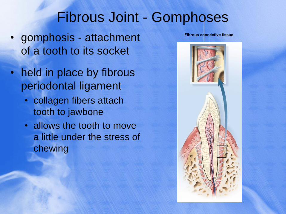
Fibrous Joint - Gomphoses
• gomphosis - attachment
of a tooth to its socket
• held in place by fibrous
periodontal ligament
• collagen fibers attach
tooth to jawbone
• allows the tooth to move
a little under the stress of
chewing
. Fibrous connective tissue
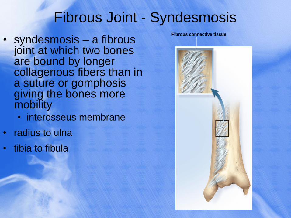
Fibrous Joint - Syndesmosis
• syndesmosis – a fibrous joint at which two bones are bound by longer collagenous fibers than in a suture or gomphosis giving the bones more mobility • interosseus membrane
• radius to ulna
• tibia to fibula
Fibrous connective tissue
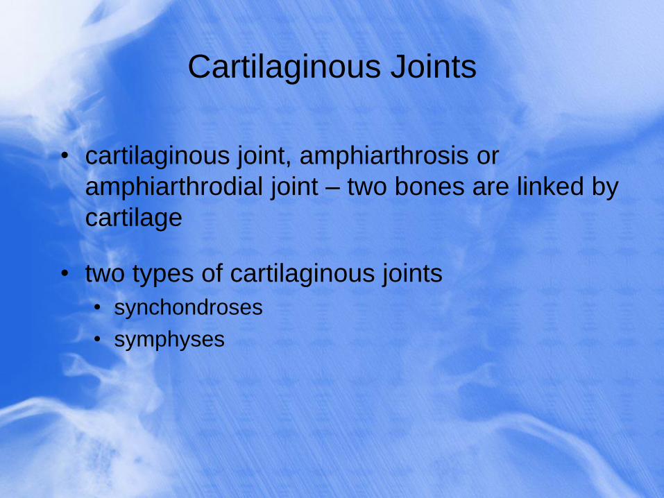
Cartilaginous Joints
• cartilaginous joint, amphiarthrosis or
amphiarthrodial joint – two bones are linked by
cartilage
• two types of cartilaginous joints
• synchondroses
• symphyses
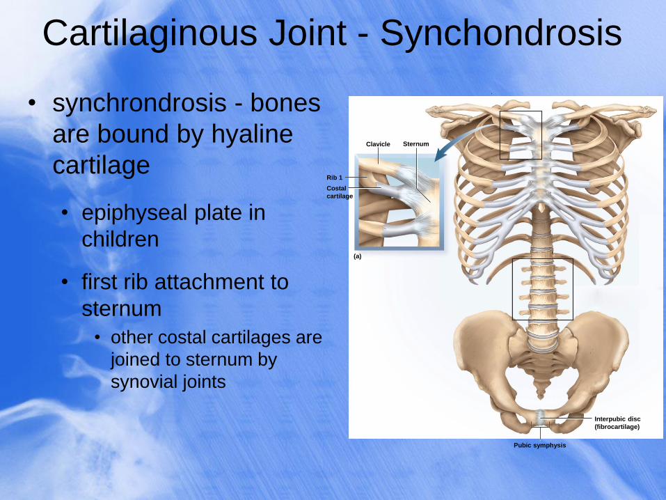
Cartilaginous Joint - Synchondrosis
• synchrondrosis - bones
are bound by hyaline
cartilage
• epiphyseal plate in
children
• first rib attachment to
sternum
• other costal cartilages are
joined to sternum by
synovial joints
Pubic symphysis
Clavicle
Rib 1
(a)
Sternum
Costal
cartilage
Interpubic disc
(fibrocartilage)
.
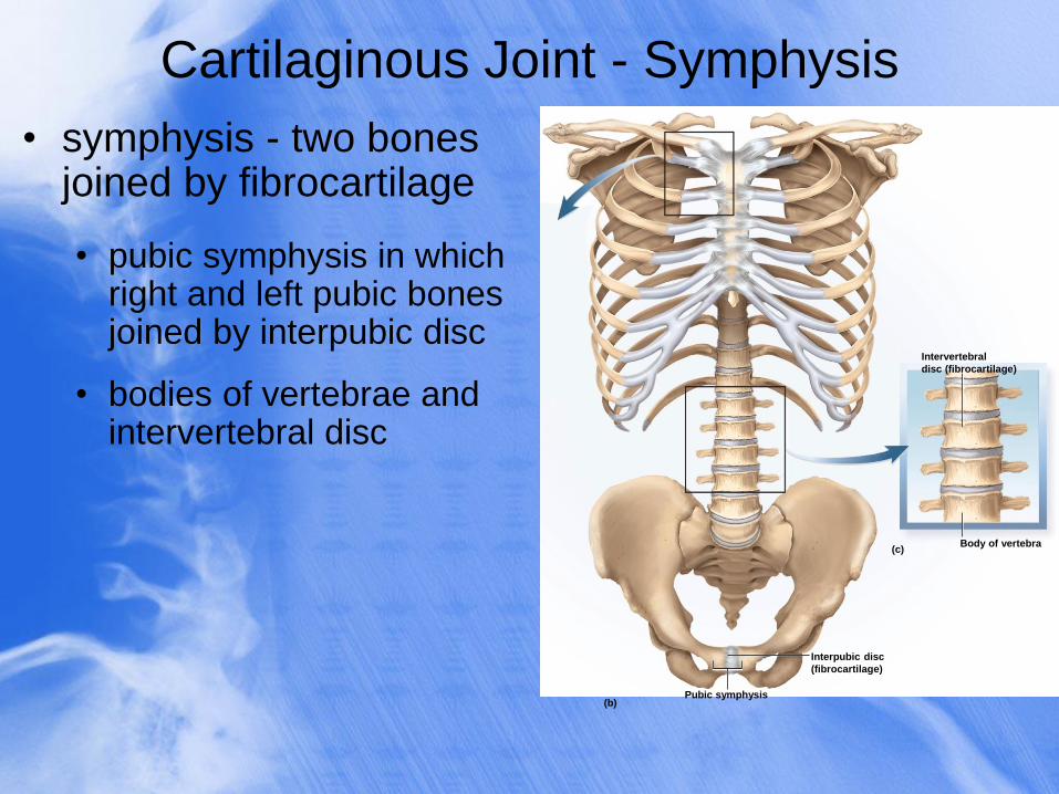
Cartilaginous Joint - Symphysis
• symphysis - two bones joined by fibrocartilage
• pubic symphysis in which right and left pubic bones joined by interpubic disc
• bodies of vertebrae and intervertebral disc
Pubic symphysis
Body of vertebra (c)
(b)
Interpubic disc
(fibrocartilage)
Intervertebral
disc (fibrocartilage)
.
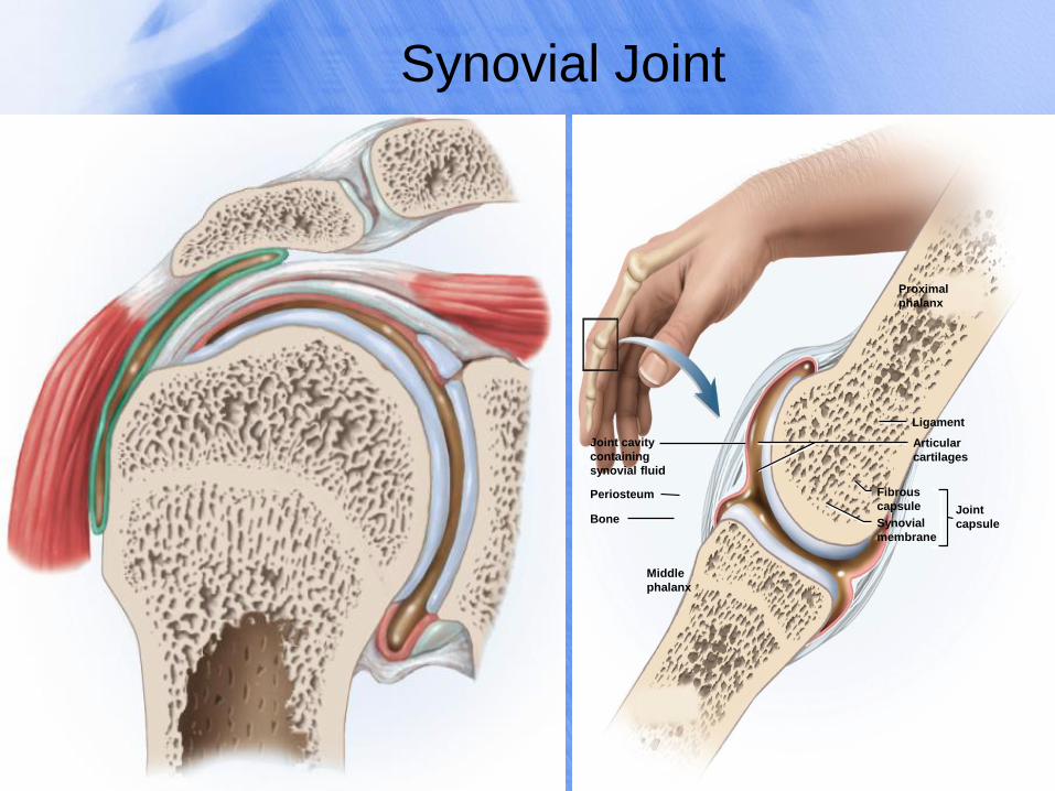
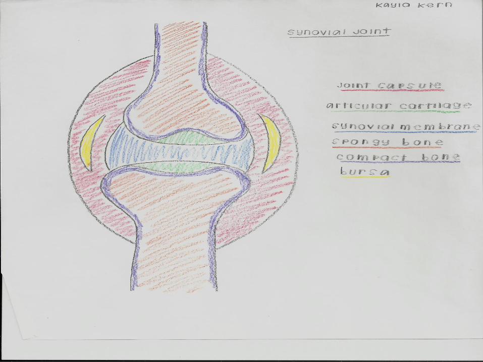
Synovial Joint
.
Periosteum
Ligament
Bone
Proximal
phalanx
Joint cavity
containing
synovial fluid
Fibrous
capsule
Articular
cartilages
Joint
capsule
Middle
phalanx
Synovial
membrane
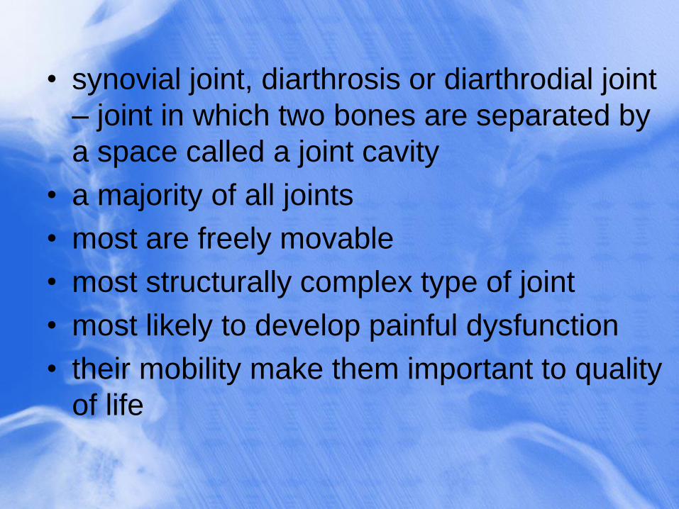
• synovial joint, diarthrosis or diarthrodial joint
– joint in which two bones are separated by
a space called a joint cavity
• a majority of all joints
• most are freely movable
• most structurally complex type of joint
• most likely to develop painful dysfunction
• their mobility make them important to quality
of life
General Anatomy
• articular cartilage – layer of hyaline cartilage
that covers the facing surfaces of two bones
• joint (articular) cavity – separates articular
surfaces
• synovial fluid – slippery lubricant in joint cavity
• gives it a viscous, slippery texture like raw
egg whites
• nourishes articular cartilage and removes
waste
• makes movement of synovial joints almost
friction free
• joint (articular) capsule – connective tissue that
encloses the cavity and retains the fluid
• outer fibrous capsule – continuous with
periosteum of adjoining bones
• Inner synovial membrane – composed mainly
of cells that secrete synovial fluid and
macrophages that remove debris from the joint
cavity
• in a few synovial joints, fibrocartilage grows inward
from the joint capsule
• articular disc forms a pad between bones
• temporomandibular joint, distal radioulnar joints,
sternoclavicular and acromioclavicular joints
• meniscus – in the knee, two cartilages extend
inward from the left and right
• these cartilages absorb shock and pressure
• guide bones across each other
• improve the fit between bones
• stabilize the joints, reducing the chance of dislocation
• accessory structures associated with synovial joints
• tendon – attaches muscle to bone
• ligament – attaches bone to bone
• bursa – a fibrous sac filled with synovial fluid,
located between adjacent muscles, where tendon
passes over bone, or between bone and skin
• cushion muscles
• helps tendons slide more easily over joints
• modify direction of tendon pull
• tendon sheaths – elongated cylindrical bursae
wrapped around a tendon
• in hand and foot
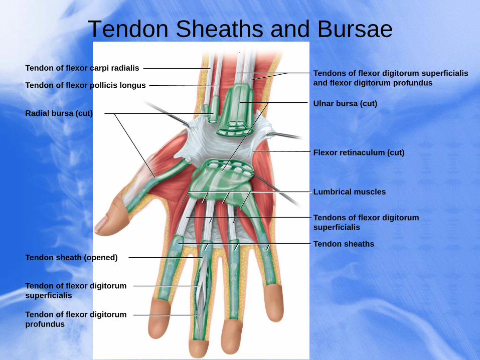
Tendon Sheaths and Bursae
.
Tendon of flexor pollicis longus
Radial bursa (cut)
Flexor retinaculum (cut)
Ulnar bursa (cut)
Lumbrical muscles
Tendon of flexor carpi radialis
Tendon sheaths
Tendon sheath (opened)
Tendon of flexor digitorum
superficialis
Tendon of flexor digitorum
profundus
Tendons of flexor digitorum superficialis
and flexor digitorum profundus
Tendons of flexor digitorum
superficialis
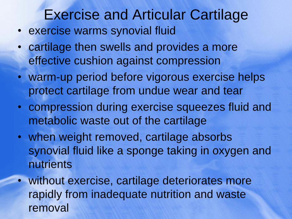
Exercise and Articular Cartilage • exercise warms synovial fluid
• cartilage then swells and provides a more
effective cushion against compression
• warm-up period before vigorous exercise helps
protect cartilage from undue wear and tear
• compression during exercise squeezes fluid and
metabolic waste out of the cartilage
• when weight removed, cartilage absorbs
synovial fluid like a sponge taking in oxygen and
nutrients
• without exercise, cartilage deteriorates more
rapidly from inadequate nutrition and waste
removal
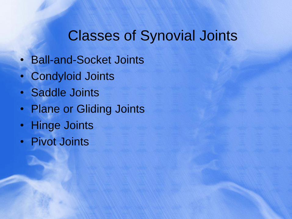
Classes of Synovial Joints
• Ball-and-Socket Joints
• Condyloid Joints
• Saddle Joints
• Plane or Gliding Joints
• Hinge Joints
• Pivot Joints
9-25
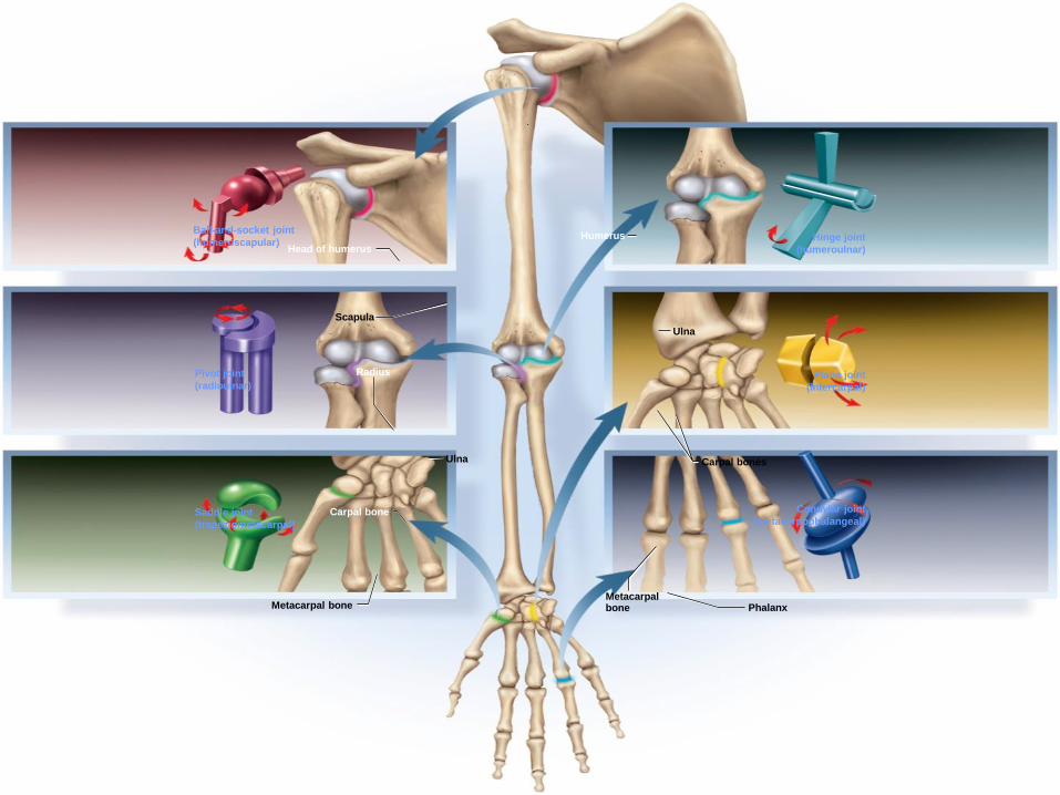
Classes of Synovial Joints
Head of humerus
Scapula
Carpal bone
Metacarpal bone Phalanx Metacarpal bone
Humerus
Ulna Carpal bones
Radius
Ulna
Ball-and-socket joint
(humeroscapular)
Pivot joint
(radioulnar)
Saddle joint
(trapeziometacarpal)
Hinge joint
(humeroulnar)
Plane joint
(intercarpal)
Condylar joint
(metacarpophalangeal)
.
Ball-and-Socket Joints
• smooth, hemispherical head fits within a cuplike
socket
• shoulder joint - head of humerus into glenoid cavity of
scapula
• hip joint - head of femur into acetabulum of hip bone
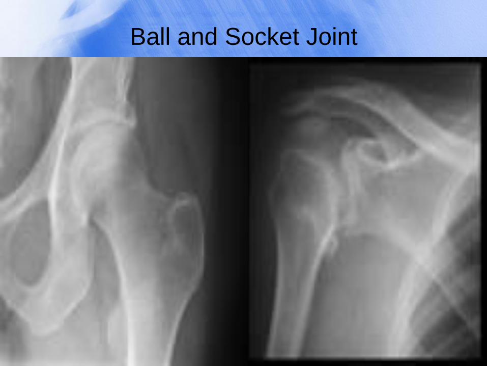
Ball and Socket Joint
Condyloid Joints
• oval convex surface on one bone fits into a
complementary shaped depression on the other
• radiocarpal joint of the wrist
• metacarpophalangeal joints at the bases of the
fingers
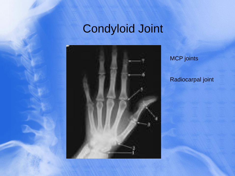
Condyloid Joint
MCP joints
Radiocarpal joint
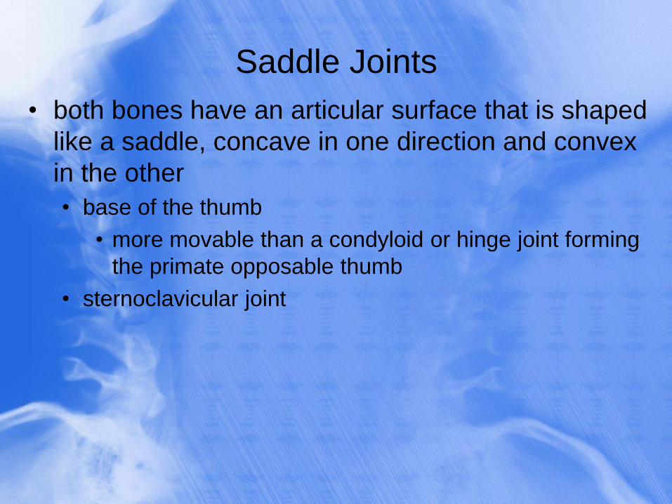
Saddle Joints
• both bones have an articular surface that is shaped
like a saddle, concave in one direction and convex
in the other
• base of the thumb
• more movable than a condyloid or hinge joint forming
the primate opposable thumb
• sternoclavicular joint
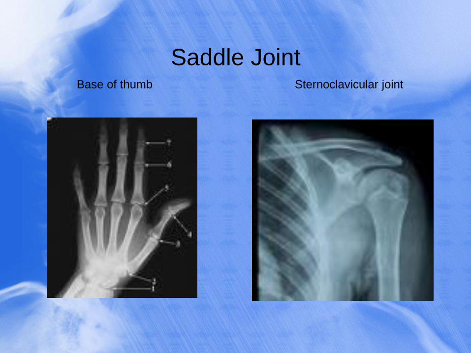
Saddle Joint Base of thumb Sternoclavicular joint
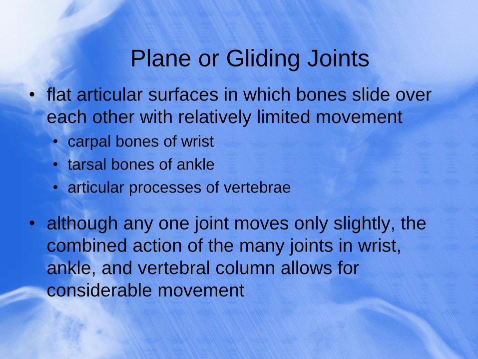
Plane or Gliding Joints
• flat articular surfaces in which bones slide over
each other with relatively limited movement
• carpal bones of wrist
• tarsal bones of ankle
• articular processes of vertebrae
• although any one joint moves only slightly, the
combined action of the many joints in wrist,
ankle, and vertebral column allows for
considerable movement
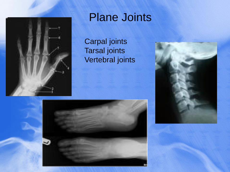
Plane Joints
Carpal joints
Tarsal joints
Vertebral joints
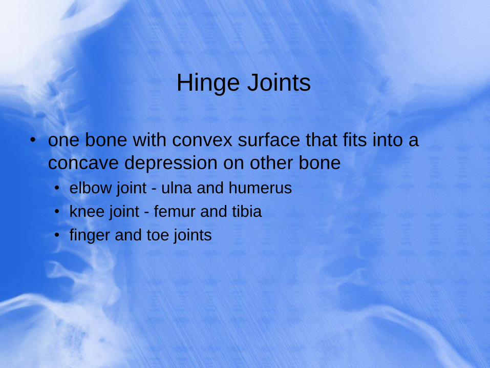
Hinge Joints
• one bone with convex surface that fits into a
concave depression on other bone
• elbow joint - ulna and humerus
• knee joint - femur and tibia
• finger and toe joints
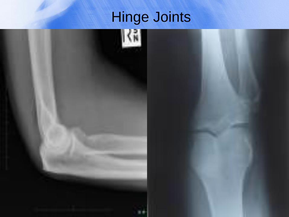
Hinge Joints
Pivot Joints
• one bone has a projection that is held in place
by a ring-like ligament
• bone spins on its longitudinal axis
• atlantoaxial joint (dens of axis and atlas)
• proximal radioulnar joint allows the radius to rotate
during pronation and supination
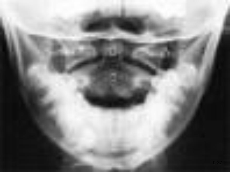
9-37
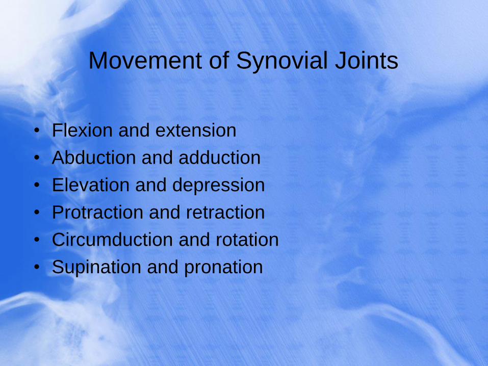
Movement of Synovial Joints
• Flexion and extension
• Abduction and adduction
• Elevation and depression
• Protraction and retraction
• Circumduction and rotation
• Supination and pronation
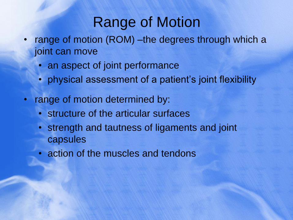
Range of Motion • range of motion (ROM) –the degrees through which a
joint can move
• an aspect of joint performance
• physical assessment of a patient’s joint flexibility
• range of motion determined by:
• structure of the articular surfaces
• strength and tautness of ligaments and joint
capsules
• action of the muscles and tendons
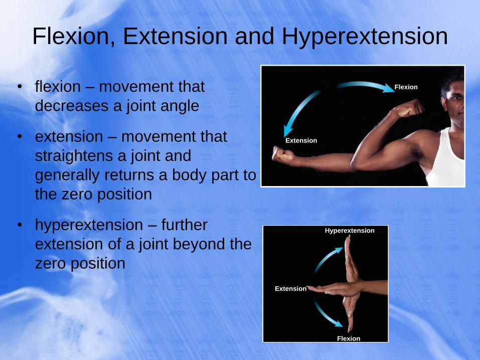
Flexion, Extension and Hyperextension
• flexion – movement that
decreases a joint angle
• extension – movement that
straightens a joint and
generally returns a body part to
the zero position
• hyperextension – further
extension of a joint beyond the
zero position
Extension
Flexion
Extension
Flexion
Hyperextension
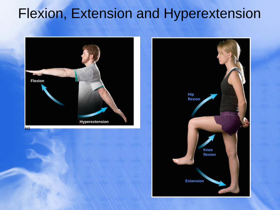
Flexion, Extension and Hyperextension
(c)
Flexion
Hyperextension
Hip
flexion
Knee
flexion
Extension
Abduction and Adduction
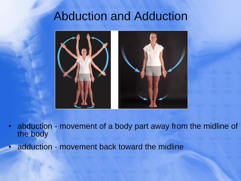
• abduction - movement of a body part away from the midline of the body
• adduction - movement back toward the midline
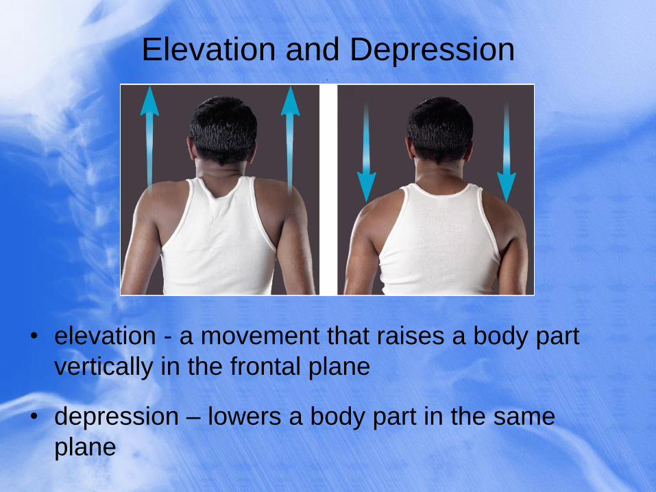
Elevation and Depression
• elevation - a movement that raises a body part
vertically in the frontal plane
• depression – lowers a body part in the same
plane
.
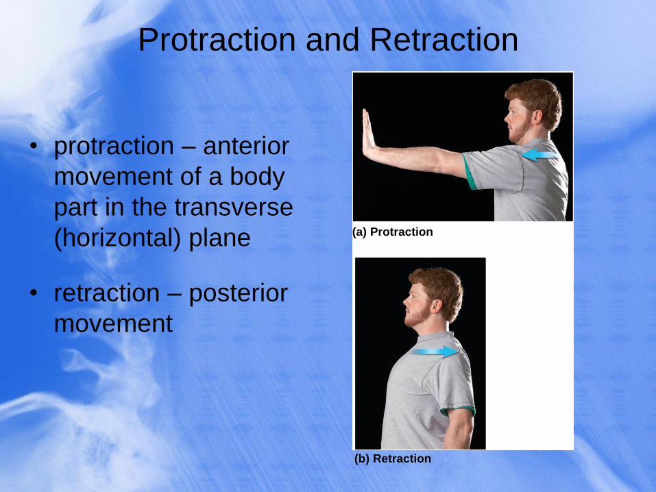
Protraction and Retraction
• protraction – anterior
movement of a body
part in the transverse
(horizontal) plane
• retraction – posterior
movement
(a) Protraction
(b) Retraction
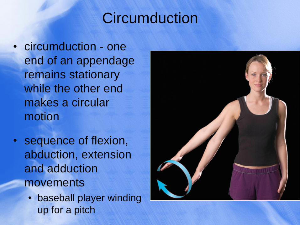
Circumduction
• circumduction - one
end of an appendage
remains stationary
while the other end
makes a circular
motion
• sequence of flexion,
abduction, extension
and adduction
movements
• baseball player winding
up for a pitch
.
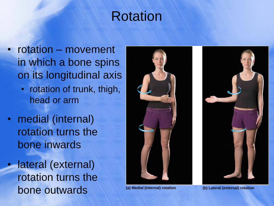
Rotation
• rotation – movement
in which a bone spins
on its longitudinal axis
• rotation of trunk, thigh,
head or arm
• medial (internal)
rotation turns the
bone inwards
• lateral (external)
rotation turns the
bone outwards (b) Lateral (external) rotation (a) Medial (internal) rotation
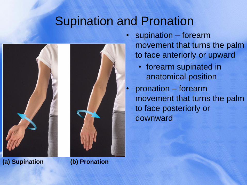
Supination and Pronation
• supination – forearm
movement that turns the palm
to face anteriorly or upward
• forearm supinated in
anatomical position
• pronation – forearm
movement that turns the palm
to face posteriorly or
downward
(a) Supination (b) Pronation
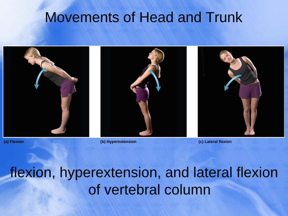
Movements of Head and Trunk
flexion, hyperextension, and lateral flexion
of vertebral column
(a) Flexion (b) Hyperextension (c) Lateral flexion
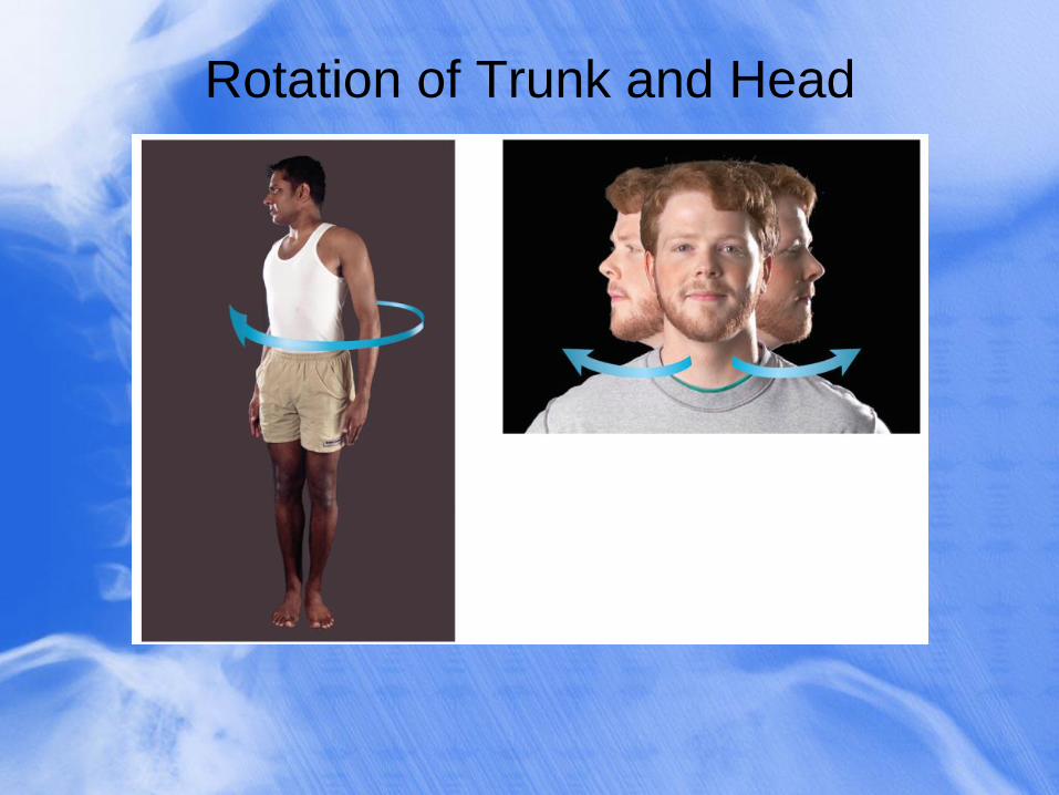
Rotation of Trunk and Head
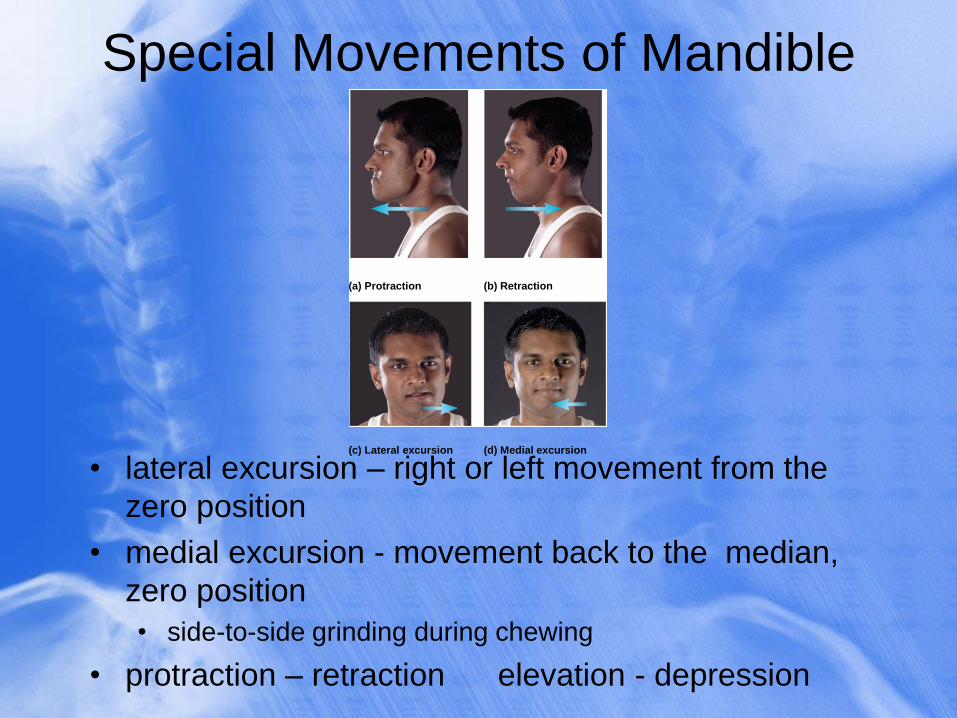
Special Movements of Mandible
• lateral excursion – right or left movement from the
zero position
• medial excursion - movement back to the median,
zero position
• side-to-side grinding during chewing
• protraction – retraction elevation - depression
(a) Protraction (b) Retraction
(c) Lateral excursion (d) Medial excursion
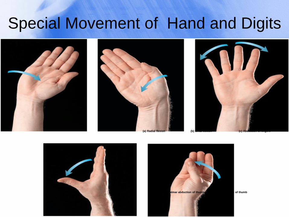
Special Movement of Hand and Digits
(a) Radial flexion (b) Ulnar flexion
(d) Palmar abduction of thumb (e) Opposition of thumb
(c) Abduction of fingers
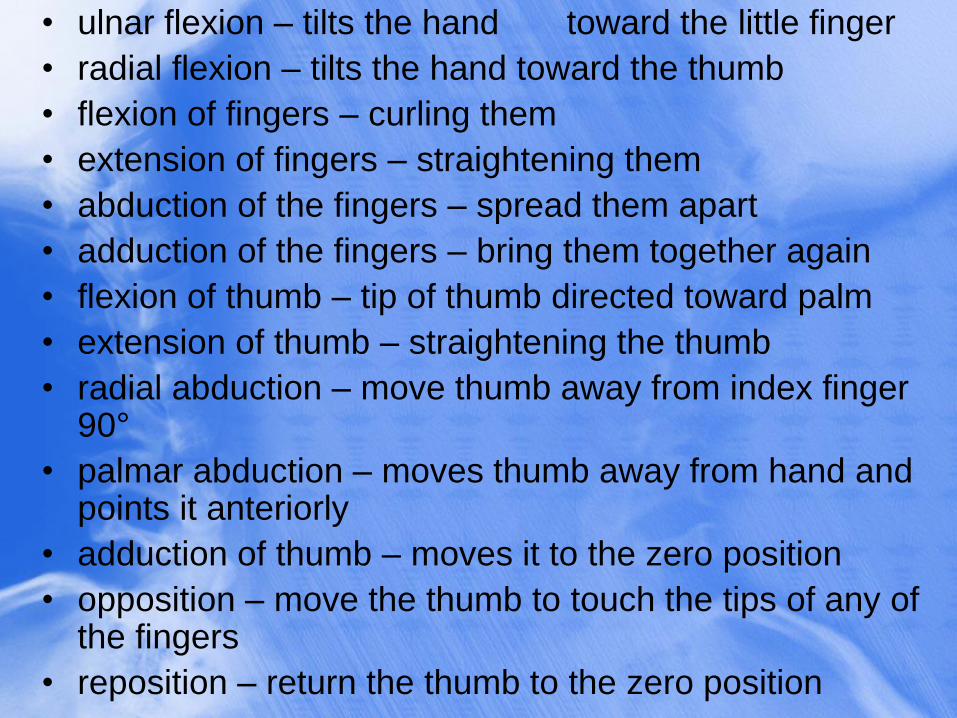
• ulnar flexion – tilts the hand toward the little finger
• radial flexion – tilts the hand toward the thumb
• flexion of fingers – curling them
• extension of fingers – straightening them
• abduction of the fingers – spread them apart
• adduction of the fingers – bring them together again
• flexion of thumb – tip of thumb directed toward palm
• extension of thumb – straightening the thumb
• radial abduction – move thumb away from index finger 90°
• palmar abduction – moves thumb away from hand and points it anteriorly
• adduction of thumb – moves it to the zero position
• opposition – move the thumb to touch the tips of any of the fingers
• reposition – return the thumb to the zero position
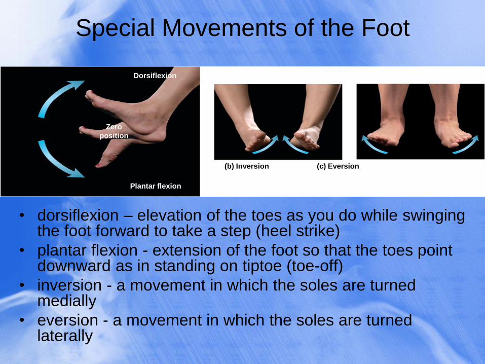
Special Movements of the Foot
• dorsiflexion – elevation of the toes as you do while swinging the foot forward to take a step (heel strike)
• plantar flexion - extension of the foot so that the toes point downward as in standing on tiptoe (toe-off)
• inversion - a movement in which the soles are turned medially
• eversion - a movement in which the soles are turned laterally
Dorsiflexion
Zero
position
(c) Eversion (b) Inversion
Plantar flexion
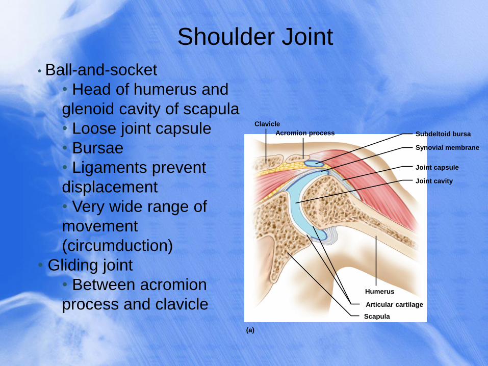
Shoulder Joint
• Ball-and-socket
• Head of humerus and
glenoid cavity of scapula
• Loose joint capsule
• Bursae
• Ligaments prevent
displacement
• Very wide range of
movement
(circumduction)
• Gliding joint
• Between acromion
process and clavicle Humerus
Articular cartilage
Scapula
Clavicle
Acromion process Subdeltoid bursa
Synovial membrane
Joint capsule
Joint cavity
(a)
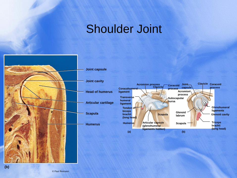
Shoulder Joint
.
© Paul Reimann
Head of humerus
Joint cavity
Joint capsule
Articular cartilage
Scapula
Humerus
(b)
Coracohumeral
ligament
Transverse
humeral
ligament
Tendon of
biceps
brachii
(long head)
Acromion process Clavicle
Coracoid
process Acromion
process
Subscapular
bursa
Joint
capsule
Coracoid
process
Clavicle
Glenohumeral
ligaments
Glenoid cavity
Triceps
brachii
(long head)
Glenoid
labrum
Scapula Humerus
Scapula
Articular capsule
(glenohumeral
ligaments hidden) (a) (b)
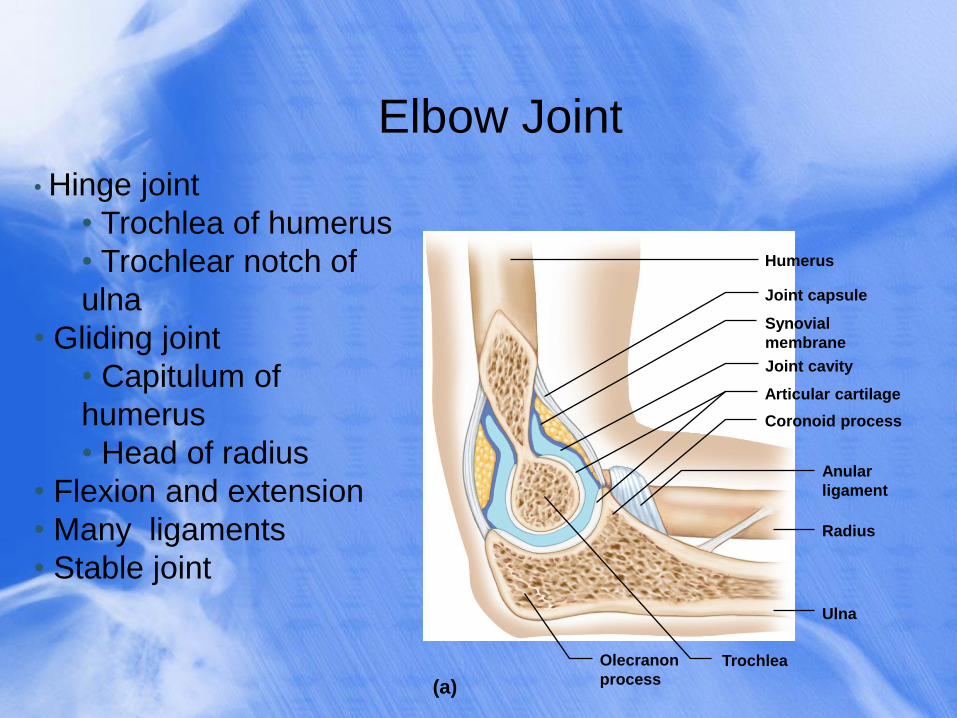
Elbow Joint
• Hinge joint
• Trochlea of humerus
• Trochlear notch of
ulna
• Gliding joint
• Capitulum of
humerus
• Head of radius
• Flexion and extension
• Many ligaments
• Stable joint
Humerus
Joint capsule
Synovial
membrane
Joint cavity
Articular cartilage
Coronoid process
Anular
ligament
Radius
Ulna
Olecranon
process Trochlea
(a)
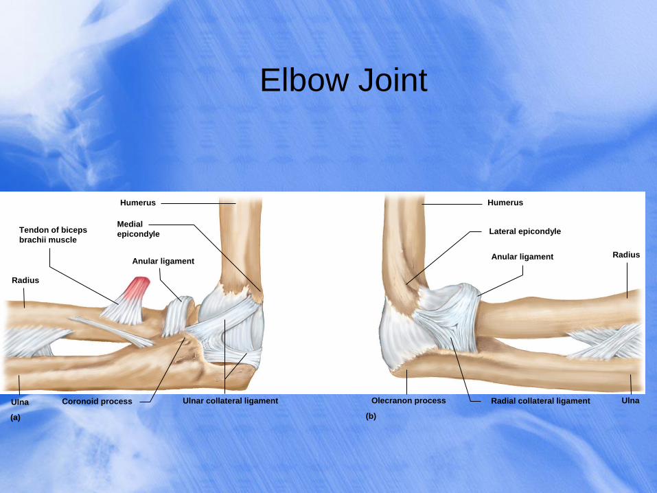
Elbow Joint
Radius
Tendon of biceps
brachii muscle
Anular ligament
Humerus
Medial
epicondyle
Ulnar collateral ligament Coronoid process Ulna
Humerus
Lateral epicondyle
Anular ligament Radius
Olecranon process Radial collateral ligament Ulna
(b) (a)
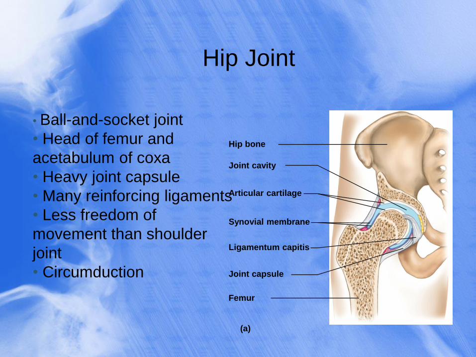
Hip Joint
• Ball-and-socket joint
• Head of femur and
acetabulum of coxa
• Heavy joint capsule
• Many reinforcing ligaments
• Less freedom of
movement than shoulder
joint
• Circumduction
Hip bone
Joint cavity
Articular cartilage
Synovial membrane
Joint capsule
Ligamentum capitis
Femur
(a)
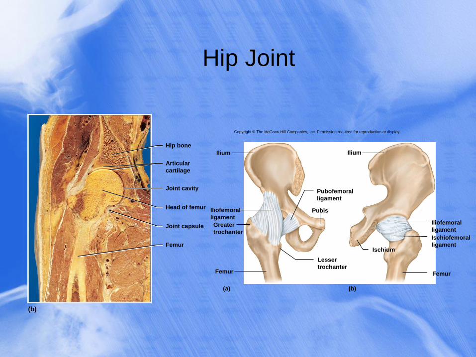
Hip Joint
.
(b)
Joint cavity
Articular
cartilage
Hip bone
Head of femur
Joint capsule
Femur
Ilium
Iliofemoral
ligament
Greater
trochanter
Femur
Lesser
trochanter
Pubis
Pubofemoral
ligament
Ischium
Iliofemoral
ligament
Ischiofemoral
ligament
Femur
Ilium
(a) (b)
Copyright © The McGraw-Hill Companies, Inc. Permission required for reproduction or display.
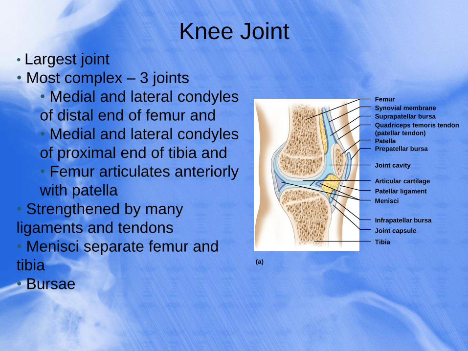
Knee Joint • Largest joint
• Most complex – 3 joints
• Medial and lateral condyles
of distal end of femur and
• Medial and lateral condyles
of proximal end of tibia and
• Femur articulates anteriorly
with patella
• Strengthened by many
ligaments and tendons
• Menisci separate femur and
tibia
• Bursae
Femur
Quadriceps femoris tendon
(patellar tendon)
Synovial membrane
Suprapatellar bursa
Patella
Prepatellar bursa
Joint cavity
Articular cartilage
Menisci
Patellar ligament
Infrapatellar bursa
Joint capsule
Tibia
(a)
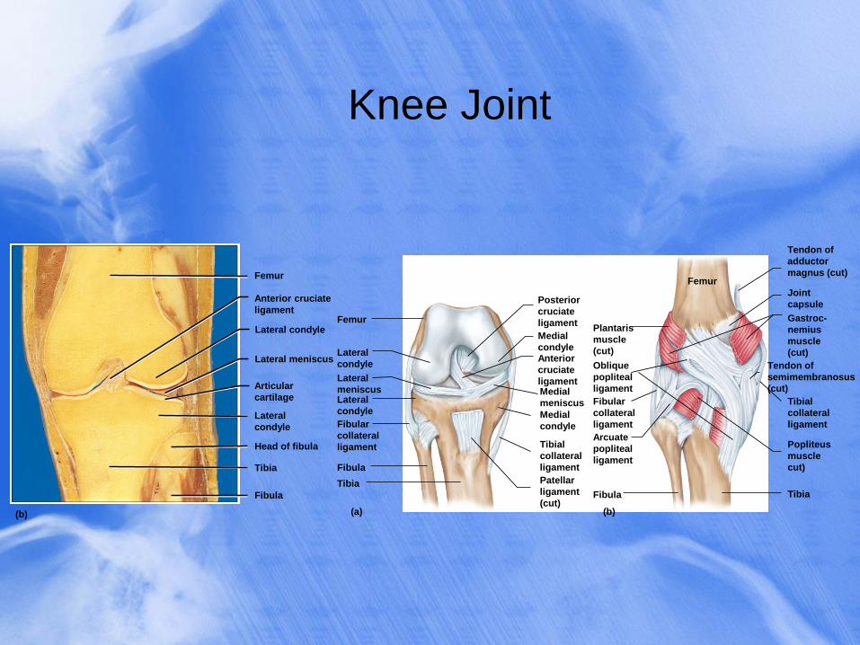
Knee Joint
.
Anterior cruciate
ligament
Femur
(b)
Lateral condyle
Lateral meniscus
Articular
cartilage
Lateral
condyle
Head of fibula
Tibia
Fibula
Gastroc-
nemius
muscle
(cut)
Popliteus
muscle
cut)
Oblique
popliteal
ligament
Arcuate
popliteal
ligament
Fibula Tibia
Femur
Joint
capsule
Fibular
collateral
ligament
Plantaris
muscle
(cut)
Tibial
collateral
ligament
Tendon of
semimembranosus
(cut)
(a) (b)
Femur
Lateral
condyle
Lateral
meniscus Lateral
condyle
Fibular
collateral
ligament
Fibula
Tibia
Medial
condyle Anterior
cruciate
ligament Medial
meniscus
Medial
condyle
Tibial
collateral
ligament
Patellar
ligament
(cut)
Posterior
cruciate
ligament
Tendon of
adductor
magnus (cut)
Temporomandibular Joint
• temporomandibular (jaw) joint (TMJ) – articulation of the
condyle of the mandible with the mandibular fossa of the
temporal bone
• combines elements of condylar, hinge, and plane
joints
• synovial cavity of the TMJ is divided into superior and
inferior chambers by an articular disc
• deep yawn or strenuous depression can dislocate the
TMJ
• condyles pop out of fossa and slip forward
• relocated by pressing down on molar teeth while
pushing the jaw backward
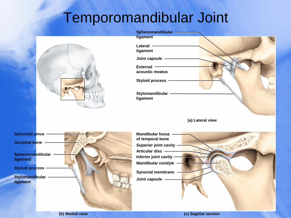
Temporomandibular Joint
Joint capsule
Styloid process
(a) Lateral view
(c) Sagittal section (b) Medial view
Occipital bone
Sphenoid sinus
Styloid process
Joint capsule
Synovial membrane
Mandibular condyle
Superior joint cavity
Inferior joint cavity
Articular disc
Sphenomandibular
ligament
Lateral
ligament
External
acoustic meatus
Stylomandibular
ligament
Mandibular fossa
of temporal bone
Sphenomandibular
ligament
Stylomandibular
ligament
TMJ Syndrome • temporomandibular joint (TMJ) syndrome
• may affect as many as 75 million Americans
• signs and symptoms • can cause moderate intermittent facial pain
• clicking sounds in the jaw
• limitation of jaw movement
• often severe headaches, vertigo (dizziness), tinnitus (ringing in the ears)
• pain radiating from jaw down the neck, shoulders, and back
• cause of syndrome • caused by combination of psychological tension and malocclusion
(misalignment of teeth)
• treatment • psychological management, physical therapy, analgesic and anti-
inflammatory drugs, corrective dental appliances to align teeth properly
Lifespan Changes
• Joint stiffness is an early sign of aging
• Fibrous joints first to change; can strengthen
however over a lifetime
• Changes in symphysis joints of vertebral
column diminish flexibility and decrease height
(remember water loss from the IVDs)
• Synovial joints lose elasticity
• Disuse hampers the blood supply
• Activity and exercise can keep joints functional
longer