Embed Size (px)
Citation preview
ANEMIAS
Dr. Farjah H. AlGahtaniConsultant Hematologist ,assistant professor
Director of transfusion medicine and blood bank department
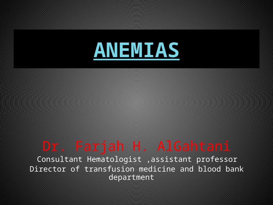
ANEMIA
•Single cell line(RBC) problem•Multiple cell line problem
( RBC,WBC,Platelet)- Bone marrow suppression
- immunologic disorders- peripheral destruction/sequestration
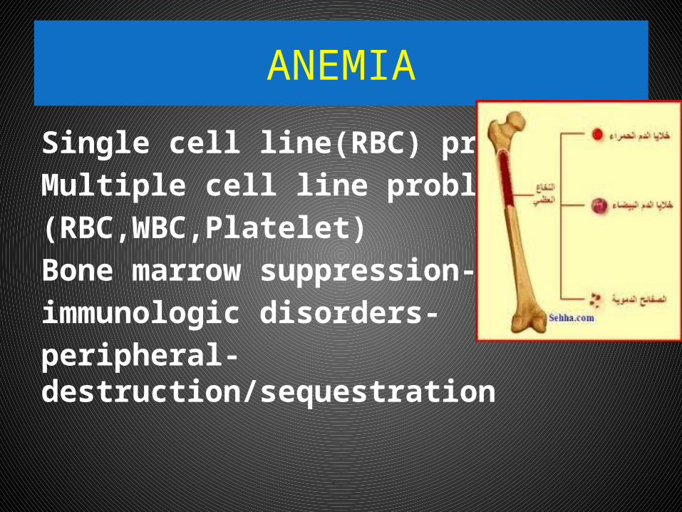
Anemia•Anemia is generally defined as a hematocrit
< 40% (hemoglobin <13.0 g/dL) in menor
< 37% (hemoglobin <12.0 g/dL) inwomen .
(WHO definition) Red blood cell (RBC) indices, which include
the mean corpuscular volume (MCV), the mean corpuscular hemoglobin (MCH),
the mean corpuscular hemoglobin content (MCHC), and the red-cell distribution width (RDW) index, are
further used to define types of anemia.
Anemia•Despite having a set of peculiar
symptoms and signs, anemia is not a disease per se, but a syndrome, as it may
arise from an extensive list of causes .•It is the chronic syndrome of highest
prevalence in clinical medicine .
Anemia Etiology
•Based on Hb, red cell indices, retic count and red cell morphology
•(1 )Inadequate response A. Hypochromic microcyctic
B. Normochromic Normocytic C. Macrocytic
•(2)Adequate response R/O blood loss---Includes Hemolytic
disorders
Anemia-symptoms
•What are the symptoms of Anemia?
•General malaise, weakness, fatigue, breathlessness on exertion, palpitations,
angina .
• Desire to eat sand and clay .
• Menorrhagia common in women.
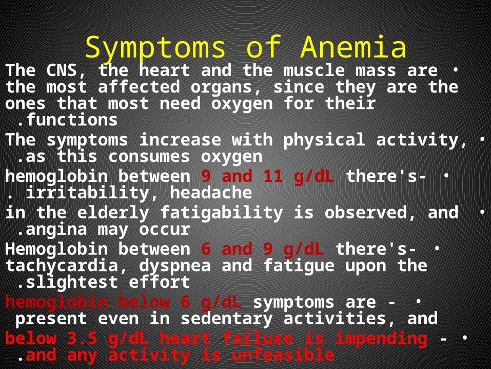
Symptoms of Anemia•The CNS, the heart and the muscle mass are the most
affected organs, since they are the ones that most need oxygen for their functions .
•The symptoms increase with physical activity, as this consumes oxygen .
•-hemoglobin between 9 and 11 g/dL there's irritability, headache.
• in the elderly fatigability is observed, and angina may occur .
•-Hemoglobin between 6 and 9 g/dL there's tachycardia, dyspnea and fatigue upon the slightest
effort .• -hemoglobin below 6 g/dL symptoms are present
even in sedentary activities, and • -below 3.5 g/dL heart failure is impending and any
activity is unfeasible.
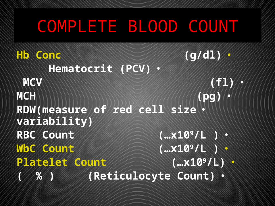
COMPLETE BLOOD COUNT
•Hb Conc (g/dl)•Hematocrit (PCV) •MCV (fl) •MCH (pg)
•RDW(measure of red cell size variability)•RBC Count (…x109/L )•WbC Count (…x109/L )•Platelet Count (…x109/L)•(Reticulocyte Count) % ( )
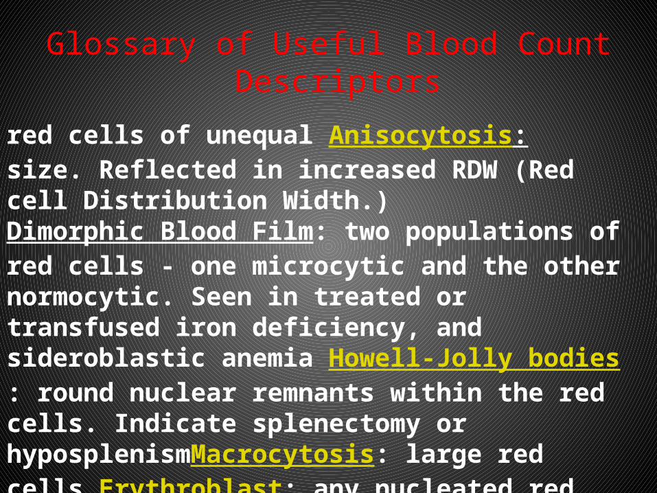
Glossary of Useful Blood Count Descriptors
Anisocytosis: red cells of unequal size. Reflected in increased RDW (Red cell Distribution Width.) Dimorphic Blood Film: two populations of red cells - one microcytic and the other normocytic. Seen in treated or transfused iron deficiency, and sideroblastic anemia Howell-Jolly bodies: round nuclear remnants within the red cells. Indicate splenectomy or hyposplenismMacrocytosis: large red cells Erythroblast: any nucleated red cell precursor Hypersegmented neutrophils: a neutrophil with six or more lobes. Usually (but not inevitably) means vitamin B12 or folate deficiency
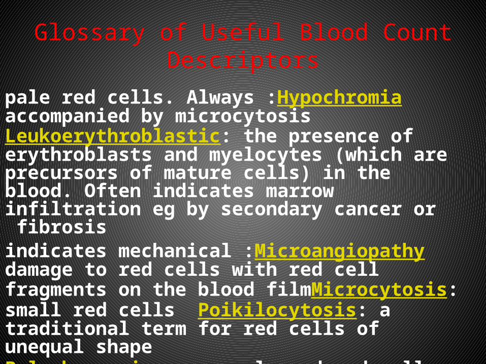
Glossary of Useful Blood Count Descriptors
Hypochromia :pale red cells. Always accompanied by microcytosis Leukoerythroblastic: the presence of erythroblasts and myelocytes (which are precursors of mature cells) in the blood. Often indicates marrow infiltration eg by secondary cancer or fibrosis
Microangiopathy :indicates mechanical damage to red cells with red cell fragments on the blood filmMicrocytosis: small red cells Poikilocytosis: a traditional term for red cells of unequal shape
Polychromasia: grey coloured red cells on film, indicating presence of increased reticulocytes
Reticulocyte: an erythrocyte newly released from the bone marrow
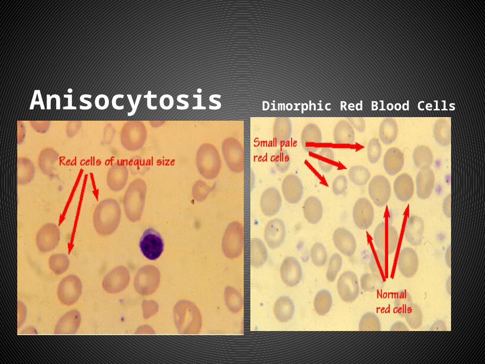
Anisocytosis Dimorphic Red Blood Cells
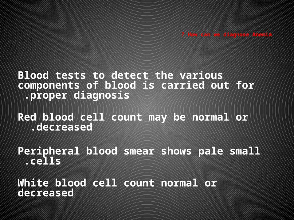
How can we diagnose Anemia ?
Blood tests to detect the various components of blood is carried out for proper diagnosis .
Red blood cell count may be normal or decreased .
Peripheral blood smear shows pale small cells .
White blood cell count normal or decreased
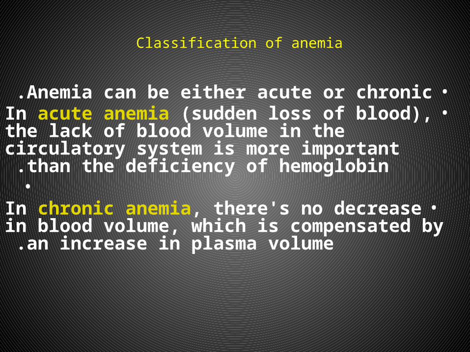
Classification of anemia
•Anemia can be either acute or chronic .•In acute anemia (sudden loss of blood), the lack of
blood volume in the circulatory system is more important than the deficiency of hemoglobin .•
•In chronic anemia, there's no decrease in blood volume, which is compensated by an increase in
plasma volume .
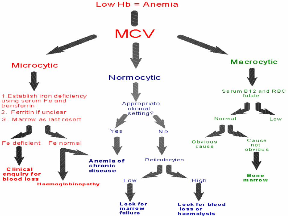
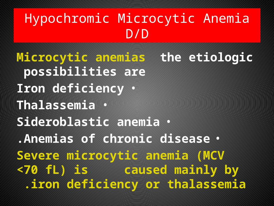
Hypochromic Microcytic AnemiaD/D
Microcytic anemias the etiologic possibilities are
•Iron deficiency•Thalassemia
•Sideroblastic anemia•Anemias of chronic disease.
Severe microcytic anemia (MCV <70 fL) is caused mainly by iron deficiency or
thalassemia .
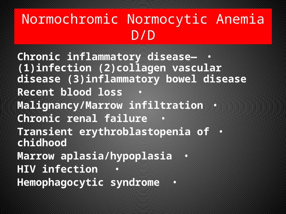
Normochromic Normocytic AnemiaD/D
•Chronic inflammatory disease—(1)infection (2)collagen vascular disease (3)inflammatory bowel disease
•Recent blood loss•Malignancy/Marrow infiltration
•Chronic renal failure•Transient erythroblastopenia of chidhood
•Marrow aplasia/hypoplasia•HIV infection
•Hemophagocytic syndrome
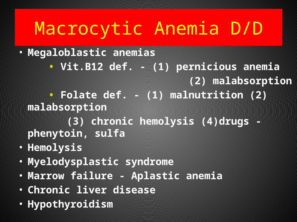
Macrocytic Anemia D/D• Megaloblastic anemias • Vit.B12 def. - (1) pernicious anemia (2) malabsorption • Folate def. - (1) malnutrition (2) malabsorption (3) chronic hemolysis (4)drugs - phenytoin,
sulfa• Hemolysis• Myelodysplastic syndrome• Marrow failure - Aplastic anemia• Chronic liver disease• Hypothyroidism
Macrocytic anemia D/D
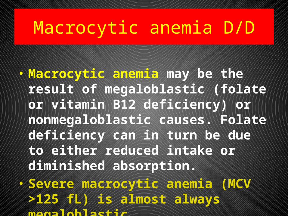
• Macrocytic anemia may be the result of megaloblastic (folate or vitamin B12 deficiency) or nonmegaloblastic causes. Folate deficiency can in turn be due to either reduced intake or diminished absorption.
• Severe macrocytic anemia (MCV >125 fL) is almost always megaloblastic.
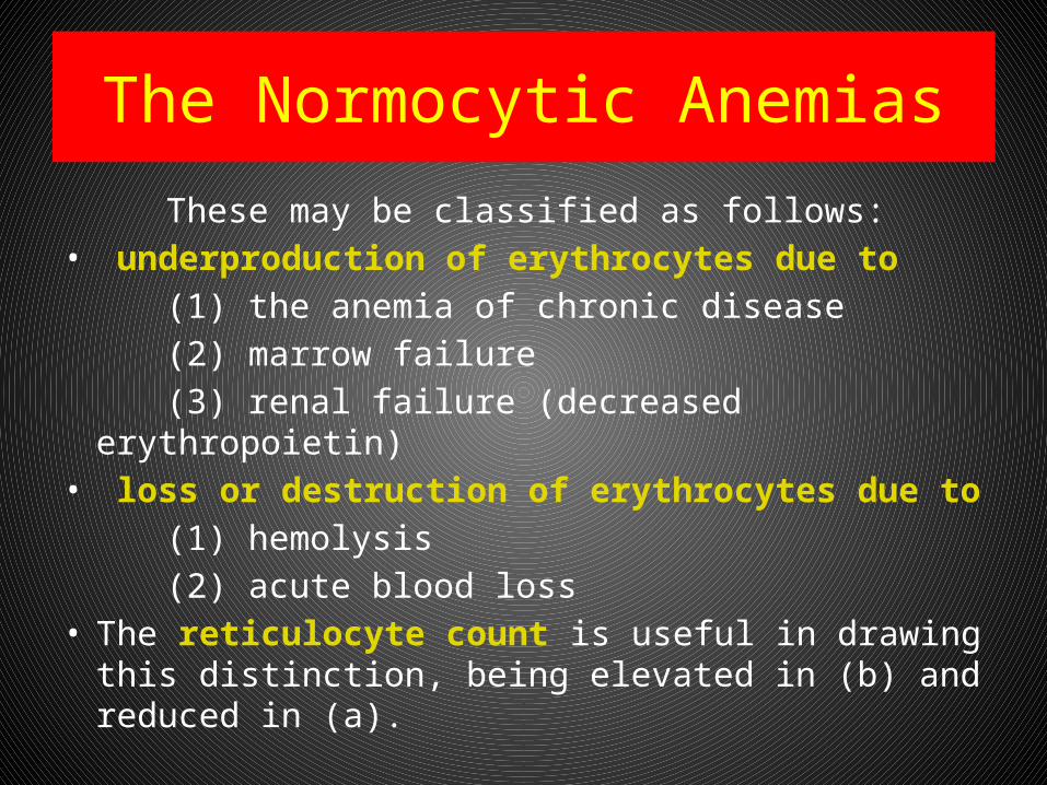
The Normocytic Anemias
These may be classified as follows:• underproduction of erythrocytes due to (1) the anemia of chronic disease (2) marrow failure (3) renal failure (decreased erythropoietin)• loss or destruction of erythrocytes due to (1) hemolysis (2) acute blood loss• The reticulocyte count is useful in drawing this
distinction, being elevated in (b) and reduced in (a).
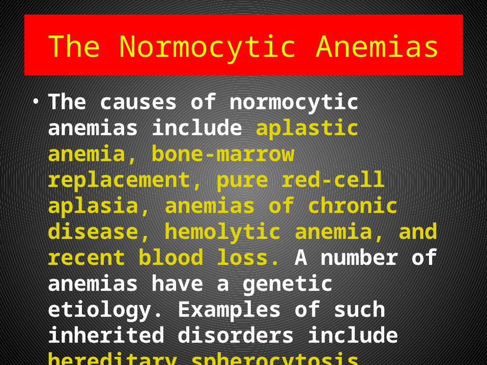
The Normocytic Anemias
• The causes of normocytic anemias include aplastic anemia, bone-marrow replacement, pure red-cell aplasia, anemias of chronic disease, hemolytic anemia, and recent blood loss. A number of anemias have a genetic etiology. Examples of such inherited disorders include hereditary spherocytosis, sickle-cell (SC) anemia, and thalassemia
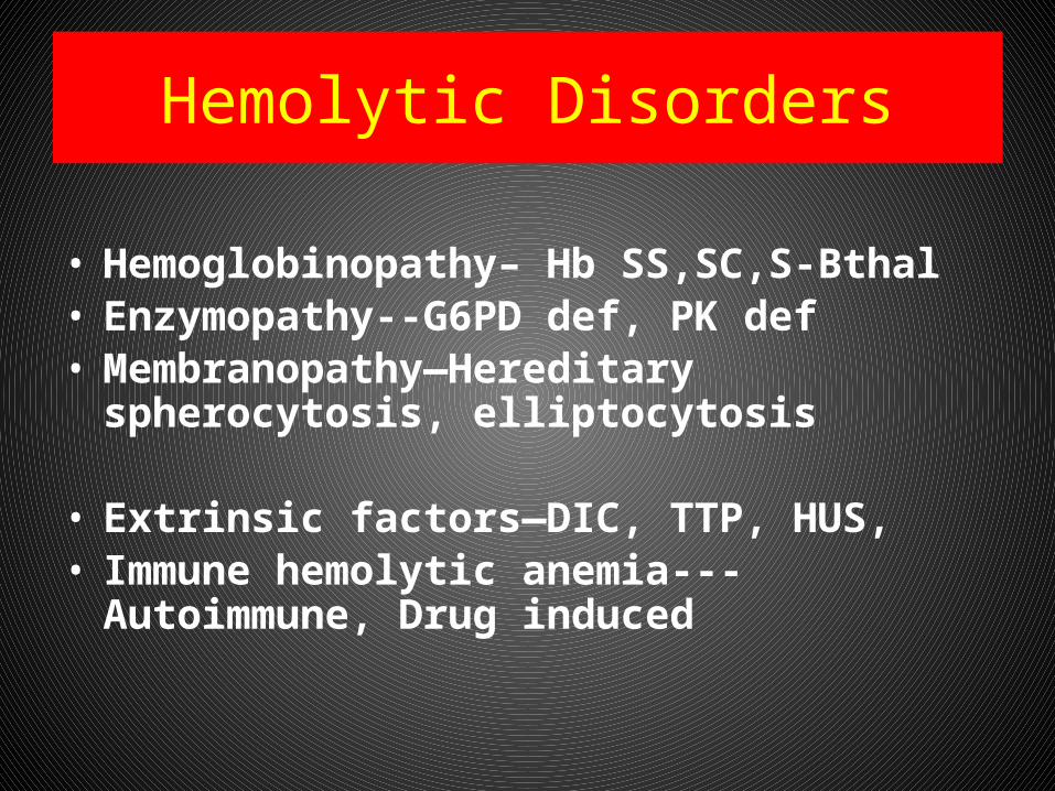
Hemolytic Disorders
• Hemoglobinopathy– Hb SS,SC,S-Bthal• Enzymopathy--G6PD def, PK def• Membranopathy—Hereditary spherocytosis,
elliptocytosis
• Extrinsic factors—DIC, TTP, HUS, • Immune hemolytic anemia---Autoimmune,
Drug induced
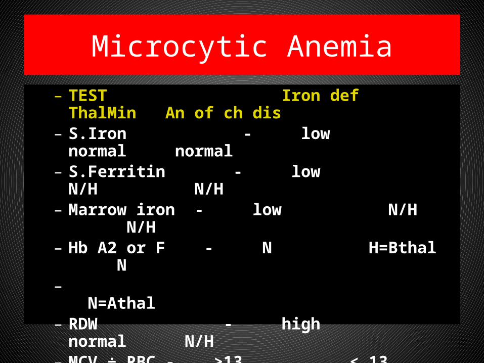
Microcytic Anemia– TEST Iron def ThalMin An of ch dis – S.Iron - low normal normal– S.Ferritin - low N/H N/H– Marrow iron - low N/H N/H– Hb A2 or F - N H=Bthal N– N=Athal– RDW - high normal N/H – MCV ÷ RBC - >13 < 13 – Sickle/B-thal – Hb S > Hb A– Absence of microcytosis in both parents excludes B-
thal or Sickle/B-thal but not A-thal
Iron deficiency anemia (IDA)
• It is a condition when supply of iron in the body to bone marrow falls short of that required for the production of red blood cells. It is the commonest cause of anemia throughout the world.

Iron Deficiency Anemia
• The incidence of anemia in the general population is about 1.5%.
• Iron deficiency related to inadequate replacement of lost iron is the most frequent cause of asymptomatic anemia and has a variety of causes.
• Iron deficiency is common among women of childbearing age; 10% to 20% of menstruating women have abnormally low concentrations of hemoglobin (usually <12 g per 100 mL).
Iron Deficiency Anemia• What are the causes of IDA ?
• Increased physiological demand for more red blood cells
eg: increased physical activity. In children, during spurts of growth.
• In women during menstruation, pregnancy, parturition and lactation.
• Inadequate dietary intake due to poor economic reasons or deficient foods. Decreased absorption due to disorders in the digestive system. GI blood loss Peptic ulcer, piles, hiatus hernia, carcinoma of stomach, carcinoma colon, chronic ingestion of a certain type of pain relievers, hookworm infestation.
Iron Deficiency Anemia
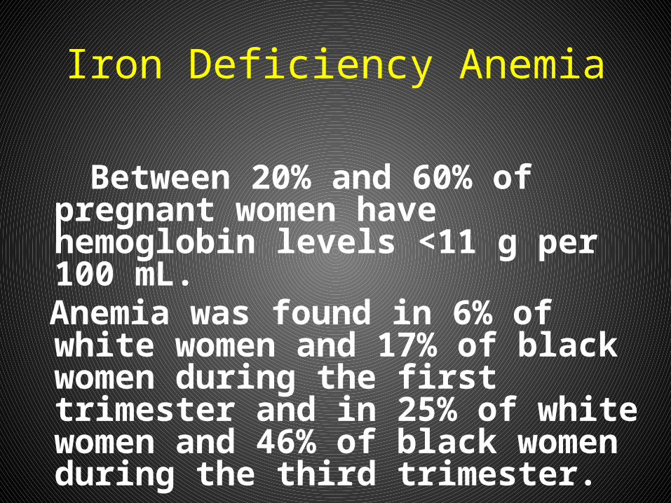
Between 20% and 60% of pregnant women have hemoglobin levels <11 g per 100 mL.
Anemia was found in 6% of white women and 17% of black women during the first trimester and in 25% of white women and 46% of black women during the third trimester.
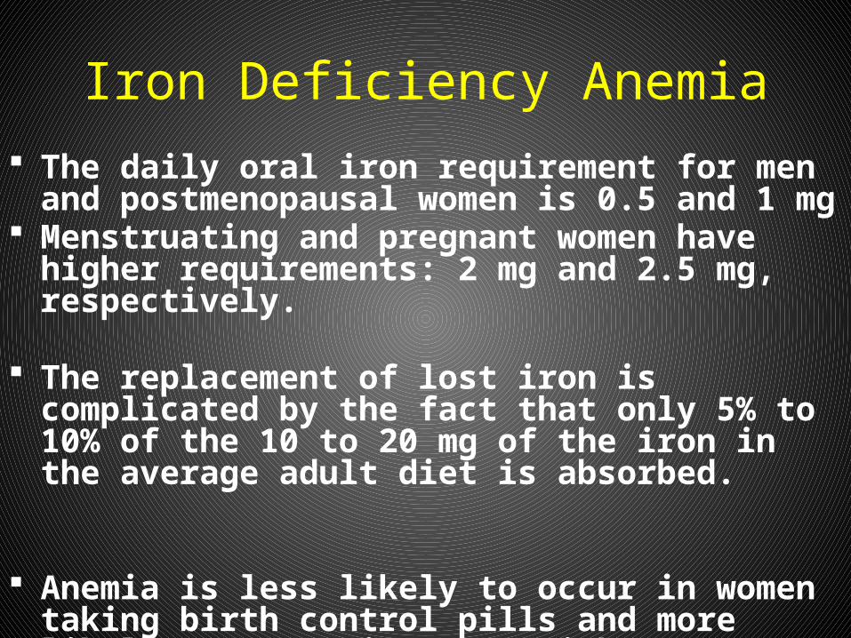
Iron Deficiency Anemia The daily oral iron requirement for men and
postmenopausal women is 0.5 and 1 mg Menstruating and pregnant women have higher
requirements: 2 mg and 2.5 mg, respectively.
The replacement of lost iron is complicated by the fact that only 5% to 10% of the 10 to 20 mg of the iron in the average adult diet is absorbed.
Anemia is less likely to occur in women taking birth control pills and more likely to occur in women with intrauterine devices.
Iron Deficiency Anemia
Because men and postmenopausal women rarely develop iron deficiency that is not related to gastrointestinal blood loss (often occult), an evaluation of gastrointestinal tract must be performed when an iron deficiency is detected in these individuals
Iron Deficiency Anemia-Diagnosis
• Red blood cell count may be normal or decreased.
• Peripheral blood smear shows pale small cells.
• Aniso-poikilocytosis• White blood cell count usually normal• Serum iron is reduced • Total iron binding capacity of blood shows
an increase.• Low serum ferritin
Iron Deficiency Anemia-Treatment
• Correction of iron deficiency - to restore hemoglobin level.
• To replenish iron stores. • Oral iron administration is advised.
(Side effects of oral iron)• Parenteral iron may be needed
occasionally• Treat the underlying cause
Anemia of Chronic Disease
• Patients with cancer, infection, or inflammation, commonly have a mild-to-moderate anemia caused by red cell underproduction.
• This 'anemia of chronic disease' is very common, and is usually normocytic. Some cases develop abnormalities of iron metabolism, in which case there may be a microcytosis.
Hemolysis
• Hemolysis is defined as the premature destruction of red blood cells, from whatever cause.
• Recognising the presence of hemolysis The simplest tests are (a) Raised reticulocyte count(b) Raised indirect (unconjugated) bilirubin(c) Raised serum LDH(d) Diminished serum haptoglobin
concentration. Further more sophisticated testing may be needed in some cases.
Hemolysis
Principal causes Inherited abnormalities * Membrane (Hereditary Spherocytosis)* Hemoglobin (Sickle Cell Anemia)* Enzymes (Glucose-6-phosphate dehydrogenase (G6PD) deficiency)
Hemolysis
Acquired causes• Immune Warm and Cold Autoimmune Hemolytic
Anemia• Non-immune * Mechanical Damage from leaky heart
valves * Microangiopathic hemolytic anemia
(MAHA) like TTP, HUS & DIC
Lead Poisoning
• Hypochromic microcytic anemia• Associated iron deficiency• Child has pica and is exposed to lead paint
or lead dust• Blood smear shows basophilic stipling and
blood lead is elevated.• Removal from exposure,chelation therapy
and correction of iron deficiency are important.
THALASSEMIA
• Normal Hb is a tetramer of 2 alpha and 2 beta chains
• Alpha-thalassemia:decrease or total lack of alpha globin synthesis
• Beta-thalassemia:decrease or total lack of beta globin synthesis
THALASSEMIA
• Clinical classification-• Silent carrier(AorB):normal CBC• Thal trait(AorB):mild anemia(HM)• HbHdisease(A-thal):moderately severe hemolytic
anemia,icterus,splenomegaly• Severe Beta-thal:severe anemia,growth
retardation,hepatosplenomegaly,bony def.• Thalassemia major:tranfusion dependent• Thal-intermedia:no regular transfusions
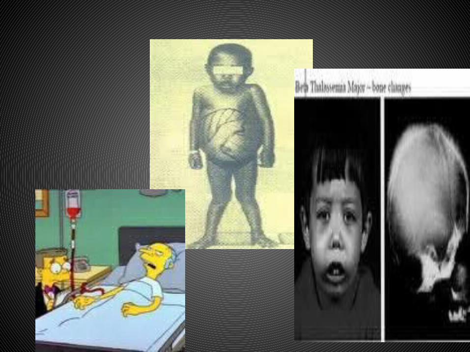
THALASSEMIA-complications
• HbH disease:severe hemolytic anemia,spenomegaly,hypersplenism
• Thal-major:poorly trasfused-skeletal abnormalities,growth retardation,CHF
• Thal-major:well transfused with iron overload-(1)Endocrine disturbances:delayed puberty,growth retardation,diabetes mellitus,hypothyroidism (2)Cardiac:arrhythmias, congestive heart failure (3)Hepatic:cirrhosis,liver failure
THALASSEMIA-complications
• HbH disease:severe hemolytic anemia,spenomegaly,hypersplenism
• Thal-major:poorly trasfused-skeletal abnormalities,growth retardation,CHF
• Thal-major:well transfused with iron overload-(1)Endocrine disturbances:delayed puberty,growth retardation,diabetes mellitus,hypothyroidism
• (2)Cardiac:arrhythmias, congestive heart failure (3)Hepatic:cirrhosis,liver failure
THALASSEMIA-Lab
• Thal trait:Hb 9-10 g/dl• HbH disease:Hb 6-7 g/dl• Thal intermedia:Hb 7-8 g/dl• Thal major:Hb less than 5 g/dl• Peripheral smear:hypochromic,microcytic,
anisopoikilocytosis,target cells• Hb electrophoresis: (1)Thal trait-HbF 1-5%,
HbA2 3.5-8%,rest HbA (2)Thal major- HbF 20-100%,HbA2 2-7%,HbA 0-60%
THALASSEMIA-therapy
• Red cell transfusion 3-4 weekly-Hb 9-10• Chelation therapy with desferrioxamine• Splenectomy if transfusion >200ml/kg/yr• Folic acid 5mg daily• Penicillin prophylaxis to all splenectomised• Pneumococcal and Hib vaccine before sply.• Cholecystectomy for gall stones• Bone marrow transplantation is curative• Genetic counselling
SICKLE CELL DISEASE
• SA :Sickle cell trait-asymptomatic• SS :Sickle cell anemia• S-Bthal:Sickle cell-beta thal• SC :Hb SC disease• Pathophysiology:Valine replaced by
glutamic acid at Beta 6 position. With deoxygenation HbS crystallises&gels
Clinical features• Anemia:chronic,onset at 3-4 mo• Aplastic crisis:parvo virus B12• Sequestation crisis:usuallyspleen• Hemolytic crisis• Dactylitis:Hand foot syndrome(infant)• Painful crisis:muscle,bone,bone marrow,lung, intestines• Cerebrovascular accidents • Acute chest syndrome:infection,infarction,emb• Chronic lung disease:pulmonary fibrosis,restictive lung
disease,cor pulmonale
Clinical features• Priapism• Ocular:retinopathy• Gall bladder disease:stones,cholecystitis• Renal:hematuria,conc.deficit,nephropathy• Cardiomyopathy• Skeletal:avascular necrosis of femoral head• Leg ulceration:in older pts• Infections:pneumococcal pneumonia,meningitis,
arthritis,Hinf sepsis,salmonella&staph osteomyelitis,mycoplasma pneumonia,viral infe
• Growth failure,delayed puberty• Psychologic problems:chronic illness,chronic pain
THERAPY• Anemia is usually chronic&compensated• Blood transfusion only given based on clinical condition,Hb
level&retic count• Crisis:Splenic sequestration crisis,aplastic
crisis,hyperhemolytic crisis-in all of these PRBC is indicated when anemia is sympto.
• Pain crisis:IVF,analgesia with narcotics,NSAIDs• Acute chest syndrome:O2,judicial use of
analgesics&fluids,antibiotics,PRBC• Stroke:O2,fluids,exchange transfusion• Hydroxyurea:decrease number&severity of VOC• Bone marrow transplantation
G6PD deficiency
• Episodic hemolysis on exposure to oxidants• Severity of hemolysis depends on the
enzyme variant• Gene for G6PD is on X chromosome• Jaundice,dark urine(bilirubin,hemoglobin),• Red cells appear blistered• G6PD levels may be normal with hemolysis • Therapy:PRBC,IVF,urine alkalinisation• Prevention:avoid oxidants,fava beans,henna