Embed Size (px)
DESCRIPTION
Anatomy Terminology. Body Regions. Axial Region (down midline of body) Appendicular Region (limbs). REGIONS OF THE BODY. 1. Axial Region ( Goes down midline of the body) a) Head b) Neck c) Trunk (has 3 parts) 1) Thorax (chest area). Above diaphragm. Contains heart and lungs. - PowerPoint PPT Presentation
Citation preview

Anatomy Terminology


Body Regions
Axial Region (down midline of body) Appendicular Region (limbs)

REGIONS OF THE BODY
1. Axial Region (Goes down midline of the body)– a) Head– b) Neck– c) Trunk (has 3 parts)
1) Thorax (chest area). Above diaphragm. Contains heart and lungs.– Pectoral Region (chest)– Costal ( rib) margin
2) Abdomen (not called the stomach!). Contains the digestive organs
Lumbar region (low back)• Gluteal region (buttocks)
3) Pelvis (area that would be covered by brief underwear) Contains urinary and reproductive organs
Inguinal region (Groin)

REGIONS OF THE BODY
2. Appendicular Region (limbs) a) Upper Limbs
1) Axilla (armpit)2) Arm (Brachium): shoulder to elbow
Antecubital fossa (inside of elbow, where blood is drawn)3) Forearm (elbow to wrist). Don’t confuse with arm!4) Wrist5) Hand: 4 fingers with 3 phalanges each; thumb with 2 phalanges;
Pollicis: ThumbPalmar surface: Palm

REGIONS OF THE BODY
2. Appendicular Region (limbs) b) Lower Limbs
1) Thigh (hip to knee). Don’t confuse with leg!2) Leg (knee to ankle).
Calf (back of the leg)Popliteal region (behind knee)Genu: the knee itself
3) Ankle4) Foot: 5 digits
Hallux: big toePlantar surface: sole of foot

Body Cavities
Figure 1.8a

Body Cavities
Figure 1.8b

Regional Terms (not on the quiz or test)
Figure 1.4a

Figure 1.4b
Regional Terms (not on the quiz or test)

Anatomical Position
The body standing erect, facing forward, feet together, toes pointed anteriorly, hands at one’s side, fingers pointing inferiorly, and palms facing forward.
Once the body is in this position (or imagined to be in this position,) the positional terms can be used correctly.

Anatomical Position
Figure 1.3
Anatomical PositionThe person is standing up straightThe palms face anteriorlyThe knees, elbow, and neck are straight (not bent)The toes point anteriorly, but the fingers point inferiorly
Left and Right: yours or the patient’s?

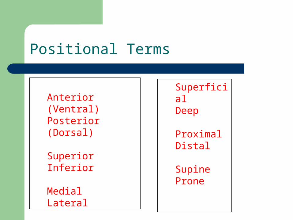
Positional Terms
These are terms used to describe the position of certain structures on the body.
Note: These are “relative terms.” This means that these words are usually used in relating the position of one body structure to another. You can’t say, “He is shorter”. You have to say, “He is shorter than John”.
Incorrect: the nose is medial Correct: the nose is medial to the ears
Positional Terms
Anterior (Ventral)Posterior (Dorsal)
SuperiorInferior
MedialLateral
SuperficialDeep
ProximalDistal
SupineProne

Positional Terminology
Anterior/Ventral: towards the front of the body (includes palms and soles)
Posterior/Dorsal: towards the back of the body Superior: towards the head Inferior: towards the feet Medial (NOT MIDDLE): towards the midline of body Lateral: away from midline Varus: inward angulation of the distal segment of a bone or joint. Valgus: outward angulation of the distal segment of a bone or
joint.

Positional Terminology
Superficial: Toward the external environment Deep: Towards the inner body Proximal: towards the heart Distal: away from the heart Supine: Laying on one’s back Prone: Laying on one’s stomach

Positional Terms

Positional Terms
(Skip)

Positional Terms

Body Planes
• Frontal (Coronal)• Sagittal• Transverse
Sagittal plane
Para-Sagittal plane

Body Planes and Sections• Frontal (coronal) plane
– Lies vertically and divides body or organ into anterior and posterior parts
• Sagittal plane– Divides right from left side of body or organ
• Midsagittal (median) plane– Specific sagittal plane that lies vertically in the midline
and divides body into EQUAL right and left sides • Parasagittal plane
– Specific sagittal plane that lies vertically in the midline and divides body into UNEQUAL right and left sides
• Transverse plane • Divides body or organ into superior-inferior parts

Body Planes and Sections
Figure 1.5
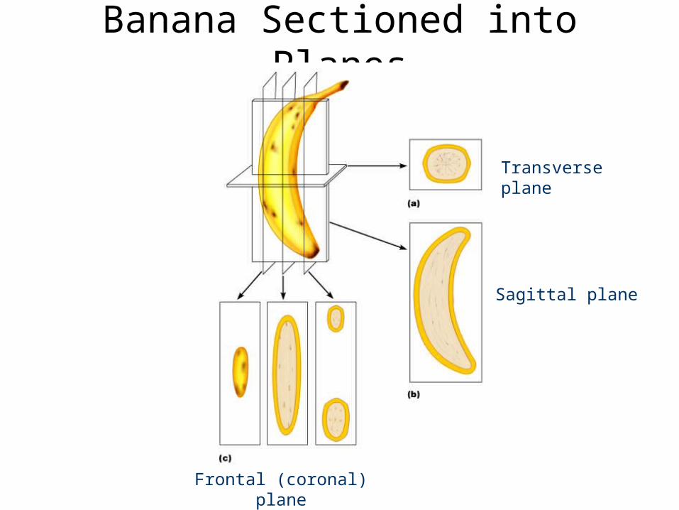
Banana Sectioned into Planes
Transverse plane
Sagittal plane
Frontal (coronal) plane

Movement Terms
Flexion: to decrease the angle of a joint
Extension: to increase the angle of a joint, returning it to anatomical position
Hyperextension: extension beyond anatomical position
In the foot, there are special terms used instead of flexion/extension:
Dorsiflexion: flexion of the ankle joint; to raise the toes up in the air. When you stand on your heels with your toes up in the air, you are dorsiflexing your ankle joints.
Plantarflexion: extension of the ankle joint; to point the toes downward. When you stand on your toes, you are plantarflexing your ankle joints.

Flexion and Extension
Flexion: to decrease the angle of a jointExtension: to increase the angle of a joint, returning it to anatomical positionHyperextension: extension beyond anatomical position

Flexion and Extension
Flexion: to decrease the angle of a jointExtension: to increase the angle of a joint, returning it to anatomical positionHyperextension: extension beyond anatomical position

Flexion, Extension, Hyperextension
ExtensionHyperextension

Movement Terminology
Internal Rotation (or medial rotation): to rotate in the transverse plane toward the midline of the body
External Rotation (or Lateral Rotation): to rotate in the transverse plane away from the midline of the body.
These two terms are usually used to describe motions of the shoulder or hips.

Internal Rotation External Rotation

Movement Terms
Abduction: to move a body part away from the midline of the body in the frontal plane
Adduction: to move a body part toward the midline of the body in the frontal plane
Circumduction: to move a body part in a circle
Rotation: to pivot a body part around an axis, as in shaking the head “no”

Abduction, Adduction, Circumduction

Movement Terminology
Inversion: to rotate in the frontal plane toward the midline of the body. Inversion puts the body part into the varus position.
Eversion: to rotate in the frontal plane away from the midline of the body. Eversion puts the body part into the valgus position.
You invert and evert your hands, but the bones of the ankle don’t move in a single plane. Rather, they move in three planes, so that motion is more properly called supination and pronation.
Movements of the Hand or Foot Only
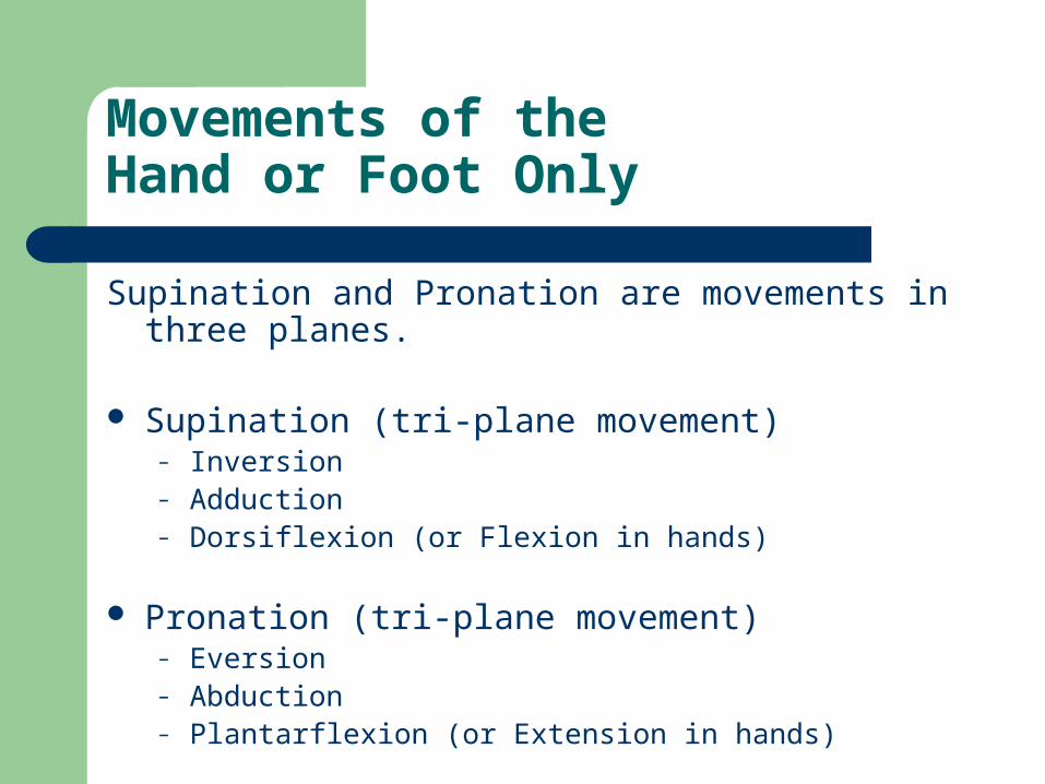
Supination and Pronation are movements in three planes.
Supination (tri-plane movement)– Inversion– Adduction– Dorsiflexion (or Flexion in hands)
Pronation (tri-plane movement)– Eversion– Abduction– Plantarflexion (or Extension in hands)

Pronation Supination

Pronation and Supination
PronationSupination

Gliding Motion

Common Confusion of POSITIONS vs. MOVEMENTS
Prone: a POSITION, not a movement; body is lying face down. Pronation: a MOVEMENT; when the palm is turned downward (in
Anatomical Position, the palm will face posterior). The foot can also be pronated; the sole turns laterally away from the body. Pronation of the foot is a tri-plane movement of plantarflexion, abduction, and eversion.
Supine: a POSITION, not a movement; body is laying on the back. Supination: a MOVEMENT; when the palm is turned upward, like
holding a bowl of soup (in Anatomical Position, the palm will face anterior). The foot can also be supinated; the sole turns medially towards the body. Supination of the foot is a tri-plane movement of dorsiflexion, adduction, and inversion.

Movement Terms
Protraction – to project a body part anteriorly in the transverse plane, such as the shoulders or jaw
Retraction – to pull a body part posteriorly

Movement Terms
Elevation – lifting a body part superiorly, such as shoulders or jaw.
Depression – lowering a body part inferiorly

Movement Terms
Opposition – movement of the thumb to touch the tips of other fingers

Regional Terminology
Thorax– Pectoral Region– Costal = rib
Abdomen Pelvis
– Inguinal (Groin)
Lumbar region Gluteal region Axilla (armpit)
Upper Extremity Arm (Brachium)
Antecubital fossa Forearm Hand
Palmar surface of handLower Extremity Thigh Leg (Calf in back)
Popliteal region (behind knee)
Genu: the knee itself Foot
Plantar surface of foot

Joint Abbreviations
MPJ: Metacarpal (or metatarsal) phalangeal joint

Joint Abbreviations
IPJ: Interphalangeal joint– DIPJ is the distal IPJ– PIPJ is the proximal IPJ
NOTE: The joint at the tip of the thumb is just called the IPJ
PIPJ
IPJ
DIPJ

Anterior-Posterior X-ray (AP view)
X-ray beam passes from anterior to posterior.

Anterior-Posterior X-ray (AP view)
X-ray beam passes from anterior to posterior.

Lateral X-ray (Lat view)
X-ray beam passes from medial to lateral

Lateral X-ray (Lat view)
X-ray beam passes from medial to lateral

Oblique X-ray
Beam enters at 45° angle; good for identifying fractures.

Long Bones
Spongy (cancellous) Bone
Compact Bone

Long Bones Contain Spongy and Compact Bone.

Histology
Histology is the study of normal tissues under a microscope.
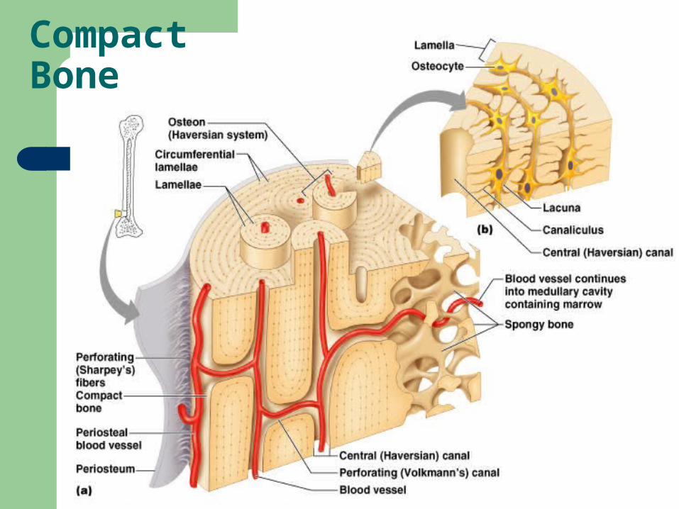
Be able to recognize a description of all the structures seen in compact bone under a microscope:– Lacunae, perforating canal, osteon (functional unit
of compact bone), central canal, canaliculi, lamellae, osteocytes, osteoblasts, osteoclasts, periosteum, hydroxyapatite.
Compact Bone

Compact Bone Structures
Osteon: functional unit of compact bone. hydroxyapatite The crystalline structure of calcium and phosphate
that make up bone matrix lamellae The circular and concentric layers of collagen fibers lacunae The pockets or cavities in which the cells are trapped Haversian (or central) canal The large channels containing a blood
vessel which run longitudinally down the center of each unit canaliculi The “tiny channels” which run transversely through the
layers of bone and allow for diffusion of nutrients and wastes to the cells
perforating canal: connects one Haversian canal to another osteocytes The mature bone cells which are trapped in the matrix and
help to maintain it Osteoblasts: bone cells that lay down new bone Osteoclasts: bone cells that reabsorb bone

Bone Terms to Know
Periosteum (secured to the bone by Sharpey’s fibers) Sharpey’s fibers (anchor the outer wrapping to the bony
matrix below it) Articular Cartilage (cap around long bone) Tendon (attaches muscle to bone) Ligament (attaches bone-to-bone) Aponeurosis (modified tendon) Epiphysis (ends of long bones) Diaphysis (shaft of long bone) Medullary Cavity (hollow area inside long bone) Spongy (cancellous) Bone (contains trabeculae instead of
osteons and lamellae) Trabeculae (appearance of a sponge)
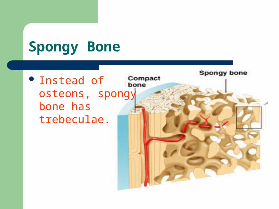
Spongy Bone
Instead of osteons, spongy bone has trebeculae.

Bone Cells
Osteoblast (makes bone) Osteocyte (mature bone cell) Osteoclast (reabsorbs bone)

Histology
Tendons and ligaments are made from what type of connective tissue?– Dense Regular Connective Tissue

Tendon: dense reg. CT

Bones Lab

The Axial Skeleton
Skull Sternum Vertebrae
– 7 Cervical– 12 thoracic– 5 lumbar– 5 sacral– 5 fused coccygeal
Ribs

Appendicular Skeleton
Humerus Radius Ulna Carpals
– Metacarpals– Phalanges (pollicis is thumb)
Femur Patella Tibia Fibula Tarsals
– Metatarsals– Phalanges (hallux is big toe)
Pelvic Girdles Os Coxae (Innominate bone)
– Ilium– Ischium– Pubis
Pectoral Girdles Clavicle Scapula