Embed Size (px)
Citation preview
British Heart Journal, 1976, 38, 186-196.
Analysis of coronary collaterals in ischaemicheart disease by angiography duringpacing induced ischaemia1
M. H. Frick, M. Valle, 0. Korhola, E. Riihimiki, and M. WiljasaloFrom the Cardiovascular Laboratory, First Department of Medicine, andfrom the First Department of Radio-logy, University Central Hospital, Helsinki, Finland
Studies were made using ordinary selective coronary angiography and angiography during ischaemia producedby right atrial pacing, on a series of 41 patients with ischaemic heart disease, to examine the response of thecollaterals to the ischaemia stimulus. Regional myocardial perfusion was determined under the same circum-stances by measuring regional l33Xenon washout curves.No collaterals were found in 8 patients, none of whom demonstrated collaterals when angiography was
repeated during ischaemia. Eleven of the 33 patients with prepacing collaterals (33%) responded to ischaemiawith an increase in the collaterals, 16 patients (49%) showed no change, 5 patients (15%) showed a decreasein the collaterals, and one patient exhibited a bidirectional change. Regional myocardial perfusion responsesclosely paralleled the angiographic changes, yielding suggestive evidence that the collaterals were intimatelyinvolved in the enhancement of the flow.
Despite different collateral andflow responses to ischaemia, the data on exercise tolerance, left ventricularend-diastolic pressure, ejection fraction, prevalence of left ventricular asynergy, and the topographic relationbetween asynergy and collaterals, were largely similar. The data show that in some patients the collateralcirculation reacts to ischaemia by enhancement, but the functional significance of this response is obscure.
Coronary collateral circulation has been the subject functional significance of these different patternsof great interest for decades (Blumgart, Schlesinger, with regard to their role in preventing ischaemia,and Davis, 1940; Zoll, Wessler, and Schlesinger, influencing left ventricular function, and modifying1951; Baroldi, Mantero, and Scomazzoni, 1956; prognosis (Demany, Tambe, and Zimmerman,Bloor and Liebow, 1965; Fulton, 1965; Paulin, 1967; Helfant, Kemp, and Gorlin, 1970; Martinez-1967; Gensini and--Da- Costa,- 1969; James, 1970). Rios et al., 1970; Tuna and Amplatz, 1970;It is now generally accepted that coronary collaterals Helfant, Vokonas, and Gorlin, 1971; Miller et al.,are congenitally determined. Large differences in 1971; Helfant and Gorlin, 1972; Miller et al., 1972;both the number and location of these vessels have Moberg, Webster, and Sones, 1972; Levin et al.,been found in mammalian species (Schaper, 1971), 1973; Banka, Bodenheimer, and Helfant, 1974;but it is not known whether there is such an in- Carroll, Verani, and Falsetti, 1974; Handa et al.,herited variation in man. Angiographic studies of 1974; Lavine et al., 1974; Williams et al., 1974).ischaemic heart disease have revealed subgroups of When there is adequate backflow during selectivepatients with varying collateral patterns which can coronary angiography, the radio-opaque material isbe roughly grouped as follows: 1) no collaterals, not forced under pressure into the high resistance2) some collaterals, and 3) rich collateral networks. coronary arterial circulation, but is merely carriedThere is conflicting evidence relating to the along with the blood stream. Therefore, only col-
Received 7 April 1975. laterals carrying flow are visualized. An additional'Supported by grants from the Finnish State Council for factor is the resolution power of the current radio-Medical Research, the Finnish Heart Association, and the graphic techniques, limiting adequate visualiza-E. Aaltonen Foundation. tion to vessels of 100 tu or more. Of the various
on 11 February 2019 by guest. P
rotected by copyright.http://heart.bm
j.com/
Br H
eart J: first published as 10.1136/hrt.38.2.186 on 1 February 1976. D
ownloaded from
Dynamics of coronary collaterals 187
mechanisms which may establish collateral flow in 1973). An Arriflex cine camera was used at a speed of 80ischaemic heart diseases, effective pressure gradients frames/s. In addition, 35 x 35 cm films were exposed increated by the luminal obstructions evidently play two projections for each coronary artery. The totalthe major role. This is illustrated by the disap- number of selective injections was thus 5 per artery. Ifpearance of the collaterals after successful bypass the patient did not have angina or had only transient andsurgery (Valle et al., 1975). During coronary angio- mild pain, he was asked to repeat the study duringsurgery (Vcalemet al 1975).dD ing coronay aongo- angina to be caused by speeding up the heart; consentgraphy, ischaemia is avoided if possible. Conse- was obtained from every subject. In our laboratory thequently, visualization of the collaterals may be pacing technique has remained essentially the same asincomplete in the absence of the postobstruction originally described (Sowton et al., 1967), consisting of avasodilatation caused by ischaemia. Thus, con- gradual increase in heart rate until the development ofclusions drawn from ordinary coronary angiograms angina and/or dyspnoea, with ischaemic ST segmentdo not necessarily apply to the circumstances pre- changes. The details of this technique combined withvailing during ischaemia. coronary angiography have been described (FrickThe present study compares collateral circula- et al., 1974). In brief, a selective injection was made
tion demonstrated by routine coronary angiography during ischaemia into the coronary artery which gaveandbyangiographdurinischa a i d b rise to the collaterals seen on the prepacing angiograms,and by angiograpny Aduring lscnaema induced by and cine filming was repeated. Thereafter a second
right atrial pacing. Additional information was selective injection was made and 35 x 25 cm films wereobtained by determining the regional myocardial exposed at a rate of 5 per injection. If the collateralsblood flow under the same circumstances. originated from two arteries, both of these were injected.
The amount of contrast material was kept constant in allthe injections. The time interval between the prepacing
Subjects and methods injections and the injections during ischaemia variedfrem 3 to 10 minutes according to the angina threshold
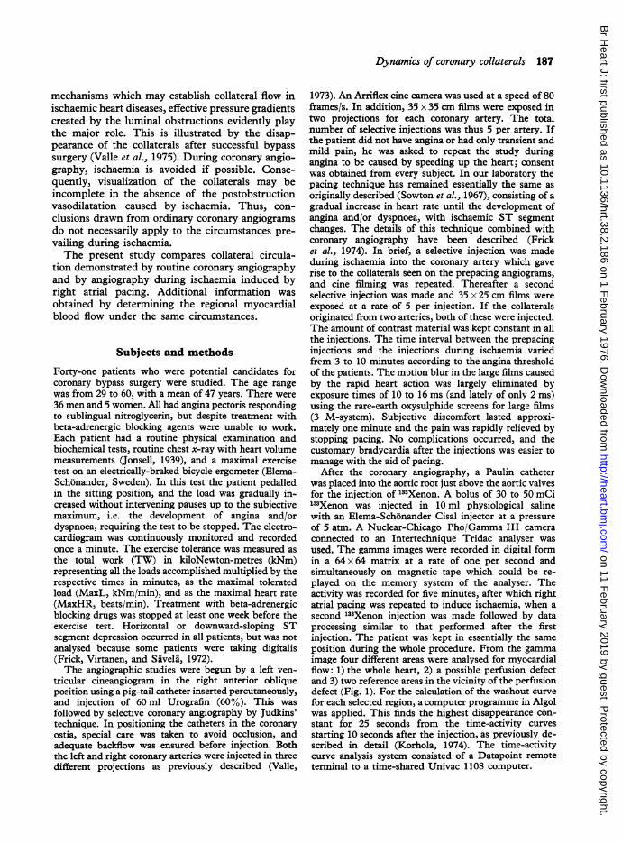
Forty-one patients who were potential candidates for of the patients. The motion blur in the large films causedcoronary bypass surgery were studied. The age range by the rapid heart action was largely eliminated bywas from 29 to 60, with a mean of 47 years. There were exposure times of 10 to 16 ms (and lately of only 2 ms)36 men and 5 women. All had angina pectoris responding using the rare-earth oxysulphide screens for large filmsto sublingual nitroglycerin, but despite treatment with (3 M-system). Subjective discomfort lasted approxi-beta-adrenergic blocking agents were unable to work. mately one minute and the pain was rapidly relieved byEach patient had a routine physical examination and stopping pacing. No complications occurred, and thebiochemical tests, routine chest x-ray with heart volume customary bradycardia after the injections was easier tomeasurements (Jonsell, 1939), and a maximal exercise manage with the aid of pacing.test on an electrically-braked bicycle ergometer (Elema- After the coronary angiography, a Paulin catheterSchonander, Sweden). In this test the patient pedalled was placed into the aortic root just above the aortic valvesin the sitting position, and the load was gradually in- for the injection of 133Xenon. A bolus of 30 to 50 mCicreased without intervening pauses up to the subjective 133Xenon was injected in 10 ml physiological salinemaximum, i.e. the development of angina and/or with an Elema-Schonander Cisal injector at a pressuredyspnoea, requiring the test to be stopped. The electro- of 5 atm. A Nuclear-Chicago Pho/Gamma III cameracardiogram was continuously monitored and recorded connected to an Intertechnique Tridac analyser wasonce a minute. The exercise tolerance was measured as used. The gamma images were recorded in digital formthe total work (TW) in kiloNewton-metres (kNm) in a 64 x 64 matrix at a rate of one per second andrepresenting all the loads accomplished multiplied by the simultaneously on magnetic tape which could be re-respective times in minutes, as the maximal tolerated played on the memory system of the analyser. Theload (MaxL, kNm/min), and as the maximal heart rate activity was recorded for five minutes, after which right(MaxHR, beats/min). Treatment with beta-adrenergic atrial pacing was repeated to induce ischaemia, when ablocking drugs was stopped at least one week before the second 133Xenon injection was made followed by dataexercise test. Horizontal or downward-sloping ST processing similar to that performed after the firstsegment depression occurred in all patients, but was not injection. The patient was kept in essentially the sameanalysed because some patients were taking digitalis position during the whole procedure. From the gamma(Frick, Virtanen, and Savela, 1972). image four different areas were analysed for myocardialThe angiographic studies were begun by a left ven- flow: 1) the whole heart, 2) a possible perfusion defect
tricular cineangiogram in the right anterior oblique and 3) two reference areas in the vicinity of the perfusionposition using a pig-tail catheter inserted percutaneously, defect (Fig. 1). For the calculation of the washout curveand injection of 60 ml Urografin (60%). This was for each selected region, a computer programme in Algolfollowed by selective coronary angiography by Judkins' was applied. This finds the highest disappearance con-technique. In positioning the catheters in the coronary stant for 25 seconds from the time-activity curvesostia, special care was taken to avoid occlusion, and starting 10 seconds after the injection, as previously de-adequate backflow was ensured before injection. Both scribed in detail (Korhola, 1974). The time-activitythe left and right coronary arteries were injected in three curve analysis system consisted of a Datapoint remotedifferent projections as previously described (Valle, terminal to a time-shared Univac 1108 computer.
on 11 February 2019 by guest. P
rotected by copyright.http://heart.bm
j.com/
Br H
eart J: first published as 10.1136/hrt.38.2.186 on 1 February 1976. D
ownloaded from
188 Frick, Valle, Korhola, Riihimaki, and Wiljasalo
Comments on methodsDepending on whether the collaterals originated from theleft or from the right coronary artery, 5 or 10 selectiveinjections were made in the prepacing state before theinjections during pacing-induced ischaemia. It could beargued that these previous injections resulted in thecollateral pattern observed during ischaemia. Analysisof the angiograms obtained during repeated prepacinginjections disclosed no systematic difference from thefirst to the last injection; the collateral pattern remainedboth quantitatively and qualitatively the same as itappeared in the first exposure. The angiographic studieswere begun by an injection of 60 ml Urografin into theleft ventricle. The circulatory effects of angiographyhave been amply documented, and have also beenobserved in our laboratory (Frick, Heikkila, andLuomanmaki, 1970). It is conceivable that the almost
FIG. 1 Gamma image showing a perfusion defect in invariable rise in left ventricular end-diastolic pressurethe anterior wall. The black spots demonstrate areas (LVEDP) persisted throughout the study, with addedselected by a light pen for detailed analysis of regional peaks caused by the selective intracoronary injections.
The aortic pressure was constantly monitored throughmyocardialperfusion. the coronary catheters but was not routinely recorded.
There were no hypotensive episodes requiring treatment.It is known that little or no change in aortic pressure
The left ventricular cineangiograms were analysed for occurs during right atrial pacing (Sowton et al., 1967;regional motion abnormalities (Herman et al., 1967), Frick, 1972), making it highly unlikely that this plays aand ejection fraction was calculated using percentage major role in the ischaemic response.changes in the diameter from multiple axes if required Except in cases with no prepacing collaterals, only(Lewis and Sandler, 1971). the arteries from which the collaterals arose were in-The luminal obstructions in the coronary arteries were jected during ischaemia. This was done to lessen the
estimated as percentages. To facilitate the statistical duration of angina, but as a result limited data werecomparison of different subgroups the scoring system of obtained on the response to ischaemia of the contra-the European Multicentre Study of aorto-coronary lateral artery from which no collaterals arose on thebypass surgery was adopted: 0=no obstruction, prepacing angiogram. The ideal approach would have1 = <25 per cent, 2=25 to 50 per cent, 3=51 to 75 per been simultaneous selective injections into both left andcent, 4= subtotal, and 5= total obstruction. The total right coronary arteries.score for each patient was used. The regional myocardial flow was determined to
In comparing the prepacing and pacing angiograms, obtain supporting data on the functional significanceboth the 35 x 35 cm and the cine films were used and the of the response of the collaterals to ischaemia. A de-comparisons were made in the same phase of the cardiac tailed analysis of regional myocardial flow in multiplecycle. Attention was paid to the calibre of the large areas is beyond the scope of this paper which is con-epicardial arteries and to the collaterals, which were cemed with the demonstration of the dynamic pro-classified as follows: 0=no collaterals, 1 =few collaterals perties of the collaterals. Yet it is essential to understandwithout opacification of postobstructive segments, that a perfusion defect in a scintigram or diffuse hetero-2= collaterals with opacification of a distal segment, and geneity of the uptake of the tracer in a particular area3= excellent filling of a postobstructive segment allowing can be caused by a reduced flow or diminished viablethe assessment of the degree of narrowing. The resolu- muscle mass or both simultaneously. Sometimes even antion power of the angiographic unit permitted analysis of increased flow can be detected in a 'cold spot' resultingvessels of 100 0 or more. In assessing the effect of from flow via large non-nutrient epicardial arteries.ischaemia induced by pacing, the following criteria were Viable muscle mass did not change between scintigramsused: no change, increase in numbers, increase in calibre, made before and during the ischaemia, and the problemdecrease in numbers, and decrease in calibre, of col- encountered in the present study was the difficulty oflaterals. A change was accepted when there was agree- measuring the 'true' myocardial flow. The presentment between three independent observers. Visual technique assesses the highest flow component and theobservations of a change in the calibre of the large changes induced by ischaemia in each area. Conse-epicardial arteries were corroborated by micrometer quently, the main emphasis in the analysis was onmeasurements at equidistant points. The rapid heart rate changes in flow rather than absolute values.during pacing was evident in the cine films and ex- An additional difficulty in gamma imaging ofthe heartcluded a 'blind' analysis of the changes. Student's lies in the fact that the image is dominated by thet-test (one-tailed) was used in determining the signi- anterior wall, the inferior wall being perpendicular to theficance of the difference of sample means. axis of detection. This has led, for example, to a poor
on 11 February 2019 by guest. P
rotected by copyright.http://heart.bm
j.com/
Br H
eart J: first published as 10.1136/hrt.38.2.186 on 1 February 1976. D
ownloaded from
Dynamics of coronary collaterals 189
correlation between inferior wall asynergy and data from artery and the left circumflex was increased. Fourgamma imaging (Korhola et al., 1975). In the present patients showed no change in this respect.series gamma imaging was performed on 30 patients. Thirty-three patients had collaterals in prepacingOnly two patients had significant lesions in either the agorams. Pacintschad resuls in gleft circumflex or the right coronary artery without a angiograms. Pacing to ischaemia resulted in threeconcomitant obstruction in the anterior descending different responses: 1) increase in collaterals, 2) noartery but both these patients had a cold spot in the change, and 3) decrease in collaterals. In the de-anterior wall, and could be analysed in the same way as scription to follow these groups are labelled positivethe other patients. responders, nonresponders, and negative re-
sponders. One patient exhibited a bidirectional
Results change and is analysed separately.Positive responders This group consisted of 11
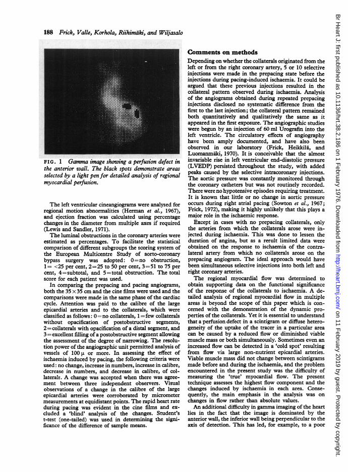
Angiographic data patients, 33 per cent of those with collaterals. InThere were 8 patients without collaterals in the all patients the collaterals were feeding seg-prepacing angiograms. Three of these had entirely ments beyond total occlusions. In 8 patients leftnormal coronary arteries and normal contraction ventricular asynergy was detected and was topo-patterns of the left ventricle. Two had an isolated graphically related to the area distal to occlusion50 per cent stenosis in the left anterior descending and to the site of collaterals; in 2 patientsartery with normal left ventricular contraction in asynergy was absent at the site of collaterals;one, and apical and low anterior hypokinesia in the and in 1 patient with total occlusions in all threeother. There was also one patient with 90 per cent arteries akinesia was observed in an apical area notstenosis in the left anterior descending and 50 supplied by collaterals, but was absent in theper cent stenosis in the right coronary artery (apical inferior wall supplied by collaterals from theakinesia), one patient with 25 per cent stenosis in left anterior descending artery and proximal rightthe right coronary artery (inferior hypokinesia), andone patient with 75 per cent stenosis in the left coronary artery.circumflex and 50 per cent stenosis in the right In the prepacing angiogram 7 patients had classcoronary artery (normal left ventricle). The 2 collaterals; in 2 of these patients, collaterals re-clinical characteristics are given in Table 1. None mained in the same class but increased in number,of these patients showed collaterals when paced to and in 5, they increased to class 3 (Fig. 2). Fourischaemia, but in one patient the calibre of the left patients had class 3 collaterals which increased inanterior descending artery was reduced and in three calibre and in number. In this group 1 patientthe calibre of both the left anterior descending showed a clear widening of the left anterior de-
TABLE 1 Clinical features
No collaterals Prepacing collateralsPositive Non- Negativeresponders responders responders
No. of patients 8 11 16 5Men/women 5/3 10/1 15/1 5/0Age (yr)* 49 47 44 52
41-55 35-55 29-57 47-60Duration of angina 49 29 48 31
pectoris (mth)* 24-168 2-84 9-120 12-60No. of patients with 2 3 7 4
previous infarction (25%) (27%) (44%) (80%)No. of patients with:
1-vessel diseaset 2 3 3 02-vessel disease 2 3 5 33-vessel disease 0 5 8 2no significant disease 4 0 0 0
Coronary score* 2-5 10-3 8-8 10-60-7 7-15 4-15 8-13
*Mean and range.t50 per cent obstruction or more.
on 11 February 2019 by guest. P
rotected by copyright.http://heart.bm
j.com/
Br H
eart J: first published as 10.1136/hrt.38.2.186 on 1 February 1976. D
ownloaded from
190 Frick, Valle, Korhola, Riihimdki, and Wiljasalo~~~~~~~~~~~~~~~~~~~~~~~~~~~~~~~~~~~~~~............... ... ... .......
2A 2B
......F.v _.>. ................._-
_w.~~~~~~~~~~~~~~~~~~~~~~~~~......_SD,~~~~~~~~~~~~~~~~~~~~~~~~~~~~~~~~~~~~~~~~~~........
.....~~gE" ._...
3A 3B
on 11 February 2019 by guest. P
rotected by copyright.http://heart.bm
j.com/
Br H
eart J: first published as 10.1136/hrt.38.2.186 on 1 February 1976. D
ownloaded from
Dynamics of coronary collaterals 191
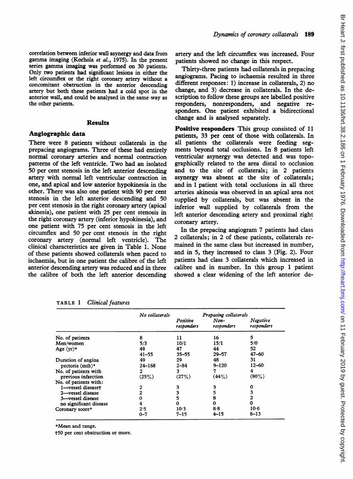
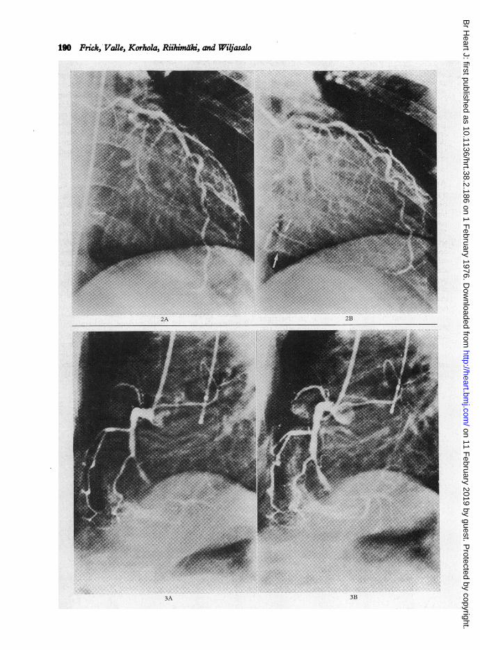
scending and right coronary arteries (Fig. 3) and 1 2 collaterals decreasing to class 1 (Fig. 4), and 1 hadpatient a widening of both the left anterior descend- class 1 collaterals not seen during ischaemia. Oneing and left circumflex arteries. The clinical patient showed an additional narrowing of thefeatures are shown in Table 1 and the circulatory right coronary artery during ischaemia. Clinicalmeasurements in Table 2. and circulatory data are shown in Tables 1
and 2.Nonresponders This group was the largest, com- One patient with total occlusions in the leftprising 16 patients, 49 per cent of those with anterior descending and right coronary arteries andcollaterals. In 12 patients the collaterals were a 90 per cent stenosis in the left circumflex arterysupplying segments distal to total occlusion, in had class 2 collaterals from the left circumflex to4 patients segments distal to 85 to 95 per cent left anterior descending and right coronary arteriesstenosis. In 2 patients no asynergy was found. In and class3 collaterals from the right coronary to13 of the remaining 14 patients left ventricular left anterior descending artery. Pacing to ischaemiaasynergy was topographically related to the ob- resulted in increase in number and calibre of col-asynergyon was topogteraphicallyrelatedatoth re
o laterals to the distal right coronary artery (inferiorstructilons and collaterals. The collaterals were hypokinesia). Coaterals from the proximal rightessentially unchanged during lschaeman The per- coronary to the left anterior descending decreasedtinent data are shown in Tables 1 and 2. in calibre.
Negative responders In 5 patients, 15 per centof those with collaterals, these decreased during Clinical and circulatory correlationsischaemia. Three of the patients had total occlu- The group with no prepacing collaterals differed insions with distal segments supplied by collaterals; several respects from the patients with collaterals.in 2 patients the obstructions were 90 and 95 per There was no patient with total occlusion of anycent. One patient had a normal left ventricular vessel and none had triple vessel disease. In only 3contraction pattern; in 4 patients the left ven- was left ventricular asynergy observed. The meantricular asynergy was related to the site of coronary score was lower than that of any of theobstructions and collaterals. Three patients had groups with collaterals (P <0O001) and the heartclass 3 collaterals decreasing to class 2, 1 had class volume was smaller (P < 0 01). Yet all had angina
TABLE 2 Circulatory measurements
No collaterals Prepacing collateralsPositive Non- Negativeresponders respond, responders
Heart volume (ml/rn BSA)* 408+28 502+19 481+20 473+ 35LVEDP (mmHg)* 15-9+2-2 17-5+1-2 18-8+2-8 14-6+1-9Ejection fraction(%)* 75+4 62+5 66+5 56+7Pacing rate at ischaemia* 127+5 126+5 127+4 146+8
*Mean+ SE of the mean.LVEDP, left ventricular end-diastolic pressure.Conversion factor Traditional to SI units: 1 mmHg- 0133 kPa.
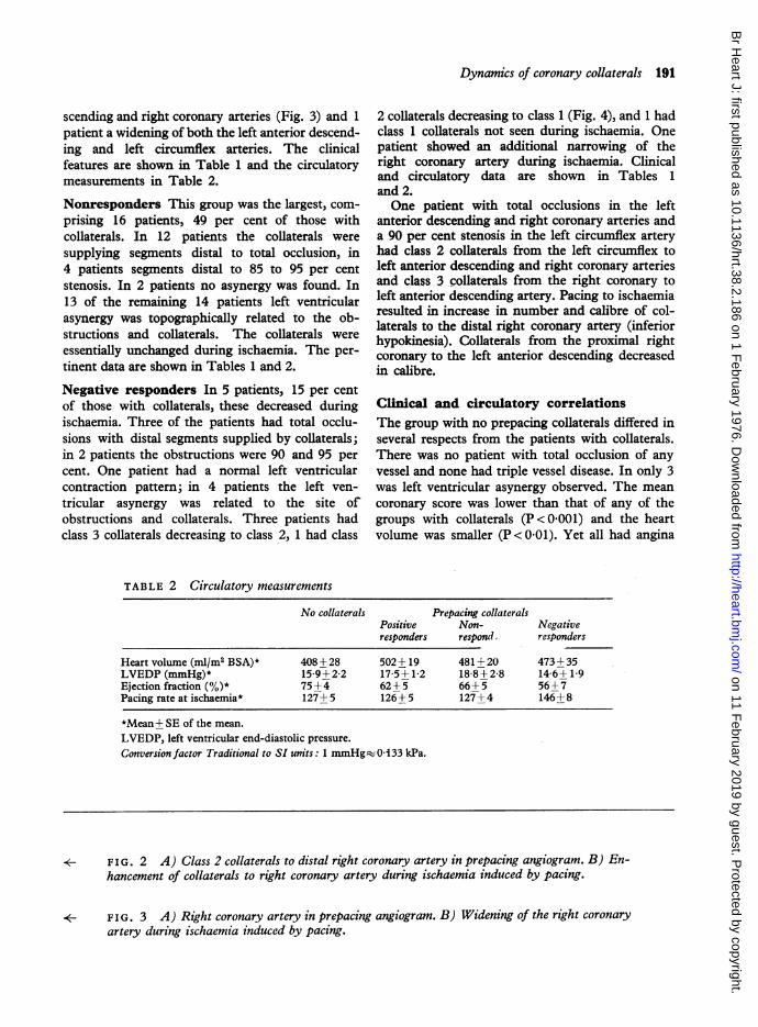
*- FIG. 2 A) Class 2 collaterals to distal right coronary artery in prepacing angiogram. B) En-hancement of collaterals to right coronary artery during ischaemia induced by pacing.
*- FIG. 3 A) Right coronary artery in prepacing angiogram. B) Widening of the right coronaryartery during ischaemia induced by pacing.
on 11 February 2019 by guest. P
rotected by copyright.http://heart.bm
j.com/
Br H
eart J: first published as 10.1136/hrt.38.2.186 on 1 February 1976. D
ownloaded from
192 Frick, Valle, Korhola, Riihimaki, and Wiljasalo
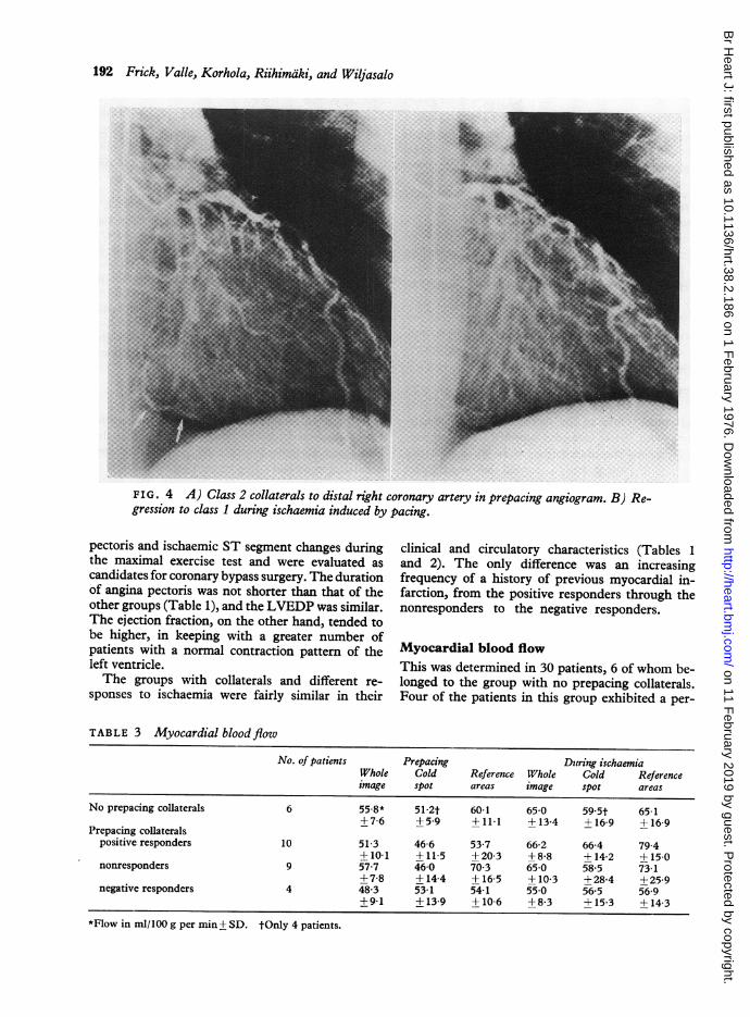
FIG. 4 A) Class 2 collaterals to distal right coronary artery in prepacing angiogram. B) Re-gression to class 1 during ischaemia induced by pacing.
pectoris and ischaemic ST segment changes during clinical and circulatory characteristics (Tables 1the maximal exercise test and were evaluated as and 2). The only difference was an increasingcandidates for coronary bypass surgery. The duration frequency of a history of previous myocardial in-of angina pectoris was not shorter than that of the farction, from the positive responders through theother groups (Tablel1), and theLVEDPwas similar. nonresponders to the negative responders.The ejection fraction, on the other hand, tended tobe higher, in keeping with a greater number ofpatients with a normal contraction pattern of the Myocardial blood flowleft ventricle. This was determined in 30 patients, 6 of whom be-The groups with collaterals and different re- longed to the group with no prepacing collaterals.
sponses to ischaemia were fairly similar in their Four of the patients in this group exhibited a per-
TABLE 3 Myocardial blood flow
No. of patients Prepacing During ischaemiaWhole Cold Reference Whole Cold Referenceimage spot areas image spot areas
No prepacing collaterals 6 55-8* 5i-2t 60-i 65-0 59-5t 65-1±7-6 ±59 ±111 +13-4 ±16-9 ±16-9
Prepacing collateralspositive responders i0 51-3 46-6 53-7 66-2 66-4 794
±10-1 ±11-5 +20-3 ±8-8 +14-2 + 15-0nonresponders 9 57-7 46-0 70 3 65-0 585 731
±7-8 ±14-4 +16-5 ±10-3 +28-4 +25-9negative respioders 4 48-3 53-1 54-1 55 0 565 569+91 +13-9 +10-6 +8-3 +15-3 +14-3
*Flow in ml/i00 g per min + SD. tOnlY 4 patients.
on 11 February 2019 by guest. P
rotected by copyright.http://heart.bm
j.com/
Br H
eart J: first published as 10.1136/hrt.38.2.186 on 1 February 1976. D
ownloaded from
Dynamics of coronary collaterals 193
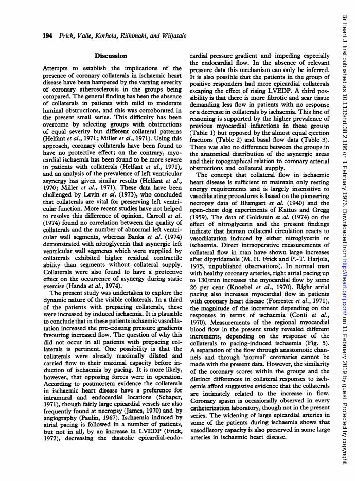
fusion defect in the scintigram, and all 24 patients WHOLE HEARTwith prepacing collaterals in which the scintigramswere made had a perfusion defect. The absolute 5_values for flow before pacing and during ischaemia 40_are given for the different groups in Table 3. Thepercentage changes caused by ischaemia in the four 30different areas used in the measurements are given 3for the various groups in Fig. 5. The data for the 20two areas in the vicinity of the defect were pooled.The exceptional case with the bidirectional
change showed an increase of 23 per cent for thewhole imaged area and a similar increase for theareas in the proximity of the defect. The flow in thecold spot decreased by 5 per cent. A% PERFUSION DEFECTThe myocardial flow of the whole heart responded 50
to ischaemia by a mean increase of 29 per cent inthe positive responder group, contrasting with the 40mean increase of 13 per cent in the nonrespondergroup and 14 per cent in the negative responder 30_group. The difference between the positive re-sponders and the pooled data of the non- and 20negative responders is significant (P<0-05). Themean response of the group without prepacing 10 _.--collaterals was + 17-0 per cent.The flow in the area of the perfusion defect in-
creased in all groups, amounting to +43 per cent L\% VICINITY OF THE DEFECTin the positive responder group. This did not differ 50 _significantly from the responses in the non- and inegative responder groups, +27 per cent, and 40+6-5 per cent respectively, nor from the pooleddata of the non- and negative responders. The 30group with no prepacing collaterals reacted toischaemia by a mean increase of 16'0 per cent. 20The positive responder group showed a mean
increase of 48 per cent in the vicinity of the per- 10 -fusion defect. This was superior to the response of+ 4 per cent in the nonresponders (P < 0-05) and c±5-5 per cent in the negative responders (P < 0 05), FIG. 5 Responses of the regional myocardial per-and the pooled data of the non- and negative re- fusion to ischaemia in relation to the collateralsponders (P < 0.02). The group without collaterals response. The columns represent from left to right:responded by a mean increase of 8 per cent. positive responders, nonresponders, negative re-
sponders, and the group without prepacing collaterals.Exercise toleranceThe exercise tolerance data are shown in Table 4.The total work was somewhat lower in the TABLE 4 Exercise tolerancegroup without prepacing collaterals, but this did notdiffer significantly from the total work in the other Total Maximal Maximalgroups. The maximum tolerated load and maximum work load heart rategroups...(kNm) (kNmlmin) (beats/mtn)heart rate were similar in all groups except for the _kN____kNm_min___beatslmin_negative responses. Three of the 5 women belonged No collaterals 27-5+6-5* 5-3+0 7 132+10to the group without prepacing collaterals and may Prepacing collateralscontribute to the slightly lower exercise tolerance. positive responders 41-9+11-3 6-3+1-1 131+7the maximum heart rates during physical exercise nonresponders 32+7+ 53 569+05 126+5and during pacing were remarkably similar except negative_responders_46_3_7_7_6_6_0_7_105_16in the group of negative responders. *=mean+SE of the mean
on 11 February 2019 by guest. P
rotected by copyright.http://heart.bm
j.com/
Br H
eart J: first published as 10.1136/hrt.38.2.186 on 1 February 1976. D
ownloaded from
194 Frick, Valle, Korhola, Riihimaki, and Wiljasalo
Discussion cardial pressure gradient and impeding especiallythe endocardial flow. In the absence of relevant
Attempts to establish the implications of the pressure data this mechanism can only be inferred.presence of coronary collaterals in ischaemic heart It is also possible that the patients in the group ofdisease have been hampered by the varying severity positive responders had more epicardial collateralsof coronary atherosclerosis in the groups being escaping the effect of rising LVEDP. A third pos-compared. The general finding has been the absence sibility is that there is more fibrotic and scar tissueof collaterals in patients with mild to moderate demanding less flow in patients with no responseluminal obstructions, and this was corroborated in or a decrease in collaterals by ischaemia. This line ofthe present small series. This difficulty has been reasoning is supported by the higher prevalence ofovercome by selecting groups with obstructions previous myocardial infarctions in these grouspof equal severity but different collateral patterns (Table 1) but opposed by the almost equal ejection(Helfant et al., 1971; Miller et al., 1971). Using this fractions (Table 2) and basal flow data (Table 3).approach, coronary collaterals have been found to There was also no difference between the groups inhave no protective effect; on the contrary, myo- the anatomical distribution of the asynergic areascardial ischaemia has been found to be more severe and their topographical relation to coronary arterialin patients with collaterals (Helfant et al., 1971), obstructions and collateral supply.and an analysis of the prevalence of left ventricular The concept that collateral flow in ischaemicasynergy has given similar results (Helfant et al., heart disease is sufficient to maintain only resting1970; Miller et al., 1971). These data have been energy requirements and is largely insensitive tochallenged by Levin et al. (1973), who concluded vasodilatating procedures is based on the pioneeringthat collaterals are vital for preserving left ventri- necropsy data of Blumgart et al. (1940) and thecular function. More recent studies have not helped open-chest dog experiments of Kattus and Greggto resolve this difference of opinion. Carroll et al. (1959). The data of Goldstein et al. (1974) on the(1974) found no correlation between the quality Of effect of nitroglycerin and the present findingscollaterals and the number of abnormal left ventri- indicate that human collateral circulation reacts tocular wall segments, whereas Banka et al. (1974) vasodilatation induced by either nitroglycerin ordemonstrated with nitroglycerin that asynergic left ischaemia. Direct intraoperative measurements ofventricular wall segments which were supplied by collateral flow in man have shown large increasescollaterals exhibited higher residual contractile after dipyridamole (M. H. Frick and P.-T. Hariola,ability than segments without collateral supply. 1975, unpublished observations). In normal manCollaterals were also found to have a protective with healthy coronary arteries, right atrial pacing upeffect on the occurrence of asynergy during static to 130/min increases the myocardial flow by someexercise (Handa et al., 1974). 26 per cent (Knoebel et al., 1970). Right atrialThe present study was undertaken to explore the pacing also increases myocardial flow in patients
dynamic nature of the visible collaterals. In a third with coronary heart disease (Forrester et al., 1971),of the patients with prepacing collaterals, these the magnitude of the increment depending on thewere increased by induced ischaemia. It is plausible responses in terms of ischaemia (Conti et al.,to conclude that in these patients ischaemic vasodila- 1970). Measurements of the regional myocardialtation increased the pre-existing pressure gradients blood flow in the present study revealed differentfavouring increased flow. The question of why this increments, depending on the response of thedid not occur in all patients with prepacing col- collaterals to pacing-induced ischaemia (Fig. 5).laterals is pertinent. One possibility is that the A separation of the flow through anastomotic chan-collaterals were already maximally dilated and nels and through 'normal' coronaries cannot becarried flow to their maximal capacity before in- made with the present data. However, the similarityduction of ischaemia by pacing. It is more likely, Of the coronary scores within the groups and thehowever, that opposing forces were in operation. distinct differences in collateral responses to isch-According to postmortem evidence the collaterals distinaffordfsuggestive evidencelthatptheecollateralin ischaemic heart disease have a preference for aemia afford suggestive evidence that the collateralsintramural and endocardial locations (Schaper, are intimately related to the increase in flow.1971), though fairly large epicardial vessels are also Coronary spasm is occasionally observed in everyfrequently found at necropsy (James, 1970) and by catheterization laboratory, though not in the presentangiography (Paulin, 1967). Ischaemia induced by series. The widening of large epicardial arteries inatrial pacing is followed in a number of patients, some of the patients during ischaemia shows thatbut not in all, by an increase in LVEDP (Frick, vasodilatory capacity is also preserved in some large1972), decreasing the diastolic epicardial-endo- arteries in ischaemic heart disease.
on 11 February 2019 by guest. P
rotected by copyright.http://heart.bm
j.com/
Br H
eart J: first published as 10.1136/hrt.38.2.186 on 1 February 1976. D
ownloaded from
Dynwnics of coronary collaterals 195
The group with no prepacing collaterals included Bloor, C. M., and Liebow, A. A. (1965). Coronary collateralthree patients with normal coronary arteries. In circulation. American Journal of Cardiology, 16, 238.twofthseaXenoscitigrm wa mad, an one
Blumgart, H. L., Schlesinger, M. J., and Davis, D. (1940).two of these a Xenon scintigram was made, and one Studies on the relation of the clinical manifestations ofshowed a clearcut perfusion defect in the anterior angina pectoris, coronary thrombosis, and myocardialwall. These patients, who had severe angina and a infarction to the pathological findings. American Heartpositive maximal exercise test, and were studied as J7ournal, 19, 1.candidates for coronary bypass surgery, presumably Cannon, P. J., Dell, R. B., and Dwyer, E. M., Jr. (1972).canddatefocornarybypss srger, pesumbly Regional myocardial perfusion rates in patients withrepresent the subset of ischaemic heart disease in coronary artery disease. J'ournal of Clinical Investigation,which the classical myocardial oxygen supply- 51, 978.demand mechanism is distorted, leading to un- Carroll, R. J., Verani, M. S., and Falsetti, H. L. (1974). TheI ~~~~~~~effect of collateral circulation on segmental left ventricularpredictable results of exercise testing and atrial contraction. Circulation, 50, 709.pacing. Conti, C. R., Pitt, B., Gundel, W. D., Friesinger, G. C., andThe similarity of the data on left ventricular Ross, R. S. (1970). Myocardial blood flow in pacing-
function and exercise tolerance in the three groups, induced angina. Circulation, 42, 815.posiivenon,ad neatie rsponers is vidnce Demany, M. A., Tambe, A., and Zimmerman, H. A. (1967).positive, non-, and negative responders, is evidence Correlation between coronary arteriography and post-
against the functional significance of the enhance- exercise electrocardiogram. American J7ournal of Cardio-ment of collaterals by ischaemia. It is likely that logy, 19, 526.there are no different subjective thresholds for the Forrester, J. S., Helfant, R. H., Pasternac, A., Amsterdam,angina triggered by a large or small ischaernic area,
E. A., Most, A. S., Kemp, H. G., and Gorlin, R. (1971).angintrigeredy a lrge o smal ischemic rea, Atrial pacing in coronary heart disease: effect on hemody-and that angina in itself may be a manifestation of namics, metabolism and coronary circulation. Americanthe run-off capacity of collateral circulation as sug- Journal of Cardiology, 27, 237.gested by Guyton (1971). The present data indicate, Frick, M. H. (1972). Right atrial pacing in coronary hearthowever, that collaterals are capable of increasing disease. Advances in Cardiology, 8, 193.Frick, M. H., Harjola, P.-T., and Valle, M. (1975). Effect ofboth their size and the amount of flow in response aorto-coronary grafts and native vessel patency on theto an appropriate stimulus. The patient with occurrence of angina pectoris after coronary bypassbidirectional response to ischaemia illustrates that surgery. British Heart Jounral, 37, 414.intealajustnent ocur, videtly avouing Frick, M. H., Heikkilai, J., and Luomanmaiki, K. (1970).internal adjustments occur, evidently favouring FriCPressure responses to left ventricular and aortic angio-
areas with greater demands. A 'stealing' effect graphy characterizing incipient left ventricular failure.(Fuster et al., 1974) could not be studied since the In Incipient Cardiac Insufficiency, p. 111. Sandoz, Basle.contralateral coronary artery was not routinely Frick, M. H., Valle, M., Wiljasalo, M., and Korhola, 0.injected during ischaemia. Though there is evidence (1974)iCm rnary anprgoapchtothe collaterals. Methodo-that collaterals significantly reduce the mortality in logical aspects. Annals of Clinical Research, 6, 253.acute myocardial infarction (Williams et al., 1974), Frick, M. H., Virtanen, K., and Savela, J. (1972). Modifica-and normal values for regional myocardial per- tion of digitalis-induced electrocardiographic changes byfusion under basal conditionshave. been observed propranolol and potassium. Annals of Clinical Research,fusion under basal conditions have been observedl 4, 213.distal to obstructions if well supplied by collaterals Fulton, W. F. M. (1965). The Coronary Arteries. Thomas,(Cannon, Dell, and Dwyer, 1972), the present data Springfield, Illinois.suggest that the ability of collaterals to meet the Fuster, V., Frye, R. L., Connolly, D. C., Danielson, M. A.,increased metabolic demands of pacing or exercise- and Mankin, H. T. (1974). Electrocardiographic-angio-graphic correlation and its influence by collaterals earlyinduced tachycardia is limited. The disappearance in the onset of the coronary syndromes (abstract). Circula-of collaterals after successful bypass grafting (Valle tion, 50, Suppl. 3, 108.et al., 1975), and the correlation of this phenomenon Gensini, G. G., and Da Costa, B. C. B. (1969). The coronarywith improved exercise tolerance after surgery collateral circulation in living man. American Journal of(Frick, Harjola, and Valle, 1975) do not contradict Cardiology, 24., 393.
Goldstein, R. E., Stinson, E. B., Scherer, J. L., Seningen,this argument, but are the result of a changed R. P., Grehl, T. M., and Epstein, S. E. (1974). Intra-distribution of effective pressure gradients. operative coronary collateral function in patients with
coronary occlusive disease. Circulation, 49, 298.Guyton, A. C. (1971). Collaterals, blood flow and tissue
nutrition. New England Journal of Medicine, 284, 1323.References Handa, S., Flessas, A., Connelly, G., Klein, M., Keefe, J.,
and Ryan, T. (1974). Ventricular asynergy during staticBanka, V. S., Bodenheimer,M. M., and Helfant, R. H. (1974). exercise: its relation to coronary lesions and collateral
Determinants of reversible asynergy. Effect of pathologic vessels (abstract). Circulation, 50, Suppl. 3, 51.Q waves, coronary collaterals, and anatomic location. Helfant, R. H., and Gorlin, R. (1972). The coronary collateralCirculation, 50, 714. circulation. Annals of Internal Medicine, 77, 995.
Baroldi, G., Mantero, O., and Scomazzoni, G. (1956). The Helfant, R. H., Kemp, H. G., and Gorlin, R. (1970). Coronarycollaterals of coronary arteries in normal and pathologic atherosclerosis, coronary collaterals, and their relation tohearts. Circulation Research, 4, 223. cardiac function. Annals of Internal Medicine, 73, 189.
Fl
on 11 February 2019 by guest. P
rotected by copyright.http://heart.bm
j.com/
Br H
eart J: first published as 10.1136/hrt.38.2.186 on 1 February 1976. D
ownloaded from

196 Frick, Valle, Korhola, Riihimdki, and Wiliasalo
Helfant, R. H., Vokonas, P. S., and Gorlin, R. (1971). functional significance of the coronary collateral circula-Functional importance of the humn coronary collateral tion in patients with coronary artery disease (abstract).circulation. New England Journal of Medicine, 284, 1277. American Journal of Cardiology, 29, 281.
Herman, M. V., Heinle, R. A., Klein, M. D., and Gorlin, R. Miller, R. R., Zelis, R., Mason, D. T., and Amsterdam, E. A.(1967). Localized disorders in myocardial contraction. (1971). Relation of coronary collateral vessels to ventricularAsynergy and its role in congestive heart failure. New function in patients with equal extent of coronary arteryEngland Journal of Medicine, 277, 222. disease (abstract). Circulation, 44, Suppl. 2, 202.
James,T. N. (1970). The delivery and distribution of coronary Moberg, C. H., Webster, J. S., and Sones, F. M. (1972).collateral circulation. Chest, 58, 183. Natural history of severe proximal coronary disease as
Jonsell, S. (1939). A method for the determination of the heart defined by cineangiography (abstract). American Journalsize by teleoroentgenography (a heart volume index). of Cardiology, 29, 282.Acta Radiologica, 20, 325. Paulin, S. (1967). Interarterial coronary anastomoses in re-
Kattus, A. A., and Gregg, D. E. (1959). Some determinants lation to arterial obstruction demonstrated in coronaryof coronary collateral blood flow in the open-chest dog. arteriography. Investigative Radiology, 2, 147.Circulation Research, 7, 628. Schaper, W. (1971). The Collateral Circulation of the Heart.
Knoebel, S. B., Elliott, W. C., Ross, E., and McHenry, P. L. North-Holland, Amsterdam.(1970). The effect of cardioacceleration by right atrial Sowton, G. E., Blacon, R., Cross, D., and Frick, M. H.pacing on myocardialbloodflowin normal human subjects. (1967). Measurement of the angina threshold using atrialCardiovascular Research, 4, 306. pacing. A new technique for the study of angina pectoris.
Korhola, 0. (1974). Myocardial scintigraphy and estimation of Cardiovascular Research, 1, 301.regional blood flow with Xenon-133. Acta Radiologica, Tuna, N., and Amplatz, K. (1970). The significance ofSuppl. 337. coronary collateral circulation. Coronary arteriographic
Korhola, O., Valle, M., Wiliasalo, M., Riihimaki, E., and electrovectorcardiographic correlations (abstract).Suoranta, H., and Frick, M. H. (1975). Myocardial per- American Journal of Cardiology, 26, 663.fusion defects in ischemic heart disease visualized by Valle, M. (1973). Postoperative coronary angiography. Actasemiselective 133Xe injections. Correlations with left Radiologica, Suppl. 333.ventricular angiography. Journal of Nuclear Medicine and Valle, M., Wiliasalo, M., Frick, M. H., Korhola, O., Suoranta,Biology. (In the press.) H., and Tallorth, K. (1975). Collateral circulation before
Lavine, P., Filip, Z., Najmi, M., Kimbiris, D., Segal, B. L., and after coronary reconstruction. Annals of Clinicaland Linhart, J. W. (1974). Clinical and hemodynamic Research, 7, 251.evaluation of coronary collateral vessels in coronary artery Williams, D. O., Amsterdam, E. A., Hughes, J. L., Miller,disease. American Heart JYournal, 87, 343. R. R., and Mason, D. T. (1974). Importance of coronary
Levin, D. C., Sos, T. A., Lee, J. G., and Baltaxe, H. A. collaterals in acute myocardial infarction: relation to(1973). Coronary collateral circulation and distal coronary pump function, cardiogenic shock and survival (abstract).runoff: the key factors in preserving myocardial contrac- Circulation, 50, Suppl. 3, 108.tility in patients with coronary artery disease. American Zoll, P. M., Wessler, S., and Schlesinger, M. J. (1951).J'ournal of Roentgenology, 119, 474. Interarterial coronary anastomoses in the human heart,
Lewis, R. P., and Sandler, H. (1971). Relationship between with particular reference to anemia and relative cardiacchanges in left ventricular dimensions and the ejection anoxia. Circulation, 4, 797.fraction in man. Circulation, 44, 548.
Martinez-Rios, M. A., Da Costa, B. C. B., Cecena-Seldner,F. A., and Gensii, G. G. (1970). Normal electrocardio- Requests for reprints to Dr. M. H. Frick, Cardiovasculargram in the presence of severe coronary artery disease. Laboratory, First Department of Medicine, UniversityAmerican Journal of Cardiology, 25, 320. Centratory , Haartmaninkat 4f 00290 H elsity
Miller, R. R., Mason, D. T., Salel, A., Zelis, R. F., Massumi, Central Hospital, Haartmaninkatu 4, 00290 Helsinki 29,R. A., and Amsterdam, E. A. (1972). Determinants and Finland.
on 11 February 2019 by guest. P
rotected by copyright.http://heart.bm
j.com/
Br H
eart J: first published as 10.1136/hrt.38.2.186 on 1 February 1976. D
ownloaded from