Embed Size (px)
Citation preview
Heart 1996;76:3-5
Editorial
Imaging the thoracic aorta
Evaluation of techniques for aortic imaging is complicatedby rapid advances in modem technology and the diversenature of pathology affecting this region. It is furtherobfuscated by the assertions of the many single-modalityzealots who trade sensitivity and specificity figures likeinsults. One thing is certain however-no single techniquecurrently offers all the answers to all the questions.
Imaging should quickly, safely, and confidently confirmor refute the presence of aortic disease and determine itsextent for treatment planning and prognosis.I A more pro-saic but no less serious consideration is to present the tho-racic surgeon with images that he or she feels comfortablewith. This will obviously differ with local surgical prefer-ence, some choosing to operate on the basis of ultrasoundfindings alone while others will not commit a patient tointervention without a cineangiogram.The nature of the suspected aortic pathology and the
clinical presentation will dictate how the patient is investi-gated almost as much as the availability of in-house equip-ment and local expertise. Acute processes such asdissection, rupture of an atheromatous aneurysm, ortrauma demand rapid and accurate imaging (preferably atthe presenting hospital), whereas the incidental discoveryof a coarctation, for example, will allow a more compre-hensive response.
Acute aortic diseaseThe urgency of presentation and potential instabilityof the condition of patients with acute aortic dissectioneffectively narrow the imaging options to angiography,computed tomography (CT), and transoesophagealechocardiography (TOE). The few centres which havedeveloped rapid response magnetic resonance imaging(MRI) protocols can justifiably advocate its primary use inthis setting.' Generally speaking, however, MRI is notreadily available and the acutely ill patient unquestionablyappears more vulnerable inside an eight ton magnet.
Angiography, long considered the gold standard, hasbeen pushed into a niche by TOE and CT, largely becauseof its invasive nature and inability to detect dissectinghaematomas where there is no intimal flap.' To considerangiography defunct, however, reveals a naivete about thesubtlety and complexity of aortic disease. Angiography isuseful where there is persisting uncertainty over data fromother tests and for those surgeons who prefer the familiaranatomy of biplane arteriograms. There is no questionthat involvement of branch vessels is more clearly resolvedby this technique. Visualisation of the carotid arteries maybe desirable if the whole arch is to be replaced, althoughwhether coronary angiography is necessary in the acutesetting is debatable.
Transthoracic echocardiography may prove useful inassessing the ascending aorta but it is severely limited dis-tally. In experienced hands, TOE is capable of precisediagnosis of dissection and has the bonus of giving addi-tional information about intracardiac function.4 Images
produced by this novel technique often show exquisitedetail and are capable of convincing the most critical scep-tic (fig 1). As a semi-invasive technique TOE is not withoutrisk in the unstable patient but probably has less potentialthan angiography for causing mishap. Although it com-pares favourably with other techniques, TOE is not fool-proof and reservations have been expressed about blindareas in the ascending aorta and arch produced by themajor airways and by considerable vascular unfolding.5These considerations are much less important than theserious issue of availability. Ultrasound is generallyaccepted as one of the most operator dependent imagingtechniques, and it is therefore important for an out ofhours service to be manned by experienced operators.Maintenance of this level of expertise will be most difficultto achieve in district general hospitals, and may also causeproblems in some tertiary centres. I think that every major
Figure 1 Cross sectional transoesophageal echocardiograms ofa type Aaortc dissection. The thickened intimalflap and compression of the truelumen (TL) by the false lumen (FL) are well demonstrated in both theascending aorta (A) and the proximal descending aorta (B). (Courtesy ofDrNE R Goodfield, Royal Infirmary Edinburgh.)
3
on Decem
ber 17, 2021 by guest. Protected by copyright.
http://heart.bmj.com
/H
eart: first published as 10.1136/hrt.76.1.3 on 1 July 1996. Dow
nloaded from
Editorial
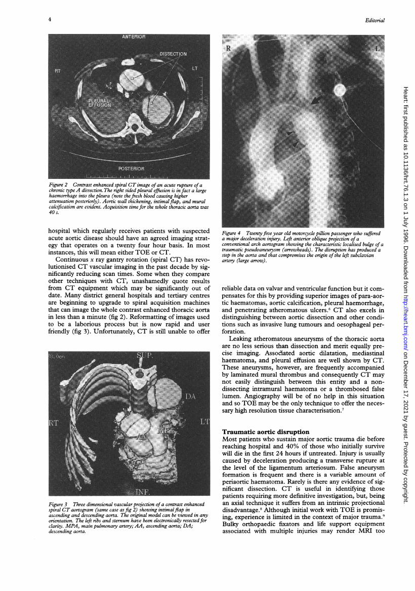
Figure 2 Contrast enhanced spiral CT image of an acute rupture of achronic type A dissection. The right sided pleural effusion is in fact a largehaemorrhage into the pleura (note the fresh blood causing higherattenuation posteriorly). Aortic wall thickening, intimalflap, and muralcalcification are evident. Acquisition time for the whole thoracic aorta was40s.
hospital which regularly receives patients with suspectedacute aortic disease should have an agreed imaging strat-egy that operates on a twenty four hour basis. In mostinstances, this will mean either TOE or CT.
Continuous x ray gantry rotation (spiral CT) has revo-lutionised CT vascular imaging in the past decade by sig-nificantly reducing scan times. Some when they compareother techniques with CT, unashamedly quote resultsfrom CT equipment which may be significantly out ofdate. Many district general hospitals and tertiary centresare beginning to upgrade to spiral acquisition machinesthat can image the whole contrast enhanced thoracic aortain less than a minute (fig 2). Reformatting of images usedto be a laborious process but is now rapid and userfriendly (fig 3). Unfortunately, CT is still unable to offer
Figure 3 Three dimensional vascular projection of a contrast enhancedspiral CT aortogram (same case as fig 2) showing intimalflap inascending and descending aorta. The original model can be viewed in anyorientation. The left ribs and sternum have been electronically resectedforclarity. MPA, main pulmonary artery; AA, ascending aorta; DA;descending aorta.
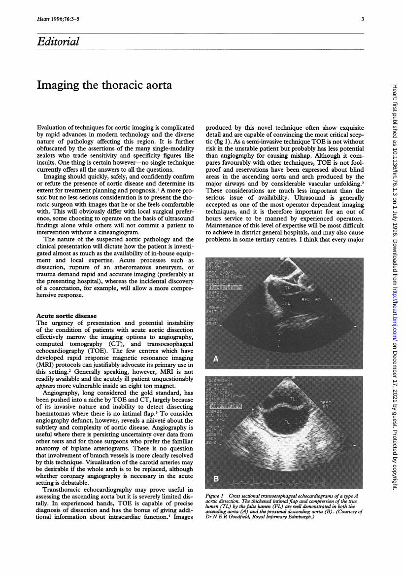
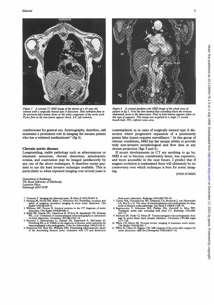
Figure 4 Twenty five year old motorcycle pillion passenger who suffereda major deceleration injury. Left anterior oblique projection of aconventional arch aortogram showing the characteristic localised bulge of atraumatic pseudoaneurysm (arrowheads). The disruption has produced astep in the aorta and that compromises the origin of the left subclavianartery (large arrow).
reliable data on valvar and ventricular function but it com-pensates for this by providing superior images of para-aor-tic haematomas, aortic calcification, pleural haemorrhage,and penetrating atheromatous ulcers.6 CT also excels indistinguishing between aortic dissection and other condi-tions such as invasive lung tumours and oesophageal per-foration.
Leaking atheromatous aneurysms of the thoracic aortaare no less serious than dissection and merit equally pre-cise imaging. Assoodiated aortic dilatation, mediastinalhaematoma, and pleural effusion are well shown by CT.These aneurysms, however, are frequently accompaniedby laminated mural thrombus and consequently CT maynot easily distinguish between this entity and a non-dissecting intramural haematoma or a thrombosed falselumen. Angiography will be of no help in this situationand so TOE may be the only technique to offer the neces-sary high resolution tissue characterisation.7
Traumatic aortic disruptionMost patients who sustain major aortic trauma die beforereaching hospital and 40% of those who initially survivewill die in the first 24 hours if untreated. Injury is usuallycaused by deceleration producing a transverse rupture atthe level of the ligamentum arteriosum. False aneurysmformation is frequent and there is a variable amount ofperiaortic haematoma. Rarely is there any evidence of sig-nificant dissection. CT is useful in identifying thosepatients requiring more definitive investigation, but, beingan axial technique it suffers from an intrinsic projectionaldisadvantage.8 Although initial work with TOE is promis-ing, experience is limited in the context of major trauma.9Bulky orthopaedic fixators and life support equipmentassociated with multiple injuries may render MRI too
4
on Decem
ber 17, 2021 by guest. Protected by copyright.
http://heart.bmj.com
/H
eart: first published as 10.1136/hrt.76.1.3 on 1 July 1996. Dow
nloaded from
Editorial
Figure 5 A coronal Tl MRI image of the thorax ofa 45year oldwoman with a surgically treated type A dissection. Slow turbulentflow inthe persistent false lumen shows as the white component of the aortic arch.Fasterflow in the true lumen appears black. LV, left ventricle.
cumbersome for general use. Arteriography, therefore, stillmaintains a prominent role in imaging the trauma patientwho has a widened mediastinum"° (fig 4).
Chronic aortic diseaseLongstanding, stable pathology such as atheromatous ortraumatic aneurysm, chronic dissection, annuloaorticectasia, and coarctation may be imaged satisfactorily byany one of the above techniques. It therefore seems pru-dent to use the least invasive technique available. This isparticularly so when repeated imaging over several years is
Figure 6 A coronal gradient echo MRI image of the whole torso ofpatient infig 5. Note the thin intimalflap extending down the tortuousabdominal aorta to the bifurcation. Flow in both lumens appears white onthis type of sequence. This image was acquired in a single 1I secondbreath hold. IVC, inferior vena cava.
contemplated, as in cases of surgically treated type A dis-section where progressive expansion of a persistentlypatent false lumen requires surveillance." In this group ofchronic conditions, MRI has the unique ability to providetruly non-invasive morphological and flow data in anychosen projection (figs 5 and 6).
If recent developments in CT are anything to go by,MRI is set to become considerably faster, less expensive,and more accessible in the near future. I predict that ifmagnet evolution is maintained there will ultimately be nocontroversy over which technique is best for aortic imag-ing.
JOHN H REID
Department ofRadiology,The Royal Infirmary ofEdinburgh,Lauriston Place,Edinburgh EH3 9YW
1 Treasure T. Imaging the dissected aorta. Br HeartJ 1993;70:497-8.2 Panting JR, Norell MS, Baker C, Nicholson AA. Feasibility, accuracy and
safety of magnetic resonance imaging in acute aortic dissection. ClinRadiol 1995;50:455-8.
3 Williams MP, Farrow R. Atypical patterns in the CT diagnosis of aorticdissection. Clin Radiol 1994;49:686-9.
4 Ballal RS, Nanda NC, Gatewood R, D'Arcy B, Samdarshi TE, HolmanWL, et al. Usefulness of transesophageal echocardiography in assessmentof aortic dissection. Circulation 1991;84: 1903-16.
5 Kronzon I, Demopoulos L, Schrem SS, Pastemack P, McCauley D,Freedberg RS, et al. Pitfalls in the diagnosis of thoracic aortic aneurysm bytransesophageal echocardiography. JAm Soc Echocardiogr 1990;3:145-8.
6 Kazerooni EA, Bree RL, Williams DM. Penetrating atherosclerotic ulcersof the descending thoracic aorta: evaluation with CT and distinction
from aortic dissection. Radiology 1992;183:759-65.7 Taams MA, Gussenhoven WJ, Schippers LA, Roelandt J, van Herwerden
LA, Bos E, et al. The value of transesophageal echocardiography for diag-nosis of thoracic aorta pathology. EurHeartJ 1988;9:1308-16.
8 Raptopoulos V, Scheiman RG, Phillips DA, Davidoff A, Silva WE.Traumatic aortic tear: screening with chest CT. Radiology 1992;182:667-73.
9 Moloney JF, Duffy CI, Plehn JF. Transesophageal echocardiographic find-ings in severe blunt chest trauma (abstract). Circulation 1991;84 suppl2:11-128.
10 White CS, Mirvis SE. Pictorial review: imaging of traumatic aortic injury.Clin Radiol 1995;50:281-7.
11 White R, Ullyot D, Higgins CB. MR imaging of the aorta after surgery foraortic dissection. AYRAm Y Roentgenol 1988;150:87-92.
5
on Decem
ber 17, 2021 by guest. Protected by copyright.
http://heart.bmj.com
/H
eart: first published as 10.1136/hrt.76.1.3 on 1 July 1996. Dow
nloaded from