Embed Size (px)
Citation preview
A comparison of the bone-like apatite
formation potency between Hydroxyapatite
and β-Tricalcium phosphate
in Glass ionomer dental luting cement.
Sang Il Yoon
The Graduate School
Yonsei University
Department of Dental Science
A comparison of the bone-like apatite
formation potency between Hydroxyapatite
and β-Tricalcium phosphate
in Glass ionomer dental luting cement.
A Dissertation Thesis
Submitted to the Department of Dental Science
and the Graduate School of Yonsei University
in partial fulfillment of the
requirements for the degree of
master of Dental Science
Sang Il Yoon
December 2008
This certifies that the dissertation
of Sang Il Yoon is approved.
Thesis Supervisor : Prof. Hyung Jun Choi
Heung Kyu Son
Yong Keun Lee
The Graduate School
Yonsei University
June 2008

감사의 글
논문이 완성되기까지 자상하게 지도해 주시고 관심을 아끼지 않고
격려해주신 최형준 지도 교수님께 감사드리며, 실험설계부터 논문이 끝
나기까지 항상 조언을 해주신 이용근 교수님과 애정어린 관심과 격려
를 아끼지 않으신 소아치과의 모든 교수님께 감사드리며, 실험실의 모
든 것 하나하나를 가르쳐주신 치과재료학 교실 여러분께도 깊은 감사
의 말씀드립니다.
모든 실험과정과 논문쓰기까지 사랑과 용기로 감싸주신 병원 가족
모두에게 감사드리며, 실험실을 항상 친구처럼 같이해준 박상욱 선생님,
채문희 선생님, 논문의 완성에 좋은 의견을 제시해준 이정진 선생님,
홍은경 선생님께도 감사드립니다.
3년을 넘게 항상 옆에서 나의 든든한 후원자가 되어주고, 격려해주며
슬픔과 기쁨을 함께해준 아내 김선희에게 감사의 뜻을 전합니다.
끝으로 지난 긴 시간동안 논문이 완성되기까지 항상 제 든든한 버팀
목이 되어주셨던 아버님, 어머님, 장모님, 형, 형수님, 처제께 제 지난
날의 사랑을 담아 이 논문의 영광을 바칩니다.
저자 씀

i
Table of contents
List of Figures and Tables····································································ii
Abstract ································································································iv
I. Introduction ·······················································································1
II. Materials and Methods ···································································4
1. Materials ·······················································································4
2. Methods ························································································7
1) Preparation of sample·································································7
2) Film thickness ············································································8
3) Setting time ················································································8
4) Compressive strength ·································································9
5) Bonding strength ······································································10
6) Surface observation··································································11
7) Statistical evaluation ································································11
III. Results ···························································································12
1. Film thickness·············································································12
2. Setting time·················································································14
3. Compressive strength ·································································16
4. Bonding strength ········································································18
5. Microstructure ············································································20
IV. Discussion······················································································23
V. Conclusion·······················································································26
VI. References·····················································································28
Abstract in Korean ·············································································30

ii
List of Figures
Fig. 1. RelyXTM GIC (3M/ESPE, USA).··············································5
Fig. 2. β-tricalcium phosphate.····························································5
Fig. 3. Calcium phosphate tribasic (HA).···········································6
Fig. 4. Samples for the investigation of bonding strength.··············10
Fig. 5. Film thickness of GIC-apatites.·············································13
Fig. 6. Setting time of GIC-apatites.·················································15
Fig. 7. Compressive strength of GIC-apatites.·································17
Fig. 8. Bonding strength of GIC-apatites.········································19
Fig. 9. Dentin surface of control group(pure GIC) under SEM.····21
Fig. 10. Dentin surface of 15% HA-GIC group under SEM. ·········21
Fig. 11. Dentin surface of 15% BCP-GIC group under SEM.········22
Fig. 12. Dentin surface of 15% ββββTCP-GIC group under SEM····22

iii
List of Tables
Table 1. The component of SBF in this study.····································6
Table 2. Sample identification of GIC in this study.··························7
Table 3. Film thickness of GIC-apatites···········································13
Table 4. Setting time of GIC-apatites.···············································15
Table 5. Compressive strength of GIC-apatites.······························17
Table 6. Bonding strength of of GIC-apatites.·································19

iv
Abstract
A comparison of the bone-like apatite formation potency
between Hydroxyapatite and ββββ-Tricalcium phosphate
in Glass ionomer dental luting cement
An increase in bonding strength due to the formation of bone-like
apatite when of glass ionomer dental luting cement(GIC) and
hydroxyapatite(HA) is bonded to human teeth has been reported in
previous studies. However, the amount of bone-like apatite formed was
rather low due to the low solubility of HA. The purpose of this study
was to determine whether a mixture of highly soluble β-tricalcium
phosphate(β-TCP) and HA added to glass ionomer cements increases
bone-like apatite formation in the tooth-cement interface. Also, this
study aims to determine whether the bioactivity of biphasic calcium
phosphate(BCP: a mixture of HA and TCP), which is known to have
both the physical properties of HA and the high solubility of β-TCP, is
affected when used in dental luting cements.
Considering the fact that 15% HA-GIC(RelyXTM luting, 3M ESPE,
USA) had the best physical properties in previous studies, the same
composition of β-TCP and HA was used. In addition, an 85/15 mixture

v
BCP of HA and β-TCP, which has been reported to have the greatest
osteoinductive potency(HUIPIN YOAN et al., 2002) was used, Total of
four groups: the control group using pure glass ionomer, and the 3 test
groups using 15% HA-GIC, 15% BCP-GIC, 15% TCP containing
85/15 mixture of HA and β-TCP, respectively, were established. The
necessary requirement to be used as a dental luting cement is the film
thickness, setting time, and compressive strength. Therefore, the
experiments were conducted with an emphasis on these three factors,
following ISO standard 9917-1:2003(E) regulations. Five samples from
each group were bonded to natural teeth and stored in simulated body
fluid(SBF) at 36.5oC. After four weeks, the bonding strength was
measured, and the luting surface was analyzed using a scanning
electronic microscope(SEM).
No significant differences in film thickness were observed between
the 15% HA-GIC, 15% BCP-GIC, 15% TCP-GIC groups, which were
thinner than that of pure GIC. The setting time of the pure GIC group
was shorter than that of the three test groups, but no significant inter-
group differences were observed. Compressive strength was
proportional to the amount of HA, with 15% HA-GIC having the
greatest value, followed by 15% BCP-GIC, 15% TCP-GIC, and GIC, in

vi
decreasing order. 15% TCP-GIC and 15% BCP-GIC had the highest
bonding strengths, and a SEM analysis of cross sections revealed that
bone-like apatite formation was proportional to the amount of β-TCP.
Improvements in physical properties were observed in the HA, BCP,
TCP-GIC groups, and in particular, bonding strength - which was the
most important aspect of interest in this study - showed remarkable
improvements. The higher bonding strengths of 15% TCP-GIC group
compared to 15% HA-GIC group may be explained by the high
solubility of β-TCP, which facilities bone-like apatite formation in the
tooth-cement interface. However, further long-term studies involving
experiments with various compositions of HA and β-TCP will pave the
way for advancements in dental cements.
Keywords: Glass ionomer cement, hydroxyapatite, BCP, β-TCP, Bone like apatite

1
A comparison of the bone-like apatite formation
potency between Hydroxyapatite and ββββ-Tricalcium
phosphate in Glass ionomer dental luting cement.
Sang Il Yoon
Department of Dental Science, The Graduate School Yonsei University
(Directed by professor Hyung Jun Choi, DDS, PhD)
I. Introduction
Dental luting cement is a medium to bond the restorative material to
the teeth. It has been studied for improvement of bonding strength with
the human teeth. Glass ionomer cement was developed(Wilson& Kent,
1972) and is being widely used clinically nowadays. Glass ionomer
cement bonds with tooth not only mechanically just like other cements,
but also chemically(Erickson& Glasspoole, 1994). However, chemical
bonding was almost insignificant in clinical application of glass
ionomer cement because of the weak nature of chemical bonding and
the majority of bonding depends on mechanical retention, just like

2
other dental luting cements. This weakness could lead to secondary
dental caries or restoration exclusion due to bacterial invasion or
microleakage(Pachuta& Meiers, 1995). In addition, restorations could
be fallen out easily when insufficient amount of tooth material is
available because bonding does not occur directly between the tooth
and the cement. Therefore, not only the physical property of the dental
luting cement itself, but its changes at the interface with the teeth are
very important. To overcome these shortcomings, for instance, various
fillers had been added to glass ionomer dental luting cement such as
silver-cermets, stainless steel powder, carbon and alumino-silicate
fibers, and hydroxyapatite(Kawano, 2001; Xu, 2000). In the previous
study, bone-like material forming ability by mixing hydroxyapatite
with several bone cements was reported in a protein-free acellular
simulated body fluid with ion concentrations nearly equal to those of
the human blood plasma. Accordingly, hydroxyapatite has been of
considerable interest as a biomaterial for implants and bone
augmentation since its chemical composition is close to that of bone
and tooth(M. Okumura, 1997). According to a previous study, 15%
hydroxyapatite, when added to the glass ionomer cement, increases the
bonding strength with human teeth by forming bone-like apatite(Y.

3
Sangil, 2004). However, due to low solubility of hydroxyapatite, only
limited amount of bone-like apatite formation was observed under SEM.
In this study, we expected to increase the formation of bone-like apatite
by using 15% β-tricalcium phosphate, the same apatite system as
hydroxyapatite but with higher solubility and smaller size of particle.
But, Pure β-tricalcium phosphate has poor physical property compared
with hydroxyapatite. Therefore, 15% biphasic calcium phosphate, a
mixture of pure hydroxyapatite and pure β-tricalcium phosphate was
used in this study. Biphasic calcium phosphate was prepared as 85/15
mixture of hydroxyapatite and β-tricalcium phosphate, the ratio
known to have the highest osteoinductive potential(Huipin Yuan et al,
2002).
The aim of this study was to enhance the bonding strength of glass
ionomer cement at the interspaces with teeth, by utilizing bioactivities
of hydroxyapatite and high solubility of β-tricalcium phosphate, which
result in increased apposition of bone-like apatite at the interspaces
with teeth. This will provide a good foundation for the wide application
of the bioactivity of apatite in other fields of the dentistry.

4
II. Materials and Methods
1. Materials
Commercially available glass ionomer cement(GIC),
hydroxyapatite(HA) and β-tricalcium phosphate(β-TCP) were
purchased to prepare the composite of glass ionomer cement and HA or
β-TCP. RelyXTM GIC (3M/ESPE, USA) was selected in this study.
Fluka(Sigma-Aldrich Inc., USA) was selected in this study as β-TCP.
It’s molecular fomula is Ca3(PO4)2 and molecular weight is 310.2.
Calcium phosphate tribasic(Sigma-Aldrich Inc., USA) was selected as
HA. It’s molecular fomula is Ca5(OH)(PO4)3 and molecular weight is
502.3. To provide an environment similar to intraoral, test specimens
were maintained at 36.5˚C similar to body temperature in simulated
body fluid(SBF) which has a composition as saliva.
5
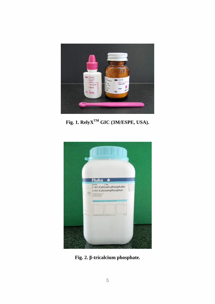
Fig. 1. RelyXTM GIC (3M/ESPE, USA).
Fig. 2. β-tricalcium phosphate.

6
Fig. 3. Calcium phosphate tribasic(HA).
Table 1. The composition of simulates body fluid in this study
List Material 1L (g) 1 NaCl 7.996 2 NaHCO3 0.35 3 KCl 0.224 4 K2HPO4.3H2O 0.174 5 MgCl2.6H2O 0.305 6 1M-HCl 40 ml 7 CaCl2 0.278 8 Na2SO4 0.071 9 NH2C(CH2OH)3 6.057

7
2. Methods
1) Preparation of sample
According to previous studies, where 15% HA-RelyXTM GIC was
shown to have most improved physical property, we limited the
concentration of apatite-GIC to 15%(Y. Sangil, 2004).
15% concentration of HA, β-TCP, BCP(85/15 HA and β-TCP) were
replaced for glass powder, keeping the water/powder ratio of 2. In other
words, the subtracted powder(GIC) was replaced with the same amount
of HA, β-TCP, BCP. For equal distribution of the mixed HA, Sonicator
(SH-2100, Saehan, Korea) was used for sonication. Air bubble was
completely removed in a vacuum oven below 50˚C.
Table 2. Sample identification of GIC in this study
Sample I.D GIC wt% of calcium phosphate
Control RelyXTM 0
HA-GIC RelyXTM 15% HA
BCP-GIC RelyXTM 15% BCP
TCP-GIC RelyXTM 15% TCP
8
2) Film thickness
After GIC, 15% HA-GIC, 15% BCP-GIC, 15% TCP-GIC were
inserted between two optically flat, square or circular glass plates with
a contact surface area of 200 mm2, a force of 150 N was loaded
vertically for 10 min. Film thickness was the difference in thickness of
the plates with and without the cement film.
3) Setting time
GIC, mixed 15% apatite-GICs were placed in the cabinet(37°C, 90%
relative humidity) right after it was mixed. Ninety seconds later, an
indentor was lowered vertically onto the surface of the cement and
remained there for five seconds. This procedure was repeated at ten
seconds intervals until the needle(indentor) failed to make a complete
circular indentation in the cement. The setting time was recorded as the
time elapsed between the time at the end of mixture to the time when
the needle failed to make a complete circular indentation.

9
4) Compressive strength
Mixed 15% apatite-GICs and GIC were preserved in the cabinet(37°C,
30% relative humidity) for 24 hr after mixture. The specimens were
loaded by a mechanical tester, which is capable of loading at the rate of
50 N/min. The applied load was recorded when the specimen fractured
and the compressive strength(C) was calculated in MPa using the
following formula.
C = 4p / πd²
p = the maximum load applied (N)
d = the measured diameter of the specimen (mm)

10
5) Bonding strength
Five specimens were fabricated for each group. First, twenty human
second molars were prepared to expose the dentin. Four groups of GIC
and apatite-GICs, prepared as mentioned above, were bonded to the
four groups of teeth with exposed dentin. The polyethylene tube
(diameter - 4mm) was used to bond apatite-GICs to the dentin. They
were immersed in simulated body fluid (SBF) at 36.5oC for four
weeks. Afterwards, the bonding strength of four groups was measured
using testing machine (Instron, UK) and compared to the control group.
Fig. 4. Samples for the measurement of bonding strength.

11
6) Surface observation
The surface and cross-section of the specimens were observed using
SEM (S 2000, Hitachi, Japan).
7) Statistical evaluation
The statistical significant differences were analyzed by Mann-
Whitney U test, one-way ANOVA, Duncan’s multiple range test with
statistical software (SAS version 8.1). One-way ANOVA determines if
a statistically significant difference exists between each group, and if
the P value was less than or equal to 0.05 it was considered as
statistically different. Duncan’s Multiple Range Test means that the
same letters are not statistically different. Data were summarized as
median and range.

12
III. Result
1. Film thickness
15% HA-GIC, 15% BCP-GIC, 15% TCP-GIC had thinner film
thickness compared with the one in pure GIC (Table 3, Fig. 5).
However, there was no statistically significant difference among the
three groups of apatite-GICs (P>0.05). All the samples containing were
satisfied the requirement of film thickness of ISO standard 9917-
1:2003(E) regulations(maximum film thickness : 25μm) .

13
Table 3. Film thickness of GIC-apatites
Film thickness(mm)
Control TCP-GIC HA-GIC BCP-GIC
1 0.020 0.013 0.011 0.012
2 0.021 0.011 0.012 0.011
3 0.025 0.013 0.010 0.011
4 0.020 0.012 0.011 0.013
5 0.021 0.011 0.012 0.011
Average 0.020 0.010 0.010 0.010
0 .0 0 0
0 .0 0 5
0 .0 1 0
0 .0 1 5
0 .0 2 0
0 .0 2 5
P u r e G IC 1 5 % T C P 1 5 % H A 1 5 % B C P
[ m m ]
Fig. 5. Film thickness of GIC-apatites.

14
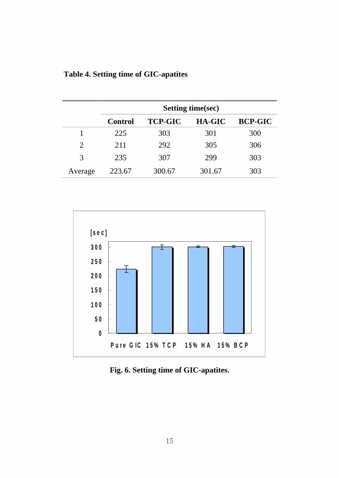
2. Setting time
15% HA-GIC, 15% BCP-GIC, 15% TCP-GIC had longer setting time
compared with the control group (Table 4, Fig. 6). However, there was
no significant difference among the three groups (P>0.05), and their
setting time was within the range of 3 to 8 minutes as indicated in the
ISO standard 9917-1:2003(E) regulations.
15
Table 4. Setting time of GIC-apatites
Setting time(sec)
Control TCP-GIC HA-GIC BCP-GIC
1 225 303 301 300
2 211 292 305 306
3 235 307 299 303
Average 223.67 300.67 301.67 303
0
5 0
1 0 0
1 5 0
2 0 0
2 5 0
3 0 0
P u r e G IC 1 5 % T C P 1 5 % H A 1 5 % B C P
[ s e c ]
Fig. 6. Setting time of GIC-apatites.
16
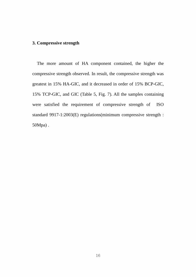
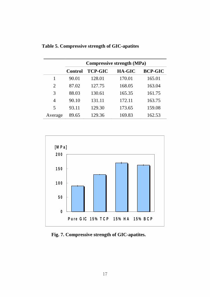
3. Compressive strength
The more amount of HA component contained, the higher the
compressive strength observed. In result, the compressive strength was
greatest in 15% HA-GIC, and it decreased in order of 15% BCP-GIC,
15% TCP-GIC, and GIC (Table 5, Fig. 7). All the samples containing
were satisfied the requirement of compressive strength of ISO
standard 9917-1:2003(E) regulations(minimum compressive strength :
50Mpa) .
17
Table 5. Compressive strength of GIC-apatites
Compressive strength (MPa)
Control TCP-GIC HA-GIC BCP-GIC
1 90.01 128.01 170.01 165.01
2 87.02 127.75 168.05 163.04
3 88.03 130.61 165.35 161.75
4 90.10 131.11 172.11 163.75
5 93.11 129.30 173.65 159.08
Average 89.65 129.36 169.83 162.53
0
5 0
1 0 0
1 5 0
2 0 0
P u r e G IC 1 5 % T C P 1 5 % H A 1 5 % B C P
[ M P a ]
Fig. 7. Compressive strength of GIC-apatites.

18
4. Bonding strength
Bonding strength was greatest in 15% TCP-GIC, the group with
highest amount of β-TCP involved (Table 6, Fig. 8). 15% HA-GIC
and 15% BCP-GIC had subsequent bonding strength, and there were no
statistically significant difference between them (P>0.05). The bonding
strength of GIC group cited Lee's 2006 results which showed the
lowest measurement(Lee, 2006).

19
Table 6. Bonding strength of of GIC-apatites
Bonding strength (mN/mm²)
Control TCP-GIC HA-GIC BCP-GIC
1 790.50 1555.1 1327.5 1507.3
2 584.90 1580.3 1502.5 1515.5
3 786.90 1532.1 1455.3 1499.7
4 803.00 1510.7 1391.4 1450.9
5 790.20 1521.6 1531.4 1478.9
Average 751.10 1539.96 1441.62 1490.46
Fig. 8. Bonding strength of GIC-apatites.
0
200
400
600
800
1000
1200
1400
1600
1800
Pure GIC 15% TCP 15% HA 15% BCP
[mN/mm 2 ]

20
5. Microstructure
On the SEM image of the sectioned dentin surface, bone-like apatite
crystal was deposited around the intertubular dentin in all three groups
of 15% HA-GIC, 15% BCP-GIC, 15% TCP-GIC (Fig. 10, 11, 12).
Among them, 15% TCP-GIC group showed the highest apatite
formation on the dentin surface, while there was no sign of bone-like
apatite formation in the GIC group (Fig. 9).

21
(a)(×5000) (b)(×20000)
Fig. 9. Dentin surface of control group(pure GIC) under SEM.
(a)(×5000) (b)(×20000)
Fig. 10. Dentin surface of 15% HA-GIC group under SEM.

22
(a) (×5000) (b) (×20000)
Fig. 11. Dentin surface of 15% BCP-GIC group under SEM.
(a) (×5000) (b) (×20000)
Fig. 12. Dentin surface of 15% ββββTCP-GIC group under SEM.
23
IV. Discussion
GIC, due to its fluoride releasing and chemical bonding with teeth
advantages, is a widely used dental luting cement. However, the weak
strength and tooth bonding strength compared to other contemporary
dental luting cement causes material fracture and secondary dental
caries between the tooth interface. In order to solve such problems
many studies have been done.
Apatite is one of many calcium phosphate minerals and a main
component of human bone and teeth. Enamel and human bones are
consisted of 97% and 65% of apatite respectively. Apatite of bone and
teeth are mainly composed of HA with extremely small amounts of
calcium ions, metal ions and fluoride ions, thus being called a
biological apatite. We in this study, tried to improve the physical
properties of GIC by using HA, β-TCP, BCP(a mixture of HA and
β-TCP) which are known to be highly bioactive(H. Yuan, 2002).
RelyXTM GIC (3M/ESPE, USA), used in this study, is resin
reinforced glass ionomer cement, but it has not light curing reaction. It
has two hardening reactions, acid-base reaction between
fluoroaluminosilicate glass and polycarboxyl acid, and polymerization
24
reaction between methacylate group of polymer and HEMA(2-
hydroxyethylmethacrylate). Therefore, to estimate biocompatibility as
dental luting cement according to ISO standard 9917-1:2003(E)
regulations, film thickness, setting time, compressive strength, bonding
strength were measured.
15% HA-GIC, 15% BCP-GIC, 15% TCP-GIC(experimental groups)
showed thinner film thickness values compared to pure GIC(control groups).
Since the average particle size of GIC is 13~19㎛(<45㎛) and 5~10㎛ in
apatites, we could predict that small apatite particles move into the inter
matrix empty spaces, thus explaining the reduction of film thickness.
Experimental groups had longer setting time compared to control
groups. Such a result is probably due to the easy bonding between
polyacrylic acid and calcium, aluminum, sodium ions of GIC powder
and the strong bond between calcium and phosphate ions of apaptite
and hydroxyl groups resulting in delayed reaction with polyacrylic
acids and apatites(A. Hideki, 2002).
Experimental groups showed higher compressive strength value
compared to control groups. Such values were higher in experimental
groups with apatite (15% HA-GIC, 15% BCP-GIC, 15% TCP-GIC).
Apatities of high calcium concentration seemed to accelerated ionic

25
bonding with polyacrylic acid(M.E. Lucas, 2003). Packed relatively
small particle sized apatite in GIC particles seemed to stabilize its
structure. Higher compressive strength of HA compared to β-TCP
explains the highest strength value of 15% HA-GIC groups.
Experimental groups showed stronger bond strength compared to
control groups. 15% TCP-GIC containing β-TCP showed the
strongest value. The formation of bond-like apatite during the ionic
change between the interface of calcium phosphate and SBF explains
the increased bonding strength with tooth structures, such results could
be proved in SEM examinations(L.L. Hench, 1996).
The strongest bonding strength of 15% TCP-GIC could be explained
by the active ionic change in SBF due to the higher solubility of β-
TCP compared to HA resulting in abundant bone-like apatite
formation(T. Kokubo, 1991).
Improvements of mechanical properties of the material itself and
tooth bonding strength by mixing apatite to GIC were discussed.
Thus, if studies on environmental factors to greatly improve
bioactivity of apatite and ideal mixing ratio and methods could be done,
improvements in longevity and problems regarding secondary dental
caries could be solved.

26
V. Conclusion
In order to investigate changes in the material itself and between the
interface with the human tooth, 15 wt % HA, BCP, β-TCP were
mixed with GIC. Film thickness, setting time, compressive strength,
bonding strength with dentin and the surface of dentin were evaluated.
Test specimens were maintained at 36.5 °C in SBF for 4 weeks. The
following conclusions were drawn from this investigation .
1. 15% HA-GIC, 15% BCP-GIC, 15% TCP-GIC had thinner film
thickness compared to pure GIC. However, there was no
statistically significant difference among the three groups of
apatite-GICs. The film thickness of the three groups were
within the range of ISO standard 9917-1:2003(E) regulations
and they did not affect the physical properties.
2. 15% HA-GIC, 15% BCP-GIC, 15% TCP-GIC had longer
setting time compared to the control group. However, there was
no significant difference among the three groups. Setting time
of the three materials were in the range of 3 to 8 minutes as
indicated in the ISO standard 9917-1:2003(E) regulations.

27
3. Higher amounts of HA composition led to higher compressive
strengths. The value was greatest in 15% HA-GIC and
decreased in order of 15% BCP-GIC, 15% TCP-GIC and GIC.
4. Bonding strength of 15% TCP-GIC, the group with the highest
amount of β-TCP was the greatest. 15% BCP-GIC and 15%
HA-GIC showed subsequent values, no statistically significant
difference between them were present. Bonding strength was
lowest in GIC.
5. In the SEM image of the sectioned dentin surface, bone-like
apatite crystal was deposited around the intertubular dentin in
all three groups of 15% HA-GIC, 15% BCP-GIC, 15% TCP-
GIC. Among them , 15% TCP-GIC group showed the highest
apatite formation on the dentin surface.

28
VI. References
Anderson P: Demineralization in enamel and hydroxyapatite aggregates
at increasing ionic strengths. Arch. Oral Biol. 49: 199-207, 2004.
Boode HE: Remineralization of artifical caries lesion. Caries Res 15 :
198-199, 1980.
Dickens SH: Mechanical properties and biochemical activity of
remineralizing resin-based CaPO4 cements. Dent. Mater. 19(6): 558-
566, 2003.
Golberg J, Tanzer J, Munster E: Cross-sectional clinical evaluation of
recurrent caries, restoration of marginal integrity and oral hygiene
status. J. Am. Dent. Assoc. 102: 653-656, 1981.
Hembree JH: In vitro microleakage of a new dental adhesive system.
J. Prosthet. Dent. 55: 442-445, 1986.

29
International organization for standardization : Dental resin-based pit
and fissure sealants ISO 6874 : 1988(E)
Jovanovic J, Adnadjevic B: The influence of hydroxyapatite
modification on cross-linking of polydimethylsiloxane /HAp
composites. Colloids and Surfaces B, Biointerfaces 39: 181-186, 2004.
Kawasaki K, Kambara M, Matsumura H: A comparison of the
adsorption of saliva proteins and some typical proteins on to the surface
of hydroxyapatite. Colloids and Surfaces B, Biointerfaces 32: 321-334,
2003.
Santos C: Water absorption characteristics of dental composites
incorporating hydroxyapatite filler. Biomaterials 23(8): 1897-1904,
2002.
Vitorino R: In vitro hydroxyapatite adsorbed salivary proteins.
Biochemical and Biophysical Research Communications 320: 342-346,
2004.
30
국문국문국문국문 요약요약요약요약
글래스아이오노머글래스아이오노머글래스아이오노머글래스아이오노머 치과용치과용치과용치과용 시멘트에시멘트에시멘트에시멘트에 대한대한대한대한
하이드록시아파타이트와하이드록시아파타이트와하이드록시아파타이트와하이드록시아파타이트와 제제제제 3 3 3 3 인산칼슘의인산칼슘의인산칼슘의인산칼슘의 골유사골유사골유사골유사 아파타이트아파타이트아파타이트아파타이트
형성능력의형성능력의형성능력의형성능력의 비교비교비교비교
연세대학교 대학원 치의학과
윤 상 일
지도교수 : 최 형 준
글래스아이오노머 시멘트와 하이드록시아파타이트를 혼합하여 인간
치아와 접착시켰을 때 골유사아파타이트(bone–like apatite)의 형성으로
인하여 결합력이 증가되었다는 보고가 있었다. 하지만, 하이드록시아파
타이트의 낮은 용해도로 인해 골유사아파타이트가 비교적 적게 형성되
었다. 이번 연구의 목적은 용해도가 높은 제3 인산칼슘과 하이드록시아
파타이트를 글래스아이오노머 시멘트에 혼합하여 치아에 접착하였을
때, 치아와 시멘트 사이의 계면에서 골유사아파타이트의 형성이 증가하
는지를 알아보는 것이다. 그리고, 하이드록시아파타이트의 강한 물리적
31
특성과 제3 인산칼슘의 높은 용해성을 모두 가지는 이상인산칼슘
(biphasic calcium phosphate)의 생체활성능(bioactivity)이 시멘트에 혼합하
였을 때도 발현되는지 알아보기 위함이다.
이전 연구에서 글래스아이오노머 시멘트 제품인 RelyXTM (3M/ESPE,
USA)에 15 wt%의 하이드록시아파타이트를 혼합하였을 때 가장 높은
물리적 성질을 보인 점을 감안하여, 같은 비율의 제3 인산칼슘과 하
이드록시아파타이트를 사용하였다. 그리고, Huipin 등의 연구에 따라
가장 골유도능이 높다고 알려진 하이드록시아파타이트와 제3 인산칼
슘이 85 : 15의 비율로 혼합된 이상인산칼슘을 사용하였다. 따라서,
순수한 글래스아이오노머 시멘트는 대조군으로, 15 wt%의 하이드록
시아파타이트, 이상인산칼슘, 제3 인산칼슘을 각각 포함하는 글래스
아이오노머 시멘트를 실험군으로 설정하였다. 치과용 시멘트의 기본
성질인 피막도, 경화시간, 압축강도는 ISO 9927 기준에 맞게 측정되
었다. 혼합된 글래스아이오노머 시멘트를 치아에 접착시킨 후, SBF
에서 36.5 oC 수조에 4주간 보관하고 결합강도를 측정하였다. 그리고,
절단면의 표면 형태를 알아보기 위해 전자현미경 사진으로 관찰하
였다.
1. 글래스아이오노머 시멘트의 피막도는 실혐군이 대조군에 비해 얇게
측정되었다. 하지만, 실험군 간에 통계학적 유의차가 없었다

32
(P>0.05).
2. 글래스아이오노머 시멘트의 경화시간은 실험군이 대조군에 비해 길
게 측정되었다. 이는 ISO 9917의 기준에 만족하였고, 실험군 간에 통
계학적 유의차가 없었다(P>0.05).
3. 하이드록시아파타이트가 많이 포함되어 있을수록 글래스아이오노머
시멘트의 압축강도가 크게 측정되었다. 즉, 15 wt% 하이드록시아파타
이트, 이상인산칼슘, 제3 인산칼슘을 포함한 글래스아이오노머 시멘
트, 순수한 글래스아이오노머 시멘트 순으로 압축강도가 크게 측정
되었다(P<0.05).
4. 제3 인산칼슘의 양이 많이 포함되어 있을수록 글래스아이오노머 시
멘트의 결합강도가 크게 측정되었다. 즉, 15 wt% 제3 인산칼슘, 이상
인산칼슘, 히이드록시아파타이트를 포함한 글래스아이오노머 시멘트,
순수한 글래스아이오노머 시멘트 순으로 결합강도가 크게 측정되었
다(P<0.05).
5. 절단면의 전자현미경 관찰 결과, 위와 같은 순으로 골유사아파타이
트 형성이 많이 관찰되었다.
이상의 실험 결과로부터 아파타이트를 첨가한 것은 기존의 글래스아
이오노머 시멘트의 물성을 향상시켰을 뿐 아니라, 결합강도 또한 증가
하였다. 특히, 용해도가 높은 제3 인산칼슘을 포함한 그룹이 하이드록

33
시아파타이트를 포함한 그룹보다 치아와의 계면에서 골유사아파타이트
의 형성이 많이 이루어졌으며, 이로 인해 결합강도가 더 높았을 것으로
생각된다. 이러한 아파타이트의 생체활성능을 최대한 활용한다면 글래
스아이오노머 시멘트 뿐 아니라, 여러 가지 다른 치과용 재료의 발전을
이룰 수 있을 것이다.

34
핵심되는 말: 글래스아이오노머 시멘트, 하이드록시아파타이트, 이상인산칼슘,
제 3 인산칼슘, 골유사아파타이트