Embed Size (px)
DESCRIPTION
Potency Unchained
Citation preview
C E L L B I O L O G Y
Potency unchainedDifferentiated cells have been reprogrammed to an embryonic-like state using a physical stimulus. This treatment generates a new cell population that contributes to both the embryo and the placenta. See Article p.641 & Letter p.676
A U S T I N S M I T H
Cell specialization in mammals is essen-tial for diverse functions, such as mus-cle contraction and nerve conduction.
These specializations become fixed during development, and conversion between dif-ferentiated cell types seems to be extremely rare. However, in this issue, two studies by Obokata et al.1,2 show that cells isolated from newborn mice lose their identity on expo-sure to mildly acidic conditions. Remarkably, instead of triggering cell death or tumour growth, as might be expected, a new cell state emerges that exhibits an unprecedented potential for differentiation into every possible cell type.
Studies on tissue regeneration in amphib-ians, reptiles and birds indicate that differenti-ated cells have some ability to dedifferentiate or to switch identity. Mammalian cells are more resistant, but fate conversion is observed in certain cancers. It was only with the cloning
of Dolly the sheep3, in which nuclear mater-ial from the mammary cell of an adult sheep was transferred into an enucleated egg cell to produce a cloned animal, that the capacity for complete reprogramming of the mammalian genome was confirmed. However, cloning does not convert whole cells.
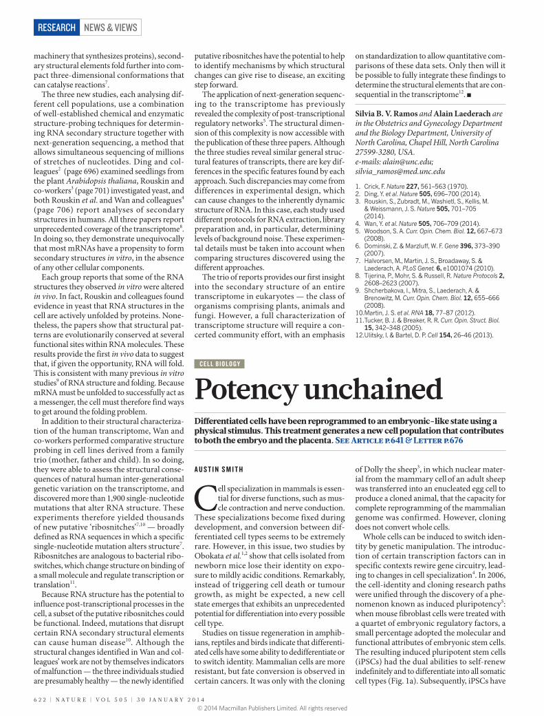
Whole cells can be induced to switch iden-tity by genetic manipulation. The introduc-tion of certain transcription factors can in specific contexts rewire gene circuitry, lead-ing to changes in cell specialization4. In 2006, the cell-identity and cloning research paths were unified through the discovery of a phe-nomenon known as induced pluripotency5: when mouse fibroblast cells were treated with a quartet of embryonic regulatory factors, a small percentage adopted the molecular and functional attributes of embryonic stem cells. The resulting induced pluripotent stem cells (iPSCs) had the dual abilities to self-renew indefinitely and to differentiate into all somatic cell types (Fig. 1a). Subsequently, iPSCs have
machinery that synthesizes proteins), second-ary structural elements fold further into com-pact three-dimensional conformations that can catalyse reactions7.
The three new studies, each analysing dif-ferent cell populations, use a combination of well-established chemical and enzymatic structure-probing techniques for determin-ing RNA secondary structure together with next-generation sequencing, a method that allows simultaneous sequencing of millions of stretches of nucleotides. Ding and col-leagues2 (page 696) examined seedlings from the plant Arabidopsis thaliana, Rouskin and co-workers3 (page 701) investigated yeast, and both Rouskin et al. and Wan and colleagues4 (page 706) report analyses of secondary structures in humans. All three papers report unprecedented coverage of the transcriptome8. In doing so, they demonstrate unequivocally that most mRNAs have a propensity to form secondary structures in vitro, in the absence of any other cellular components.
Each group reports that some of the RNA structures they observed in vitro were altered in vivo. In fact, Rouskin and colleagues found evidence in yeast that RNA structures in the cell are actively unfolded by proteins. None-theless, the papers show that structural pat-terns are evolutionarily conserved at several functional sites within RNA molecules. These results provide the first in vivo data to suggest that, if given the opportunity, RNA will fold. This is consistent with many previous in vitro studies9 of RNA structure and folding. Because mRNA must be unfolded to successfully act as a messenger, the cell must therefore find ways to get around the folding problem.
In addition to their structural characteriza-tion of the human transcriptome, Wan and co-workers performed comparative structure probing in cell lines derived from a family trio (mother, father and child). In so doing, they were able to assess the structural conse-quences of natural human inter-generational genetic variation on the transcriptome, and discovered more than 1,900 single-nucleotide mutations that alter RNA structure. These experiments therefore yielded thousands of new putative ‘ribosnitches’7,10 — broadly defined as RNA sequences in which a specific single-nucleotide mutation alters structure7. Ribosnitches are analogous to bacterial ribo-switches, which change structure on binding of a small molecule and regulate transcription or translation11.
Because RNA structure has the potential to influence post-transcriptional processes in the cell, a subset of the putative ribosnitches could be functional. Indeed, mutations that disrupt certain RNA secondary structural elements can cause human disease10. Although the structural changes identified in Wan and col-leagues’ work are not by themselves indicators of malfunction — the three individuals studied are presumably healthy — the newly identified
putative ribosnitches have the potential to help to identify mechanisms by which structural changes can give rise to disease, an exciting step forward.
The application of next-generation sequenc-ing to the transcriptome has previously revealed the complexity of post-transcriptional regulatory networks5. The structural dimen-sion of this complexity is now accessible with the publication of these three papers. Although the three studies reveal similar general struc-tural features of transcripts, there are key dif-ferences in the specific features found by each approach. Such discrepancies may come from differences in experimental design, which can cause changes to the inherently dynamic structure of RNA. In this case, each study used different protocols for RNA extraction, library preparation and, in particular, determining levels of background noise. These experimen-tal details must be taken into account when comparing structures discovered using the different approaches.
The trio of reports provides our first insight into the secondary structure of an entire transcriptome in eukaryotes — the class of organisms comprising plants, animals and fungi. However, a full characterization of transcriptome structure will require a con-certed community effort, with an emphasis
on standardization to allow quantitative com-parisons of these data sets. Only then will it be possible to fully integrate these findings to determine the structural elements that are con-sequential in the transcriptome12. ■
Silvia B. V. Ramos and Alain Laederach are in the Obstetrics and Gynecology Department and the Biology Department, University of North Carolina, Chapel Hill, North Carolina 27599-3280, USA.e-mails: [email protected]; [email protected]
1. Crick, F. Nature 227, 561–563 (1970).2. Ding, Y. et al. Nature 505, 696–700 (2014).3. Rouskin, S., Zubradt, M., Washietl, S., Kellis, M.
& Weissmann, J. S. Nature 505, 701–705 (2014).
4. Wan, Y. et al. Nature 505, 706–709 (2014).5. Woodson, S. A. Curr. Opin. Chem. Biol. 12, 667–673
(2008).6. Dominski, Z. & Marzluff, W. F. Gene 396, 373–390
(2007).7. Halvorsen, M., Martin, J. S., Broadaway, S. &
Laederach, A. PLoS Genet. 6, e1001074 (2010).8. Tijerina, P., Mohr, S. & Russell, R. Nature Protocols 2,
2608–2623 (2007).9. Shcherbakova, I., Mitra, S., Laederach, A. &
Brenowitz, M. Curr. Opin. Chem. Biol. 12, 655–666 (2008).
10. Martin, J. S. et al. RNA 18, 77–87 (2012).11. Tucker, B. J. & Breaker, R. R. Curr. Opin. Struct. Biol.
15, 342–348 (2005).12. Ulitsky, I. & Bartel, D. P. Cell 154, 26–46 (2013).
6 2 2 | N A T U R E | V O L 5 0 5 | 3 0 J A N U A R Y 2 0 1 4
NEWS & VIEWSRESEARCH
© 2014 Macmillan Publishers Limited. All rights reserved
been produced from a range of adult cell types, fostering enthusiasm worldwide for develop-ing customized disease-modelling and cell-therapy applications.
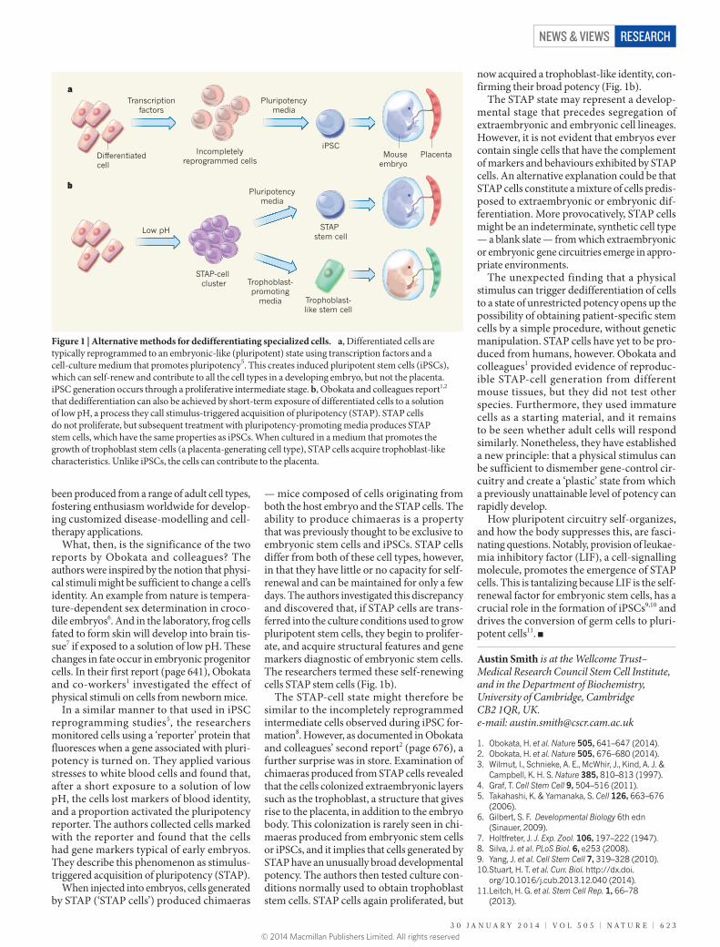
What, then, is the significance of the two reports by Obokata and colleagues? The authors were inspired by the notion that physi-cal stimuli might be sufficient to change a cell’s identity. An example from nature is tempera-ture-dependent sex determination in croco-dile embryos6. And in the laboratory, frog cells fated to form skin will develop into brain tis-sue7 if exposed to a solution of low pH. These changes in fate occur in embryonic progenitor cells. In their first report (page 641), Obokata and co-workers1 investigated the effect of physical stimuli on cells from newborn mice.
In a similar manner to that used in iPSC reprogramming studies5, the researchers monitored cells using a ‘reporter’ protein that fluoresces when a gene associated with pluri-potency is turned on. They applied various stresses to white blood cells and found that, after a short exposure to a solution of low pH, the cells lost markers of blood identity, and a proportion activated the pluripotency reporter. The authors collected cells marked with the reporter and found that the cells had gene markers typical of early embryos. They describe this phenomenon as stimulus- triggered acquisition of pluri potency (STAP).
When injected into embryos, cells generated by STAP (‘STAP cells’) produced chimaeras
— mice composed of cells originating from both the host embryo and the STAP cells. The ability to produce chimaeras is a property that was previously thought to be exclusive to embryonic stem cells and iPSCs. STAP cells differ from both of these cell types, however, in that they have little or no capacity for self-renewal and can be maintained for only a few days. The authors investigated this discrepancy and discovered that, if STAP cells are trans-ferred into the culture conditions used to grow pluri potent stem cells, they begin to prolifer-ate, and acquire structural features and gene markers diagnostic of embryonic stem cells. The researchers termed these self-renewing cells STAP stem cells (Fig. 1b).
The STAP-cell state might therefore be similar to the incompletely reprogrammed intermediate cells observed during iPSC for-mation8. However, as documented in Obokata and colleagues’ second report2 (page 676), a further surprise was in store. Examination of chimaeras produced from STAP cells revealed that the cells colonized extraembryonic layers such as the trophoblast, a structure that gives rise to the placenta, in addition to the embryo body. This colonization is rarely seen in chi-maeras produced from embryonic stem cells or iPSCs, and it implies that cells generated by STAP have an unusually broad developmental potency. The authors then tested culture con-ditions normally used to obtain trophoblast stem cells. STAP cells again proliferated, but
now acquired a trophoblast-like identity, con-firming their broad potency (Fig. 1b).
The STAP state may represent a develop-mental stage that precedes segregation of extraembryonic and embryonic cell lineages. However, it is not evident that embryos ever contain single cells that have the complement of markers and behaviours exhibited by STAP cells. An alternative explanation could be that STAP cells constitute a mixture of cells predis-posed to extraembryonic or embryonic dif-ferentiation. More provocatively, STAP cells might be an indeterminate, synthetic cell type — a blank slate — from which extraembryonic or embryonic gene circuitries emerge in appro-priate environments.
The unexpected finding that a physical stimulus can trigger dedifferentiation of cells to a state of unrestricted potency opens up the possibility of obtaining patient-specific stem cells by a simple procedure, without genetic manipulation. STAP cells have yet to be pro-duced from humans, however. Obokata and colleagues1 provided evidence of reproduc-ible STAP-cell generation from different mouse tissues, but they did not test other species. Furthermore, they used immature cells as a starting material, and it remains to be seen whether adult cells will respond similarly. Nonetheless, they have established a new principle: that a physical stimulus can be sufficient to dismember gene-control cir-cuitry and create a ‘plastic’ state from which a previously un attainable level of potency can rapidly develop.
How pluripotent circuitry self-organizes, and how the body suppresses this, are fasci-nating questions. Notably, provision of leukae-mia inhibitory factor (LIF), a cell-signalling molecule, promotes the emergence of STAP cells. This is tantalizing because LIF is the self-renewal factor for embryonic stem cells, has a crucial role in the formation of iPSCs9,10 and drives the conversion of germ cells to pluri-potent cells11. ■
Austin Smith is at the Wellcome Trust–Medical Research Council Stem Cell Institute, and in the Department of Biochemistry, University of Cambridge, Cambridge CB2 1QR, UK.e-mail: [email protected]
1. Obokata, H. et al. Nature 505, 641–647 (2014).2. Obokata, H. et al. Nature 505, 676–680 (2014).3. Wilmut, I., Schnieke, A. E., McWhir, J., Kind, A. J. &
Campbell, K. H. S. Nature 385, 810–813 (1997).4. Graf, T. Cell Stem Cell 9, 504–516 (2011).5. Takahashi, K. & Yamanaka, S. Cell 126, 663–676
(2006).6. Gilbert, S. F. Developmental Biology 6th edn
(Sinauer, 2009).7. Holtfreter, J. J. Exp. Zool. 106, 197–222 (1947).8. Silva, J. et al. PLoS Biol. 6, e253 (2008).9. Yang, J. et al. Cell Stem Cell 7, 319–328 (2010).10. Stuart, H. T. et al. Curr. Biol. http://dx.doi.
org/10.1016/j.cub.2013.12.040 (2014).11. Leitch, H. G. et al. Stem Cell Rep. 1, 66–78
(2013).
Transcriptionfactors
Pluripotencymedia
Di�erentiatedcell
Incompletelyreprogrammed cells
iPSCMouse
embryoPlacenta
a
Low pH
Pluripotencymedia
STAP-cell cluster
STAP stem cell
b
Trophoblast-promoting
media Trophoblast-like stem cell
Figure 1 | Alternative methods for dedifferentiating specialized cells. a, Differentiated cells are typically reprogrammed to an embryonic-like (pluripotent) state using transcription factors and a cell-culture medium that promotes pluripotency5. This creates induced pluripotent stem cells (iPSCs), which can self-renew and contribute to all the cell types in a developing embryo, but not the placenta. iPSC generation occurs through a proliferative intermediate stage. b, Obokata and colleagues report1,2 that dedifferentiation can also be achieved by short-term exposure of differentiated cells to a solution of low pH, a process they call stimulus-triggered acquisition of pluripotency (STAP). STAP cells do not proliferate, but subsequent treatment with pluripotency-promoting media produces STAP stem cells, which have the same properties as iPSCs. When cultured in a medium that promotes the growth of trophoblast stem cells (a placenta-generating cell type), STAP cells acquire trophoblast-like characteristics. Unlike iPSCs, the cells can contribute to the placenta.
3 0 J A N U A R Y 2 0 1 4 | V O L 5 0 5 | N A T U R E | 6 2 3
NEWS & VIEWS RESEARCH
© 2014 Macmillan Publishers Limited. All rights reserved