Embed Size (px)
Citation preview

Hindawi Publishing CorporationBioMed Research InternationalVolume 2013, Article ID 490946, 12 pageshttp://dx.doi.org/10.1155/2013/490946
Research ArticleApatite Formation: Why It May Not Work as Planned, andHow to Conclusively Identify Apatite Compounds
Christophe Drouet
CIRIMAT Carnot Institute-UMR CNRS/INPT/UPS 5085, University of Toulouse, Ensiacet, 4 Allee Emile Monso,31030 Toulouse Cedex 4, France
Correspondence should be addressed to Christophe Drouet; [email protected]
Received 19 April 2013; Revised 24 June 2013; Accepted 2 July 2013
Academic Editor: Stanley J. Stachelek
Copyright © 2013 Christophe Drouet. This is an open access article distributed under the Creative Commons Attribution License,which permits unrestricted use, distribution, and reproduction in any medium, provided the original work is properly cited.
Calcium phosphate apatites are inorganic compounds encountered in many different mineralized tissues. Bone mineral, forexample, is constituted of nanocrystalline nonstoichiometric apatite, and the production of “analogs” through a variety of methodsis frequently reported. In another context, the ability of solid surfaces to favor the nucleation and growth of “bone-like” apatiteupon immersion in supersaturated fluids such as SFB is commonly used as one evaluation index of the “bioactivity” of suchsurfaces. Yet, the compounds or deposits obtained are not always thoroughly characterized, and their apatitic nature is sometimesnot firmly assessed by appropriate physicochemical analyses. Of particular importance are the “actual” conditions in which theprecipitation takes place. The precipitation of a white solid does not automatically indicate the formation of a “bone-like carbonateapatite layer” as is sometimes too hastily concluded: “all that glitters is not gold.” The identification of an apatite phase shouldbe carefully demonstrated by appropriate characterization, preferably using complementary techniques. This review considersthe fundamentals of calcium phosphate apatite characterization discussing several techniques: electron microscopy/EDX, XRD,FTIR/Raman spectroscopies, chemical analyses, and solid state NMR. It also underlines frequent problems that should be kept inmind when making “bone-like apatites.”
1. Introduction
Synthetic-calcium-phosphate-apatite-based compounds arefrequently encountered in the literature dealing with bonetissue engineering. This fact is not surprising though, giventhe apatitic nature of bone mineral itself [1]. Detailed inves-tigations on this biomineral (e.g., [2–8]) have revealed itsnanocrystalline, nonstoichiometric, and hydrated characters,as well as specific surface features involving labile ions innonapatitic chemical environments, potentially exchangeablewith ions contained in surrounding fluids. Similar character-istics were also pointed out for synthetic biomimetic analogs(e.g., [9]), allowing one to prepare bioactive ceramics [10].
Such biomimetic nanocrystalline apatites exhibit an over-all chemical composition that can generally be described byformulas of the type Ca
10−𝑥(PO4)6−𝑥(HPO
4)𝑥(OH)2−𝑥
[11] orCa10−𝑥−𝑍(PO4)6−𝑥(HPO
4)𝑥(OH)2−𝑥−2𝑍
[12], where 𝑥 and 𝑍depend on conditions of formation and state of ageing (mat-uration). It should be stressed here that the physico-chemical
characteristics of nanocrystalline apatites—whether of bio-logical or synthetic origin—differ significantly from thoseof stoichiometric hydroxyapatite (HA), Ca
10(PO4)6(OH)2,
especially in terms of stoichiometry, crystal size, crystaldisorder, unit cell parameters, surface features, and hydrationstate [13].
While the synthesis of apatite materials for the prepara-tion of implantable bioceramics is bound to occupy a privi-leged place in bone applications thanks to (sub)structural andcompositional features close to those of bone mineral, it isnot suited—in a self-supported way—for load-bearing appli-cations that require stronger mechanical properties. Metalimplants then appear as the most commonly used candidates(e.g., hip or knee prostheses), despite potential drawbackssuch as poor bone-bonding abilities or allergy issues [14].In this case, the chemical quality of the interface betweenthe implant and the surrounding bone tissue is of primeimportance for the success and durability of implantation[15]. This point has led many researchers and industrial

2 BioMed Research International
companies to investigate and develop deposition processes ofapatite compounds on metal implants (e.g., by plasma spraytechnologies) in view of favoring bone-bonding properties[16–18]. Also, more generally, tests are often carried out insupersaturated solutions so as to evaluate the ability of asurface to induce the nucleation and growth of a “bone-like”apatite layer [19]. In these cases, the term “supersaturated”implicitly refers to hydroxyapatite (solubility product 𝑝𝐾sp(25∘C) = 117 [20] for the formula Ca
10(PO4)6(OH)2). Several
types of supersaturated solutions have been used, the mostfamous probably being the so-called SBF solution (SimulatedBody Fluid) [19, 21] or its multiples (e.g., SBF × 1.5) [22, 23].Tests are generally performed for several days and often at37∘C to approach physiological conditions. Although careshould be taken when drawing conclusive statements on amaterial “bioactivity” on the sole basis of such laboratorytests (run out of the body and in the absence of cells or oftenof proteins), such assays may however be informative, forexample, for a preliminary “ranking” of implants capabilities[21], but it has to be kept in mind that additional dataare needed (e.g., cell behavior assessments and then in vivoconfirmations) to draw a more accurate picture of the actualbioactivity or expected biological behavior of the samplestested.
In all the preceding statements, the formation of anapatite phase is a specific concern, whether for the productionof self-supported bioactive ceramics, for the setup of acoating on implants for improving bone-bonding abilities, orelse for drawing relative “bioactivity” information. A greatdeal of papers and congress communications refer to theseaspects, and conclusive statements are commonly drawn.Unfortunately, adequate physico-chemical characterizationdata are not always attached to such statements, and theestablishment of the “apatitic” nature of samples or depositsis sometimes unconvincing or even dubious. The announcedconditions of preparation of a compound considered to beapatite sometimes depart from the conventional conditionsof stability of apatite, requiring characterization proofs. Inother instances, the sole observation of a white deposit afterimmersion in supersaturated media is considered as a solidproof for concluding that a “bone-like carbonated apatitelayer” has been formed, which is obviously not scientificallysatisfactory.
Various aspects should in contrast be taken into accountbefore making conclusive statements. The chemistry ofcalcium phosphates is indeed very rich, with numerouspossible phases (beside apatite) able to form depending onexperimental conditions [24]. Also, the “real” conditionsof treatments are sometimes slightly different from the“intended” ones, due, for example, to experimental mistakesor approximations, and some steps in the synthesis/coatingprocess may not be adequately performed. Furthermore, thesubstrate itself might be able to react with the immersionsolution, for instance, by releasing/capturing some ions ormolecular entities into/from the surrounding medium, thusmodifying its actual surface state or the external conditions(e.g., effect on the pH of the medium). Finally, inadequate
conditions may lead to variations in supersaturation of pHalong the process.
All of these considerations point to a situation that ismore complex than may be initially thought. All in all, theconclusion that an apatite phase has been obtained requires,as in every reliable materials science study, adequate physico-chemical characterization. This contribution is intended toreview themain characteristics to be expected from a calciumphosphate apatite compound, drawn from complementarycharacterization techniques. The cases of hydroxyapatite andof a nanocrystalline apatite of rather low maturation state areexamined for establishing typical “identity cards” for suchmaterials. Characteristics of other minerals that may possiblyalso be encountered in the above-mentioned contexts are alsoreviewed, and some frequent issues are commented upon.
2. Results and Discussion
2.1. Apatite Formation: Why It May Not Work as Planned. . ..In this first section, the idea is to provide readers withelements pointing out that apatite formation is not a trivialphenomenon. Whether due to inexact concentration cal-culations, unexpected alteration/equilibration of a substratesurface upon immersion, or else because of incompleteprocessing steps, several factors could in fact lead to otheroutcomes than planned. The following paragraphs describesome of the most likely causes of experimental failure ormistakes to avoid.
2.1.1.The Impact of IncompleteWashing. Most laboratory testsaiming at evaluating the ability of solid surfaces to get coveredby an apatite layer upon immersion are carried out in “close-to-physiological” media. Generally, though, only the mineralfraction of body fluids is considered (e.g., in SBF solution[19]), meaning in particular in the absence of proteins orcells. This situation indeed allows a greater ease of use andleads already to some informative results. However, it shouldbe kept in mind at this point that the adsorption of organicsubstances such as proteins or peptides, or the activity of cellsonce in actual in vivo environments, could lead to somewhatmodified outcomes.
In cases when synthetic simulated body fluids (onlymineral) are prepared, it is customary to use chemicalcompositions that are more or less close to that of bloodplasma (reported in Table 1, as reported by Krebs [25], alongwith speciation data determined in this work using theVisual Minteq 3.0 software). As can be seen, blood plasma(or synthetic SBF) contains in particular large amounts ofsodium and chloride ions, which are predominant ionicspecies. The observation of a white deposit on the substrateafter withdrawal of the piece is therefore not an absoluteproof of the formation of apatite at this stage; indeed, in caseswhere the washing step has not been thoroughly performed,the formation/residual presence of NaCl aggregates couldexplain this white coating phenomenon. Figure 1 reportsthe illustrative example of a titanium alloy disk (TA6V)immersed in SBF × 10 for 5 days at room temperature(RT) in the case of either incomplete or more advanced

BioMed Research International 3
Table 1: Chemical composition of blood plasma (inorganic species) from Krebs [25], at pH 7.4 and 37∘C, and speciation data (calculatedusing the Visual Minteq 3.0 software).
Main ion types Average globalconcentration (mM)
Main species (and % oftotal concentration∗)
Corresponding activities∗∗(mM)
Sodium 137.4 Na+ (97%) 100.900Chloride 103 Cl− (96%) 75.427
Carbonate 27HCO3
− (90%) 18.509H2CO3(aq) (5%) 1.486
Potassium 4.4 K+ (97%) 3.246
Calcium 2.5Ca2+ (79%) 0.655
CaHCO3+ (9%) 0.177
CaCl+ (7%) 0.132
Phosphate (inorganic) 1
HPO42− (45%) 0.151
NaHPO4− (30%) 0.226
H2PO4− (12%) 0.090
CaHPO4(aq) (5%) 0.054
Magnesium 0.8Mg2+ (78%) 0.207MgCl+ (11%) 0.066
MgHCO3+ (7%) 0.042
Sulfate (inorganic) 0.5SO42− (75%) 0.125
NaSO4
− (19%) 0.071∗
For species in amount ≥5%.∗∗Using Davies’ equation for calculation of activity coefficients (total ionic strength ∼0.14M).
washing. Although SEMobservations evidenced the presenceof deposited crystals in both cases, EDX analyses pointedout that the poorly washed sample was (as one could indeedexpect) mostly covered by NaCl crystals. In the context ofimmersion tests, the absence of NaCl crystals should thenprobably be systematically verified (by EDX and/or XRDanalyses).
2.1.2. Role of “Real” Experimental Conditions. In cases whereadequate sample preparation was done prior to analysis,thus including thorough washing steps, the observation of adeposit after immersion in a hydroxyapatite-supersaturatedmedium still does not necessarily imply the formation ofapatite. Experimental mistakes (e.g., due to the weighing ofhygroscopic starting salts for preparing the supersaturatedsolution) may lead to undesired immersion conditions andthen to nucleated phases other than apatite. This stressesthe fact that parameters such as the medium pH shouldprobably be monitored during the immersion step to verifythat no significant pH change has occurred. This is all morerelevant as the precipitation of (hydroxy) apatite impliesthe incorporation of OH− ions and theoretically implies anacidification of the medium: this acidification phenomenonmay remain of low amplitude if the liquid-to-solid ratio ischosen sufficiently high, but this underlines the fact that insome cases the pH may turn out to be inappropriate to formapatite. The incorporation of pH-buffering agents like Triscan be found effective, but this implies the addition to themedium of a “foreign” organic compound, which may notalways appear appropriate. In cases when the pH followsuncontrolled variations, other phasesmay stably form instead
of apatite (depending on the exact conditions), such as acidiccalcium phosphate compounds like brushite CaHPO
4⋅2H2O
(DCPD), monetite CaHPO4(DCPA) or octacalcium phos-
phate (OCP). Indeed, these phases become more stable thanhydroxyapatite in acidic pH conditions [24].
The variation of supersaturation during immersion testsmay also lead to a modification in deposited phase withtime. For information, the saturation index relative to variousphases of interest and considering as initial conditions theionic concentrations of blood plasma (inorganic species, seeTable 1) is reported in Table 2. A negative value of this indexis indicative of a situation where the medium is supersatu-rated with respect to the phase concerned. These data werecalculated using the Visual Minteq 3.0 software, taking intoaccount the solubility products that are generally accepted forthese various phases (added in Table 2). As may be noted,such conditions (if correctly set up from an experimentalpoint of view) lead to a high supersaturation with respectto HA and to a moderate supersaturation for octacalciumphosphate (OCP), calcite or vaterite, among other phases. Incontrast, they are moderately undersaturated with respect tomagnesium-containing phases such as MgHPO
4⋅3H2O and
more undersaturated with respect to brushite or monetite. Ifundesirable (or variations in) ionic concentrations are presentduring the tests, however, the ionic product is going to evolveand the overall saturation scheme is expected to change.Supersaturation aspects are thus also of prime importanceduring apatite precipitation plans.
In this regard, the use of constant composition crystalgrowth (C3G) systems [30, 31], which keeps the pH, ionic

4 BioMed Research International
SodiumChloride
Poorly washed
C Ca VTiO Mg Al P Mo
Na
Ca
Cl
1 2 3(keV)
(a)
C CaTi
VO
Na Mg
Al
P
Mo Cl
Ca V
Ti
Washed
PhosphorusCalcium
Titanium
2 4 6(keV)
(b)
Figure 1: SEM and EDX observations for a TA6V disk immersed in SBF × 10 for 5 days at RT—role of washing.
Table 2: Saturation index relative to various phases of interest and considering as initial conditions the ionic concentrations of blood plasmareported in Table 1.
Selected phases 𝑝𝐾sp (25∘C) Reference
Saturation index(c) forblood
plasma conditions (kJ/mol)Hydroxyapatite (HA) Ca10(PO4)6(OH)2 116.8(a)
[26]
−15.673Octacalcium phosphate (OCP) Ca8(PO4)4(HPO4)2⋅5H2O 96.6(b) −1.540Monetite (DCPA) CaHPO4 6.9 24.875Brushite (DCPD) CaHPO4⋅2H2O 6.59 25.381Calcite c-CaCO3 8.48
[27]−2.377
Vaterite v-CaCO3 7.91 −0.788Magnesium hydrogenphosphate MgHPO4⋅3H2O 18.18 [28] 3.171(a)Expressed relative to HA formula in Ca10.(b)Expressed relative to OCP formula in Ca8.(c)Saturation index (Δ𝐺𝑠) [29] calculated as:Δ𝐺𝑠 = −(𝑅𝑇/𝑁)Ln(𝐼𝑃/𝐾sp)where𝑁 is the number of ions involved, IP is the ion product, and𝐾sp is the solubilityproduct. A negative value of Δ𝐺𝑠 means that the medium is supersaturated with respect to the concerned phase.
strength, temperature, and relative concentrations constant,can be seen as an advantageous way to control all experi-mental parameters and derive relevant data on the ability ofsubstrates to provoke the nucleation and growth of apatite.
Finally, especially in the case of long immersion testsrun in supersaturated fluids or for precipitations in mediacontaining organic molecules (glucose, proteins. . .), thedevelopment of microorganisms may become significantfor rather long periods of treatment. In this case, bacterialactivity may then contribute to modifying the initial mediumin a significant way. In order to avoid such issues, theincorporation of additives such as sodium azide NaN
3in the
medium remains an option. Antibacterial agents such as pen-strep or fungizones may also be included.
2.1.3. Potential Impact of Substrate Equilibration with theMedium. Thenature and behavior of the substrate itself, oncein solution, may also play a nonnegligible role in the outcomeof immersion tests. For instance, substrates exposing ionicor functional molecular grafted groups (e.g., after specifictreatments aiming to alter the surface roughness or intendingto improve cellular activity)may endure a preliminary surfaceequilibration with the medium, potentially modifying theinitially determined immersion conditions [13]. If the pH
of the medium, or another relevant factor, is altered by theconditioning of the substrate upon contact with the medium,this could in turn lead to uncontrolled or undesirabletest conditions. Preliminary immersion tests should thusprobably be run for samples for which the inert characterhas not been clearly established, so as to guarantee theabsence of noticeable change in the medium. In this view,pH monitoring could again be seen as relevant during theprocess.
2.1.4. Incomplete Reactions. Hydroxyapatite is a mineralphase that gathers a large number of ions. As such, its precip-itation—especially at moderate temperatures—is bound toremain a rather slow phenomenon [24, 32]. This can explainthe low degree of crystallinity of precipitated apatites, whichsubsequently mature in solution (evolution towards stoi-chiometry) at a pace that depends on experimental condi-tions [13]. In this context, the transient formation of precursorphases (metastable but exhibiting a faster formation rate) isalso possible despite conditions that are thermodynamicallyfavorable to apatite formation, in linewithOstwald’s rule [33].
Among possible precursors, amorphous calcium phos-phate (ACP) and octacalcium phosphate (OCP, triclinic) aretwo potential candidates [29, 34] that have already been

BioMed Research International 5
suggested as precursors of bone-apatite in vivo [35, 36]. Aftertheir formation, both compounds may then be hydrolyzedto apatite in a subsequent step. ACP, although amorphous,is not totally exempt from structuration; it involves buildingblocks based on Ca
9(PO4)6⋅nH2O clusters (so-called Posner
clusters) [37], which can undergo partial internal hydrolysisthrough the reaction 𝑥PO
4
3− + 𝑥H2O → 𝑥HPO
4
2− + 𝑥OH−[38], leading to Ca
9(PO4)6−𝑥(HPO
4)𝑥(OH)𝑥and allowing in
particular the filling byOH− ions of what will become apatiticchannels. The hydrolysis of OCP probably involves a ratherdifferent mechanism. Taking into account the similaritiesbetween the structures of OCP andHA (detailed below) [39],the possibility of undergoing the OCP-to-apatite transforma-tion via a topotactic mechanism appears as a possible scheme[40–42]. In this case, the usual plate-likemorphology of OCPis expected to be rather conserved upon transformation intoapatite. This illustrates then the difficulty of assessing phaseidentification uniquely on the basis ofmorphological features(as discussed below).
This fact points again to the necessity to adequatelycharacterize formed phases, as even conditions that are ther-modynamically consistent with the formation of an apatitephase may (at least transiently) allow the formation andsometimes stabilization of precursor compounds.
2.2. On the Characterization of Apatite Compounds. Theabove statements have unveiled various possible causes forimmersion tests failure or mistaken precipitation situations.They illustrate in particular the possibility of obtainingdeposited crystals other than apatitic, due to uncontrolledparameters like pH, supersaturation or substrate reactivity.This emphasizes the need for appropriate physico-chemicalcharacterization of the phase formed (either on the occasionof immersion tests or more generally when intending toprecipitate apatite compounds in any situation), in order toconfirm or not the formation of an apatite phase. Several dis-tinctive features (structural, compositional. . .) can be identi-fied by the way of complementary techniques so as to drawconclusive statements. In this context, the following sectionaims to review the fundamentals of calciumphosphate apatitecharacterization. Comparisons with other phases that mayalso be encountered in humid conditions have also beenincluded in the discussion.
2.2.1. Microscopy Observations—TEM, SEM-EDX Techniques.Microscopy observations are often carried out, for example,for assessing the eventual presence of a deposit on the surfaceof a substrate immersed in supersaturated fluids. This isalso the way to determine the morphological features of aprecipitated compound.
The detection of differentmorphologies during the analy-sis of a single sample should not be considered as insignificantas it may be indicative of the simultaneous presence of severalphases in the specimen. This may, for example, be noticedon the occasion of immersion tests for which the solution-to-solid ratio has been underestimated, leading to strongvariations in supersaturation levels along the test.
In cases when a single morphology is observed (the best-case scenario), care should still be taken before drawing
conclusions on the nature of the phase. Figure 2 reportsthe usual plate-like or petal-like morphology of biomimeticapatite formed on the occasion of immersion tests in super-saturated solutions. Although the observation of this type ofmorphology is a promising factor in favor of the precipitationof bone-like apatite, this morphology should not be con-sidered as an absolutely conclusive criterion. Indeed, othercalcium phosphates may also exhibit anisotropic or plate-like morphologies. Platelets are also characteristic, for exam-ple, of monetite (CaHPO
4) or brushite (CaHPO
4⋅2H2O),
although the particle size is generally larger than for bone-like nanocrystalline apatites. Triclinic OCP also exhibitsa petal-like morphology. Moreover, unexpected remainingimpurities (e.g., due to poor washing) may also lead tomorphological similarities with biomimetic apatite.
EDX elemental analysis can be considered as a precious(although still not sufficient) tool for unveiling composi-tional aspects. It may, for example, rule out the presenceof NaCl crystals (Figure 2). The observation of calcium andphosphorus lines is then in favor of the formation of acalcium phosphate phase. However, although quantitativeCa/P molar ratios may be drawn from EDX analyses (ifappropriately compared with elemental standards), it shouldbe remembered that bone-like apatites are often largelynonstoichiometric and exhibit a nonapatitic surface layerhostingmostly bivalent ions such asHPO
4
2− instead of PO4
3−
(which may be related to the conditions of formation ofsuch apatites in solution) [2, 13, 43–48]. Therefore, the Ca/Pratio cannot be exploited to unequivocally determine thenature of the calcium phosphate phase (e.g., noncarbonatednanocrystalline apatites with a Ca/P ratio as low as 1.30 havebeen prepared [13])—this point will be addressed again in the“chemical analyses” subsection.
Consequently, while morphological and elemental EDXanalyses are valuable for first characterization statements,they cannot be exclusively used to confirm the apatitic natureof the precipitated phase, and additional characterization isneeded.
2.2.2. X-Ray Diffraction (XRD). XRD is undeniably a centralcharacterization tool for identifying precipitated crystallizedphases. On the contrary, amorphous compounds (sometimespresent beside a crystallized phase) will only lead to largehalos, which are unfortunately sometimes overlooked. Inthe case of calcium phosphates, the halos are due to amor-phous tricalcium phosphate (ACP) and arise as backgrounddeformations under the main diffraction lines of apatiticcompounds (see Figure 3), corresponding to interreticulardistances approximately in the ranges 1.60–2.26 and 2.30–4.07A. Care should consequently be taken when inspectingthese regions so as to verify the absence of broad halos.
The XRD pattern of well-crystallized stoichiometrichydroxyapatite (HA) is also reported in Figure 3 (relativeto both cobalt and copper anticathode K
𝛼1wavelengths).
In cases when acidic pH values may be reached, othercalcium phosphate phases such as monetite or brushitemay form, the patterns of which have also been added inFigure 3. At this point, it should also be remembered that theacquisition of XRD diagrams often favors preferential crystal

6 BioMed Research International
(a)
OCa
Ti
P
Ag
Ca
Ti
42(keV)
(b)
Figure 2: Usual plate-like morphology (SEM) for biomimetic apatite (example obtained after immersion of a titanium-based disk in Kim’ssupersaturated fluid [18] at 4∘C for 4 days then 37∘C for 4 days) and corresponding EDX spectrum.
10 20 30 40 50
Cou
nts (
a.u.)
10 20 30 40 50 60 70
0
5000
10000
15000
20000
(100)
Amorphous, ACP
Stoichiometric HA
Nanocrystalline apatite (matur. 1 day)
OCP (triclinic)
Brushite, DCPD
Monetite, DCPA
(020)(002)
2𝜃∘ (relative to CoK𝛼1wavelength)
2𝜃∘ (relative to CuK𝛼1wavelength)
Figure 3: Characteristic XRD patterns for a well-crystallized stoichiometric hydroxyapatite (HA), a nanocrystalline apatite (matured 1 day),octacalcium phosphate (OCP), amorphous calcium phosphate (ACP), monetite (DCPA), and brushite (DCPD) (relative to either 𝜆CoK
𝑎1or
𝜆CuK𝑎1).
orientations. This is especially true for phases exhibitinghighly anisotropicmorphologies like monetite or brushite. Insuch cases, experimental XRDpatternsmay then significantlydepart from calculated ones, by variations in peak intensities.A noticeable modification of the XRD pattern is, for example,found for brushite, for which the (020) experimental linebecomes noticeablymore intense than expected for randomlyoriented crystals. Despite such pattern modifications, theXRD profiles of monetite and brushite distinctively departfrom that of apatite compounds, allowing one to rule out ornot the presence of such phases.
Preferential orientations and related peak intensity mod-ifications are also observable for precipitated OCP, but in thiscase the distinction from an apatite profile is more subtle
(Figure 3). The relative similarity of experimental patternsobtained for OCP and a poorly-crystalline apatite may infact be related to (partial) structural resemblances; the OCPstructure can indeed be described as the alternative stackingof “apatitic” layers (with crystallographic positing of ions veryclose to those in HA) and “hydrated” layers (which enclose inparticular all theHPO
4
2− ions contained inOCP) [39]. In thiscontext, the distinctive presence of the (100) line of OCP atvery low 2𝜃 angles (see Figure 3) then becomes particularlyhelpful for distinguishing, on the basis of XRD analyses,precipitated OCP from an apatite phase. Unfortunately, lowangle ranges are not always examined or reported in theliterature studies (the diffractometer setup must sometimesbe specifically adapted to access this low-angle region).

BioMed Research International 7
XRD analysis is thus one major stage in sample char-acterization. But limitations to this technique also existas illustrated above (difficulty in revealing the presence ofamorphous compounds, existence of preferential orienta-tions, and necessity to analyze specific angle ranges). Also,in the case of immersion tests, the deposit to be expectedoften remains of very limited depth, making XRD analysesmore difficult to perform (the analysis of thin deposits beingmore effectively carried out with glazing angle analyses);complementary analysis may then be required, including inparticular spectroscopy techniques that are addressed below.
2.2.3. Vibrational Spectroscopies: FTIR and Raman. In viewof the preceding statements, crystallographic data are oftenhelpful but not always conclusive in terms of phase identifi-cation. Vibrational spectroscopies (FTIR, Raman) can thenbe seen as useful tools for gaining more insight on theconstitutive ions of a sample and related ionic environments.Indeed, phosphate and hydroxide groups—which involvecovalent bonds—lead to very specific vibrational featureswhen involved in an apatitic system, which may be exploitedfor phase identifications.
Table 3 reports the typical vibrational features of PO4
3−
and OH− ions in well-crystallized stoichiometric HA, andFigure 4 reports characteristic FTIR spectra for various cal-cium phosphates of interest in this study (obtainable in wetconditions), namely, stoichiometric HA, a nanocrystallineapatite matured for 1 day (freeze dried), OCP, ACP, monetite,and brushite. This figure may be used to identify some majorspectral differences among these phases. In the case of HA,two bands due to OH− ions in apatitic environments arein particular detectable at 632 ± 2 (as a shoulder to the]4(PO4) band) and 3572 ± 2 cm−1 (narrow peak). However,
for nonstoichiometric poorly-crystallized apatites obtainedby precipitation at moderate temperatures, these OH bandsare often difficult to detect by IR spectroscopy (see Figure 4);the low degree of crystallinity tends to enlarge vibrationalbands, covering weak OH signals, and the nonstoichiometrydisfavors the presence of OH− ions in apatitic channels.Also, such precipitated apatites are generally associated withwater molecules, leading to strong absorption in the 3000–3700 cm−1 region. The peak at 3572 cm−1 is, however, alsoactive in Raman spectroscopy, where water molecules are notsignificantly perceptible.The detection of this band (not to beconfusedwith the strong absorption of brushite in this region,Figure 4) is one factor attesting to the presence of an apatitephase.
Besides, although the ]1(PO4) band at 962 ± 2 cm−1
is only of minor intensity in IR spectroscopy (Table 3), itspresence is also of interest. It is indeed observed at thisposition in apatite phases or OCP but not in ACP (bandshifted to lower wavenumbers, leading to a shoulder to the]3(PO4) band, around 950 cm−1), monetite (band shifted to
ca. 995 cm−1), nor brushite (band shifted to ca. 985 cm−1), asshown in Figure 4. Besides, this ]
1(PO4) band is very active in
Raman spectroscopy, which may allow further identificationvalidation.
FTIR spectra of nanocrystalline (biomimetic) apatitesshow modified features compared with well-crystallized
stoichiometric HA. First of all, spectra are often composedof enlarged bands due to rather low degrees of crystallinity(Figure 4). In these conditions, bands that appeared isolatedin HA may only give rise to merged groups of bands (thedecomposition of which requires mathematical treatment).Moreover, as mentioned above, nanocrystalline apatites aregenerally nonstoichiometric—with calcium and hydroxidevacancies—, and the limited amount of OH− ions in apatiticchannels leads to added difficulty in detecting such ions.However, in addition to the “regular” apatite spectral fea-tures observed in HA, nanocrystalline apatites also exhibitsupplementary bands which are due to the presence ofnonapatitic ionic environments within a surface layer onthe nanocrystals, as mentioned in the introduction section;bands due to nonapatitic (labile) HPO
4
2− and PO4
3− as wellas CO
3
2− ions have in particular been identified [7, 13, 46,47, 49–52], and spectral decompositions are then needed toexplore in further details each vibrational contribution (seee.g., [13]). Also, in addition to nonapatitic HPO
4
2− ions, someapatitic HPO
4
2− ions are generally present in the lattice.From a “practical” viewpoint, some modifications of
IR spectra can be identified in the case of nanocrystallinebiomimetic apatites—as compared to HA—due to the pres-ence of HPO
4
2− ions associated with the apatite crystals, asindicated by arrows in Figure 4. This includes (1) a broadshoulder on the low wavenumbers region of the ]
4(PO4)
band (shoulder typically expanding from 500 to 550 cm−1that in fact comprises two subcomponents centered around530–534 and 550 cm−1 assignable, respectively, to nonap-atitic and apatitic HPO
4
2−) and (2) a shoulder on the highwavenumbers region of the ]
3(PO4) band (visible on spectra
by a burst around 1145 cm−1). A thin band at 875 cm−1 isalso detected for HPO
4-bearing apatites due to the P-OH
stretching. However, if the apatite is partly carbonated (whichis noticeable for instance from a broad ]
3(CO3) absorption
band in the region 1350–1570 cm−1), then the HPO4band at
875 cm−1 becomes almost superimposed with the ]2(CO3)
band (around 872 cm−1 for B-type carbonate ions), makingit difficult to exploit.
The above statements indicate that several parametersmay allow one to distinguish apatites from other calciumphosphates. Similarities, however, do remain between thespectral features of nanocrystalline apatites and precipitatedOCP, which can be attributed to the above-mentioned struc-tural relationship. Such similarities are even more obviouswhen analyzing fresh precipitates; indeed, in these conditionsthe nonapatitic layer on the surface of the nanocrystals islikely to remain rather intact (i.e., not altered by dryingprocesses), which gives rise to a better-defined IR spectrumthan for dried specimens (Figure 5). However, a closer lookat the spectra, especially in the ]
3(PO4) region, indicates that
two distinct bands at ca. 1195 and 916 cm−1 (assignable toHPO4
2− ions in the OCP lattice configuration) are detectablefor OCP but not for apatite. These differences may thenbe considered as other elements for distinguishing betweenthese two phases by IR spectroscopy.
Finally, additional identification information can comefrom the possible presence of carbonate vibration bands, the

8 BioMed Research International
Table 3: FTIR characteristic bands (PO43− and OH−) for stoichiometric HA.
Hydroxyapatite (HA) Ca10(PO4)6(OH)2Band position (cm−1) (±2 cm−1) Relative intensity Attributions
PO43− groups (apatitic)
474 (isolated) very weak ]2(PO4): OPO bending562 and 575 (merged together) moderate ]4(PO4): OPO bending603 (isolated) moderate ]4(PO4): OPO bending962 (isolated) weak ]
1(PO4): PO stretching
1048, 1090 (both isolated) intense ]3(PO4): PO stretchingOH− groups (apatitic)
632 (shouldered to ]4(PO4)) moderate/weak ]𝐿(OH): OH libration
3572 (shouldered to water O–H) moderate/weak ]𝑠(OH): OH stretching
3500 3000 1400 1200 1000 800 600 400
0.0
0.5
1.0
1.5
2.0
2.5
3.0
3.5
1145875
Abso
rban
ce (a
.u.)
Amorphous, ACP
Stoichiometric HA
Nanocrystalline apatite (freeze dried, matur. 1 day)
OCP (triclinic)
Brushite, DCPD
Monetite, DCPA
962
500–550
Wavenumbers (cm−1)
�s(OH) �L(OH)�1(PO4) �2(PO4)
�3(PO4) �4(PO4)
Figure 4: Characteristic FTIR spectra (400–1500 cm−1 range) for a well-crystallized stoichiometric hydroxyapatite (HA), a nanocrystallineapatite (matured 1 day, freeze dried), octacalcium phosphate (OCP), amorphous calcium phosphate (ACP), monetite (DCPA), and brushite(DCPD).
position of which has been cited above. Indeed, while theapatite structure can accommodate carbonate CO
3
2− ions,this is not an option for OCP.The detection of such carbonatebands can then be taken as an additional confirmation ofapatite formation.
Vibrational spectroscopies, and in particular FTIR, thusrepresent particularly well-suited tools for phase identifica-tions in the Ca-PO
4-H2O system, especially for confirming
or not the “apatite” nature of a given sample.
2.2.4. Chemical Analyses. Thedetection of calcium and phos-phorus by EDX analyses has been mentioned previously. Theoxidation state of phosphorus in orthophosphate ions usedin starting salts or in phosphoric acid is +V; since highlyreductive conditions are generally not found for the type ofexperiments of interest in this paper, this oxidation state is
bound to remain unchanged; the observation of phosphorusby EDX may be considered in such cases as a mark of thepresence of phosphate ions. The titration of phosphate ionscan, however, be performedmore accurately using a chemicalmethod, for example, via visible spectrophotometry (usingthe phosphovanadomolybdenum complex formed in acidicconditions) [53, 54]. Other techniques may also be usedto this end, such as ICP-AES, but our experience showsthat levels of phosphate ions obtained by this techniqueoften tend to be underestimated and the analysis of a goodreference sample such as calcined stoichiometric HA shouldprobably always be concomitantly done for cross-checkingand potentially applying data correction.
The calcium content of the formed compound can bereached without significant issues, for example, either bycomplexometry (e.g., with EDTA), via atomic absorption, or
BioMed Research International 9
1300 1200 1100 1000 900 800
0.00
0.25
0.50
0.75
1.00
1195
Nanocrystalline apatite (air dried, RT)
OCP
Wet freshly preparednanocrystalline apatite
Abso
rban
ce (a
.u.)
916
Wavenumber (cm−1)
Figure 5: Comparison of FTIR features for nanocrystalline apatite(dried and undried) and OCP in the ]
3(PO4) vibration range 800–
1300 cm−1.
DCPD
OCP
Wet sample
Lyoph. sample
8 6 4 2 0 −2
(ppm/H3PO4)
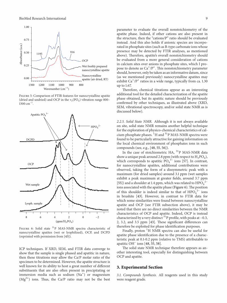
Apatitic PO4
3−
HPO4
2−
Figure 6: Solid state 31P MAS-NMR spectra characteristic ofnanocrystalline apatites (wet or lyophilized), OCP, and DCPD(reprinted with permission from [43]).
ICP techniques. If XRD, SEM, and FTIR data converge toshow that the sample is single phased and apatitic in nature,then these titrations may allow the Ca/P molar ratio of thespecimen to be determined. However, the apatite structure iswell known for its ability to host a great number of differentsubstituents that are also often present in precipitating orimmersion media such as sodium (Na+) or magnesium(Mg2+) ions. Thus, the Ca/P ratio may not be the best
parameter to evaluate the overall nonstoichiometry of theapatite phase. Indeed, if other cations are also present inthe structure, then the “cations/P” ratio should be evaluatedinstead. And this also holds if anionic species are incorpo-rated in phosphate sites (such as B-type carbonate ions whosepresence may be detected by FTIR analyses, as mentionedabove). Therefore, apatite’s overall nonstoichiometry shouldbe evaluated from a more general consideration of cationsin calcium sites over anions in phosphate sites, which I pro-pose to denote as Ca∗/P∗. This nonstoichiometry parametershould, however, only be taken as an informative datum, since(as we mentioned previously) nanocrystalline apatites mayexhibit Ca∗/P∗ ratios in a wide range, typically from ca. 1.30up to 1.67.
Therefore, chemical titrations appear as an interestingadditional tool for the detailed characterization of the apatitephase obtained, but its apatitic nature should be previouslyconfirmed by other techniques, as illustrated above (XRD,SEM, vibrational spectroscopy, and/or solid state NMR as isdiscussed below).
2.2.5. Solid State NMR. Although it is not always availableon site, solid state NMR remains another helpful techniquefor the exploration of physico-chemical characteristics of cal-cium phosphate phases. 1H and 31PMAS-NMR spectra werefound to be particularly attractive for gaining information onthe local chemical environment of phosphates ions in suchcompounds (see, e.g., [48, 55, 56]).
In the case of stoichiometric HA, 31P MAS-NMR datashow a unique peak around 2.8 ppm (with respect toH
3PO4),
which corresponds to apatitic PO4
3− ions [57]. In contrast,for nanocrystalline apatites, additional contributions wereobserved, taking the form of a dissymmetric peak with amaximum (for dried samples) around 3.1 ppm (wet samplesexhibit a peak maximum at greater fields, around 3.7 ppm[55]) and a shoulder at 1.4 ppm, which was related to HPO
4
2−
ions associatedwith the apatite phase (Figure 6).The positionof this shoulder is indeed similar to that of HPO
4
2− ionsin brushite [43]. However, in contrast to FTIR data forwhich some similarities were found between nanocrystallineapatite and OCP (see FTIR subsection above), it may benoted that there are no direct similarities between the NMRcharacteristics of OCP and apatite. Indeed, OCP is insteadcharacterized by a very distinct 31P profile, with peaks at−0.3,2, 3.2, and 3.5 ppm [43]. These significant differences cantherefore be exploited for phase identification purposes.
Finally, proton 1H NMR spectra can also be useful forapatite phase identification due to the presence of a charac-teristic peak at 0.1-0.2 ppm (relative to TMS) attributable toapatitic OH− ions [48, 55, 58].
The solid state NMR technique therefore appears as an-other interesting tool, especially for distinguishing betweenOCP and apatite.
3. Experimental Section
3.1. Compounds Synthesis. All reagents used in this studywere reagent grade.
10 BioMed Research International
Stoichiometric hydroxyapatite (HA), Ca10(PO4)6(OH)2,
was prepared by adding dropwise and underreflux a solutionof diammonium hydrogenphosphate into a boiling solu-tion of calcium nitrate, in stoichiometric proportions andin ammoniated conditions. Note that for this preparation,decarbonatedwater (previously boiled up)was used through-out the process. After 4 hours (at pH∼10), the precipitate wasfiltered, thoroughly washed with deionized water, and driedat 100∘C prior to calcination at 1000∘C for 1 h.
The nanocrystalline apatite samples studied in this workwere prepared by precipitation from mixing aqueous solu-tions of diammonium hydrogenphosphate in excess (0.6M)and calcium nitrate (0.3M), at 22∘C and at pH = 7.2 closeto the physiological value. The excess of phosphate ions wasused to provide an internal pH buffer without any additionof external buffer agent. After rapid mixing (1min), theprecipitates were left to mature in solution for 1 day and thenfiltered, washed, and freeze dried.
Brushite, CaHPO4⋅2H2O, was synthesized via precipita-
tion by stoichiometric addition of an ammoniated calciumnitrate solution into a solution of ammonium dihydro-genphosphate, followed by a 2-hour maturation stage. Theprecipitate was then filtered, washed, and dried at 37∘Covernight.
Monetite, CaHPO4, was obtained by heat treatment of
brushite (at 180∘C) in air.Amorphous calcium phosphate (ACP), am-Ca
3(PO4)2,
was prepared by rapidly adding an ammoniated solution ofcalciumnitrate into an ammoniated solution of diammoniumhydrogenphosphate, followed by a direct filtration, washingwith ammoniated water, and freeze drying for 24 hours.
Octacalcium phosphates (OCP, triclinic), Ca8(PO4)4
(HPO4)2⋅5H2O, was obtained by hydrolysis, under stirring,
of brushite crystals in the presence of an identical weightof ammonium hydrogenphosphate, at 37∘C for 24 hours.This was followed by a step of filtration, washing, and freezedrying. The sample was then conserved at low temperature(−18∘C) to avoid any evolution.
When appropriate (mentioned in the text), simulatedbody fluid (SBF) and 10-times concentrated SBF (10 × SBF)were prepared by following Kokubo’s protocol [19] or bymultiplying all concentrations by a factor of 10, respectively.
3.2. Physicochemical Characterization. X-ray diffraction(XRD) analyses were carried out on an INEL 120 CPS curvedcounter diffractometer using the CoK
𝛼1radiation (with 𝜆 =
1.78892 A).Fourier transform infrared (FTIR) spectra were obtained
on a Nicolet 5700 spectrometer, using the KBr pellet method,in the range 400–4000 cm−1 (64 scans, resolution 4 cm−1).
SEM-EDX analyses were performed on a LEO 435 VPmicroscope operated at 10–15 kV.
4. Conclusions
This review was aimed at remembering the necessity and themeans to carry out adequate physico-chemical characteriza-tion prior to drawing conclusive results on the apatitic natureof a given sample or deposit.
Several possible reasons for experimental failure werebrought up in the first part of this paper, where variousaspects that are not always taken into account were under-lined (possible experimental mistakes, poor washing step,evolution of a substrate in solution, contamination of amedium by microorganisms, residual presence of precursorphases, etc.).
The second part of this review considered the funda-mentals of apatite characterization based on various com-plementary techniques. Even in thermodynamically adequateconditions, some kinetic aspects or other factors—such asthe presence of a stabilizing impurity in the medium—couldmislead the experimentalist and give rise to unexpectedresults. In the case of immersion tests in supersaturatedsolutions, for example, the formation of a white deposit,even characterized by a plate-like morphology, does notnecessarily indicate the formation of apatite. Characterizationtools like XRD, FTIR/Raman, or solid state NMR prove to beespecially well suited to reliably identify apatite compounds,including distinguishing between OCP and nanocrystallineapatite.
All that glitters is not gold. . . all that is white is not apatiteeither.
References
[1] J. Gomez-Morales, M. Iafisco, J. M. Delgado-Lopez, S. Sarda,and C. Drouet, “Progress on the preparation of nanocrystallineapatites and surface characterization: overview of fundamentaland applied aspects,” Progress in Crystal Growth and Character-ization of Materials, vol. 59, no. 1, pp. 1–46, 2013.
[2] S. Cazalbou, C. Combes, D. Eichert, C. Rey, andM. J. Glimcher,“Poorly crystalline apatites: evolution and maturation in vitroand in vivo,” Journal of Bone and Mineral Metabolism, vol. 22,no. 4, pp. 310–317, 2004.
[3] M. Grynpas, “The crystallinity of bone mineral,” Journal ofMaterials Science, vol. 11, no. 9, pp. 1691–1696, 1976.
[4] R. G. Handschin and W. B. Stern, “Crystallographic latticerefinement of human bone,” Calcified Tissue International, vol.51, no. 2, pp. 111–120, 1992.
[5] R. G. Handschin and W. B. Stern, “X-ray diffraction studieson the lattice perfection of human bone apatite (Crista iliaca),”Bone, vol. 16, supplement 4, pp. S355–S363, 1995.
[6] E. Johansen andH. F. Parks, “Electronmicroscopic observationson the three-dimensional morphology of apatite crystallitesof human dentine and bone,” The Journal of Biophysical andBiochemical Cytology, vol. 7, pp. 743–746, 1960.
[7] C. Rey, J. Lian, M. Grynpas, F. Shapiro, L. Zylberberg, and M. J.Glimcher, “Non-apatitic environments in bone mineral: FT-IRdetection, biological properties and changes in several diseasestates,” Connective Tissue Research, vol. 21, no. 1–4, pp. 267–273,1989.
[8] C. Rey, C. Combes, C. Drouet, and M. J. Glimcher, “Bonemineral: update on chemical composition and structure,”Osteo-porosis International, vol. 20, no. 6, pp. 1013–1021, 2009.
[9] C. Drouet, M. Carayon, C. Combes, and C. Rey, “Surfaceenrichment of biomimetic apatites with biologically-active ionsMg2+ and Sr2+: a preamble to the activation of bone repairmaterials,” Materials Science and Engineering C, vol. 28, no. 8,pp. 1544–1550, 2008.

BioMed Research International 11
[10] C.Drouet, J. Gomez-Morales,M. Iafisco, and S. Sarda, “Calciumphosphate surface tailoring technologies for drug deliveringand tissue engineering and applied aspects,” in E-Book: SurfaceTailoring of Inorganic Materials for Biomedical Applications, L.Rimondini, C. L. Bianchi, and E. Verne, Eds., 2012.
[11] L. Winand, “Etude physic-chimique du phosphate tricalciquehydrate et de l’hydroxyapatite,” Annali di Chimica, vol. 6, pp.941–967, 1961.
[12] G.Kuhl andW.H.Nebergall, “Hydrogenphosphat- und carbon-atapatite,” Zeitschrift fur Anorganische und Allgemeine Chemie,vol. 324, p. 313, 1963.
[13] N. Vandecandelaere, C. Rey, and C. Drouet, “Biomimetic ap-atite-based biomaterials: on the critical impact of synthesisand post-synthesis parameters,” Journal of Materials Science-Materials in Medicine, vol. 23, no. 11, pp. 2593–2606, 2012.
[14] N. Hallab, K. Merritt, and J. J. Jacobs, “Metal sensitivity inpatients with orthopaedic implants,” Journal of Bone and JointSurgery A, vol. 83, no. 3, pp. 428–436, 2001.
[15] T. Albrektsson, P. I. Branemark, and H. A. Hansson, “Theinterface zone of inorganic implants in vivo: titanium implantsin bone,”Annals of Biomedical Engineering, vol. 11, no. 1, pp. 1–27,1983.
[16] R. Bidar, P. Kouyoumdjian, E. Munini, and G. Asencio, “Long-term results of the ABG-1 hydroxyapatite coated total hiparthroplasty: analysis of 111 cases with a minimum follow-upof 10 years,” Orthopaedics and Traumatology, vol. 95, no. 8, pp.579–587, 2009.
[17] K. De Groot, R. Geesink, C. P. A. T. Klein, and P. Serekian,“Plasma sprayed coatings of hydroxylapatite,” Journal ofBiomedicalMaterials Research, vol. 21, no. 12, pp. 1375–1381, 1987.
[18] H. Kim, Y. Kim, S. Park et al., “Thin film of low-crystallinecalcium phosphate apatite formed at low temperature,” Bioma-terials, vol. 21, no. 11, pp. 1129–1134, 2000.
[19] A. Oyane, H. Kim, T. Furuya, T. Kokubo, T. Miyazaki, and T.Nakamura, “Preparation and assessment of revised simulatedbody fluids,” Journal of Biomedical Materials Research A, vol. 65,no. 2, pp. 188–195, 2003.
[20] B. D. Ratner, A. S. Hoffman, F. J. Schoen, and J. E. Lemons,Biomaterials Science: An Introduction to Materials in Medicine,Academic Press, San Diego, Calif, USA, 1996.
[21] Y. Abe, T. Kokubo, and T. Yamamuro, “Apatite coating onceramics, metals and polymers utilizing a biological process,”Journal of Materials Science, vol. 1, no. 4, pp. 233–238, 1990.
[22] S. Jalota, A. C. Tas, and S. B. Bhaduri, “In vitro comparison ofthe apatite inducing ability of three different SBF solutions onTi6Al4V,” in Proceedings of the 29th International Conference onAdvanced Ceramics and Composites, pp. 111–118, The AmericanCeramic Society, Cocoa Beach, Fla, USA, January 2005.
[23] A. C. Tas, “Synthesis of biomimetic Ca-hydroxyapatite powdersat 37∘C in synthetic body fluids,” Biomaterials, vol. 21, no. 14, pp.1429–1438, 2000.
[24] J. C. Elliott, Structure and Chemistry of the Apatites and OtherCalcium Orthophosphates, Elsevier Science, Amsterdam, TheNetherlands, 1994.
[25] H. A. Krebs, “Chemical composition of blood plasma and se-rum,”Annual Review of Biochemistry, vol. 19, pp. 409–430, 1950.
[26] M. S. Tung, “Calcium phosphates: structure, composition,solubility, and stability,” in Calcium Phosphates in Biological andIndustrial Systems, Z. Amjad, Ed., pp. 1–19, Kluwer AcademicPublishers, London, UK, 1998.
[27] L. N. Plummer and E. Busenberg, “The solubilities of calcite,aragonite and vaterite in CO
2-H2O solutions between 0 and
90∘C, and an evaluation of the aqueous model for the systemCaCO
3-CO2-H2O,” Geochimica et Cosmochimica Acta, vol. 46,
no. 6, pp. 1011–1040, 1982.[28] R.M. Smith, A. E.Martell, and R. J. Motekaitis,NIST Crititically
Selected Stability Constants of Metal Complexes Database, NISTStandard Reference Database, Gaithersburg, Md, USA, 2003.
[29] E. D. Eanes and J. L. Meyer, “The maturation of crystallinecalcium phosphates in aqueous suspensions at physiologic pH,”Calcified Tissue International, vol. 23, no. 3, pp. 259–269, 1977.
[30] N. Spanos, D. Y. Misirlis, D. G. Kanellopoulou, and P. G.Koutsoukos, “Seeded growth of hydroxyapatite in simulatedbody fluid,” Journal of Materials Science, vol. 41, no. 6, pp. 1805–1812, 2006.
[31] M. B. Tomson and G. H. Nancollas, “Mineralization kinetics: aconstant composition approach,” Science, vol. 200, no. 4345, pp.1059–1060, 1978.
[32] G. H. Nancollas and B. Tomazic, “Growth of calcium phosphateon hydroxyapatite crystals. Effect of supersaturation and ionicmedium,” Journal of Physical Chemistry, vol. 78, no. 22, pp. 2218–2225, 1974.
[33] T. P. Feenstra and P. L. De Bruyn, “The ostwald rule of stagesin precipitation from highly supersaturated solutions: a modeland its application to the formation of the nonstoichiometricamorphous calcium phosphate precursor phase,” Journal ofColloid And Interface Science, vol. 84, no. 1, pp. 66–72, 1981.
[34] J. L. Meyer and E. D. Eanes, “A thermodynamic analysis of theamorphous to crystalline calcium phosphate transformation,”Calcified Tissue International, vol. 25, no. 1, pp. 59–68, 1978.
[35] J. Mahamid, A. Sharir, L. Addadi, and S. Weiner, “Amorphouscalcium phosphate is a major component of the forming finbones of zebrafish: indications for an amorphous precursorphase,” Proceedings of the National Academy of Sciences of theUnited States of America, vol. 105, no. 35, pp. 12748–12753, 2008.
[36] W. E. Brown, J. R. Lehr, J. P. Smith, and A. William Frazier,“Crystallography of octacalcium phosphate,” Journal of theAmerican Chemical Society, vol. 79, no. 19, pp. 5318–5319, 1957.
[37] A. S. Posner and F. Betts, “Synthetic amorphous calciumphosphate and its relation to bone mineral structure,” Accountsof Chemical Research, vol. 8, no. 8, pp. 273–281, 1975.
[38] J. C. Heughebaert, Contribution a l’Etude de l’Evolution desOrthophosphates de Calcium Precipites en OrthophosphatesApatitiques, Institut National Polytechnique de Toulouse,Toulouse, France, 1977.
[39] W. E. Brown, L. W. Schroeder, and J. S. Ferris, “Interlayering ofcrystalline octacalciumphosphate and hydroxylapatite,” Journalof Physical Chemistry, vol. 83, no. 11, pp. 1385–1388, 1979.
[40] J. Christoffersen, M. R. Christoffersen, W. Kibalczyc, and F.A. Andersen, “A contribution to the understanding of theformation of calcium phosphates,” Journal of Crystal Growth,vol. 94, no. 3, pp. 767–777, 1989.
[41] W. E. Brown, “Crystal growth of bone mineral,” ClinicalOrthopaedics and Related Research, vol. 44, pp. 205–220, 1966.
[42] M. S. Tung and W. E. Brown, “An intermediate state in hy-drolysis of amorphous calcium phosphate,” Calcified TissueInternational, vol. 35, no. 6, pp. 783–790, 1983.
[43] D. Eichert, H. Sfihi, C. Combes, and C. Rey, “Specific char-acteristics of wet nanocrystalline apatites. Consequences onblomaterials and bone tissue,” Key Engineering Materials, vol.254–256, pp. 927–930, 2004.

12 BioMed Research International
[44] D. Eichert, M. Salome, M. Banu, J. Susini, and C. Rey, “Pre-liminary characterization of calcium chemical environmentin apatitic and non-apatitic calcium phosphates of biologicalinterest by X-ray absorption spectroscopy,” Spectrochimica ActaB, vol. 60, no. 6, pp. 850–858, 2005.
[45] C. Rey, C. Combes, C. Drouet, A. Lebugle, H. Sfihi, and A.Barroug, “Nanocrystalline apatites in biological systems: char-acterisation, structure and properties,” Materialwissenschaftund Werkstofftechnik, vol. 38, no. 12, pp. 996–1002, 2007.
[46] C. Rey, C. Combes, C. Drouet, H. Sfihi, and A. Barroug,“Physico-chemical properties of nanocrystalline apatites: impli-cations for biominerals and biomaterials,”Materials Science andEngineering C, vol. 27, no. 2, pp. 198–205, 2007.
[47] C. Rey, C. Combes, C. Drouet, and M. J. Glimcher, “Bonemineral: update on chemical composition and structure,”Osteo-porosis International, vol. 20, no. 6, pp. 1013–1021, 2009.
[48] C. Jager, T. Welzel, W. Meyer-Zaika, and M. Epple, “A solid-state NMR investigation of the structure of nanocrystallinehydroxyapatite,” Magnetic Resonance in Chemistry, vol. 44, no.6, pp. 573–580, 2006.
[49] C. Rey, B. Collins, T. Goehl, I. R. Dickson, and M. J. Glimcher,“The carbonate environment in bone mineral: a resolution-enhanced Fourier Transform Infrared Spectroscopy study,”Calcified Tissue International, vol. 45, no. 3, pp. 157–164, 1989.
[50] C. Rey, M. Shimizu, B. Collins, and M. J. Glimcher, “Resolutionenhanced Fourier transform infrared spectroscopy study of theenvironment of phosphates ions in the early deposits of a solidphase of calcium phosphate in bone and enamel, and theirevolution with age.1. Investigations in the ]4 domain,” CalcifiedTissue International, vol. 46, no. 6, pp. 384–394, 1990.
[51] C. Rey, V. Renugopalakrishnan, M. Shimizu, B. Collins, and M.J. Glimcher, “A resolution-enhanced fourier transform infraredspectroscopic study of the environment of the CO32- ion in themineral phase of enamel during its formation and maturation,”Calcified Tissue International, vol. 49, no. 4, pp. 259–268, 1991.
[52] C. Rey, V. Renugopalakrishnan, B. Collins, and M. J. Glim-cher, “Fourier transform infrared spectroscopic study of thecarbonate ions in bone mineral during aging,” Calcified TissueInternational, vol. 49, no. 4, pp. 251–258, 1991.
[53] G. Charlot, Chimie Analytique Quantitative, Masson, Paris,France, 1974.
[54] D. Eichert, C. Drouet, H. Sfihi, C. Rey, and C. Combes, “Nano-crystalline apatite based biomaterials: synthesis, processingand characterization,” in Biomaterials Research Advances, J. B.Kendall, Ed., pp. 93–143, Nova Science Publishers, New York,NY, USA, 2008.
[55] D. Eichert, Etude de la Reactivite de Surface d’Apatites deSynthese Nanocristallines, Institut National Polytechnique deToulouse, Toulouse, France, 2001.
[56] Y. Wu, M. J. Glimcher, C. Rey, and J. L. Ackerman, “A uniqueprotonated phosphate group in bone mineral not present insynthetic calcium phosphates. Identification by phosphorus-31solid stateNMR spectroscopy,” Journal ofMolecular Biology, vol.244, no. 4, pp. 423–435, 1994.
[57] J. Tropp, N. C. Blumenthal, and J. S. Waugh, “Phosphorus NMRstudy of solid amorphous calcium phosphate,” Journal of theAmerican Chemical Society, vol. 105, no. 1, pp. 22–26, 1983.
[58] J. P. Yesinowski and H. Eckert, “Hydrogen environments incalcium phosphates: 1H MAS NMR at high spinning speeds,”Journal of the American Chemical Society, vol. 109, no. 21, pp.6274–6282, 1987.

Submit your manuscripts athttp://www.hindawi.com
ScientificaHindawi Publishing Corporationhttp://www.hindawi.com Volume 2014
CorrosionInternational Journal of
Hindawi Publishing Corporationhttp://www.hindawi.com Volume 2014
Polymer ScienceInternational Journal of
Hindawi Publishing Corporationhttp://www.hindawi.com Volume 2014
Hindawi Publishing Corporationhttp://www.hindawi.com Volume 2014
CeramicsJournal of
Hindawi Publishing Corporationhttp://www.hindawi.com Volume 2014
CompositesJournal of
NanoparticlesJournal of
Hindawi Publishing Corporationhttp://www.hindawi.com Volume 2014
Hindawi Publishing Corporationhttp://www.hindawi.com Volume 2014
International Journal of
Biomaterials
Hindawi Publishing Corporationhttp://www.hindawi.com Volume 2014
NanoscienceJournal of
TextilesHindawi Publishing Corporation http://www.hindawi.com Volume 2014
Journal of
NanotechnologyHindawi Publishing Corporationhttp://www.hindawi.com Volume 2014
Journal of
CrystallographyJournal of
Hindawi Publishing Corporationhttp://www.hindawi.com Volume 2014
The Scientific World JournalHindawi Publishing Corporation http://www.hindawi.com Volume 2014
Hindawi Publishing Corporationhttp://www.hindawi.com Volume 2014
CoatingsJournal of
Advances in
Materials Science and EngineeringHindawi Publishing Corporationhttp://www.hindawi.com Volume 2014
Smart Materials Research
Hindawi Publishing Corporationhttp://www.hindawi.com Volume 2014
Hindawi Publishing Corporationhttp://www.hindawi.com Volume 2014
MetallurgyJournal of
Hindawi Publishing Corporationhttp://www.hindawi.com Volume 2014
BioMed Research International
MaterialsJournal of
Hindawi Publishing Corporationhttp://www.hindawi.com Volume 2014
Nano
materials
Hindawi Publishing Corporationhttp://www.hindawi.com Volume 2014
Journal ofNanomaterials