Embed Size (px)
Citation preview
254
The atomic structure of fluor-apatite and its relation to that of tooth and bone material.
(With Plates X V I - - X V I I I . )
]~y C. A. BEEVERS, D.Sc., F.Inst.P., F.R.S.E., and D. B. MCINTYRE, B.Sc.
Dewar Crystallographic Laboratory, University of Edinburgh.
[Read June 7, 1945.]
I T is well established by X-ray crystal analysis that the mineral constituent of bone and of the enamel and dentine of teeth is essentially hydroxy-apatite
Ca~OH(PO4)3, and that hydroxy-apatite has a structure differing only in small details from that of the well-crystallized mineral fluor-apatite CasF(PO4) 3. The main difference, and in fact the only fully established one, between the two struc- tures is that the hydroxy-apatite has a slightly larger unit cell than that of the fluor-apatite. The structure of the latter has been determined by Ns and by Mehmel ~ who finally agrees with Ns We have made what is probably a more accurate determination of the structure, using more extensive X-ray data. We find that the biggest change required in the atomic co-ordinates given by Ns is a small alteration in the phosphorus position. This and other changes from N~ray-Szab5's structure are only of significance when accurate values of interatomic distances are required, and no changes are made in the general arrangement of the bonding.
The structure is of considerable importance for the understanding of the properties of bone and tooth, and it is thought that the following description of it will be of interest and value. Previous accounts of the structure have been inadequate even to those well versed in the understanding of complex structures.
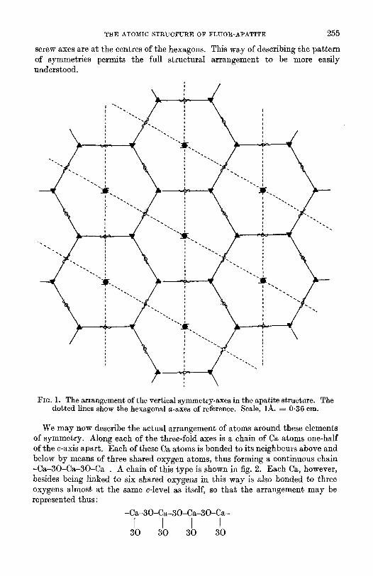
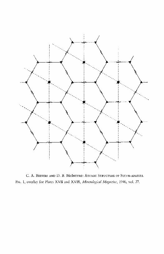
The unit cell of the apatite structure has two equal edges inclined at 120 ~ to one another. These edges are of length a 9.37~_. in the case of fluor-apatite and a 9-41.~. in the case of hydroxy-apatite. The third edge is at right angles to these and has a length of 6.88A. in both fluor- and hydroxy-apatites. In fig. 1 the dotted lines show the a-axes of the cell. The cell possesses vertical symmetry- axes of three kinds: (1) passing through the corners of the dotted unit cell are hexagonal screw-axes, these being equivalent to a three-fold rotation axis with a two-fold screw axis superposed; (2) passing through the points (2/3, 1/3) and (1/3, 2/3) of the cell are three-fold rotation axes ; and (3) passing through the half- way points of the cell edges and through the centre are two-fold screw axes. There are also centres of symmetry, and mirror planes parallel to the plane of the paper: these symmetry elements are not represented in fig. 1.
This arrangement of symmetries means that there are three-fold axes at the corners of a sheet of linked hexagons, as shown by the full lines of fig. 1. The two- fold screw axes are at the centres of the edges of the hexagons, and the six-fold
1 S. Ns Zeits. Krist., 1930, vol. 75, p. 387. [M.A. 4-462.] 2 M. Mehmel, Zeits. Krist., 1930, vol. 75, p. 323, and Zeits. Physikal. Chem., A, 1931, vol.
15, p. 223. [M.A. 4-462.]
T H E A T O M I C S T R U C T U R E O F ~ F L U O R - A P A T I T E 255
screw axes are a t the centres of the hexagons. This way of describing the pat tern of symmetries permits the full s tructural arrangement to be more easily understood.
r ~ - . l
�9 k _ ~ . 7 . v . r . , % - ~ ' . 7 -
/- \ - / : ', "-- l \
i B " ' . i
FIG. 1. The arrangement of the vertical symmetry-axes in the apatite structure. The dotted lines show the hexagonal a-axes of reference. Scale, 1/~. ~ 0.36 cm.
We may now describe the actual arrangement of atoms around these elements of symmetry. Along each of the three-fold axes is a chain of Ca atoms one-half of the c-axis apart . Each of these Ca atoms is bonded to its neighbours above and below by means of three shared oxygen atoms, thus forming a continuous chain - C a - 3 0 - C a - 3 0 - C a - . A chain of this type is shown in fig. 2. Each Ca, however, besides being linked to six shared oxygens in this way is also bonded to three oxygens almost a t the same c-level as itself, so tha t the arrangement may be represented thus:
- C a - 3 0 - C a - 3 0 - C a - 3 0 - C a -
I I I I 30 30 30 30
256 c.A. BEEVERS AND D. B. MCINTYRE ON
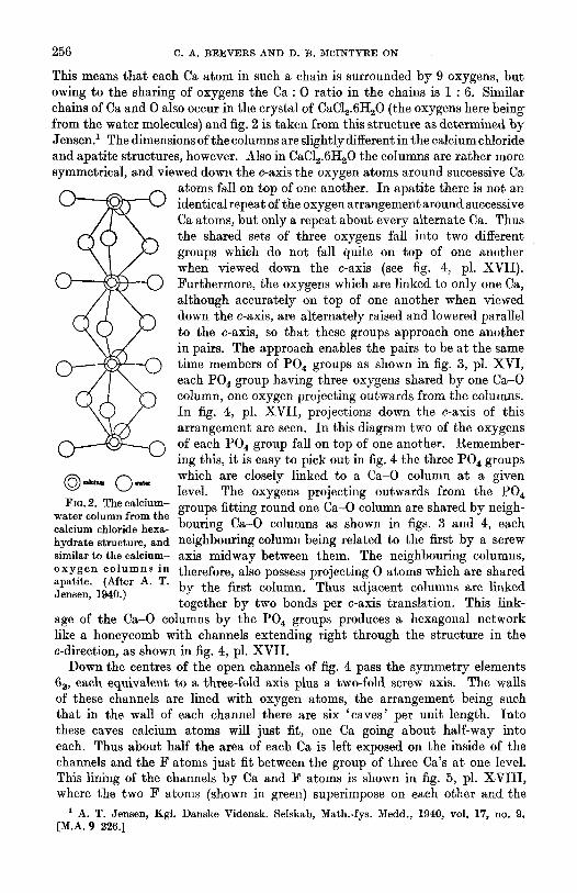
This means that each Ca atom in such a chain is surrounded by 9 oxygens, but owing to the sharing of oxygens the Ca : 0 ratio in the chains is 1 : 6. Similar chains of Ca and 0 also occur in the crystal of CaC12.6tI20 (the oxygens here being from the water molecules) and fig. 2 is taken from this structure as determined by Jensen3 The dimensions of the columns are slightly different in the calcium chloride and apatite structures, however. Also in CaCI~.6H~O the columns are rather more symmetrical, and viewed down the c-axis the oxygen atoms around successive Ca
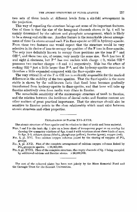
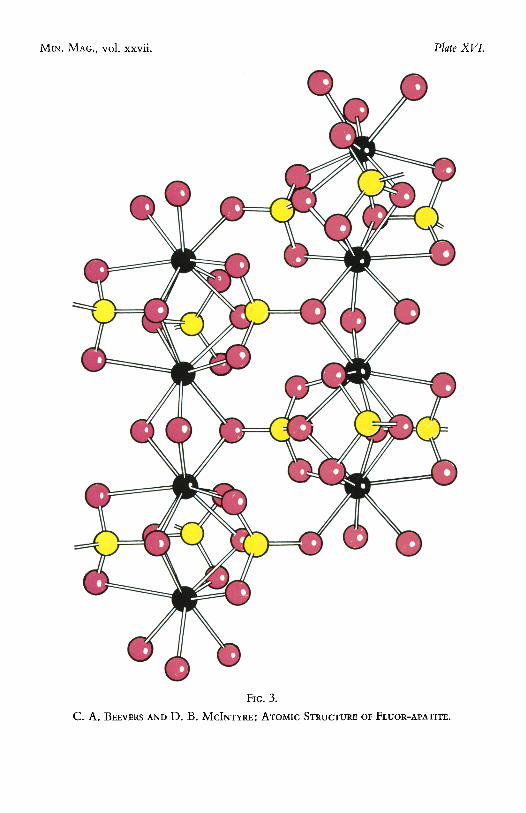
atoms fall on top of one another. In apatite there is not an identical repeat of the oxygen arrangement around successive Ca atoms, but only a repeat about every alternate Ca. Thus the shared sets of three oxygens fall into two different groups which do not fall quite on top of one another when viewed down the c-axis (see fig. 4, pl. XVII). Furthermore, the oxygens which are linked to only one Ca, although accurately on top of one another when viewed down the c-axis, are alternately raised and lowered parallel to the c-axis, so that these groups approach one another in pairs. The approach enables the pairs to be at the same time members of PO 4 groups as shown in fig. 3, pl. XVI, each PO4 group having three oxygens shared by one Ca-O column, one oxygen projecting outwards from the columns. In fig. 4, pl. XVII , projections down the c-axis of this arrangement are seen. In this diagram two of the oxygens of each POt group fall on top of one another. Remember- lng this, it is easy to pick out in fig. 4 the three PO 4 groups
( D , ~ m C),,~, which are closely linked to a Ca-O column at a given level. The oxygens projecting outwards from the PO 4
Fio. 2. The calcium- groups fitting round one Ca-O column are shared by neigh- water column from the calcium chloride hexa- bouring Ca-O columns as shown in figs. 3 and 4, each hydrate structure, and neighbouring column being related to the first by a screw similar to the calcium- axis midway between them. The neighbouring columns, oxygen columns in therefore, also possess projecting 0 atoms which are shared apatite. (After A. T. by the first column. Thus adjacent columns are linked Jensen, 1940.)
together by two bonds per c-axis translation. This link- age of the Ca-O columns by the PO 4 groups produces a hexagonal network like a honeycomb with channels extending right through the structure in the c-direction, as shown in fig. 4, pl. XVII .
Down the centres of the open channels of fig. 4 pass the symmetry elements 63, each equivalent to a three-fold axis plus a two-fold screw axis. The walls of these channels are lined with oxygen atoms, the arrangement being such that in the wall of each channel there are six 'caves ' per unit length. Into these caves calcium atoms will just fit, one Ca going about half-way into each. Thus about half the area of each Ca is left exposed on the inside of the channels and the F atoms just fit between the group of three Ca's at one level. This lining of the channels by Ca and F atoms is shown in fig. 5, pl. XVII I , where the two F atoms (shown in green) superimpose on each other and the
1 A. T. Jensen, Kgl. Danske Vidensk. SeIskab, Math.-fys. Medd., 1940, vol. 17, no. 9. [M.A. 9 226.]
THE ATOMIC STRUCTURE OF FLUOR-APATITE 2 5 7
two sets of three bonds at different levels form a six-fold arrangement in the projection.
This way of regarding the structure brings out some of its important features. One of these is that the size of the hexagonal channels shown in fig. 4 will be mainly determined by the calcium and phosphate arrangement, which is likely to be a strong and stable one. Another feature is the remarkable planar arrange- ment of three Ca atoms around each F in fluor-apatite or OH in hydroxy-apatite. From these two features one would expect that the structure would be very selective in its choice of ions to occupy the position of the F ions in fluor-apatite. The only ions definitely known to occupy these positions are the ions F -1 and OH -1, and these two are, of course, very nearly the same size. Each has two K and eight L electrons, bu t F -z has one nucleus with charge q-9, whilst OH -1 possesses two nuclear charges + 8 and q-1 respectively. This has the effect of making OH -1 just a little larger than F -1, and the hydroxy-apatite structure is therefore a little expanded compared with the fluor-apatite one.
The very critical fit of the F or OH ion is evidently responsible for the marked difference in the stability of the two apatites. That the fluor=apatite is the more stable is shown by the well-known facts that fossil bone becomes gradually transformed from hydroxy-apatite to fluor-apatite, and that bone will take up fuorine selectively even from media very dilute in fluorine.
The remarkable sensitivity of the macroscopic structure of teeth to fluorine, and the relation between the incidence of dental caries and fluorine content are other matters of great practical importance. That the structure should also be sensitive to fluorine points to the close relationship which must exist between atomic structure and other properties.
EXPLANATION OF PLATES XVI-XVIII.
The atomic structure of fluor-apatite and its relation to that of tooth and bone material. FIGS. 1 and 2 in the text ; fig. 1 also on a loose sheet of transparent paper as an overlay for
showing the symmetl2r relations of figs. 4 and 5 with rotations about three kinds of axes. In figs. 3-5, calcium atoms (black), phosphorus (yellow), fluorine (green), oxygen (red).
FIG. 3, pl. XVI. Two calcium-oxygen columns joined by the sharing of oxygens of P04 groups.
FrG. 4, pl. XVt I . Plan of the comp~te arrangement of calcium-oxygen columns linked by POa groups in apatite. • 36,000,000.
FIG. 5, pl. XVIII. Plan of the complete structure, the empty channels of fig. 4 being occupied by additional calcium and fluorine atoms, x 36,000,000.
The cost of the coloured plates has been met jointly by the Miers Memorial Fund and the Carnegie Trust for the Scottish Universities.
M~N. MAC., vol. xxvii. Plate XVL
FIG. 3.
C. A. ]~EEVERS AND D. B. MCINTYRE: ATOMIC STRUCTURE OF FLUOR-APATITE.
MIN. MAG., vol. xxvii. Plate XVI1.
FIG. 4.
C. A. BEEVERS AND D. B. MCINTYRE: ATOMIC STRUCTURE OF FLUOR-APATITE.
C. A. BEEVERS AND 1). B. MCINIYRE: ATOMIC STRUCTURE OF FLUOR-APATITE.
FiG. 1, overlay for Plates XVII and XVII[, Mineralq~:ical Magazine, 1946, vol. 27.
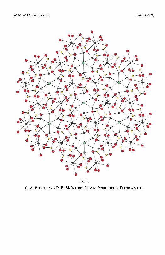
MIN. MAG., vol. xxvii. Plate XVIII.
FIG. 5.
C. A. BEEVERS AND D. B. MCINTYRE: ATOMIC STRUCTURE OF FLUOR-APATITE.
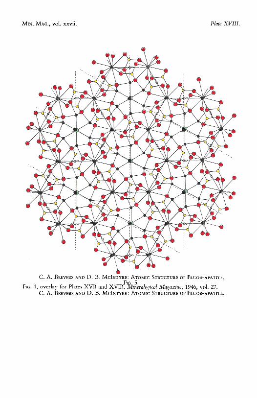
MIN. MAG., vol. xxvii. Plate XVIII.
FIG. 5.
C. A. BEEVERS AND D. B. MCINTYRE: ATOMIC STRUCTURE OF FLUOR-APATITE.