Embed Size (px)
Citation preview
1
Muscles and Muscle TissueChapter 9
2
Overview of Muscle Tissues• Compare and Contrast the three basic types of muscle tissue
• List four important functions of muscle tissue
3
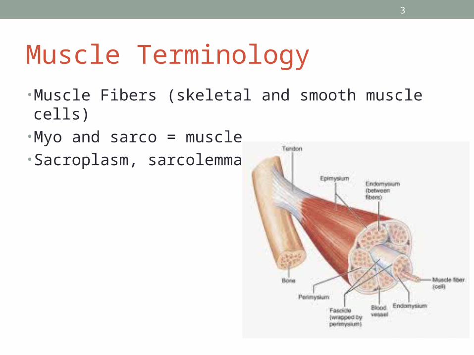
Muscle Terminology• Muscle Fibers (skeletal and smooth muscle cells)• Myo and sarco = muscle• Sacroplasm, sarcolemma
4
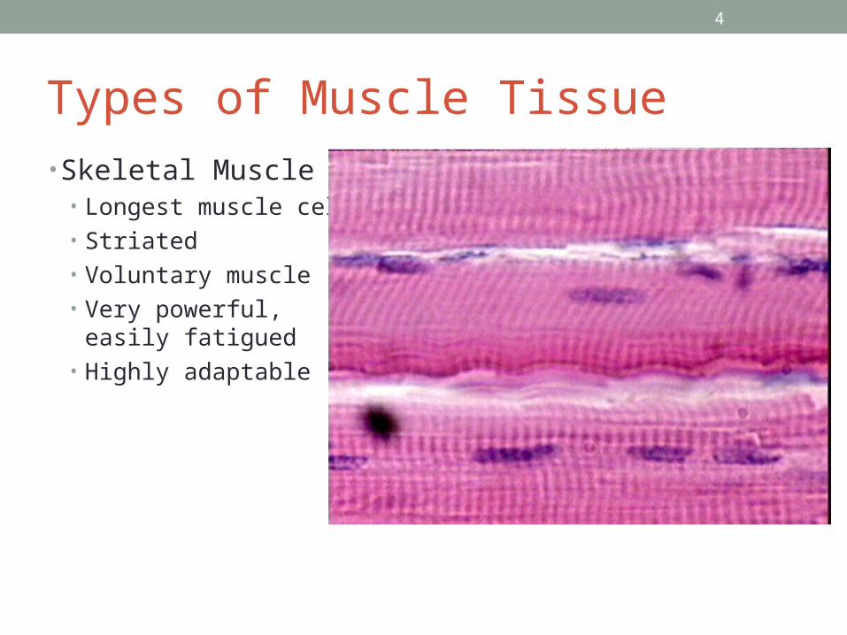
Types of Muscle Tissue• Skeletal Muscle
• Longest muscle cells• Striated• Voluntary muscle• Very powerful, easily
fatigued• Highly adaptable
5
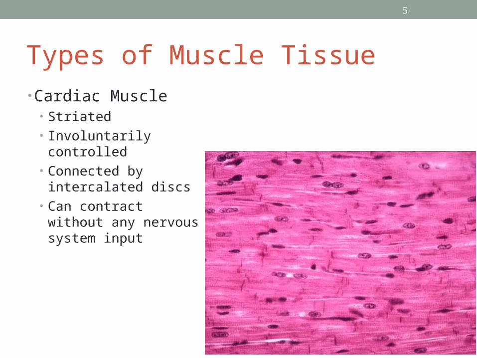
Types of Muscle Tissue• Cardiac Muscle
• Striated• Involuntarily controlled• Connected by intercalated
discs• Can contract without any
nervous system input
6
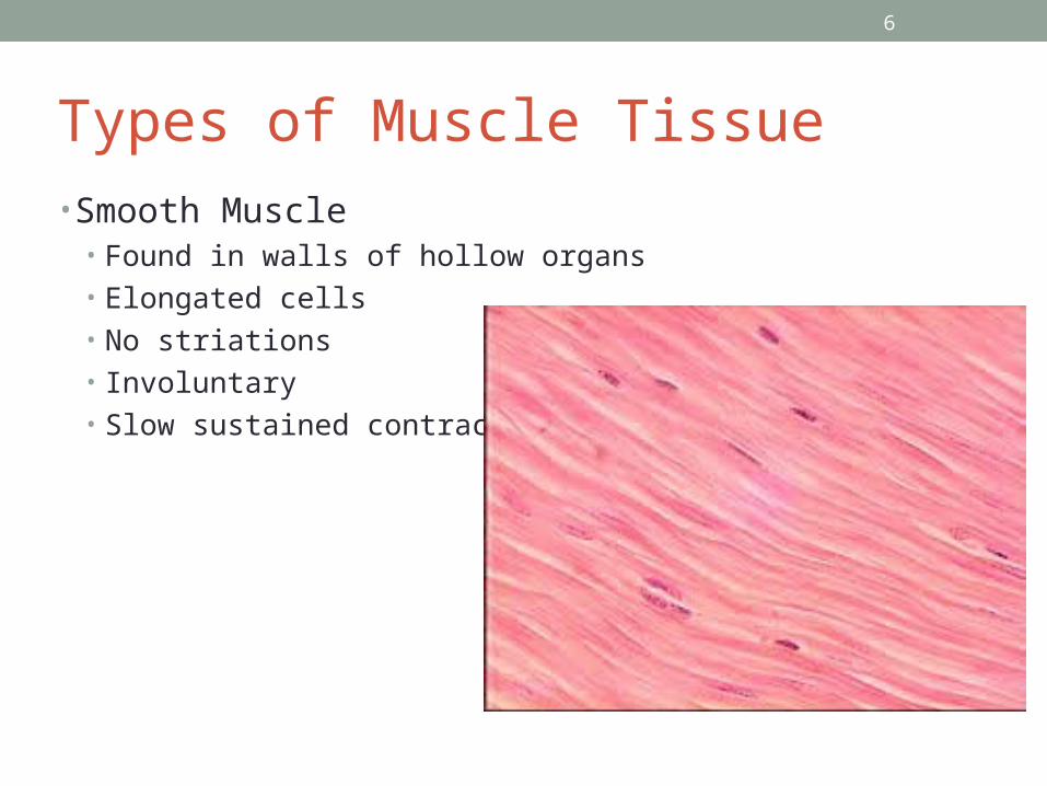
Types of Muscle Tissue• Smooth Muscle
• Found in walls of hollow organs• Elongated cells• No striations• Involuntary• Slow sustained contractions
7
Special Characteristics of Muscle Tissue• 1. Excitability• 2. Contractility• 3. Extensibility• 4. Elasticity
8
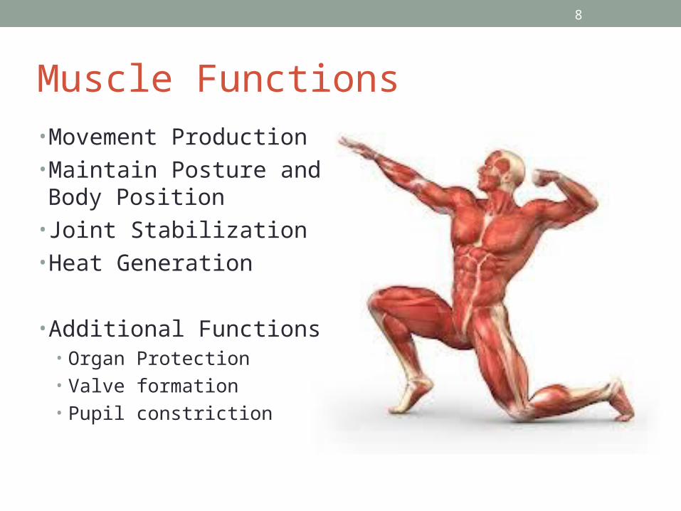
Muscle Functions• Movement Production• Maintain Posture and Body Position
• Joint Stabilization• Heat Generation
• Additional Functions• Organ Protection• Valve formation• Pupil constriction
9
Check Your Understanding• When describing muscle, what does striated mean?• Andrew is pondering an exam question that asks, Which muscle type has elongated cells and is found in the walls of the urinary bladder? How should he respond Reed?
10
Skeletal Muscle• Describe the Gross Structure of a Skeletal Muscle• Describe the microscopic structure and functional roles of the myofibrils, sarcoplasmic reticulum, and T tubules of skeletal muscle fibers
• Describe the sliding filament model of muscle contraction
11
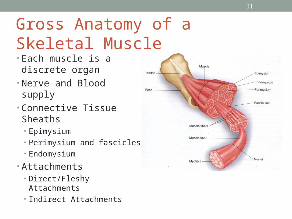
Gross Anatomy of a Skeletal Muscle• Each muscle is a discrete
organ• Nerve and Blood supply• Connective Tissue Sheaths
• Epimysium• Perimysium and fascicles• Endomysium
• Attachments• Direct/Fleshy Attachments• Indirect Attachments
12
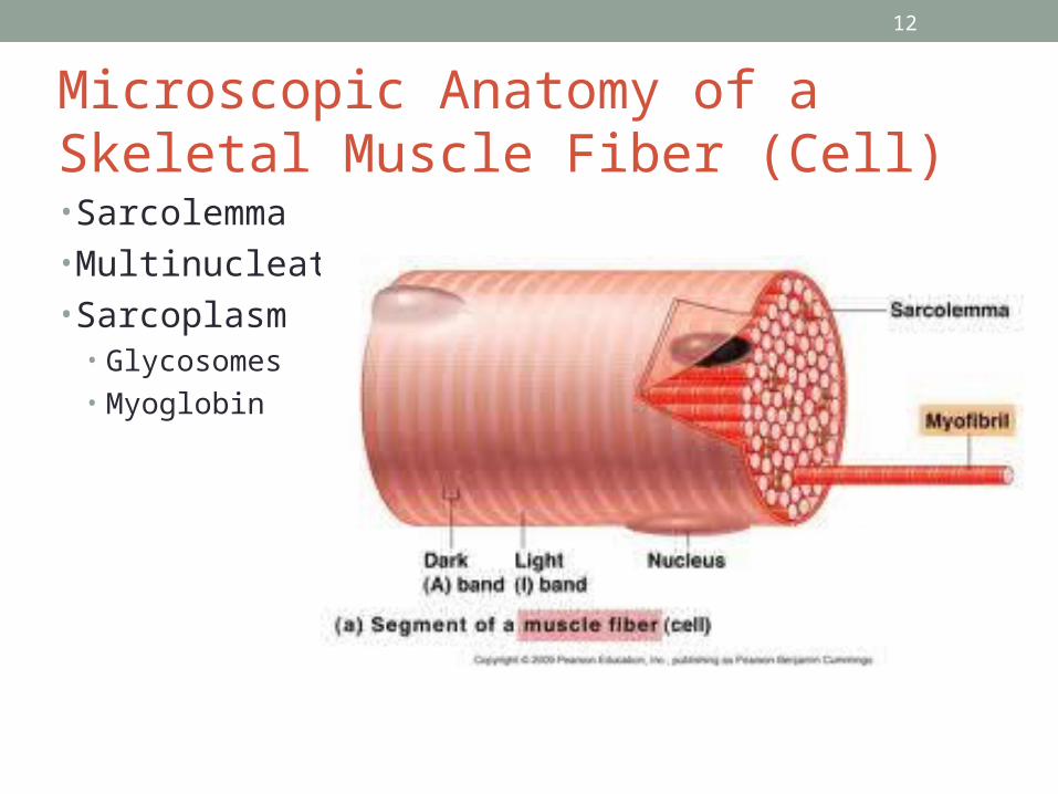
Microscopic Anatomy of a Skeletal Muscle Fiber (Cell)• Sarcolemma• Multinucleate• Sarcoplasm
• Glycosomes• Myoglobin
13
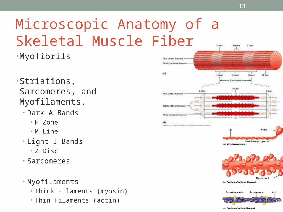
Microscopic Anatomy of a Skeletal Muscle Fiber• Myofibrils
• Striations, Sarcomeres, and Myofilaments.• Dark A Bands
• H Zone• M Line
• Light I Bands• Z Disc
• Sarcomeres
• Myofilaments• Thick Filaments (myosin)• Thin Filaments (actin)
14
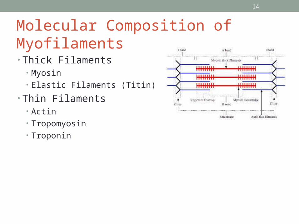
Molecular Composition of Myofilaments
• Thick Filaments• Myosin• Elastic Filaments (Titin)
• Thin Filaments• Actin• Tropomyosin• Troponin
14
15
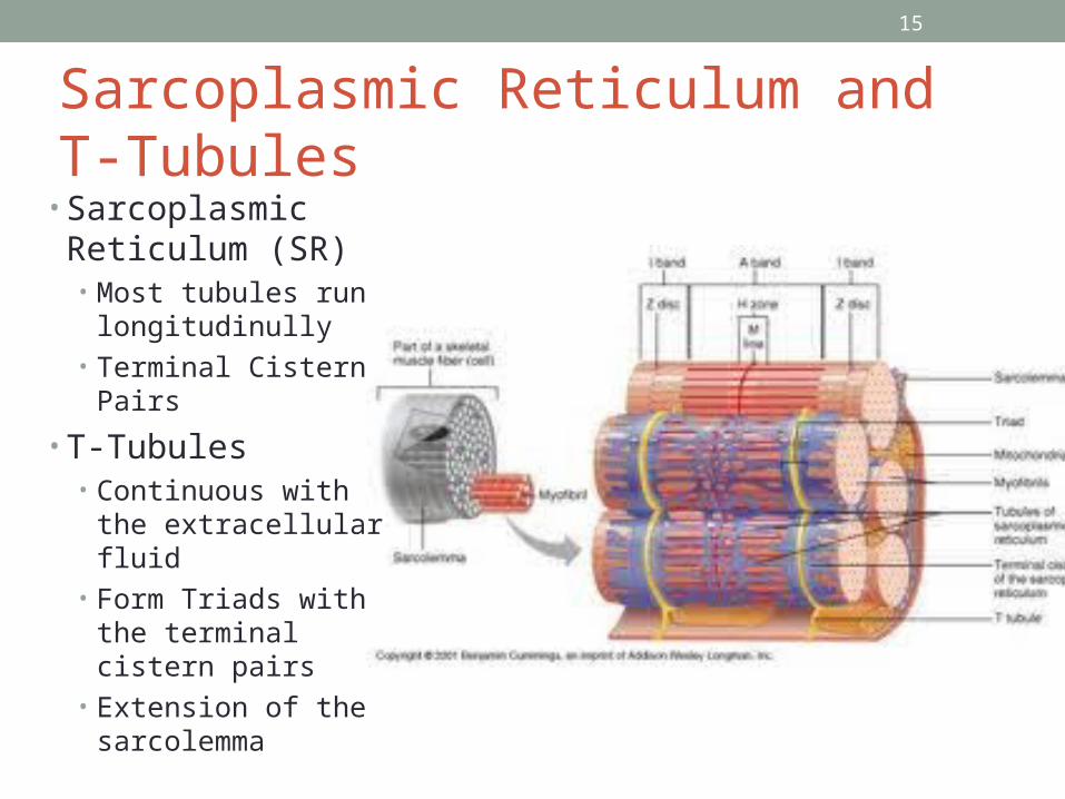
Sarcoplasmic Reticulum and T-Tubules
• Sarcoplasmic Reticulum (SR)• Most tubules run
longitudinully• Terminal Cistern Pairs
• T-Tubules• Continuous with the
extracellular fluid• Form Triads with the
terminal cistern pairs• Extension of the
sarcolemma
15
16
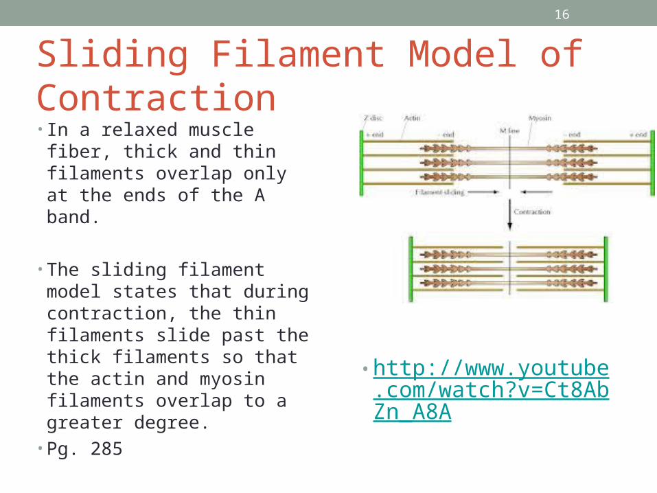
Sliding Filament Model of Contraction• In a relaxed muscle fiber,
thick and thin filaments overlap only at the ends of the A band.
• The sliding filament model states that during contraction, the thin filaments slide past the thick filaments so that the actin and myosin filaments overlap to a greater degree.
• Pg. 285
16
• http://www.youtube.com/watch?v=Ct8AbZn_A8A
17
Check Your Understanding• How does the Term Epimysium relate to the role and
position of this connective tissue sheath?• Which Myofilaments have binding sites for calcium? What
specific molecule binds calcium?• Which region or organelle -cytosol, mitochondrion, or SR-
contains the highest concentration of calcium ions in a resting muscle fiber? Which structure provides the ATP needed for muscle activity?
17
18
Physiology of Skeletal Muscle Fibers
• Explain how muscle fibers are stimulated to contract by describing events that occur at the neuromuscular junction.
• Describe how an Action Potential is Generated• Follow the events of excitation-contraction coupling that
lead to cross bridge activity.
18
19
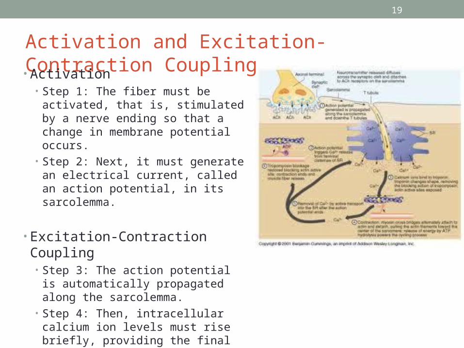
Activation and Excitation-Contraction Coupling• Activation
• Step 1: The fiber must be activated, that is, stimulated by a nerve ending so that a change in membrane potential occurs.
• Step 2: Next, it must generate an electrical current, called an action potential, in its sarcolemma.
• Excitation-Contraction Coupling• Step 3: The action potential is
automatically propagated along the sarcolemma.
• Step 4: Then, intracellular calcium ion levels must rise briefly, providing the final trigger for contraction.
19
20
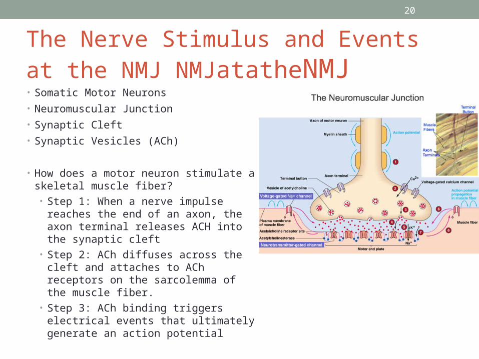
The Nerve Stimulus and Events at the NMJ NMJatatheNMJ• Somatic Motor Neurons
• Neuromuscular Junction
• Synaptic Cleft
• Synaptic Vesicles (ACh)
• How does a motor neuron stimulate a skeletal muscle fiber?• Step 1: When a nerve impulse reaches
the end of an axon, the axon terminal releases ACH into the synaptic cleft
• Step 2: ACh diffuses across the cleft and attaches to ACh receptors on the sarcolemma of the muscle fiber.
• Step 3: ACh binding triggers electrical events that ultimately generate an action potential
20
21
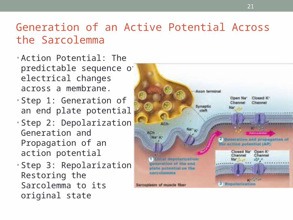
Generation of an Active Potential Across the Sarcolemma
• Action Potential: The predictable sequence of electrical changes across a membrane.
• Step 1: Generation of an end plate potential
• Step 2: Depolarization: Generation and Propagation of an action potential
• Step 3: Repolarization: Restoring the Sarcolemma to its original state
21
22
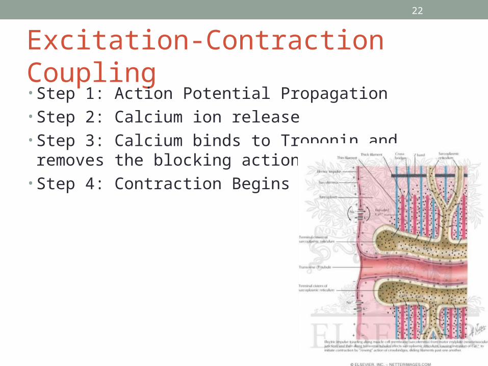
Excitation-Contraction Coupling• Step 1: Action Potential Propagation• Step 2: Calcium ion release• Step 3: Calcium binds to Troponin and removes the
blocking action of tropomyosin• Step 4: Contraction Begins
22

23
Cross Bridge Cycling• http://www.youtube.com/watch?v=Ct8AbZn_A8A
23
24
Check Your Understanding• What are the three structural components of a
neuromuscular junction?• What is the final trigger for contraction? What is the initial
trigger?• What prevents the filaments from sliding back to their
original position each time a myosin cross bridge detaches from actin?
• What would happen if a muscle fiber suddenly ran out of ATP when sarcomeres had only partially contracted?
24
25
Contraction of Skeletal Muscle• Define motor unit and muscle twitch, and describe the
events occurring during the three phases of muscle twitch.• Explain how smooth, graded contractions of a skeletal
muscle are produced.• Differentiate between isometric and isotonic contractions.
25
26
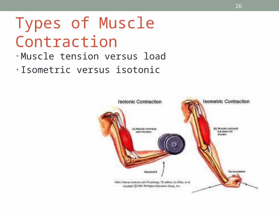
Types of Muscle Contraction• Muscle tension versus load• Isometric versus isotonic
26
27
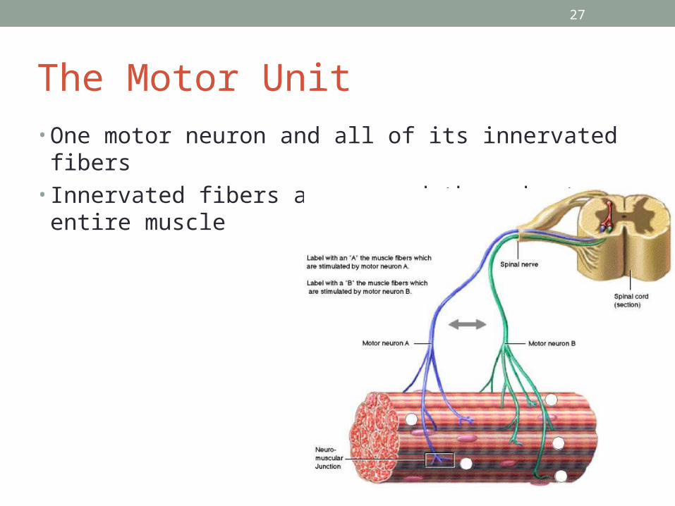
The Motor Unit• One motor neuron and all of its innervated fibers• Innervated fibers are spread throughout entire muscle
27
28
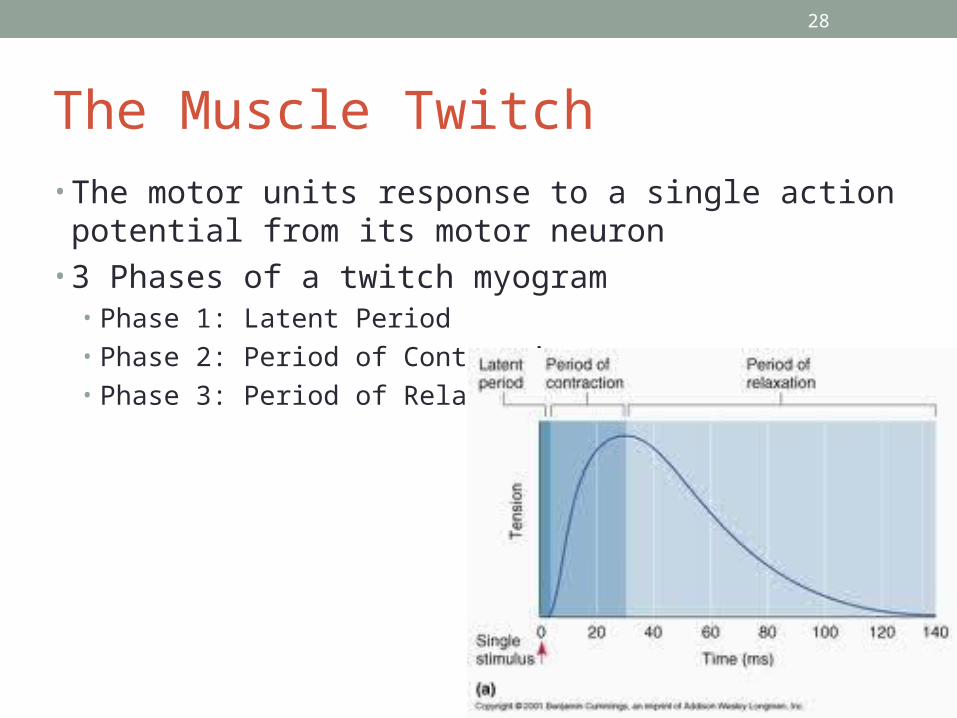
The Muscle Twitch• The motor units response to a single action potential from
its motor neuron• 3 Phases of a twitch myogram
• Phase 1: Latent Period• Phase 2: Period of Contraction• Phase 3: Period of Relaxation
28
29
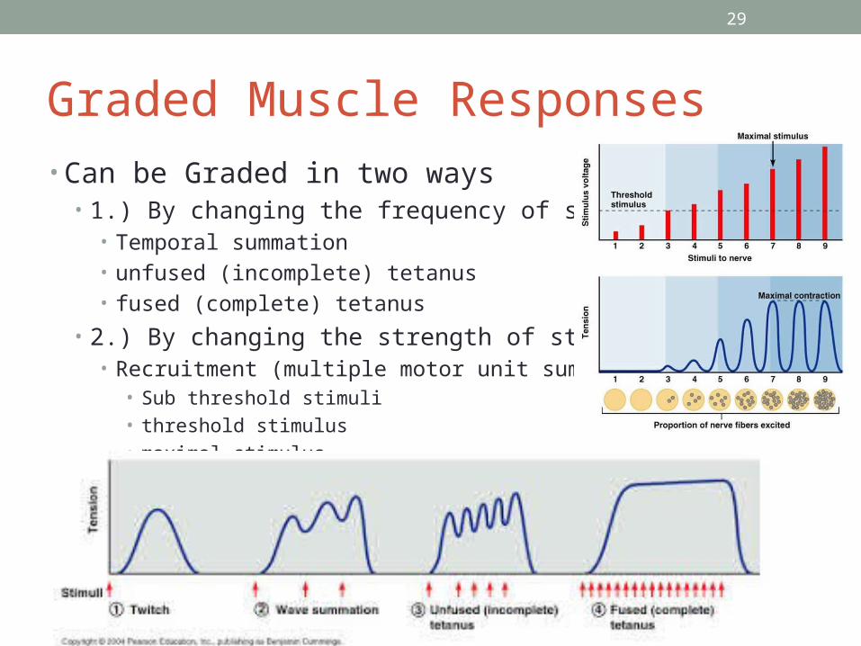
Graded Muscle Responses• Can be Graded in two ways
• 1.) By changing the frequency of stimulation• Temporal summation• unfused (incomplete) tetanus• fused (complete) tetanus
• 2.) By changing the strength of stimulation• Recruitment (multiple motor unit summation)
• Sub threshold stimuli• threshold stimulus• maximal stimulus
29
30
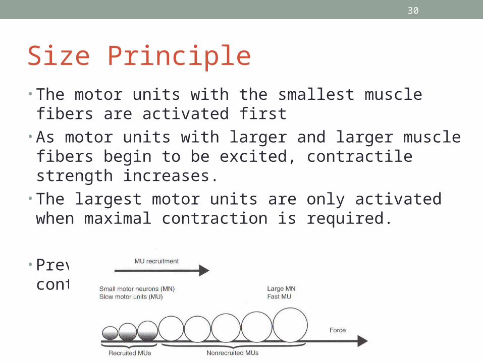
Size Principle• The motor units with the smallest muscle fibers are activated
first • As motor units with larger and larger muscle fibers begin to
be excited, contractile strength increases.• The largest motor units are only activated when maximal
contraction is required.
• Prevents fatigue due to asynchronous contraction
30
31
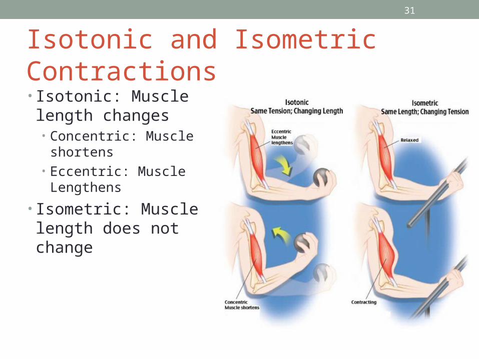
Isotonic and Isometric Contractions• Isotonic: Muscle length
changes• Concentric: Muscle
shortens• Eccentric: Muscle
Lengthens
• Isometric: Muscle length does not change
31
32
Check your understanding• What is a motor unit• What is happening in a muscle during the latent period of a
twitch contraction?• Matt is competing in a chin up competition, What type of
muscle contractions are occurring in his biceps muscles?
32
33
Muscle Metabolism
• Describe three ways in which ATP is regenerated during skeletal muscle contraction.
• Define EPOC and muscle fatigue. List possible causes of muscle fatigue.
33
34
Providing Energy for Muscle Contraction
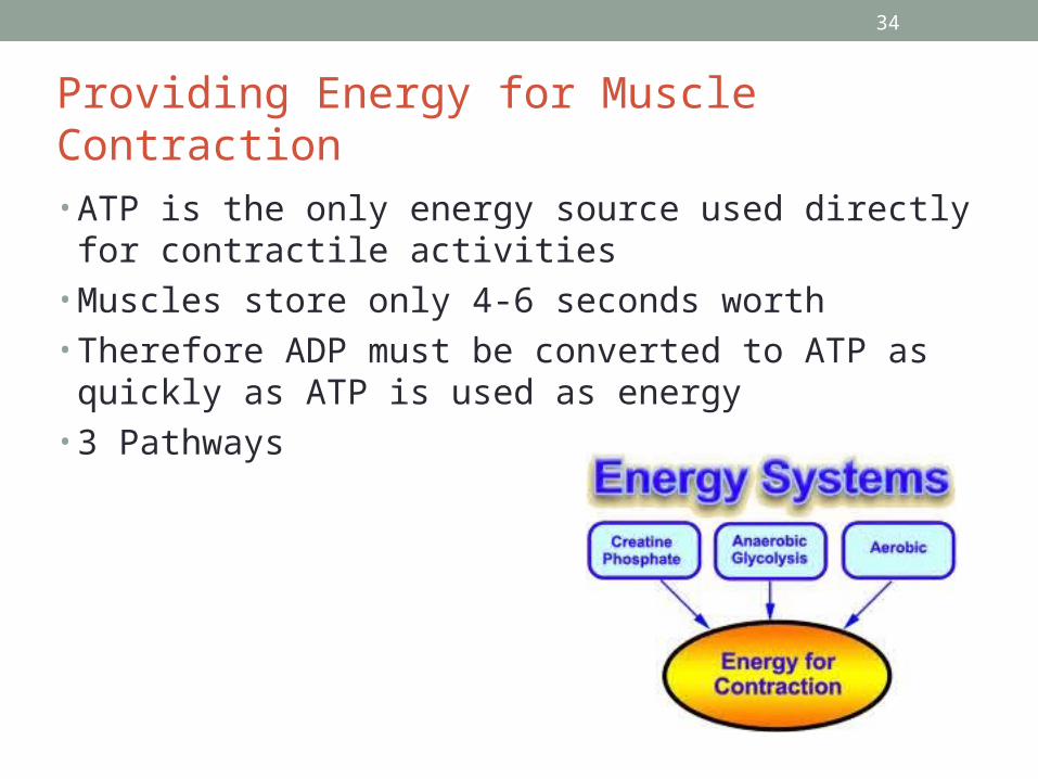
• ATP is the only energy source used directly for contractile activities
• Muscles store only 4-6 seconds worth• Therefore ADP must be converted to ATP as quickly as
ATP is used as energy• 3 Pathways
34
35
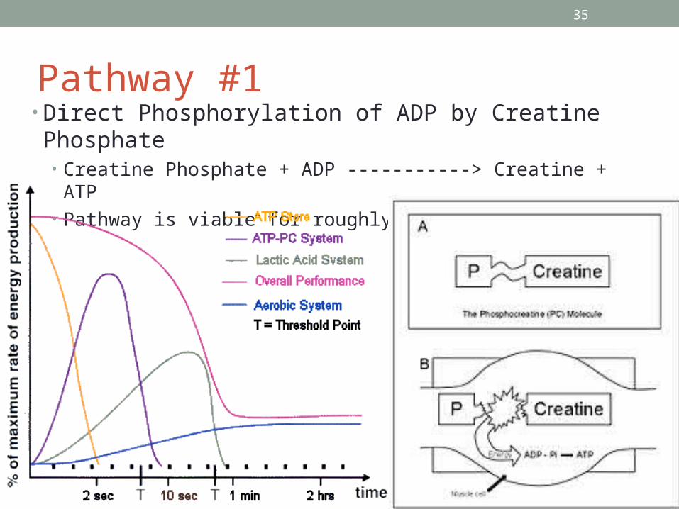
Pathway #1• Direct Phosphorylation of ADP by Creatine Phosphate
• Creatine Phosphate + ADP -----------> Creatine + ATP• Pathway is viable for roughly 15 seconds
35
36
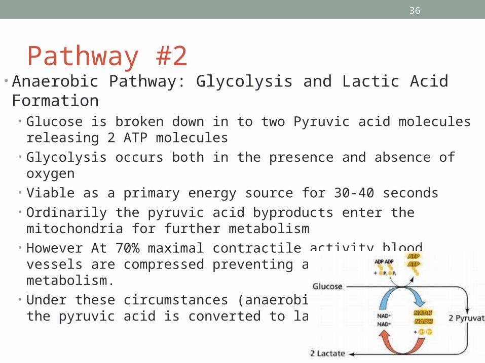
Pathway #2• Anaerobic Pathway: Glycolysis and Lactic Acid Formation
• Glucose is broken down in to two Pyruvic acid molecules releasing 2 ATP molecules
• Glycolysis occurs both in the presence and absence of oxygen• Viable as a primary energy source for 30-40 seconds• Ordinarily the pyruvic acid byproducts enter the mitochondria for further
metabolism• However At 70% maximal contractile activity blood vessels are compressed
preventing aerobic mitochondrial metabolism. • Under these circumstances (anaerobic glycolysis) most of the pyruvic acid
is converted to lactic acid
36
37
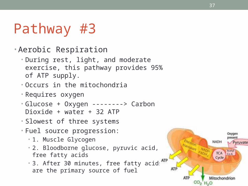
Pathway #3• Aerobic Respiration
• During rest, light, and moderate exercise, this pathway provides 95% of ATP supply.
• Occurs in the mitochondria• Requires oxygen• Glucose + Oxygen --------> Carbon Dioxide +
water + 32 ATP• Slowest of three systems• Fuel source progression:
• 1. Muscle Glycogen• 2. Bloodborne glucose, pyruvic acid, free fatty
acids• 3. After 30 minutes, free fatty acids are the
primary source of fuel
37
38
Energy Systems During Sport• Aerobic Endurance• Anaerobic Threshold
• Weightlifting: Direct Phosphorylation
• On off activities such as tennis, soccer, 100m swim: Anaerobic
• Prolonged jogging: Aerobic
38
39
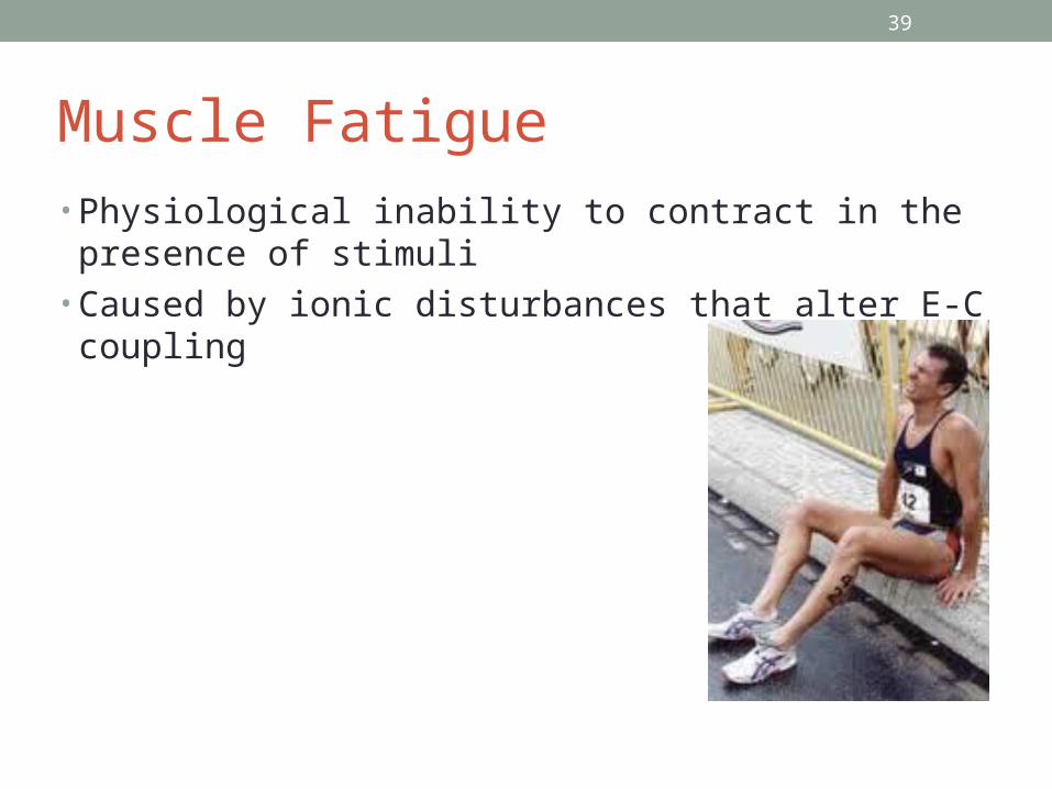
Muscle Fatigue• Physiological inability to contract in the presence of stimuli• Caused by ionic disturbances that alter E-C coupling
39
40
Excess Post-exercise Oxygen Consumption (EPOC)
• Post exercise, muscle tissue must• replenish its myoglobin bound oxygen reserves• convert excess lactic acid into pyruvic acid• replace glycogen stores• Resynthesize ATP and creatine phosphate reserves
• The increased oxygen demand during this recovery period is referred to as the EPOC or oxygen debt
40
41
Heat Production• Only 40% of energy used during muscle contraction is
converted into useful work• 60% is converted into heat
41
42
Check Your Understanding• Clayton has just finished jogging and is breathing heavily.
Why is Clayton breathing heavily? What metabolic product might account for his sore muscles and muscle weakness?
42
43
Forces of Muscle Contraction• Describe factors that influence the force, velocity, and
duration of skeletal muscle contraction• Describe three types of skeletal muscle fibers and explain
the relative value of each type
43
44
Muscle Contraction Force• Influencing Factors
• Number of fibers recruited• Size of muscle fibers• Frequency of stimulation• Degree of muscle stretch
44
45
Velocity and Duration of Contraction• Influencing factors
• Muscle Fiber Type• Load• Recruitment
45
46
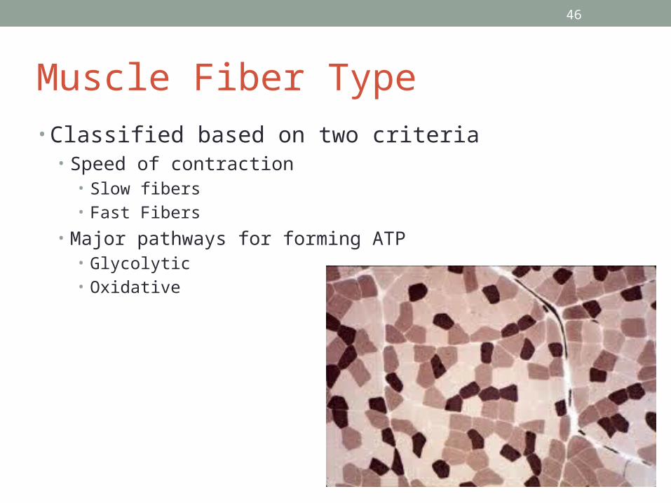
Muscle Fiber Type• Classified based on two criteria
• Speed of contraction• Slow fibers• Fast Fibers
• Major pathways for forming ATP• Glycolytic• Oxidative
46
47
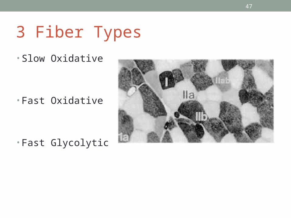
3 Fiber Types
• Slow Oxidative
• Fast Oxidative
• Fast Glycolytic
47
48
Load• Greater load results in
• a longer latent period• a slower contraction• a shorter duration of muscle contraction
48
49
Recruitment• The greater number of motor units recruited
• The faster the contraction• The more prolonged the contraction
49
50
Check Your Understanding• List two factors that influence contractile force and two that
influence velocity of contraction
50
51
Adaptations to Exercise• Compare and Contrast the effects of aerobic and
resistance exercise on skeletal muscles and on other body systems
51
52
Aerobic (endurance) Exercise• Number of capillaries surrounding the muscle fibers
increases• Number of mitochondria within the muscle fibers increases• Concentration of myoglobin increases
• Affects all fiber types, conversion is possible
52
53
Resistance Exercise• Causes muscle hypertrophy• Muscle fibers increase in diameter, not number
53

54
Study guide
54