Embed Size (px)
Citation preview
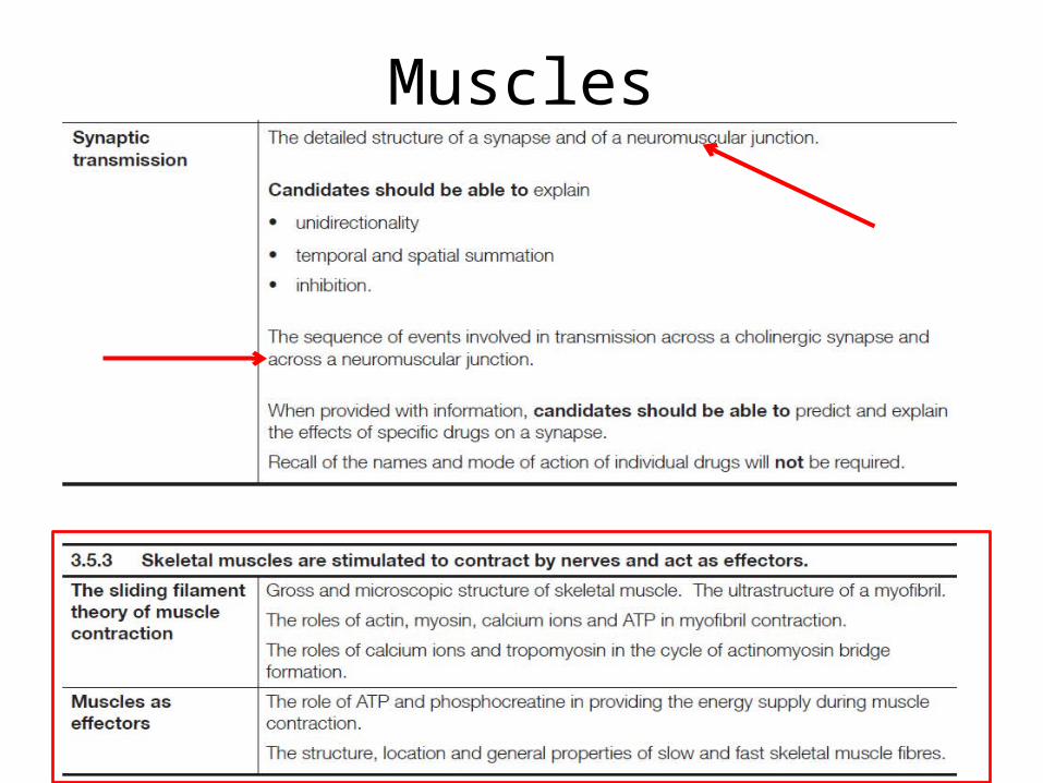
Muscles
2 of 36 © Boardworks Ltd 2009
Muscles
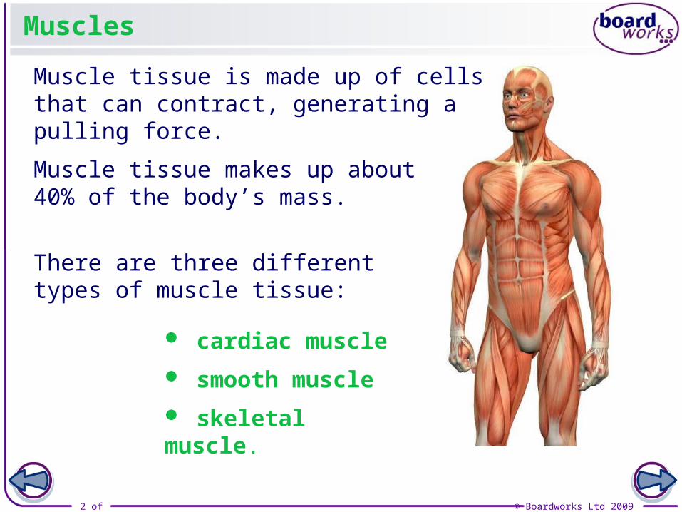
Muscle tissue makes up about 40% of the body’s mass.
There are three different types of muscle tissue:
Muscle tissue is made up of cells that can contract, generating a pulling force.
cardiac muscle
smooth muscle
skeletal muscle.

3 of 36 © Boardworks Ltd 2009
Skeletal muscle
Skeletal muscle is essential for voluntary movement, but is also constantly used for maintaining posture. It covers the skeleton and allows bones to be moved relative to one another.
The voluntary nervous system controls skeletal muscle by sending messages from the central nervous system to the muscle tissue.
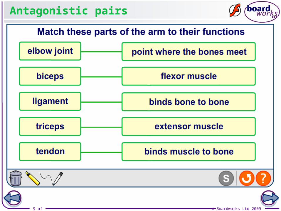
Muscles are usually attached to bones by a form of inelastic tissue called a tendon.
tendon attaches the muscle to
the bone
4 of 36 © Boardworks Ltd 2009
Cardiac muscle
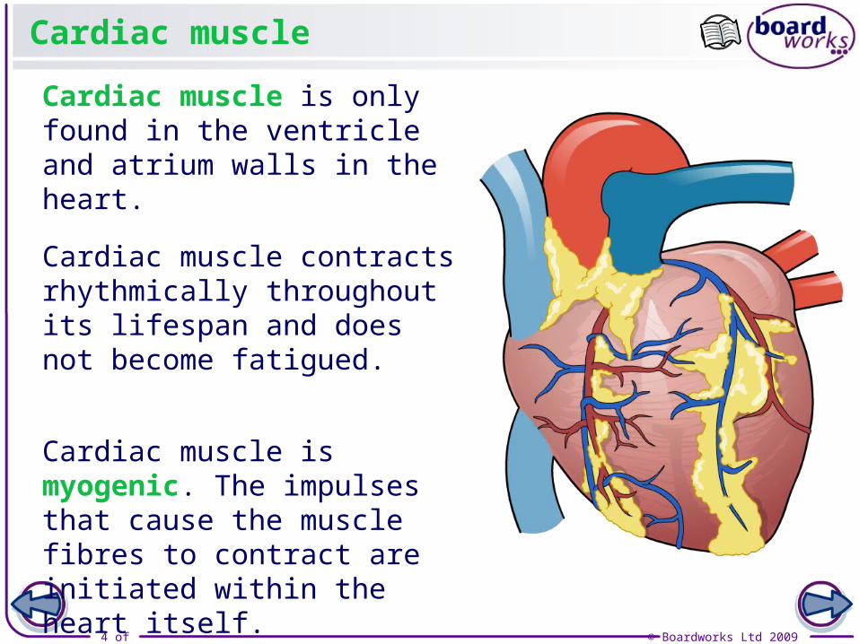
Cardiac muscle is only found in the ventricle and atrium walls in the heart.
Cardiac muscle contracts rhythmically throughout its lifespan and does not become fatigued.
Cardiac muscle is myogenic. The impulses that cause the muscle fibres to contract are initiated within the heart itself.
5 of 36 © Boardworks Ltd 2009
Smooth muscle
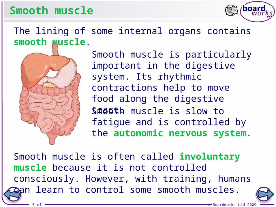
The lining of some internal organs contains smooth muscle.
Smooth muscle is often called involuntary muscle because it is not controlled consciously. However, with training, humans can learn to control some smooth muscles.
Smooth muscle is particularly important in the digestive system. Its rhythmic contractions help to move food along the digestive tract.
Smooth muscle is slow to fatigue and is controlled by the autonomic nervous system.

6 of 36 © Boardworks Ltd 2009
Question 2
• How many of the major skeletal muscles can you label?
7 of 36 © Boardworks Ltd 2009
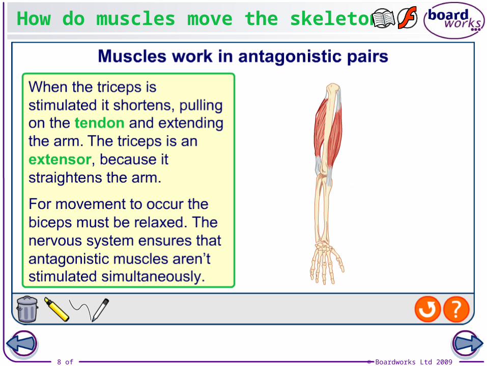
Producing movement
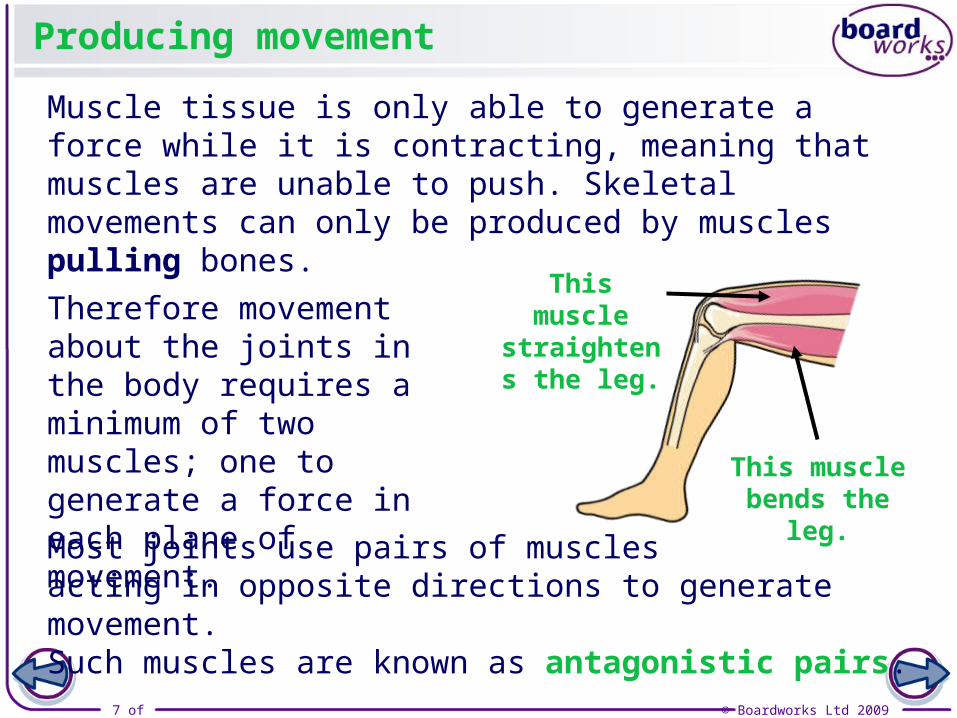
Muscle tissue is only able to generate a force while it is contracting, meaning that muscles are unable to push. Skeletal movements can only be produced by muscles pulling bones.
Therefore movement about the joints in the body requires a minimum of two muscles; one to generate a force in each plane of movement.
Most joints use pairs of musclesacting in opposite directions to generate movement. Such muscles are known as antagonistic pairs.
This muscle straightens
the leg.
This muscle bends the leg.
8 of 36 © Boardworks Ltd 2009
How do muscles move the skeleton?
9 of 36 © Boardworks Ltd 2009
Antagonistic pairs
10 of 36 © Boardworks Ltd 2009
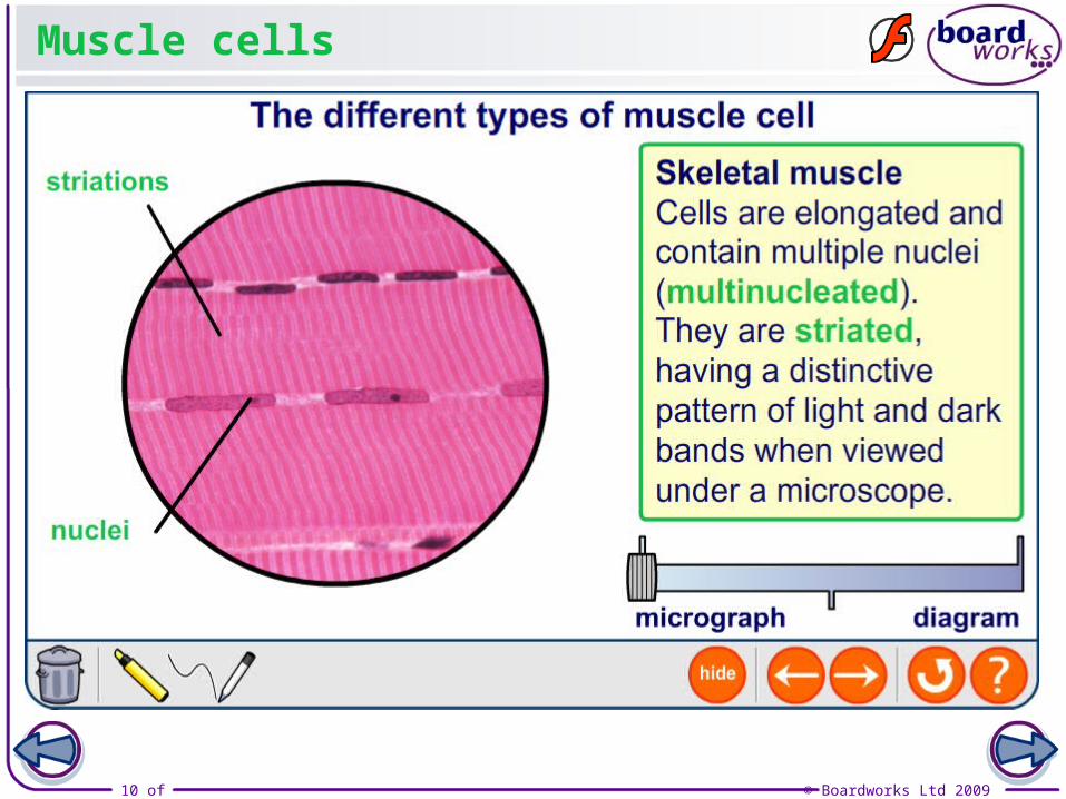
Muscle cells
11 of 36 © Boardworks Ltd 2009
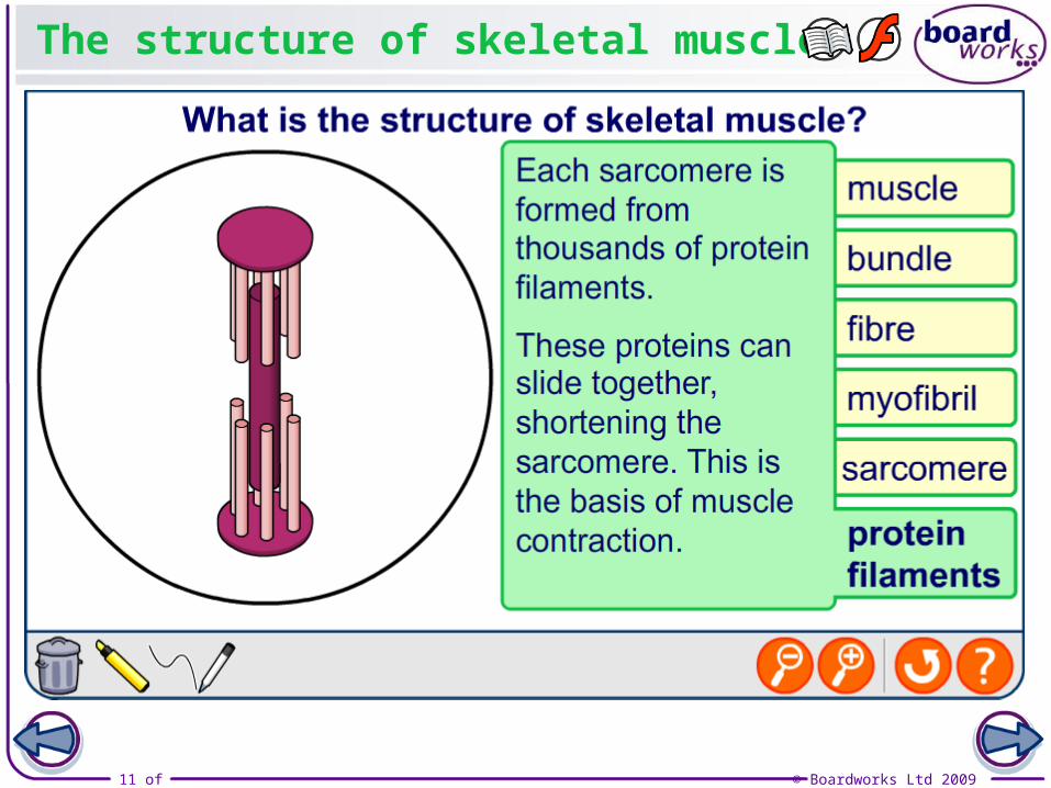
The structure of skeletal muscle

12 of 36 © Boardworks Ltd 2009
Questions 6, 7, 8
• Use the textbook to help you answer these questions.
13 of 36 © Boardworks Ltd 2009
Observing myofibrils
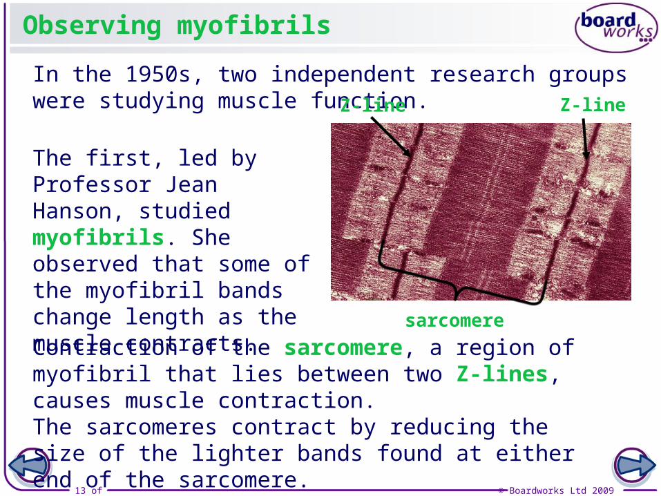
In the 1950s, two independent research groups were studying muscle function.
The first, led by Professor Jean Hanson, studied myofibrils. She observed that some of the myofibril bands change length as the muscle contracts.
Contraction of the sarcomere, a region of myofibril that lies between two Z-lines, causes muscle contraction. The sarcomeres contract by reducing the size of the lighter bands found at either end of the sarcomere.
Z-lineZ-line
sarcomere
14 of 36 © Boardworks Ltd 2009
The sarcomere
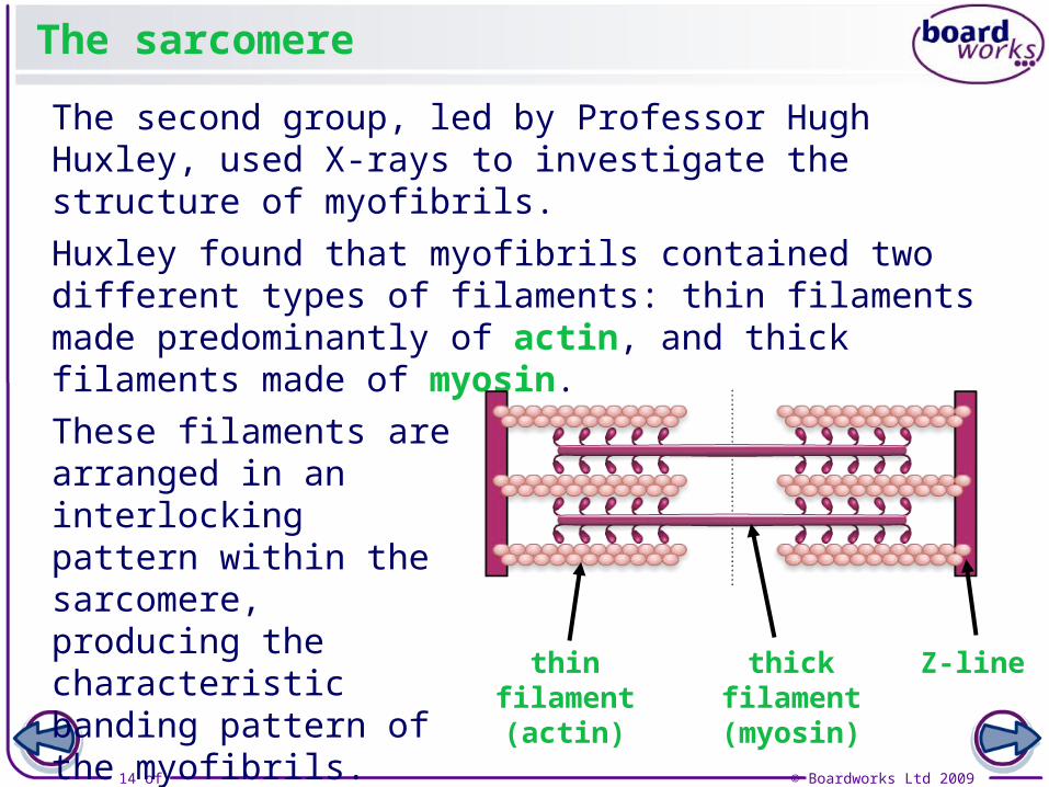
The second group, led by Professor Hugh Huxley, used X-rays to investigate the structure of myofibrils.
Huxley found that myofibrils contained two different types of filaments: thin filaments made predominantly of actin, and thick filaments made of myosin.
These filaments are arranged in an interlocking pattern within the sarcomere, producing the characteristic banding pattern of the myofibrils.
thin filament (actin)
thick filament (myosin)
Z-line

15 of 36 © Boardworks Ltd 2009
Questions 9 and 10
• Use the next slide to help you complete these questions.

16 of 36 © Boardworks Ltd 2009
The structure of the sarcomere
17 of 36 © Boardworks Ltd 2009
The structure of myosin
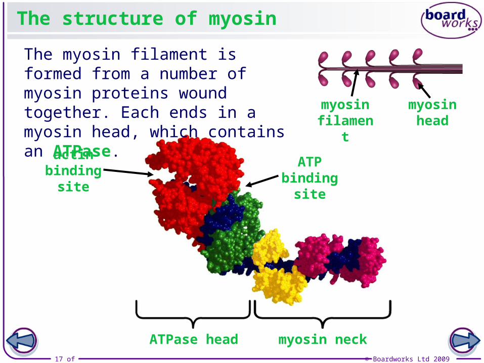
The myosin filament is formed from a number of myosin proteins wound together. Each ends in a myosin head, which contains an ATPase. myosin
filamentmyosin
head
actin binding
site
ATP binding
site
ATPase head myosin neck
18 of 36 © Boardworks Ltd 2009
The structure of actin
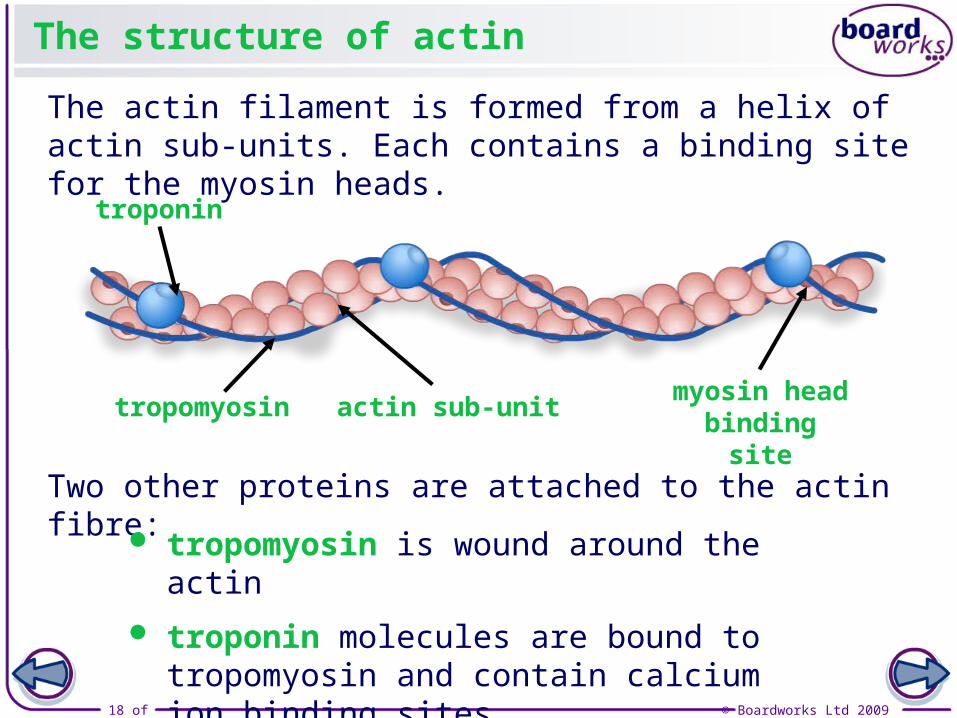
The actin filament is formed from a helix of actin sub-units. Each contains a binding site for the myosin heads.
Two other proteins are attached to the actin fibre:
troponin
tropomyosinmyosin head binding site
actin sub-unit
tropomyosin is wound around the actin
troponin molecules are bound to tropomyosin and contain calcium ion binding sites.
19 of 36 © Boardworks Ltd 2009
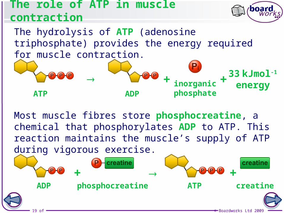
The role of ATP in muscle contraction
The hydrolysis of ATP (adenosine triphosphate) provides the energy required for muscle contraction.
Most muscle fibres store phosphocreatine, a chemical that phosphorylates ADP to ATP. This reaction maintains the muscle’s supply of ATP during vigorous exercise.
+ + 33 kJmol-1
energyATP ADP
inorganicphosphate
ADP
+ +phosphocreatine ATP creatine
20 of 36 © Boardworks Ltd 2009
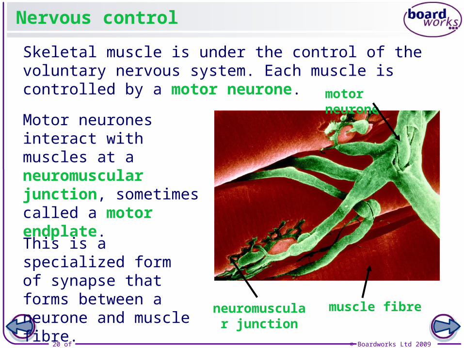
Nervous control
Skeletal muscle is under the control of the voluntary nervous system. Each muscle is controlled by a motor neurone.
Motor neurones interact with muscles at a neuromuscular junction, sometimes called a motor endplate.
This is a specialized form of synapse that forms between a neurone and muscle fibre. muscle fibre
motor neurone
neuromuscular junction

21 of 36 © Boardworks Ltd 2009
Muscle Contraction – How does it work?
Describe the differences between the two sarcomeres on your sheet.
22 of 36 © Boardworks Ltd 2009
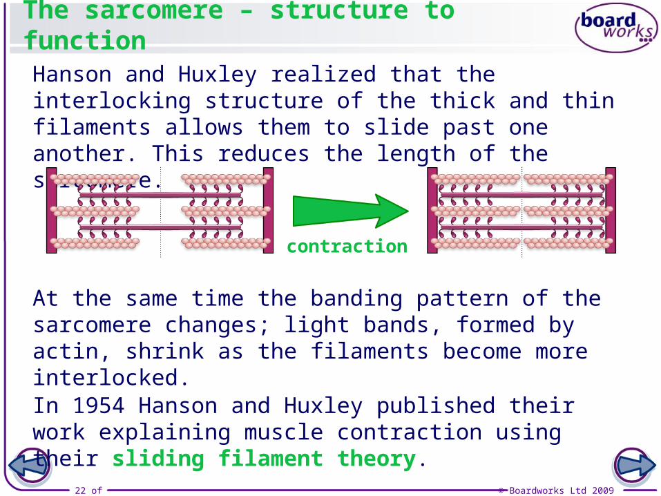
The sarcomere – structure to function
Hanson and Huxley realized that the interlocking structure of the thick and thin filaments allows them to slide past one another. This reduces the length of the sarcomere.
In 1954 Hanson and Huxley published their work explaining muscle contraction using their sliding filament theory.
At the same time the banding pattern of the sarcomere changes; light bands, formed by actin, shrink as the filaments become more interlocked.
contraction

23 of 36 © Boardworks Ltd 2009
The sliding filament theory

24 of 36 © Boardworks Ltd 2009
What controls the sliding filaments?

25 of 36 © Boardworks Ltd 2009
Now try the cut-and-stick activity.

26 of 36 © Boardworks Ltd 2009
The neuromuscular junction

27 of 36 © Boardworks Ltd 2009
Fast-twitch and slow-twitch muscle fibres
Use your textbook to research the answers to the questions on the sheet.
28 of 36 © Boardworks Ltd 2009
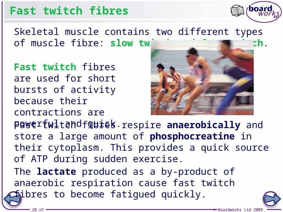
Skeletal muscle contains two different types of muscle fibre: slow twitch and fast twitch.
Fast twitch fibres
Fast twitch fibres are used for short bursts of activity because their contractions are powerful and quick.
Fast twitch fibres respire anaerobically and store a large amount of phosphocreatine in their cytoplasm. This provides a quick source of ATP during sudden exercise.
The lactate produced as a by-product of anaerobic respiration cause fast twitch fibres to become fatigued quickly.
29 of 36 © Boardworks Ltd 2009
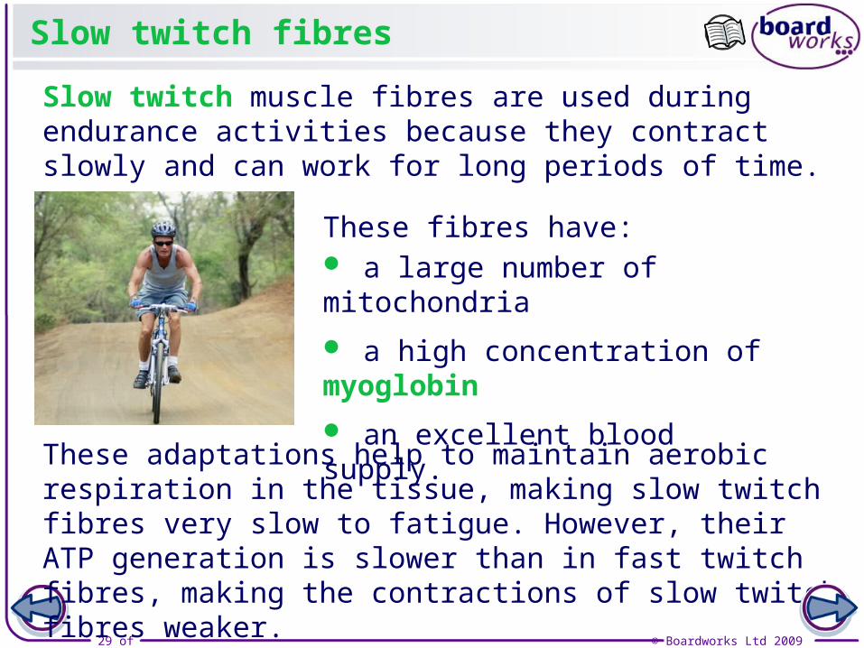
Slow twitch fibres
Slow twitch muscle fibres are used during endurance activities because they contract slowly and can work for long periods of time.
These fibres have: a large number of mitochondria
a high concentration of myoglobin
an excellent blood supply.
These adaptations help to maintain aerobic respiration in the tissue, making slow twitch fibres very slow to fatigue. However, their ATP generation is slower than in fast twitch fibres, making the contractions of slow twitch fibres weaker.