Embed Size (px)
Citation preview
Muscles and Muscle Tissue
LAB 6
Muscle Overview• Muscle tissue makes up nearly half the body
mass.
• The most distinguishing functional characteristic of muscles is their ability to transform chemical energy ATP into directed mechanical energy
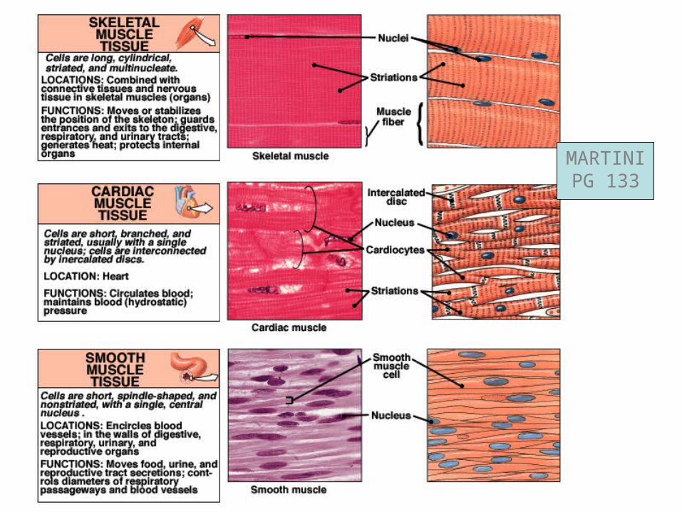
• The three types of muscle tissue are: skeletal, cardiac, and smooth
• These types differ in structure, location, function, and means of activation
MARTINIPG 133
Muscle Similarities
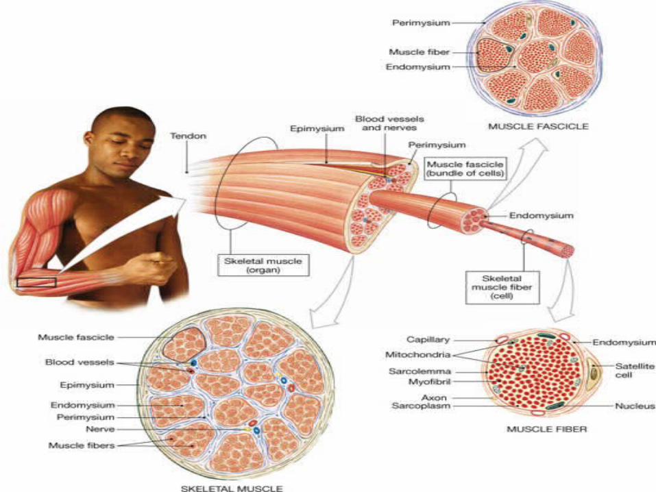
• Skeletal and smooth muscle cells are elongated and are called muscle fibers
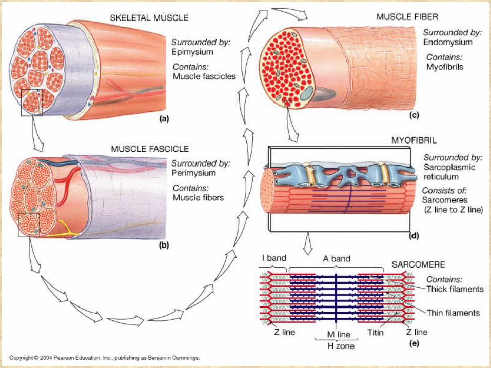
• Muscle contraction depends on two kinds of myofilaments – actin and myosin
• Muscle terminology is similar– Sarcolemma – muscle plasma membrane– Sarcoplasm – cytoplasm of a muscle cell– Prefixes – myo, mys, and sarco all refer to
muscle
Functional Characteristics of Muscle Tissue
• Excitability, or irritability – the ability to receive and respond to stimuli
• Contractility – the ability to shorten forcibly
• Extensibility – the ability to be stretched or extended
• Elasticity – the ability to recoil and resume the original resting length

Muscle Function
• Skeletal muscles are responsible for all locomotion
• Cardiac muscle is responsible for coursing the blood through the body
• Smooth muscle helps maintain blood pressure, and squeezes or propels substances (i.e., food, feces) through organs
• Muscles also maintain posture, stabilize joints, and generate heat
Muscle Classification: Functional Groups
• Prime movers – provide the major force for producing a specific movement
• Antagonists – oppose or reverse a particular movement
• Synergists– Add force to a movement– Reduce undesirable or unnecessary movement
• Fixators – synergists that immobilize a bone or muscle’s origin
Naming Skeletal Muscles
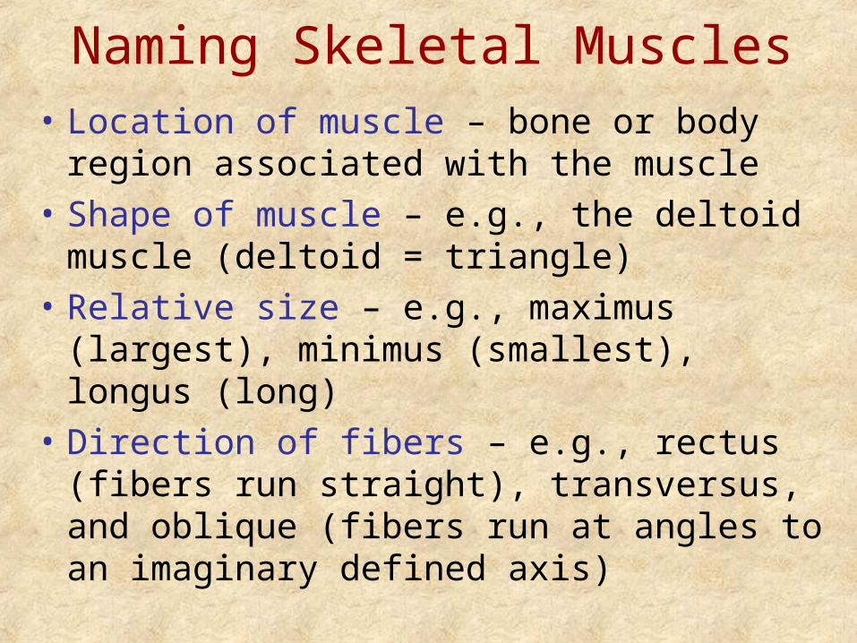
• Location of muscle – bone or body region associated with the muscle
• Shape of muscle – e.g., the deltoid muscle (deltoid = triangle)
• Relative size – e.g., maximus (largest), minimus (smallest), longus (long)
• Direction of fibers – e.g., rectus (fibers run straight), transversus, and oblique (fibers run at angles to an imaginary defined axis)
Naming Skeletal Muscles
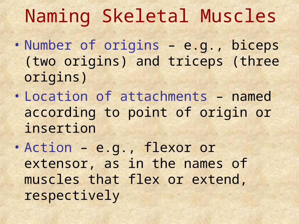
• Number of origins – e.g., biceps (two origins) and triceps (three origins)
• Location of attachments – named according to point of origin or insertion
• Action – e.g., flexor or extensor, as in the names of muscles that flex or extend, respectively
Bone-Muscle Relationships: Lever Systems
• Lever – a rigid bar that moves on a fulcrum, or fixed point
• Effort – force applied to a lever
• Load – resistance moved by the effort
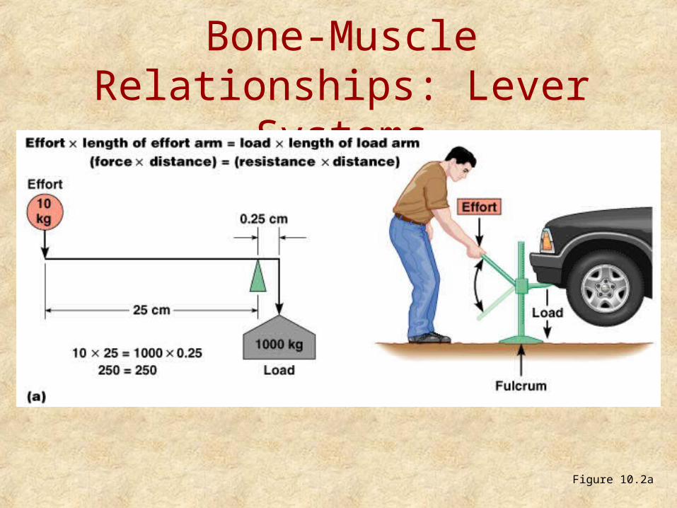
Bone-Muscle Relationships: Lever Systems
Figure 10.2a
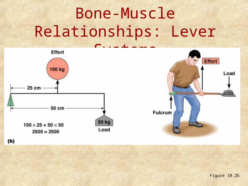
Bone-Muscle Relationships: Lever Systems
Figure 10.2b
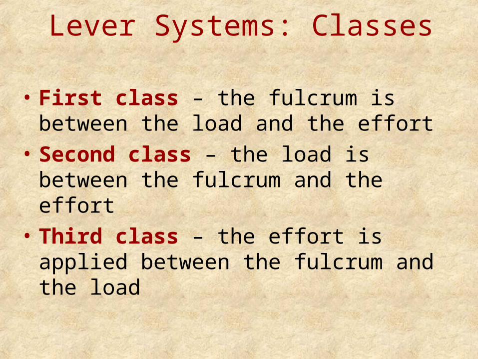
Lever Systems: Classes
• First class – the fulcrum is between the load and the effort
• Second class – the load is between the fulcrum and the effort
• Third class – the effort is applied between the fulcrum and the load
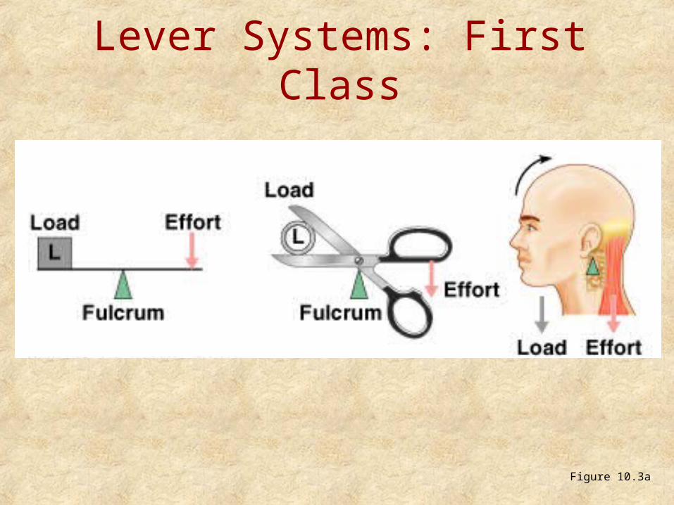
Lever Systems: First Class
Figure 10.3a
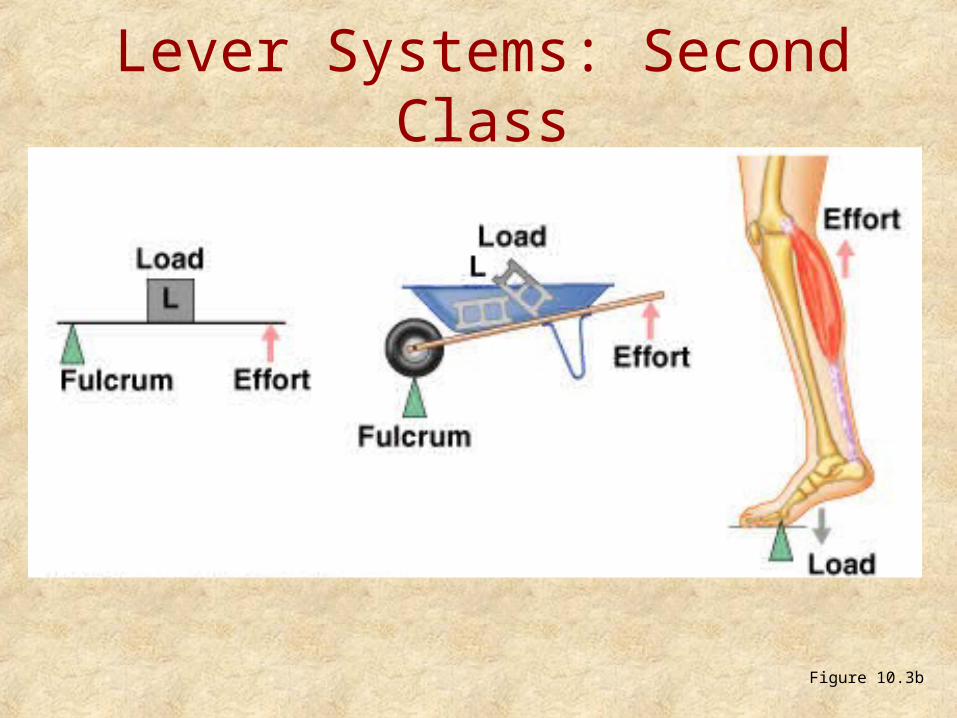
Lever Systems: Second Class
Figure 10.3b
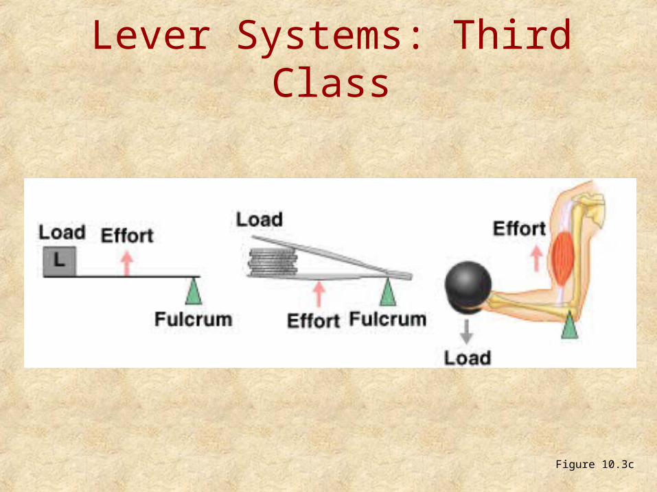
Lever Systems: Third Class
Figure 10.3c
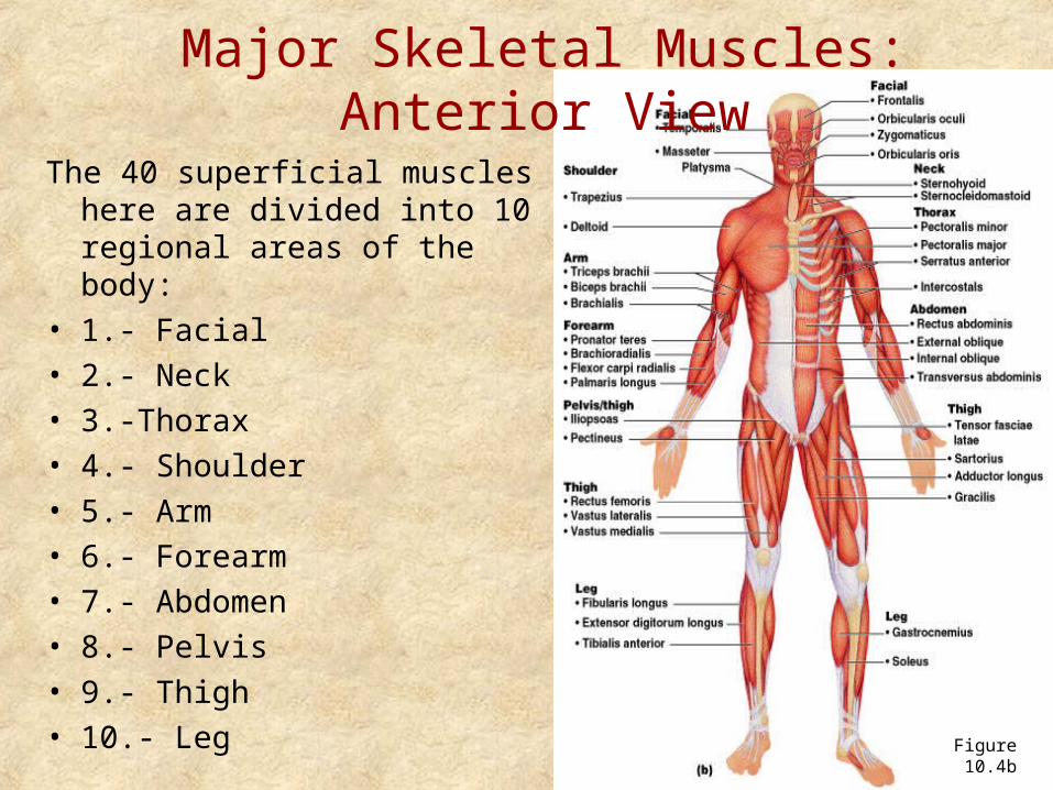
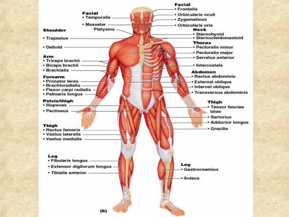
Major Skeletal Muscles: Anterior View
The 40 superficial muscles here are divided into 10 regional areas of the body:
• 1.- Facial• 2.- Neck• 3.-Thorax• 4.- Shoulder• 5.- Arm• 6.- Forearm• 7.- Abdomen• 8.- Pelvis• 9.- Thigh• 10.- Leg Figure 10.4b
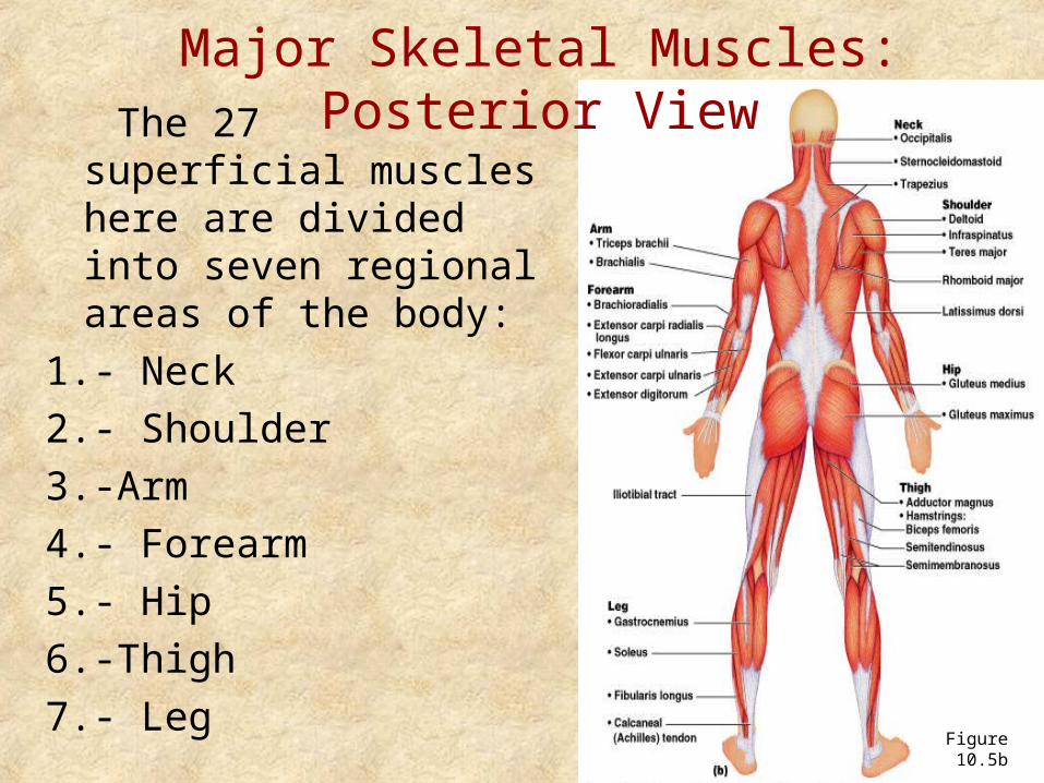
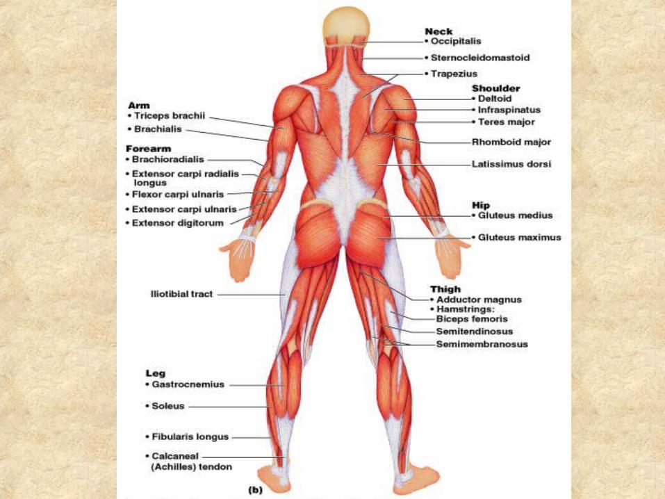
Major Skeletal Muscles: Posterior View
The 27 superficial muscles here are divided into seven regional areas of the body:
1.- Neck
2.- Shoulder
3.-Arm
4.- Forearm
5.- Hip
6.-Thigh
7.- LegFigure 10.5b
Muscles of the Face
• 11 muscles are involved in lifting the eyebrows, flaring the nostrils, opening and closing the eyes and mouth, and smiling
• All are innervated by cranial nerve VII (facial nerve)
• Usually insert in skin (rather than bone), and adjacent muscles often fuse
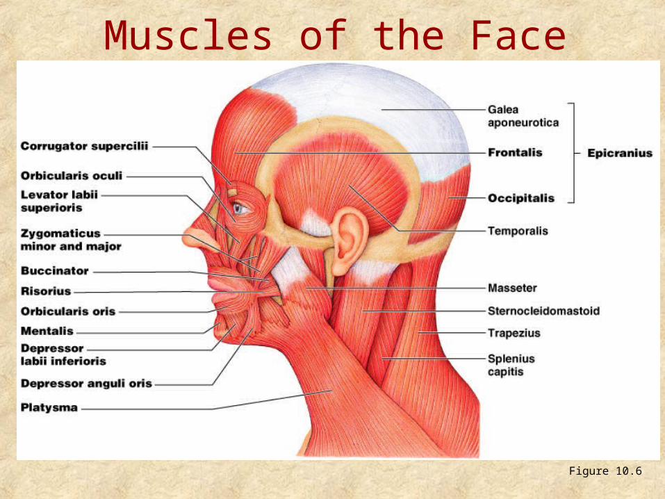
Muscles of the Face
Figure 10.6
Muscles of Mastication
• There are four pairs of muscles involved in mastication– Prime movers – temporalis and masseter– Grinding movements – pterygoids and
buccinators
• All are innervated by cranial nerve V (trigeminal nerve)
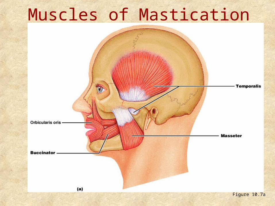
Muscles of Mastication
Figure 10.7a
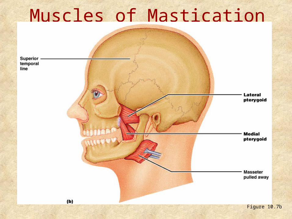
Muscles of Mastication
Figure 10.7b
Extrinsic Tongue Muscles
• Three major muscles that anchor and move the tongue
• All are innervated by cranial nerve XII (hypoglossal nerve)
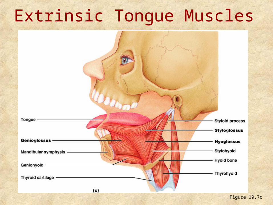
Extrinsic Tongue Muscles
Figure 10.7c
Homeostatic Imbalance• Many toxins, drugs and diseases interfere
with events at the neuromuscular junction
Ex: Myastenia gravis: Characterize by:
1.- Drooping of the upper eyelids
2.- Difficulty of swallowing and talking
3.- Muscle weakness
4.- Serum antibodies against acetilcholine (Ach)
receptor
Developmental Aspects: Male and Female
• There is a biological basis for greater strength in men than in women
• Women’s skeletal muscle makes up 36% of their body mass
• Men’s skeletal muscle makes up 42% of their body mass
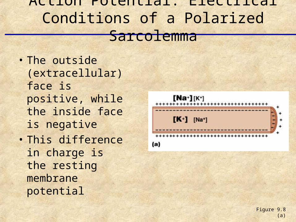
• The outside (extracellular) face is positive, while the inside face is negative
• This difference in charge is the resting membrane potential
Figure 9.8 (a)
Action Potential: Electrical Conditions of a Polarized Sarcolemma
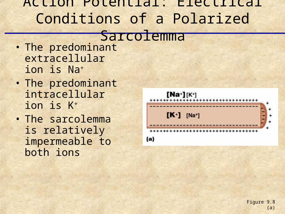
• The predominant extracellular ion is Na+
• The predominant intracellular ion is K+
• The sarcolemma is relatively impermeable to both ions
Figure 9.8 (a)
Action Potential: Electrical Conditions of a Polarized Sarcolemma
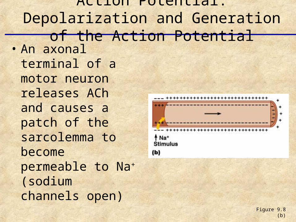
• An axonal terminal of a motor neuron releases ACh and causes a patch of the sarcolemma to become permeable to Na+ (sodium channels open)
Figure 9.8 (b)
Action Potential: Depolarization and Generation of the Action Potential
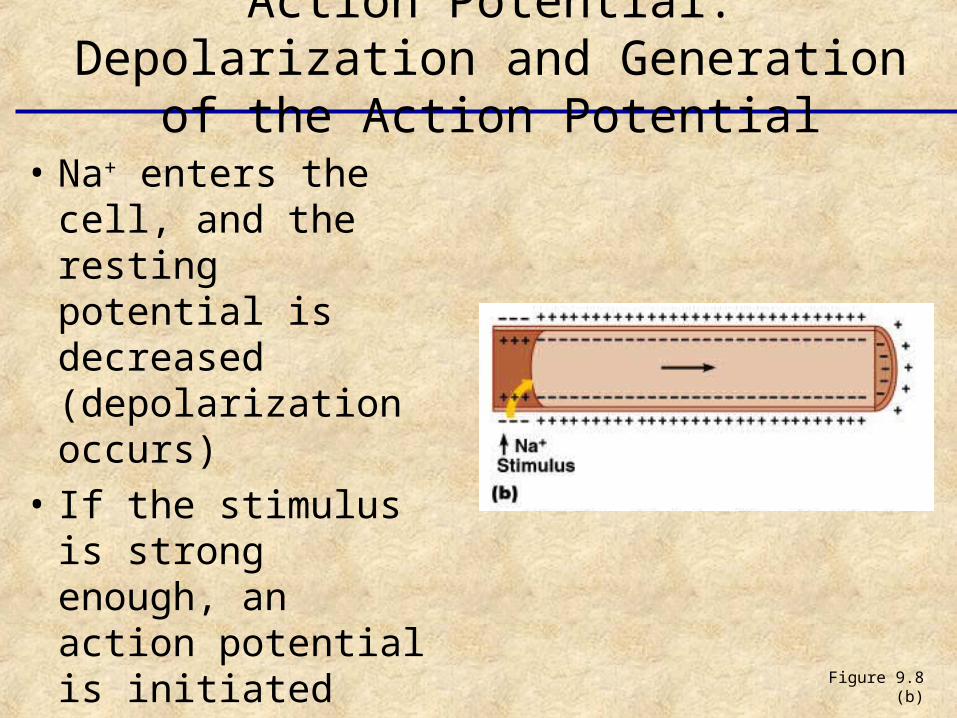
• Na+ enters the cell, and the resting potential is decreased (depolarization occurs)
• If the stimulus is strong enough, an action potential is initiated
Figure 9.8 (b)
Action Potential: Depolarization and Generation of the Action Potential
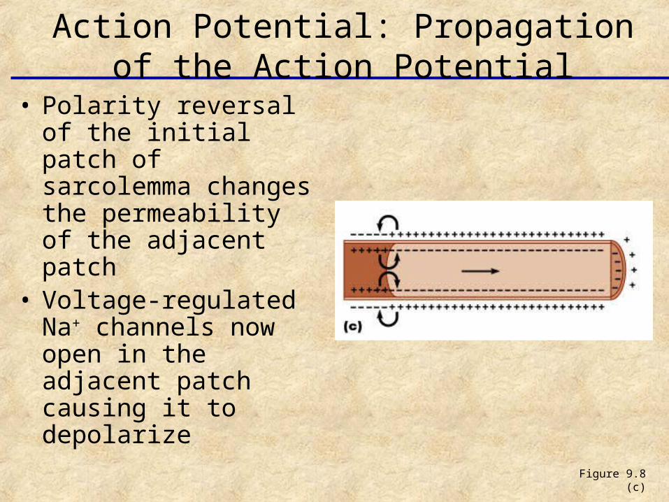
• Polarity reversal of the initial patch of sarcolemma changes the permeability of the adjacent patch
• Voltage-regulated Na+ channels now open in the adjacent patch causing it to depolarize
Figure 9.8 (c)
Action Potential: Propagation of the Action Potential
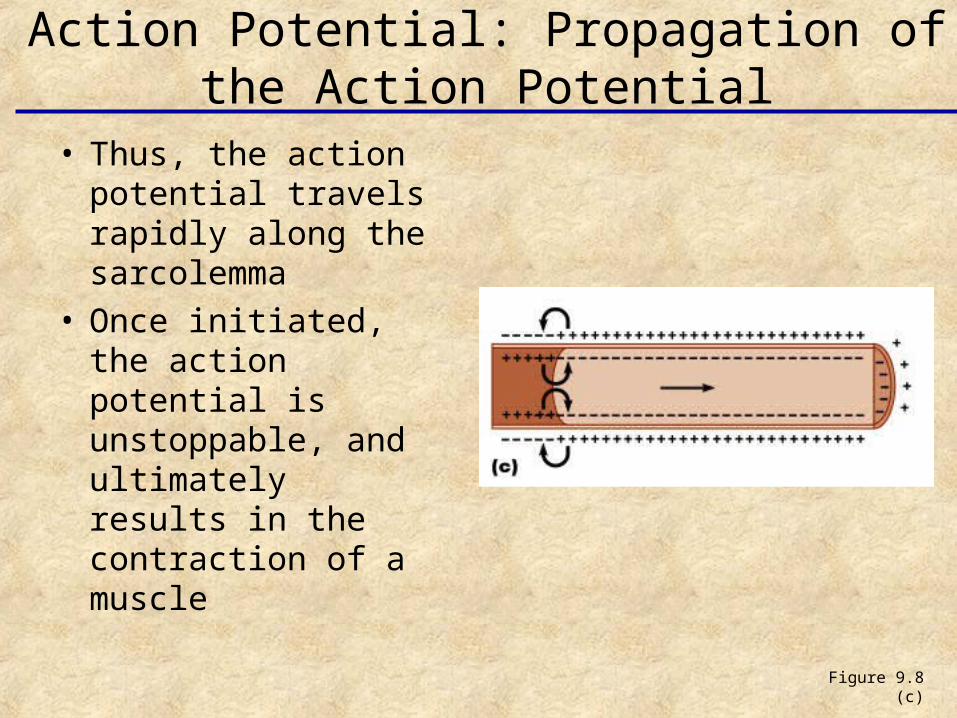
• Thus, the action potential travels rapidly along the sarcolemma
• Once initiated, the action potential is unstoppable, and ultimately results in the contraction of a muscle
Figure 9.8 (c)
Action Potential: Propagation of the Action Potential
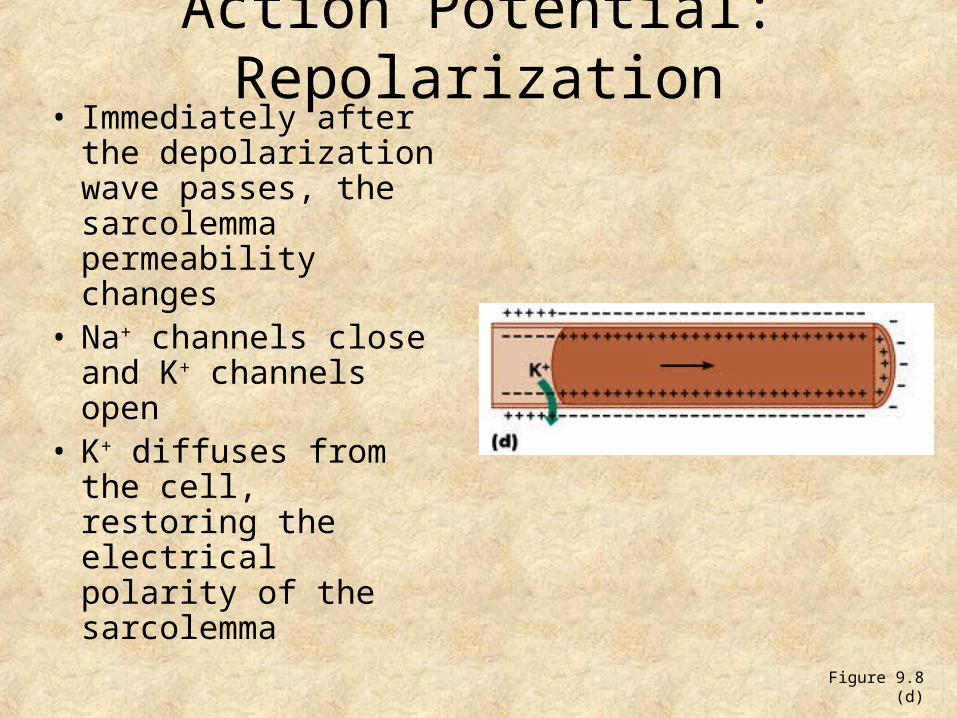
Action Potential: Repolarization• Immediately after the
depolarization wave passes, the sarcolemma permeability changes
• Na+ channels close and K+ channels open
• K+ diffuses from the cell, restoring the electrical polarity of the sarcolemma
Figure 9.8 (d)
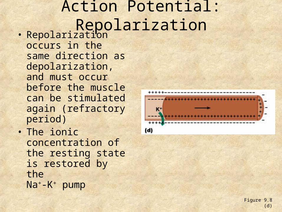
Action Potential: Repolarization• Repolarization
occurs in the same direction as depolarization, and must occur before the muscle can be stimulated again (refractory period)
• The ionic concentration of the resting state is restored by the Na+-K+ pump
Figure 9.8 (d)
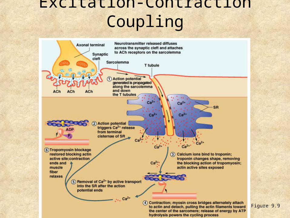
Excitation-Contraction Coupling
• Once generated, the action potential:– Is propagated along the sarcolemma– Travels down the T tubules– Triggers Ca2+ release from terminal cisternae
• Ca2+ binds to troponin and causes: – The blocking action of tropomyosin to cease– Actin active binding sites to be exposed
Excitation-Contraction Coupling
• Myosin cross bridges alternately attach and detach
• Thin filaments move toward the center of the sarcomere
• Hydrolysis of ATP powers this cycling process
• Ca2+ is removed into the SR, tropomyosin blockage is restored, and the muscle fiber relaxes
Excitation-Contraction Coupling
Figure 9.9
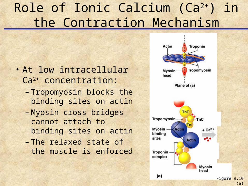
• At low intracellular Ca2+ concentration:– Tropomyosin blocks the
binding sites on actin– Myosin cross bridges
cannot attach to binding sites on actin
– The relaxed state of the muscle is enforced
Role of Ionic Calcium (Ca2+) in the Contraction Mechanism
Figure 9.10 (a)
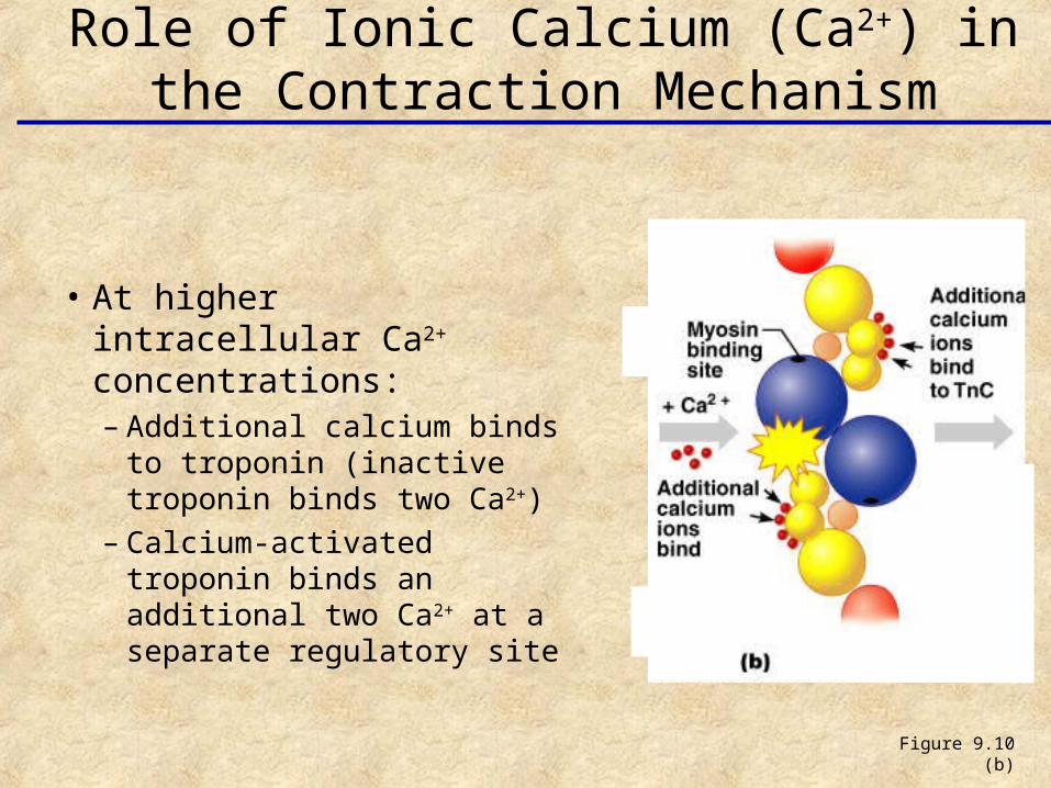
Figure 9.10 (b)
• At higher intracellular Ca2+ concentrations:– Additional calcium binds
to troponin (inactive troponin binds two Ca2+)
– Calcium-activated troponin binds an additional two Ca2+ at a separate regulatory site
Role of Ionic Calcium (Ca2+) in the Contraction Mechanism
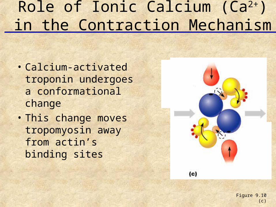
• Calcium-activated troponin undergoes a conformational change
• This change moves tropomyosin away from actin’s binding sites
Figure 9.10 (c)
Role of Ionic Calcium (Ca2+) in the Contraction Mechanism
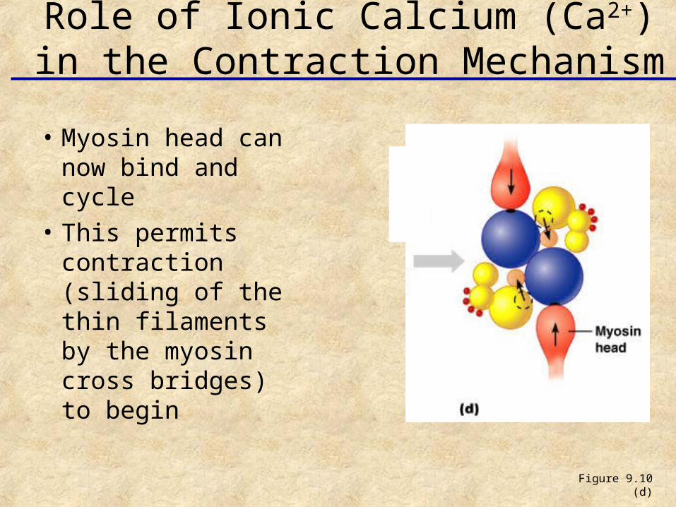
• Myosin head can now bind and cycle
• This permits contraction (sliding of the thin filaments by the myosin cross bridges) to begin
Figure 9.10 (d)
Role of Ionic Calcium (Ca2+) in the Contraction Mechanism
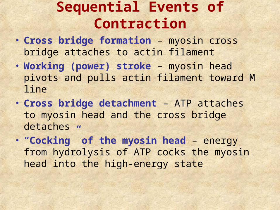
Sequential Events of Contraction
• Cross bridge formation – myosin cross bridge attaches to actin filament
• Working (power) stroke – myosin head pivots and pulls actin filament toward M line
• Cross bridge detachment – ATP attaches to myosin head and the cross bridge detaches
• “Cocking” of the myosin head – energy from hydrolysis of ATP cocks the myosin head into the high-energy state
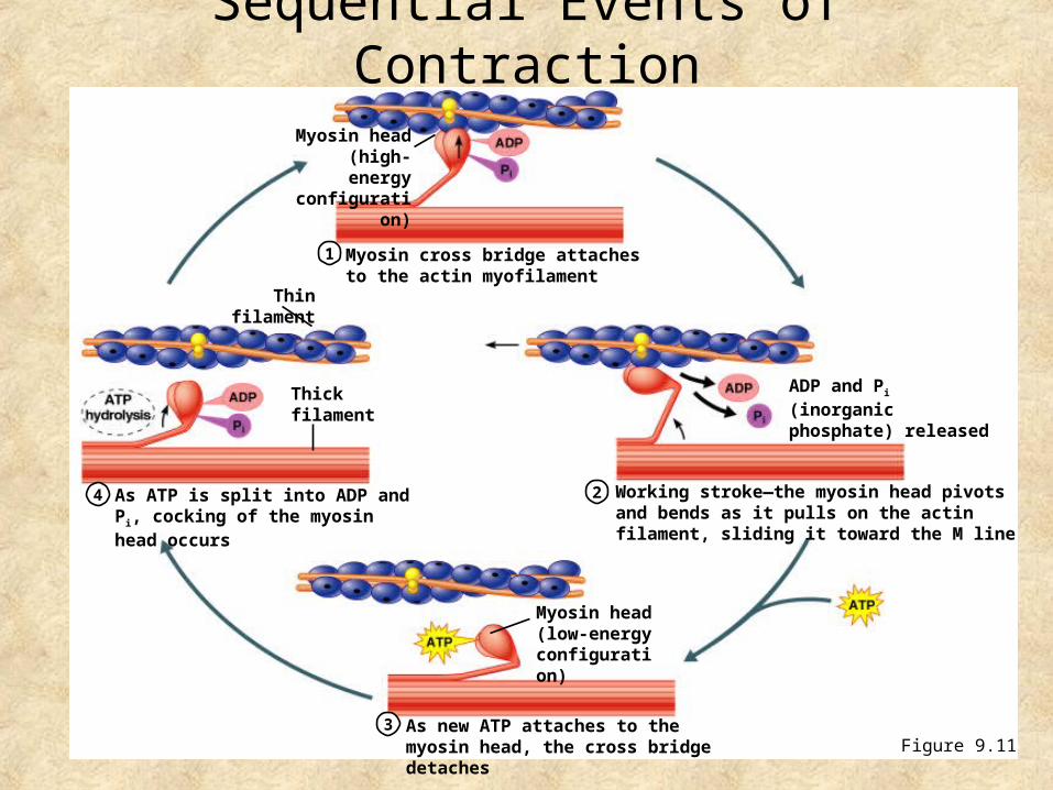
Myosin cross bridge attaches to the actin myofilament
1
2
3
4 Working stroke—the myosin head pivots and bends as it pulls on the actin filament, sliding it toward the M line
As new ATP attaches to the myosin head, the cross bridge detaches
As ATP is split into ADP and Pi, cocking of the myosin head occurs
Myosin head (high-energy
configuration)
Thick filament
Myosin head (low-energy configuration)
ADP and Pi (inorganic phosphate) released
Sequential Events of Contraction
Figure 9.11
Thin filament
Motor Unit: The Nerve-Muscle Functional Unit
• Large weight-bearing muscles (thighs, hips) have large motor units
• Muscle fibers from a motor unit are spread throughout the muscle; therefore, contraction of a single motor unit causes weak contraction of the entire muscle
Motor Unit: The Nerve-Muscle Functional Unit
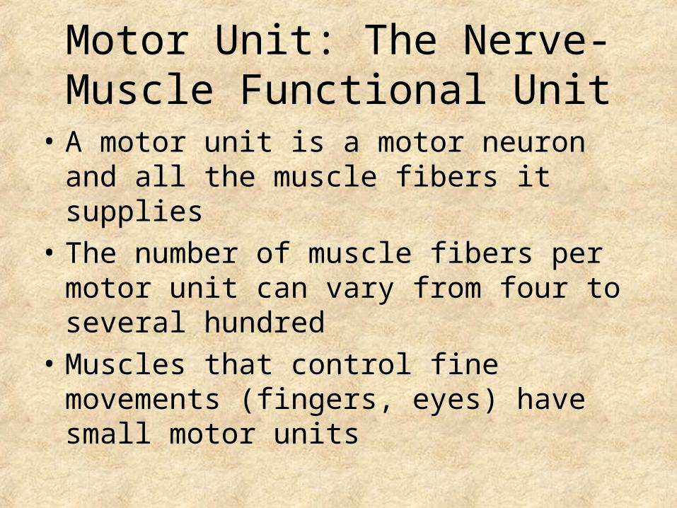
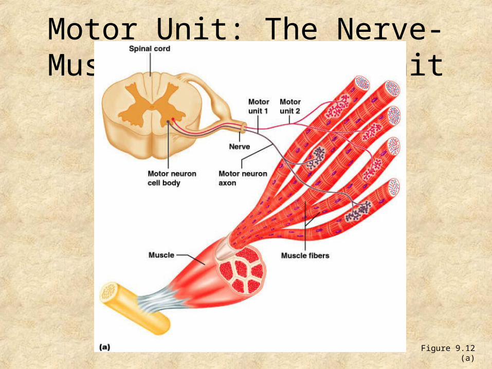
• A motor unit is a motor neuron and all the muscle fibers it supplies
• The number of muscle fibers per motor unit can vary from four to several hundred
• Muscles that control fine movements (fingers, eyes) have small motor units
Motor Unit: The Nerve-Muscle Functional Unit
Figure 9.12 (a)