Embed Size (px)
Citation preview
Ch. 9 – Muscles and Muscle Tissue
1
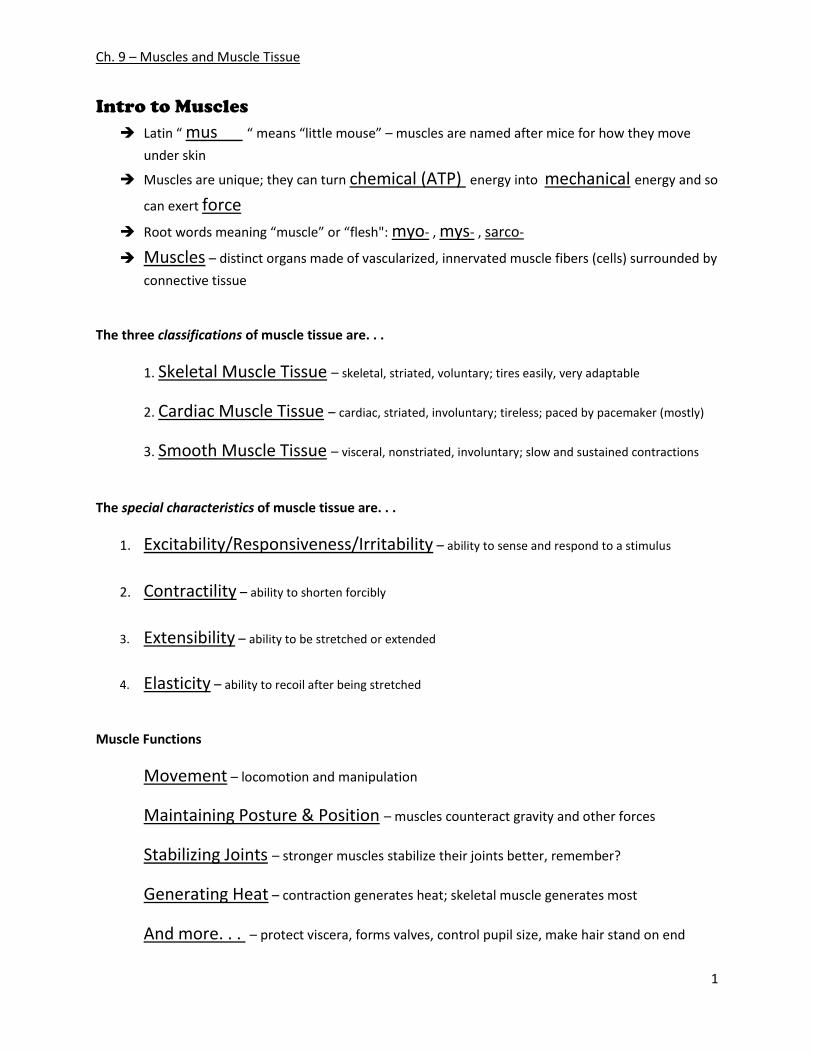
Intro to Muscles
Latin “ mus “ means “little mouse” – muscles are named after mice for how they move
under skin
Muscles are unique; they can turn chemical (ATP) energy into mechanical energy and so
can exert force
Root words meaning “muscle” or “flesh": myo- , mys- , sarco-
Muscles – distinct organs made of vascularized, innervated muscle fibers (cells) surrounded by
connective tissue
The three classifications of muscle tissue are. . .
1. Skeletal Muscle Tissue – skeletal, striated, voluntary; tires easily, very adaptable
2. Cardiac Muscle Tissue – cardiac, striated, involuntary; tireless; paced by pacemaker (mostly)
3. Smooth Muscle Tissue – visceral, nonstriated, involuntary; slow and sustained contractions
The special characteristics of muscle tissue are. . .
1. Excitability/Responsiveness/Irritability – ability to sense and respond to a stimulus
2. Contractility – ability to shorten forcibly
3. Extensibility – ability to be stretched or extended
4. Elasticity – ability to recoil after being stretched
Muscle Functions
Movement – locomotion and manipulation
Maintaining Posture & Position – muscles counteract gravity and other forces
Stabilizing Joints – stronger muscles stabilize their joints better, remember?
Generating Heat – contraction generates heat; skeletal muscle generates most
And more. . . – protect viscera, forms valves, control pupil size, make hair stand on end
Ch. 9 – Muscles and Muscle Tissue
2
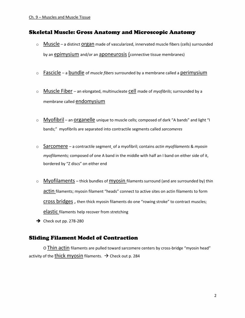
Skeletal Muscle: Gross Anatomy and Microscopic Anatomy
o Muscle – a distinct organ made of vascularized, innervated muscle fibers (cells) surrounded
by an epimysium and/or an aponeurosis (connective tissue membranes)
o Fascicle – a bundle of muscle fibers surrounded by a membrane called a perimysium
o Muscle Fiber – an elongated, multinucleate cell made of myofibrils; surrounded by a
membrane called endomysium
o Myofibril – an organelle unique to muscle cells; composed of dark “A bands” and light “I
bands;” myofibrils are separated into contractile segments called sarcomeres
o Sarcomere – a contractile segment of a myofibril; contains actin myofilaments & myosin
myofilaments; composed of one A band in the middle with half an I band on either side of it,
bordered by “Z discs” on either end
o Myofilaments – thick bundles of myosin filaments surround (and are surrounded by) thin
actin filaments; myosin filament “heads” connect to active sites on actin filaments to form
cross bridges , then thick myosin filaments do one “rowing stroke” to contract muscles;
elastic filaments help recover from stretching
Check out pp. 278-280
Sliding Filament Model of Contraction
O Thin actin filaments are pulled toward sarcomere centers by cross-bridge “myosin head”
activity of the thick myosin filaments. Check out p. 284
Ch. 9 – Muscles and Muscle Tissue
3
Muscle Types (Review)
-3 types of muscle tissue: skeletal, cardiac, and smooth
-types differ in structure, location, function, and means of activation
Muscle Similarities
-Skeletal and smooth muscle cells are elongated muscle fibers
-Muscle contraction uses two kinds of myofilaments – actin and myosin
-Muscle terminology is similar
-Sarcolemma – muscle plasma membrane
-Sarcoplasm – cytoplasm of a muscle cell
-Prefixes – myo, mys, and sarco all refer to muscle (or flesh)
Skeletal Muscle: Nervous and Circulatory Connections
-Each muscle is served by one nerve, an artery, and one or more veins
-Each skeletal muscle fiber has a nerve ending which controls contraction
-Contracting fibers obtain oxygen and nutrients via arteries
-Wastes are removed via veins
Skeletal Muscle: Attachments
-Most skeletal muscles span joints and are attached to bone in 2 or more places
-When muscles contract, the muscle’s insertion (the more movable bone) moves
toward the muscle’s origin (the less movable bone)
-Muscles attach:
Directly – epimysium of the muscle is fused to the periosteum of a bone
Indirectly – connective tissue wrappings extend beyond the muscle as a
tendon or aponeurosis
Microscopic Anatomy of a Skeletal Muscle Fiber (Review/Overview)
fibers: long, cylindrical cells with multiple nuclei; surrounded by a plasma
membrane called a sarcolemma and filled with cytoplasm called sarcoplasm;
fibers contain the usual organelles + sarcoplasmic reticula, T tubules, and
myofibrils (myofibrils contain microfilaments actin and myosin bundled into
segments called sarcomeres) check out p. 279-280
Ch. 9 – Muscles and Muscle Tissue
4
Sarcoplasmic Reticulum (SR)
-an elaborate, smooth endoplasmic reticulum that surrounds each myofibril
-regulates intracellular levels of Ca2+
-controlled by signals from T tubules check out p.283
T Tubules
-continuous with the sarcolemma
-penetrate into the cell’s interior at each A band–I band junction
-function to conduct impulses to the deepest regions of the muscle by forming
“triads” with paired terminal cisternae of SR
-impulses from T tubules signal for the release of Ca2+
from SR’s terminal
cisternae
check out p.283
Triad Relationships
-T tubules and SR provide tightly linked signals for muscle contraction
-A double zipper of integral membrane proteins protrudes into the intermembrane
space
-T tubule proteins act to sense voltage from nerves
-SR “foot proteins” are receptors that regulate Ca2+ release from the SR cisternae
Skeletal Muscle Contraction
-In order to contract, a skeletal muscle must:
-Be stimulated by a nerve ending
-Propagate (broadcast) an electrical current, called an action potential, along its sarcolemma
-Have a rise in intracellular Ca2+ levels (the final trigger for contraction)
-Linking the electrical signal to the contraction is called excitation-
contraction coupling
Ch. 9 – Muscles and Muscle Tissue
5
Nerve Stimulus of Skeletal Muscle
-stimulation is enacted by motor neurons of the somatic nervous system
-Axons of these neurons travel to muscle cells in nerves
-Axons of motor neurons begin to branch profusely as they enter muscles
-Each axonal branch forms a neuromuscular junction with a single muscle
fiber (muscle cell), ~halfway along fiber’s length
Neuromuscular Junctions
-A neuromuscular junction is formed from:
-Axonal endings, which have small membranous sacs (synaptic
vesicles) that contain the neurotransmitter acetylcholine ( ACh )
-The motor end plate of a muscle, a specific part of the sarcolemma that
contains receptors sensitive to ACh
-Though exceedingly close, axonal ends and motor end plates of muscle fibers are
always separated by a space called the synaptic cleft
Phases leading to muscle fiber contraction (p.285)
Phase 1: When a nerve impulse reaches the end of an axon at the
neuromuscular junction. . .
-Voltage-regulated calcium channels open and allow Ca2+ to enter the axon
-Ca2+ inside the axon terminal causes axonal vesicles to fuse with the axonal
membrane, releasing ACh into the synaptic cleft via exocytosis
-ACh diffuses across the synaptic cleft to bind to ACh receptors on the
sarcolemma
- Binding of ACh on sarcolemma initiates an action potential in the muscle
Phase 2: Excitation-Contraction Coupling
-Once generated, the action potential:
-Is propagated (continued) along the sarcolemma
-Travels down the T tubules into cell/fiber
Ch. 9 – Muscles and Muscle Tissue
6
-Triggers release of Ca2+ from terminal cisternae
-Ca2+ binds to troponin and causes:
-The blocking action of tropomyosin to cease
-Actin active binding sites to be exposed
-Myosin cross bridges alternately attach and detach
-Thin filaments move toward the center of the sarcomere (hydrolysis of ATP
powers this cycling process)
-Ca2+ is removed into the SR, tropomyosin blockage is restored, and the muscle
fiber relaxes
check out p. 290-291
Destruction of Acetylcholine
-ACh bound to ACh receptors is quickly destroyed by the enzyme
acetylcholinesterase
-This destruction prevents / allows (circle one) continued muscle fiber contraction
in the absence of additional stimuli
check out homeostatic imbalance myasthenia gravis, p. 285
Action Potential -A transient depolarization event that includes polarity
reversal of a sarcolemma (or nerve cell membrane) and the propagation of an
action potential along the membrane
Role of Acetylcholine (Ach)
-ACh binds to its receptors at the motor end plate
-Binding opens chemically-gated (ligand-gated) channels
-Na+ and K+ diffuse out and the interior of the sarcolemma becomes less negative
(this event is called depolarization)
Depolarization
-Initially, this is a local electrical event called end plate potential -Later, it ignites an action potential that spreads in all directions across the
sarcolemma and deep into the cell/fiber through T tubules
Ch. 9 – Muscles and Muscle Tissue
7
Role of Ionic Calcium (Ca2+) in the Contraction Mechanism
-At low intracellular Ca2+ concentration:
-Tropomyosin blocks the binding sites on actin
-Myosin cross bridges cannot attach to binding sites on actin
Result: The relaxed state of the muscle is enforced
-At higher intracellular Ca2+ concentrations:
-hyper-calcium-activated troponin undergoes a conformational change
-This change moves tropomyosin away from actin’s binding sites
Result: Myosin head can now bind and cycle
Sequential Events of Contraction
- Cross bridge formation – myosin heads attach to actin filaments
- Working (power) stroke – myosin heads pivot and pull actin filaments
toward sarcomere center (M line)
- Cross bridge detachment – ATP attaches to myosin head and the cross
bridge detaches
- “Cocking” of the myosin head – energy from hydrolysis of ATP cocks
the myosin head into the high-energy state
Contraction of Skeletal Muscle Fibers
-Contraction – refers to the activation of myosin’s cross bridging activity
(force-generating sites)
-Shortening occurs when the tension generated by the cross bridge exceeds
forces opposing shortening
-Contraction ends when cross bridges become inactive, the tension generated
declines, and relaxation is induced
Contraction of Skeletal Muscle (Organ Level)
-Contraction of muscle fibers (cells) and muscles (organs) is similar
-The two types of muscle contractions are:
-Isometric contraction – increasing muscle tension (muscle does not
shorten during contraction)
-Isotonic contraction – decreasing muscle length (muscle shortens during
contraction)
Ch. 9 – Muscles and Muscle Tissue
8
Motor Unit: The Nerve-Muscle Functional Unit
- motor unit - a motor neuron and all the muscle fibers it supplies
- number of muscle fibers per motor unit: four to hundreds
- fine movement control (fingers, eyes) requires small motor units
- large, powerful movements (thighs, hips) require large motor units
-Muscle fibers from a motor unit are spread throughout the muscle; therefore,
contraction of a single motor unit causes weak contraction of the entire muscle
Muscle Twitch
muscle twitch - the response of a muscle to a single, brief threshold stimulus
-The three phases of a muscle twitch are:
1. Latent period – first few milli-seconds after stimulation when
excitation contraction coupling is taking place
2. Period of contraction – cross bridges actively form and the
muscle shortens
3. Period of relaxation – Ca2+ is reabsorbed into the SR, and
muscle tension goes to zero
Muscle Tone
Muscle tone - the constant, slightly contracted state of all muscles, which
does not produce active movements
Purpose of muscle tone - Keeps the muscles firm, healthy, and ready to respond
to stimulus
Spinal reflexes are responsible for muscle tone because they:
-Activate one motor unit after another
-Respond to activation of stretch receptors in muscles and tendons
Ch. 9 – Muscles and Muscle Tissue
9
Muscle Metabolism: Energy for Contraction
- ATP - the only source used directly for contractile activity
- Regeneration of ATP - As soon as available stores of ATP are
hydrolyzed (4-6 seconds), they are regenerated by:
-The interaction of ADP with creatine phosphate (CP)
-Anaerobic glycolysis
-Aerobic respiration
Muscle Metabolism: Anaerobic Glycolysis
Anaerobic Glycolysis – energy metabolism in the absence of Oxygen (O2)
-Occurs when muscle contractile activity reaches 70% of maximum:
-Effects:
-Bulging muscles compress blood vessels
-Oxygen delivery is impaired
-Pyruvic acid is converted into lactic acid
-The lactic acid:
-Diffuses into the bloodstream
-Is picked up and used as fuel by the liver, kidneys, and heart
-Is converted back into pyruvic acid by the liver
Muscle Fatigue
Muscle fatigue – muscle in a state of physiological inability to contract
-Occurs when:
-ATP production < ATP use
-relative deficits of ATP occur, causing contractures
-Lactic acid accumulates in muscle
-Ionic imbalances are present
Intense exercise produces rapid muscle fatigue (with rapid recovery)
-Na+-K+ pumps cannot restore ionic balances quickly enough
Low-intensity exercise produces slow-developing fatigue
-SR is damaged and Ca2+ regulation is disrupted
Ch. 9 – Muscles and Muscle Tissue
10
Oxygen Debt
Oxygen debt – the extra amount of O2 needed in order to restore a vigorously
exercised muscle to a normal, resting state
To recover:
-Oxygen reserves must be replenished
-Lactic acid must be converted to pyruvic acid
-Glycogen stores must be replaced
-ATP and CP reserves must be resynthesized
Heat Production During Muscle Activity
Amount of energy released as useful work during muscle activity: 40%
Amount of energy released as heat during muscle activity: 60%
Overheating – dangerously high body temp; usu. prevented by radiation of
heat from the skin, made more effective by sweating
Smooth Muscle
-shape of smooth muscle fibers: spindle-shaped
-diameter of smooth muscle fibers: 2-10 mm
-lengths of smooth muscle fibers: several hundred mm
-connective tissue wrapping: fine endomysium
Compared to skeletal muscle, smooth muscle. . .
-Lacks coarse connective tissue sheaths
-Is organized into two layers of fibers: longitudinal and circular
-Is found in walls of hollow organs (except the heart)
-Has no neuromuscular junctions (has diffuse junctions instead)
-Has essentially the same contractile mechanisms as skeletal muscle
-Diagonally arranged microfilaments (corkscrew contraction)
check out table on p.310 for most in-depth comparison of muscle types
Ch. 9 – Muscles and Muscle Tissue
11
Peristalsis
Peristalsis – alternating contractions and relaxations of smooth muscle layers
that mix and squeeze substances through the lumen of hollow organs
-When the longitudinal layer contracts, the organ dilates and shortens
-When the circular layer contracts, the organ elongates
Regulation of Contraction in Smooth Muscle
Nervous:
-no neuromuscular junctions
-Innervating nerves have bulbous swellings called varicosities
-Varicosities release neurotransmitters into wide synaptic clefts called
diffuse junctions Chemical:
- some smooth muscle lack nerves and are only activated by chemicals
Special Features of Smooth Muscle Contraction
-Unique characteristics of smooth muscle include:
-Smooth muscle tone
-Slow, prolonged contractile activity
-Low energy requirements
-stress-relaxation response to stretch
Response to Stretch
The stress-relaxation response:
-smooth muscle resists stretch only briefly, then relaxes at its new length
-new length retains its ability to contract
-enables organs such as stomach and bladder to temporarily store contents
Developmental aspects of Muscles
Embryonic development: muscles form from myoblasts; growth is
regulated by complex electrochemical interactions with other developing muscle
fibers and nerves
Ch. 9 – Muscles and Muscle Tissue
12
Childhood: at birth, movement is mostly reflexive; nerve-muscle coordination
develops in a head-to-to and proximal-to-distal direction
Adolescence: peak natural neural control is reached; can be improved via
training, exercise
Adulthood: muscles respond to use, but are otherwise unchanged in healthy
individuals
Aging: Connective tissue replaces skeletal tissue gradually, sarcopenia occurs;
smooth muscle is largely unaffected (except by atherosclerosis in blood vessels);
muscles still respond positively to moderate use
Homeostatic Imbalance: Muscular Dystrophy
Muscular dystrophy – group of inherited muscle-destroying diseases
where muscles enlarge due to fat and connective tissue deposits, but muscle fibers
atrophy
Duchenne muscular dystrophy (DMD)
-World’s most common genetic disorder
-Inherited, sex-linked disease carried by females and expressed in males (1/3500)
-Diagnosed between the ages of 2-10
-Victims become clumsy and fall frequently as their muscles fail
-Progresses from the extremities upward, and victims die of respiratory failure in
their 20s
-Caused by a lack of the cytoplasmic protein dystrophin
-There is no cure, but myoblast transfer therapy shows promise