Embed Size (px)
Citation preview
Muscles
Muscle tissue (general):
Muscle tissue is composed of differentiated cells containing contractile proteins.
Muscle cells may function as single contractile units, or may be aggregated to form muscles for the movement of large structures.
These fibrillar proteins are arranged in an organized manner in theCytoplasm, and are linked by intermolecular bonds.
Energy is used to rearrange the intermolecular bonds and inducecontraction.
In this mechanism, contraction is the “active” phase, whereas the subsequent lengthening is passive. Lengthening occurs when musclesrelax because of other forces (e.g. contraction of opposing muscles,the weight of a limb, etc.)
Muscles
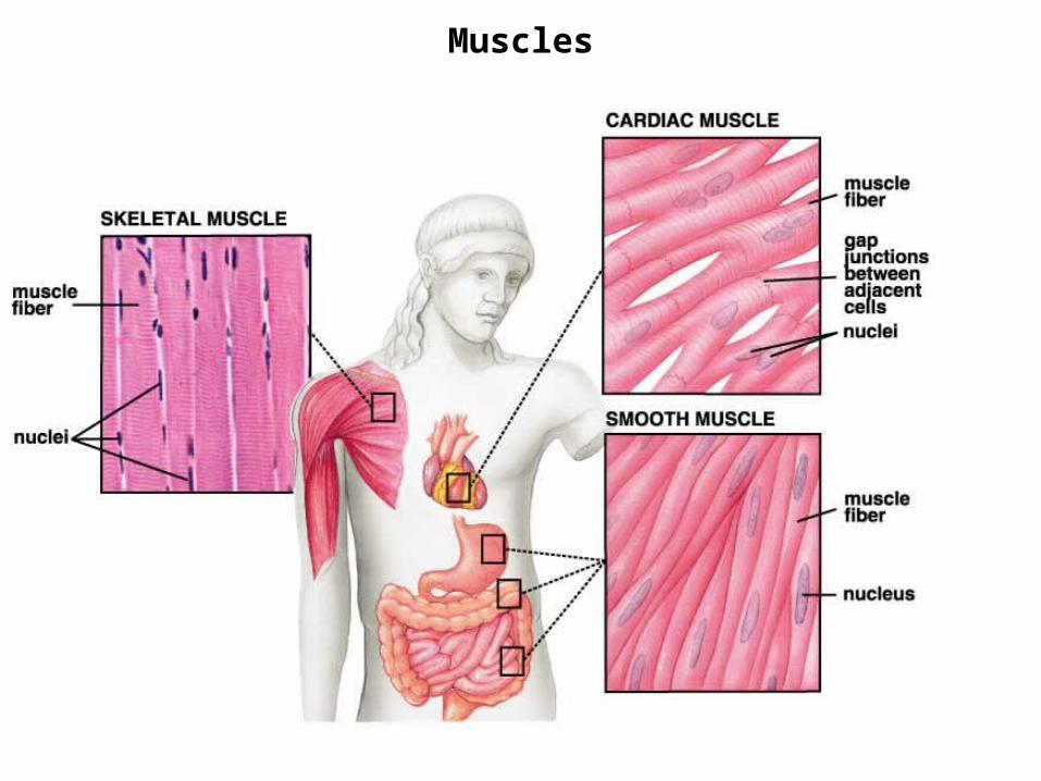
There are three types of muscle tissue:
1. Skeletal muscle:
Responsible for movement of skeleton and organs.
Sometimes referred to as voluntary muscle, because it can becontrolled voluntarily.
May also be referred to as striated muscle, because of thearrangement of the contractile proteins which give it a cross-striated appearance.
2. Visceral muscle (smooth muscle):
Muscular component of visceral structures like blood vessels,gastrointerstinal tract, uterus, the urinary bladder, etc.
Sometimes referred to as involuntary muscle, because it is underinherent, autonomic, and hormonal control.
May also be referred to as smooth muscle, because the arrangementOf the contractile proteins do not give it the “cross-stiated appearance”.
Muscles
There are three types of muscle tissue (cont.):
3. Cardiac muscle:
Structurally, appears to be in between skeletal and visceralmuscles. Although striated in appearance, they are distinguishablefrom skeletal muscles.
Provides for continuous and rhythmic contractility of the heart.
Additional notes:
There is variation within each muscle type, particularly in skeletal muscles.
Whereas skeletal and cardiac muscles are involved in relatively “forceful” contractions with a short duration, visceral muscles are specialized for continuous contractions of relatively low force production.
Muscle cell organelles have different names from their counterparts in other cells;The cytoplasm of muscle cells is called the sarcoplasm, the smooth ER is called the Sarcoplasmic reticulum, and the cell membrane is referred to as the sarcolemma.
Muscles
Muscles - Organization of Skeletal Muscle
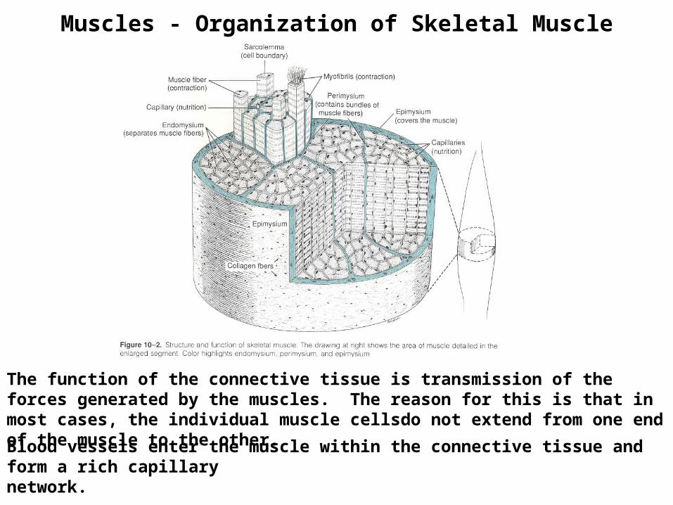
Muscle fibers are arranged in regular bundles surrounded by a dense connectivetissue surrounding the entire muscle. This layer is called the epimysium.
The individual bundles are surrounded by connective tissue called the perimysium.
Each muscle fiber itself is surrounded by a delicate layer of connective tissue calledthe endomysium.
Muscles - Organization of Skeletal Muscle
The function of the connective tissue is transmission of the forces generated by the muscles. The reason for this is that in most cases, the individual muscle cellsdo not extend from one end of the muscle to the other.
Blood vessels enter the muscle within the connective tissue and form a rich capillary network.
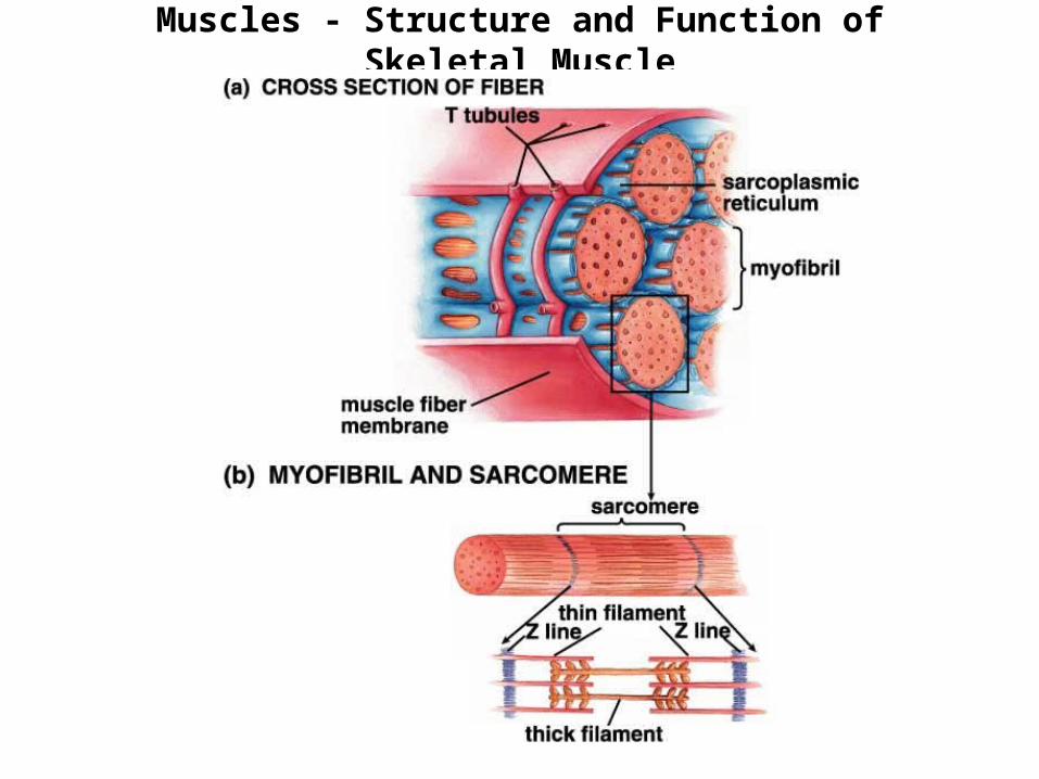
Muscles - Structure and Function of Skeletal Muscle
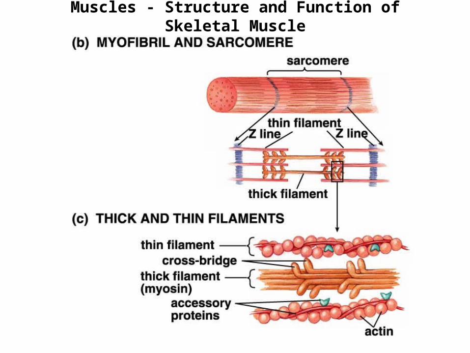
Muscles - Structure and Function of Skeletal Muscle
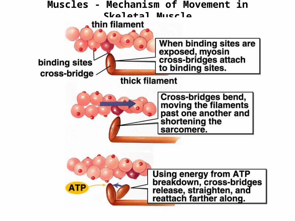
Muscles - Mechanism of Movement in Skeletal Muscle
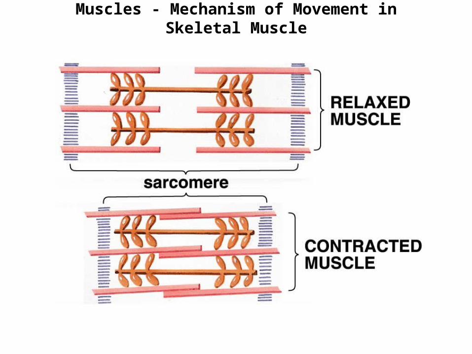
Muscles - Mechanism of Movement in Skeletal Muscle
Muscles - Mechanism of Movement in Skeletal Muscleimportance of calcium
Motor neurons activate skeletal muscle by sending messages (action potential)down into the cell’s interior, through the T tubules.
This message releases calcium stored within the sarcoplasmic reticulum.
The calcium then interacts with the accessory proteins of the thin filament,exposing the myosin binding sites. The thick filament can now form cross-bridges, and as long as there is ATP available, the muscles will contract.
Calcium is then actively (using ATP) pumped back into the sarcoplasmic reticulum.
The individual “messages” sent down motor neurons usually activate more than onemuscle fiber. The group of fibers that get activated are referred to as a motor unit.The “size” of the motor unit depends on the type of activity and the amount of musclesrequired for that activity.
Muscles - Mechanism of Movement
Cardiac muscle:
Contraction occurs in a similar way as the skeletal muscle (with somedifferences), but “message” to contract is generated by the cardiac muscle fibers themselves (not the motor neurons) .
The fibers in the sinoatrial (SA) node are specialized for this task and actas the heart’s pacemaker.
Smooth muscle:
Again, mechanism of contraction is similar although there are some majordifferences in structure, activation, and position of calcium stores...
The contractions are induced by stretching, hormones, nervous signals,or some combination of one or more of these stimuli.
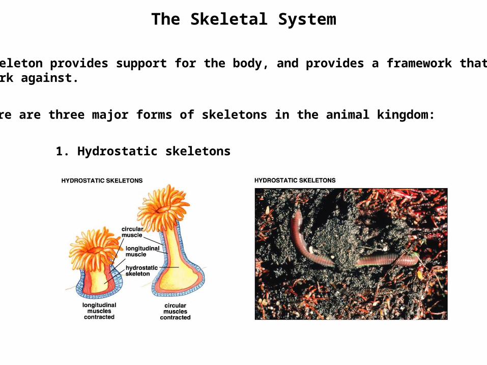
The Skeletal System
The skeleton provides support for the body, and provides a framework that musclescan work against.
There are three major forms of skeletons in the animal kingdom:
1. Hydrostatic skeletons
The Skeletal System
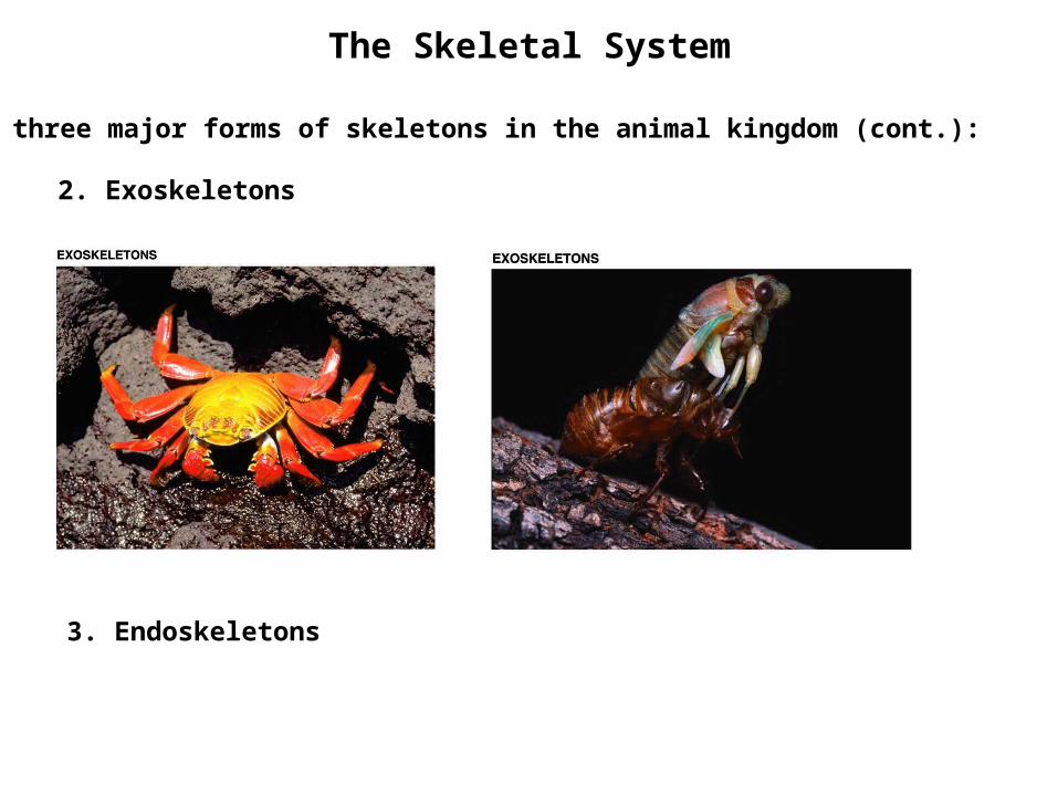
2. Exoskeletons
3. Endoskeletons
There are three major forms of skeletons in the animal kingdom (cont.):
The Skeletal System
Functions of the vertebrate skeleton:
1. Overall support and protection of internal organs.
2. Locomotion
3. The red bone marrow produces red blood cells, white blood cells,and platelets.
4. Storage of calcium and phosphate. These minerals will be released and re-absorbed as needed to maintain a constant blood concentration.
5. Sensory transduction (e.g. bones of the middle ear)
The Skeletal System
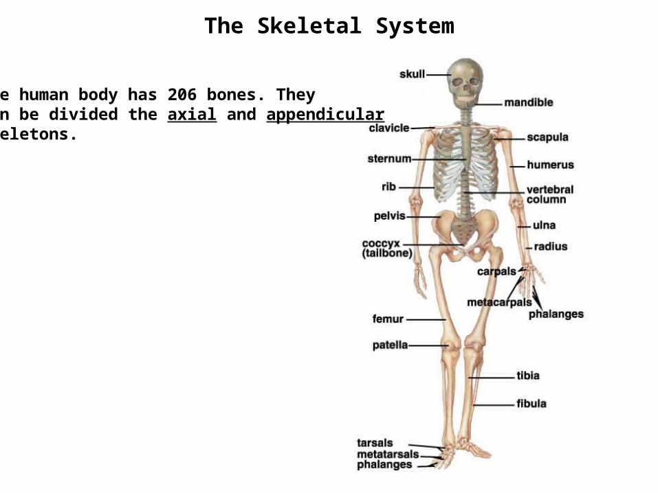
The human body has 206 bones. They can be divided the axial and appendicular Skeletons.
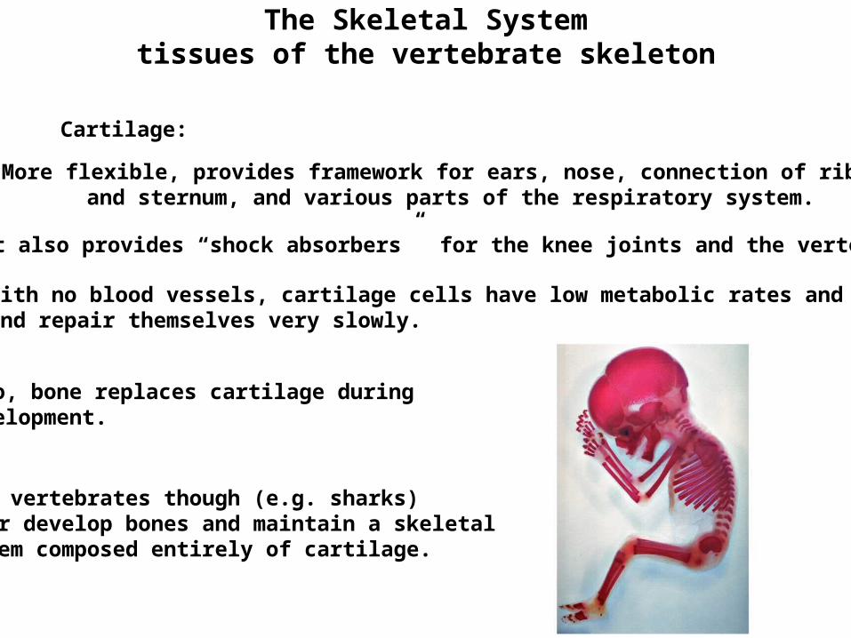
The Skeletal Systemtissues of the vertebrate skeleton
Cartilage:
More flexible, provides framework for ears, nose, connection of ribsand sternum, and various parts of the respiratory system.
It also provides “shock absorbers” for the knee joints and the vertebrae.
With no blood vessels, cartilage cells have low metabolic rates and growand repair themselves very slowly.
Also, bone replaces cartilage duringdevelopment.
Some vertebrates though (e.g. sharks)never develop bones and maintain a skeletalsystem composed entirely of cartilage.
The Skeletal Systemtissues of the vertebrate skeleton
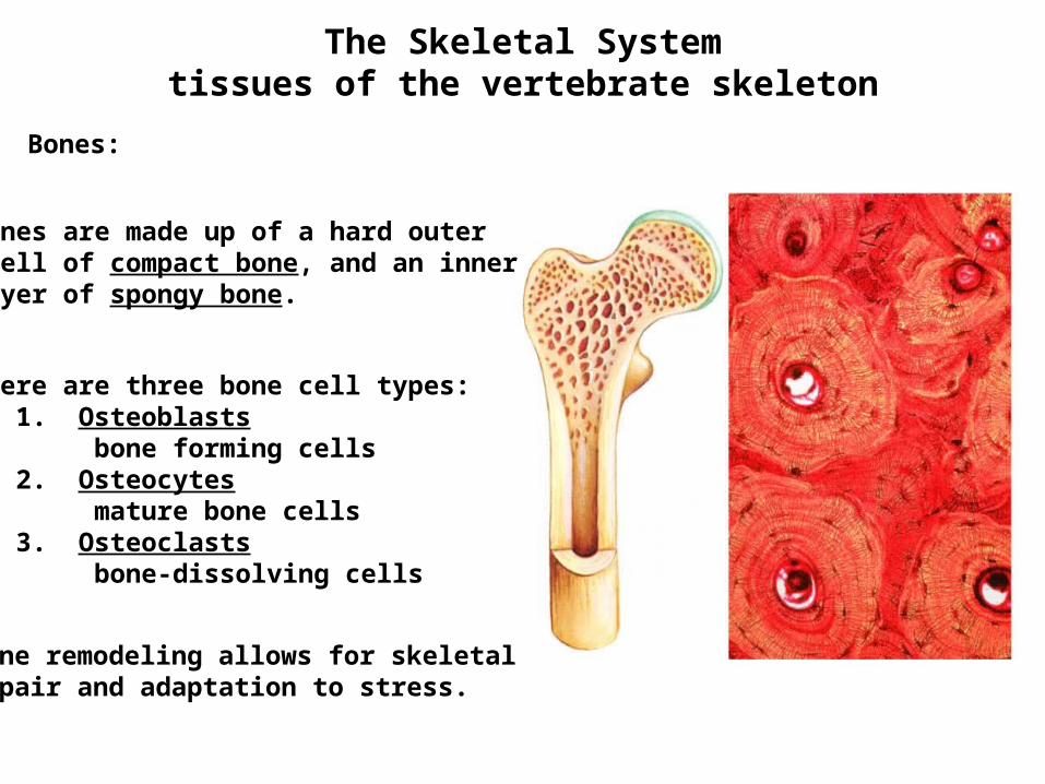
Bones:
Bones are made up of a hard outershell of compact bone, and an innerlayer of spongy bone.
There are three bone cell types: 1. Osteoblasts bone forming cells 2. Osteocytes mature bone cells 3. Osteoclasts bone-dissolving cells
Bone remodeling allows for skeletalrepair and adaptation to stress.
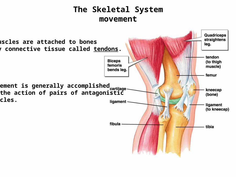
The Skeletal Systemmovement
Muscles are attached to bonesby connective tissue called tendons.
Movement is generally accomplishedby the action of pairs of antagonisticmuscles.
The Skeletal Systemmovement
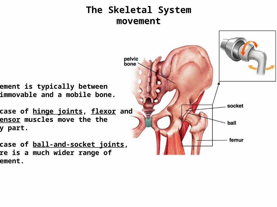
Movement is typically betweenan immovable and a mobile bone.
In case of hinge joints, flexor and extensor muscles move the the body part.
In case of ball-and-socket joints, there is a much wider range ofmovement.
The Skeletal System
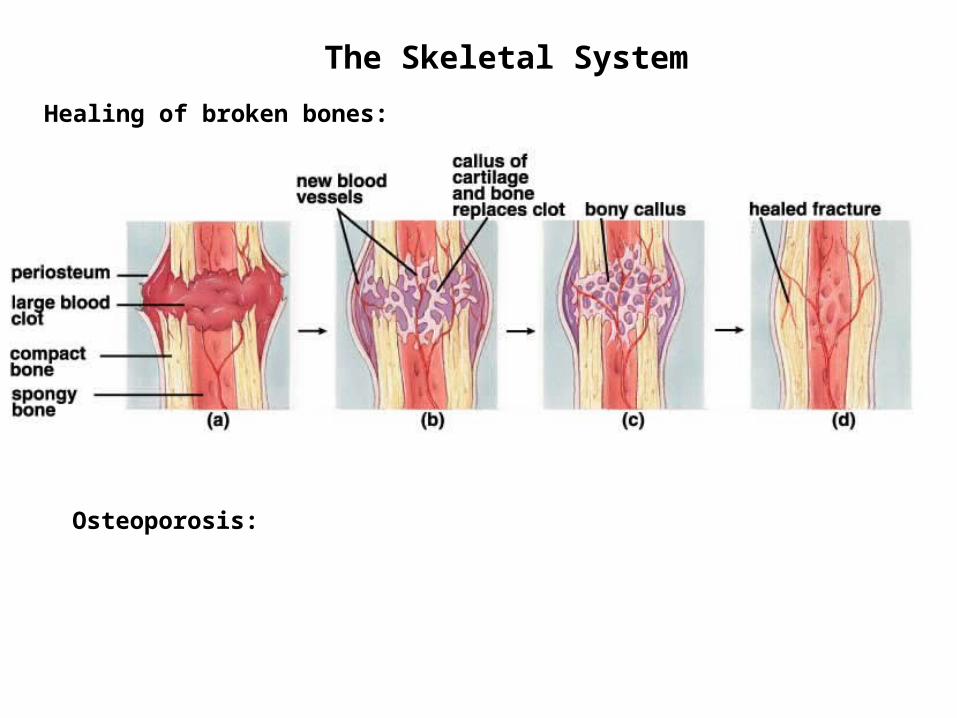
Healing of broken bones:
Osteoporosis: